Carrier Detection of Ovine Hemophilia A
Using an RFLP Marker, and Mapping of the
Factor VIM Gene on the Ovine X-Chromosome
W. Backfisch, S. Neuenschwander, U. Giger, G. Stranzlnger, and V. Pliska
From the Department of Animal Science, Swiss Federal Institute of Technology (ETH), CH-8092 ZOrich, Swit-zerland. The research was supported by the SANDOZ Stlftung Basel, AGROFONDS Zurich, and by the Swiss National Science Foundation (V P ) Our thanks are due to Drs. E. BOhler (Basel, Switzerland), J. Gltschler (San Francisco, U.S A ), and I. Oberle (Strasbourg, France) for the gifts of gene probes Critical comments by Dr. P. Driscoll are gratefully acknowledged.
Journal of Heredity 199435:474-478, 0022-1503/94/S5.00
Ovine hemophilia A is an X-linked recessive bleeding disorder. For diagnostic pur-poses, restriction fragment length polymorphism (RFLP) analysis in the region of the factor VIII (F-VIII) gene was carried out using human F-VIII gene probes. The probe St14, known to detect a highly polymorphic region that is closely linked to the F-VIII gene in humans, hybridized nonspecifIcally with DNA from sheep. Search-ing for Intragenic RFLPs, the entire 9.0-kb codSearch-ing sequence of the human F-VIII was used as a probe. Using the 1.8-kb Sstl/Kpnl F-VIII cDNA probe for hybridization, an Mspl-RFLP with allelic bands of 5.8 kb (A1) and 4.2 kb (A2) was detected. A1 was in linkage phase with the mutated allele responsible for hemophilia A. The F-VIII locus in the sheep genome was assigned to the long arm of the X-chromosome in the region Xq24-q33, using In situ hybridization with a 3-kb human F-VIII cDNA probe to QFQ banded sheep metaphase chromosomes.
Recently, we have described an inherited coagulopathy caused by a deficiency of the blood coagulation factor VIII (F-VIII) in a family of White Alpine Sheep. Investiga-tions of affected animals showed that clin-ical signs of bleeding (including severe hemarthrosis), low plasma activity of the F-VIII (<1%), high mortality rate, and X-chromosomal recessive mode of inheri-tance are comparable to that seen in hu-man patients with severe hemophilia A. Replacement therapy of hemophilic sheep with human F-VIII concentrate resulted in the remission of the coagulopathy and in rapid clinical improvement. Thus, these sheep may become a suitable animal mod-el of human hemophilia A (Neuenschwan-der et al. 1992; Neuenschwan(Neuenschwan-der and Plis-ka 1990; PlisPlis-ka et al. 1982). We have preserved this hemophilia model by es-tablishing an animal colony from one fe-male carrier. Because the determination of plasma F-VIII activity does not reliably dif-ferentiate between normal females and carriers (Neuenschwander and Pliska 1990), only the birth of a hemophilic lamb confirmed the carrier status of the ewes; however, this breeding and selection prac-tice was lengthy and costly. Heterozygote detection of hemophilia A in humans has been successfully achieved by using re-striction fragment length polymorphism (RFLP) and a variable number of tandem repeat (VNTR) markers within or adjacent to the F-VIII gene (Antonarakis and
Kaza-zian 1988). In the study presented here, we used RFLP markers to detect carriers of the F-VIII mutation in the established sheep colony. Furthermore, the F-VIII gene was assigned to a specific region of the ovine X-chromosome by in situ hybridiza-tion using a human F-VIII cDNA probe. Materials and Methods
Animals
A hemophilic ovine colony was estab-lished from one carrier ewe (White Alpine Sheep, ewe No. 157; Neuenschwander et al. 1992); it consisted of 99 offspring with-in four generations. Twelve unrelated nor-mal rams were used for matings. Carrier ewe 711 (Figure 1) and three unrelated rams (B, D, I) were used to test the suit-ability of individual restriction enzymes for RFLP analysis.
Blood Coagulation Tests
Diagnosis of hemophilia A rested, on one hand, on the occurrence of typical clinical signs of hemophilia A and/or postmortem findings (subcutaneous hematomas, hemo-peritoneum, hemarthrosis, prolonged um-bilical bleeding, etc.; Neuenschwander and Pliska 1990) and, on the other hand, on coagulation tests using citrated plasma from 2-3-week-old animals. Activated par-tial thromboplastin time (PTT) and plas-ma F-VIII activity, relative to a pool of normal ovine plasma, were routinely
de-F1
F2
1568
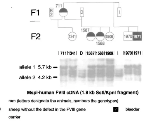
711113411 D I1587I1568I1909I I 11970119711
allele 1 5.7 kb
allele 2 4.2 kb
Mspl-human FVIII cDNA (1.8 kb Sstl/Kpnl fragment) ram (letters designate the animals, numbers the genotypes)
sheep without the defect in the FVIII gene Q bleeder carrier
Figure 1. Pedigree of the ewe 711 (F, generation) and Its offspring Animal numbers correspond to the scheme employed at the Experimental Station of the ETH Zurich, In Chamau ZG, Switzerland. Southern blot hybridization patterns for Individual animals are shown. The A-type of the ram B could be deduced from the heterozygous status of Its female offspring 1341 (A1-A2) and the homozygous status of the mother ewe 711 (Al) (DNA of this animal was no longer available at the time of our hybridization experiments.)
termined by methods described earlier (Neuenschwander et aJ. 1992; Neuensch-wander and Pliska 1994). Based on these tests, male offspring could be separated into two distinct groups: hemophiliacs with F-V11I activity of <5% of control and prolonged PTT, and normal males with normal F-VIII activity as well as normal PTT. Female "carriers" were defined as an-imals that produced at least one hemo-philic male, had intermediary to normal
F-VIII activity, and had normal to slightly
prolonged PTT (Neuenschwander et al. 1992).
DNA Probes
Stl4, a 3.0-kb EcoRI insert in pBR 322, de-tects a hypervariable VNTR locus in hu-mans that is closely linked to the F-VIII gene (Oberle et al. 1985). The hybrid plas-mid pF8.1 contains the entire human F-VIII 9.0-kb cDNA, inserted blunt-ended into the Smal sites of pUC12. These DNA probes were provided by Dr. I. Oberle, Institut de Chemie Biologique, F-67085 Strasbourg, France (Stl4), and by Dr. J. Gitschier, How-ard Hughes Medical Institute, University of California at San Francisco, San Francisco, CA 94143-0724 (pUC12). Digestion of pF8.1 with SstI and Sail yields a 3.0-kb and a 6.0-kb fragment (inserts) as well as a 2.7-6.0-kb
fragment (vector). Additional digestion with Kpnl cleaves the 3-kb fragment into 1.8-kb and 1.2-kb fragments. The probes were isolated from the vector by prepar-ative low melting agarose gel electropho-resis (Gitschier et al. 1984).
DNA Analysis
DNA samples were prepared by the pro-cedure of Jeanpierre (1987) from fresh or frozen venous blood collected in EDTA-containing tubes. DNA samples (10 ^g) were completely digested using the re-striction enzymes Apal, BamHI, Bell, BgHI, EcoRI, Haelll, HindlH, Hinfl, Kpnl, Mspl, PstI, PvuII, Rsal, Sail, and SstI, under the conditions specified by the enzyme sup-plier (Boehringer Mannheim). The result-ing fragments were separated accordresult-ing to their molecular weight by agarose gel elec-trophoresis (0.6-1.5%). Southern blotting to Hybond-N nylon filters (Amersham) and hybridization were carried out as de-scribed by Davies et al. (1988). The filters were washed three times in 2 x SSC (Man-iatis et al. 1982) and 1% SDS (sodium do-decylsulphate) at 65°C. Autoradiography was carried out at -80°C using Fuji R X-ray film for 0.5-5 days, with intensifying screens. Data analysis for the detection of genetic linkage in families was performed
by the lod-score method according to Morton (1955).
Chromosome Preparation and Identification
Metaphase chromosomes from cultured peripheral blood lymphocytes of a normal ram (G in Figure 2) were prepared accord-ing to Fries et al. (1986). Chromosomes were stained by QFQ banding technique (Caspersson et al. 1968) and metaphase spreads were photographed before hy-bridization. The identification of the chro-mosomes was based on the ovine karyo-types presented by Long (1985) and DiBerardino et al. (1989).
In situ Hybridization
Radioactive labeling of the human 3.0-kb
F-VIII cDNA probe was performed by
ran-dom primed method (Feinberg and Vogel-stein 1983, 1984) modified for tritium la-beling (Lin et al. 1985). The labeled DNA was separated from nonincorporated nu-cleoside triphosphates on a Sephadex G 50 column. The DNA was recovered by ethanol precipitation in the presence of salmon sperm DNA (10 jig). The specific radioactivity of the DNA probe was 2.5 x 10 dpm/|j,g. The procedure for in situ hy-bridization was based on the method of Harper and Saunders (1981) as modified by Fries et al. (1988). The concentration of the DNA probe was 20 ng/ml.
Analysis of the Silver Grain Distribution
After the QFQ banding, metaphases of the preparations were photographed. The au-toradiographic silver grains which con-tacted individual chromosomes were plot-ted on the ideogram of the haploid ovine genome (Gunawardana 1991). The differ-ent hybridization information concerning autosomes and the sex chromosomes in a histogram was taken into account. Results
Southern Blot Analysis
Southern blot analysis with the DNA probe Stl4 yielded only unspecific hybridization with sheep DNA. Genomic blots of digest-ed sheep DNA with 15 different enzymes showed five to 17 distinct bands after hy-bridization with the full-length F-VIII cDNA probe, depending on the type of the en-zymes used (data not shown). The F-VIII gene in sheep seemed to be "hypopoly-morphic," similar to that of human (An-tonarakis et al. 1985; Gitschier et al. 1985a; Wion et al. 1986). An MspI-RFLP could be
1587.
F4
ftll
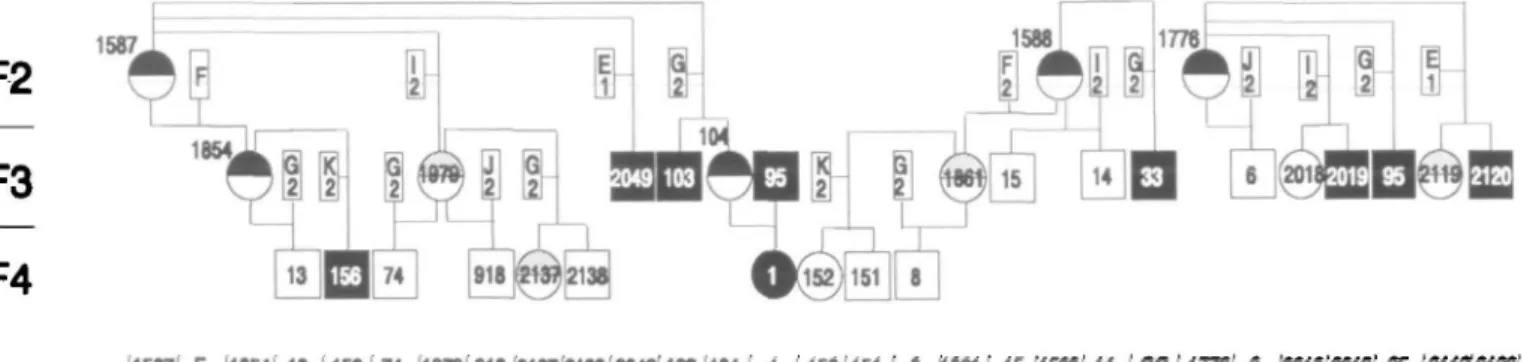
1587i F I1B54! 13 '156174 197B 918 2137 2138 2049" 103 104 1 I 1521151 ' 8 (1881' 15(1588(14 (33(17781 6 (2018'2019'K (21192120 alkHel 5.7 kb — altele2 4.2 kb —H"if
ft
I
™-Mspl-human FV1II cDNA (1.8 kb Sstl/Kpnl fragment)
D ram (letters designate the animals, numbers the genotypes) B 0 sheep wtth hemophilia A @ © sheep without the defect In the FVIII gene Q probable carrier
carrier
Figure 2. Pedigree of the ewes 1587, 1588, and 1776 (F, generation) and their offspring, and the Southern blot hybridization patterns of Individual animals
detected using the 1.8-kb Sstl/Kpnl F-VIII cDNA probe (Figures 2 and 3). The lengths of the allelic RFLP bands were 5.8 kb (al-lele 1, Al) and 4.2 kb (al(al-lele 2, A2), respec-tively.
Pedigree Analysis
The entire colony consisted of 60 females and 39 males. Ten male bleeders have oc-curred within the male progeny. Within the female progeny, four ewes were iden-tified as carriers of the defective F-V1I1 gene, based on the occurrence of hemo-philic males in their progeny. A female he-mophilic animal was produced by mating a hemophilic ram (No. 95; cf. Figure 2) with a female carrier (No. 104).
Based on the Southern blot analysis shown in Figure 1, the carrier ewe 711 has only one band at 5.8 kb (Al) and no band at 4.2 kb (A2), suggesting that this animal is homozygous for the allele Al. The healthy rams D and I have only the 4.2-kb band. The segregation of the Al allele marker to the clinically normal son 1970 and the bleeder 1971 without an A2 allele
Mspl Mspl Mspl
A1 A1
indicates a typical X-chromosomal inheri-tance pattern: (1) Both sons show the same banding pattern as their homozy-gous mother, and (2) the father's allele A2 does not appear in the profile of the sons. Thus, one of the three combinations of the alleles A1/A2 and H/h (H stands for the wild type and h for the mutant allele) shown in Figure 3 exists on the X chro-mosome within the bleeder family: Al-h, Al-H, and A2-H.
As can be concluded from the segregation of the MspI-RFLP marker and the disease in 14 informative meioses, he-mophilia A always cosegregated together with Al (Figures 1 and 2): thus, Al in the bleeder family is in linkage phase with the hemophilia locus. No recombination events were observed. Six bleeders (95, 2019, 2120, 33, 103, and 2049) and two car-riers (104, 1854) in the F3 generation in-herited the defective F-VIII gene, together with allele Al, from their mothers. Fur-thermore, in informative matings ewes 1854 and 104 transmitted the disease to-gether with Al to the offspring 1 and 156
Mspl Mspi Mspl Mspl H A2 H X1 X2 X3 mutated locus A 7-/1 A1-H
wild type wild type
A2-H
Figure 3. Possible haplotypes Al-h. Al-H. and A2-H on the X chromosomes in the aifected sheep family. The polymorphic cleavage site of the restriction enzyme Mspl defines the alleles A l and A2; the wild type of the F-VIII gene Is designated by H, and the mutated site by h
(Al-h). In contrast, the four male lambs 6, 13, 14, and 15 inherited the normal F-VIII allele together with the A2 allele (A2-H). As predicted, the mutated site in the F-VIII gene and the Mspl-RFLP marker are close-ly linked. The lod-score was Z = 4.21. The recombination frequency of the linkage group was with 95% probability within the interval (ql; q2) = [0.0; 0.15]. The statistic confidence to predict the status of a car-rier or a noncarcar-rier ewe was 85%. This low predictive value was due to the limited number of studied animals. Considering the fact that the MspI-RFLP is an intragen-ic marker, the confidence should be much higher.
In situ Hybridization
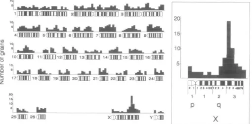
The silver grain distribution in 88 meta-phases from a normal ram was analyzed. In 40 metaphases (45.5%), the X chromo-some was labeled with one or more grains. From 934 grains analyzed, 67 (7.2%) were located on the X chromosome. The histo-gram of the grain distribution is shown in Figure 4. The relative length of the X chro-mosome in the diploid chrochro-mosome set in males is 2.64% (Hediger 1988). Thirty-six grains (3.9%) were concentrated in the re-gion Xq24-q33, with the relative length of roughly 0.6% of the total chromosome length in a male diploid chromosome set. A maximum of 19 grains were located at the passage of band q31 to q32. The sec-ond highest not flanking columns with eight grains were found at various loci of several autosomes. Consequently, the
1 i l l i i i r m i i inn 2ii[iiin:Tiiiinii 3HHEIXILIILIE
<uuiim SUI_LU B iimifl 7
• •II • • i l l
t j 4 » m 4 jit « mtd
26(00] 26 H E
Figure 4. Localization of the F-VIII gene In sheep Histogram of silver grain distribution in 88 metaphases after hybridization with a human 3kb F-VIII cDNA probe A peak was found on the X chromosome In the region Xq24-q33
highest grain column was 2.4 times higher than the second highest nonflanking col-umn.
Discussion
Cloning of the F-VIII genes of human he-mophiliacs within individual families re-veals many different mutations associated with this disease (Tuddenham et al. 1991). The type of mutation correlates with the severity of clinical signs. Point mutations causing incomplete transcription of the gene by introduction of a stop codon into the coding sequence (Antonarakis et al. 1985; Gitschier et al. 1985b; Youssoufian et al. 1986) resulted in a severe form of he-mophilia A. Substitution of a single amino acid by a point mutation found at least in one patient caused only a mild coagulo-pathy (Gitschier et al. 1986). Base inser-tions (Kazazian et al. 1988) and deleinser-tions (Gitschier et al. 1985b; Youssoufian et al. 1987), causing mostly severe coagulopa-thies, have also been detected. The molec-ular basis of mutations in hemophilic ani-mals, on the other hand, has not yet been determined. Undoubtedly, the comparison of wild types as well as mutant F-VIII gene in humans and animals may offer an inter-esting contribution to the phylogeny and structure-function relationship of coagu-lation factors. Results presented in this communication document at least a cer-tain homology within the F-VIII gene in hu-man and in sheep: the huhu-man probe used in our study interacted with the sheep DNA both in Southern blot analysis and in in situ hybridization, also indicating a con-servative evolution of this blood clotting factor.
The results of this family study
demon-strated the value of RFLP markers for the carrier detection of X-linked, recessively inherited hemophilia A. Programmed mat-ing and indirect genotype diagnosis allow one to predict the status of each ewe with-in the family shortly after birth, with a high degree of reliability. No recombina-tion between an intragenic marker and a mutated locus in the F-VIII gene was de-scribed in humans (Antonarakis and Ka-zazian 1988). The distance of two loci of lcM (i.e., 10 bp) is equivalent to q = 0.01. The recombination frequency between the 5' and 3' end of the human F-VIII gene with about 200 kb length corresponds to the value q = 0.002. If the length of the F-VIII gene of sheep is similar to that in humans, the status of the animals, based on the MspI-RFLP, could be predicted with a min-imal confidence of 99.8%. Clearly, the use of this RFLP marker has greatly improved the accuracy of carrier detection as well as permitted a neonatal diagnosis of he-mophilia in this colony. This is, however, valid solely within the investigated family. Mapping the ovine F-VIII gene by in situ hybridization we have found the highest cumulation of silver grains in the Xq24-q33 chromosomal region, following the band numbering by Hediger (1988). This result confirms the localization of F-VIII on the X-chromosome. Confirmation of the spe-cific assignment should be done with a ho-mologous F-VIII probe.
The MspI-RFLP is the first marker phys-ically mapped on the X chromosome in sheep. Using this marker, the F-VIII gene could be genetically mapped in the future, provided that further polymorphic loci will be located in its vicinity.
Reference*
Antonarakls SE and Kazazian HH, 1988. The molecular basis of hemophilia A in man Trends Genet 4:233-237. Antonarakis SE, Waber PG, Klttur SD, Patel AS, Kazazian HH, Mellls MA, Counts RB, Stamatoyannopoulos G, Bowie EJW, Fass DN, Pittman D, Wozney JM, and Toole JJ. 1985. Hemophilia A. detection of molecular delects and of carriers by DNA analysis. N Engl J Med
313:842-Caspersson T, Farber S, Foley GE, Kudynowsld J, Mod-est EJ, Slmonsson E, Wagh U, and Zech L, 1968. Chem-ical differentiation along metaphase chromosomes. Exp Cell Res 49 219-222.
Davtes W. Harbquitz I, Fries R, Stranzlnger R, and Hau-ge JG, 1988. Porcine malignant hyperthermla carrier detection and chromosomal assignment using a linked probe. Anlm Genet 19:203-212
DlBerardlno D, Hayes DH, Fries R, and Long S (eds), 1989 ISCNDA- International System for Cytogenetic No-menclature of Domestic Animals Cytogenet Cell Genet 53 65-79
Felnberg AP and Vogelsteln B, 1983 A technique for radiolabeling DNA restriction endonuclease fragments to high specific activity. Anal Blochem 132:6-13 Felnberg AP and Vogelsteln B, 1984 A technique for radiolabeling DNA restriction endonuclease fragments to high specific activity [Addendum] Anal Blochem 137.266-267.
Fries R, Hediger R, and Stranzlnger G, 1986. Tentative chromosomal localization of the bovine major hlsto-compatiblllty complex by m situ hybridization Anlm Genet 17:287-294.
Fries R, Hediger R, and Stranzinger G, 1988 The loci for parathyroid hormone and b-globln are closely linked and map to chromosome 15 In cattle Genomlcs 3.302-307.
Gitschier J. Drayna D, Tuddenham EGD, White RL, and Lawn RM, 1985a Genetic mapping and diagnosis of haemophilia A achieved through a Bell polymorphism in the factor VIII gene. Nature 314738-740.
Gitschier J, Wood Wl. Goralka TM, Wion KL, Chen EY, Eaton DH, Vehar GA, Capon DJ, and Lawn RM, 1984. Characterization of the human factor VIII gene. Nature 312326-330
Gitschier J, Wood WI, Shuman MA, and Lawn RM, 1986 Identification of a missense mutation In the factor VIII gene of a mild hemophiliac Science 232 1415-1416 Gitschier J, Wood Wl, Tuddenham EGD, Shuman MA, Goralka TM, and Chen EY, 1985b. Detection and se-quence of mutations In the factor VIII gene of hemo-philiacs. Nature 315:427^430
Gunawardana A, 1991. Physical mapping of polymor-phic DNA markers In the Bovidae family (a compara-tive study in cattle, sheep and goat) (PhD dissertation) ETH Zurich
Harper ME and Saunders GF, 1981 Localization of sin-gle copy DNA sequences on G-oanded human chro-mosomes by in situ hybridization Chromosoma 83' 431-439
Hediger R, 1988. Die in situ Hybrldlsierung zur Genkar-tlerung belm Rind und Schal (PhD dissertation) ETH Zurich.
Jeanplerre M, 1987 A rapid method for the purification of DNA from blood. Nucleic Acids Res 15561 Kazazian HH Jr, Wong C, Youssoufian H, Scott AF, Phil-lips DG, and Antonarakis SE, 1988. Haemophilia A re-sulting from de novo Insertion of LI sequences repre-sents a novel mechanism of mutation In man Nature 332:164-166.
LJn CC, Draper PN, and De Breakeleer M, 1985 High resolution chromosomal localization of the b-gene of the human b-globin gene complex by in situ hybridiza-tion Cytogenet Cell Genet 39269-274
Long M, 1985. Committee for standardized karyotype of Ovies aries: standard nomenclature for the G-band
karyotype of the domestic sheep (Ovles arles). Heredi- Neuenschwander S and PHSka V, 1994. Factor VIII In substitutions, deletions, Insertions and rearrangements tas 103.165-170 blood plasma of haemophllic sheep-analysis ol clotting of the factor VIII gene. Nucleic Acids Res 19 4821-4833. Maniatls T, Fritsch EF, and Sambrook j , 1982 Molecular 'l me - p l a s m a dilution curves. Haemostasis 24 27-35 W | Q n ^ T u d d e n n a m E G D | ^ ^ RM, 1 9 8 6 A new cloning: a laboratory manual. Cold Spring Harbor, NY: Oberle 1, Camerlno G, HelHg R, Grunebaum L, Craze- polymorphism In the factor VIII gene for prenatal di-Cold Spring Harbor Laboratory naare JP, Crapanzano C, Manuccl PM, and Mandel JL, agnosis of hemophilia A Nucleic Acids Res 11:4535-Morton NE, 1955 Sequential tests for the detection of 1 9 8 5- G e n e t l c screening for hemophilia A (classic he- 4542
linkage. Am J Human Genet 7277-318 mophllia) with a polymorphic DNA probe N EngI J Med Youssoufian H, Antonarakls SE, Aronls S, Tsiftis G,
Phll-w . . c „ , „ ... . . , u , , , 312:682-686 M p s QQ and Kazazlan HH Jr, 1987. Characterization of
Neuenschwander S, Klssllng-Albrecht L, Helniger J, c ' , , . . „ , ,u , , , , „ , _„ D M t l
Backfisch W, Stranzlnger G, and PliSka V, 1992. Inherit- ™ k a V, Schwander B, Allmendinger W, and MQIIer- ^e?£?KV£w,w? **"* ed defect of blood clotting factor VIII (haemophilia A) Lhotsky A, 1982 Erbllche HSmophllle beim Schaf Un- A c a a :>cl ub* W^iu-silt,
in sheep. Thromb Haemostasis 68 618-620 tersuchung an potentlellen Konduktorinnen Schwelz Youssoufian H, Kazazlan HH Jr, Phillips DG, Aronls S,
w u . c . „ , . „ , , i n Q n u 1.11, , Landw Monatshefte 60:284-295 Tslftls G, Brown VA, and Antonarakis SE, 1986 Recur-Neuenschwander S and PliSka V, 1990 Hemophilia In . ' , . , , , . . , , „ „ sheep and the use of sheep in blood coagulation re- Tuddenham EGD, Cooper DN, Gltschler j , Higuch, M, r e n m u t a'l o n s ' " h™p W 5 A K ' ^
search In Farm animals In blomedlcal research (PliSka Hoyer LW, Yoshloka A, Peake IR, Schwaab R, Olek K, m u t a t l o n notspots mature JZ
V and Stranzlnger G, eds). Hamburg: Verlag Paul Parey; Kazazlan HH, Lavergne J-M, Glannelll F, and Antonara- Received April 14, 1993 155-164. Ids SE, 1991. Haemophilia A database of nucleotlde Accepted March 30, 1994