THE JOURNAL OF INFECTIOUS DISEASES. VOL. 142, NO.1· JULY 1980 ©1980 by The University of Chicago. 0022-1899/80/4201-0015$00.75
Microneutralization of Cytomegalovirus
Hans Stalder and Alice Ehrensberger From the Infectious Disease Division, Department of Internal Medicine, University of Geneva, Switzerland
A simple microneutralization test for cytomegalovirus (CMV) is presented. Using a laboratory adapted stock virus, definite results were obtained after 10 days. All patients with primary CMV infection showed an antibody rise and/or seroconversion; however, neutralizing antibody appeared only seven weeks after the onset of clinical symptoms. In control patients without evidence of recent primary infection, there was a complete concordance of the presence of complement-fixing and neutralizing antibodies.
Neutralization tests [1-5] with cytomegalovirus (CMV) have not been very popular because CMV is a slow-growing virus and not easy to obtain as high titered, cell-free virus. However, the deter-mination of neutralizing antibodies may be more significant in clinical situations than, for example, levels of CF antibodies. This situation is especially true when we seek to evaluate the responses of in-dividuals to natural infection or CMV vaccines. Furthermore, neutralization tests, as has been shown for herpes simplex viruses (HSV) [6, 7], can permit us to define and identify viral subtypes. This paper describes the adoption to CMV of a simple microneutralization test[7]which has prov-en its value for typing HSV isolates and antisera.
Materials and Methods
Cells. Diploid human fibroblast cells derived from newborn foreskin (FS-9; initiated at the Center of Disease Control, Atlanta, Ga.) were used. These cells were propagated as described previously [7].
Virus. CMV (strain 268a) was originally iso-lated from the urine of a kidney transplant recipi-ent. The original isolate was characterized as CMV by its focal, slowly progressing CPE, with eosinophilic intranuclear and rare cytoplasmic in-clusions as well as mononuclear giant cells after staining with hematoxylin and eosin. The virus was identified as CMV by the immunofluorescence technique using a highly specific human antiserum (performed by Dr. U. Krech, Institut fur
Medizi-Received for publication April 30, 1979, and in revised form January 29, 1980.
This work was partially supported by a research grant from the Swiss Cancer League.
Please address requests for reprints to Dr. Hans Stalder, Medizinische Klinik, Kantonsspital, 4410 Liestal, Switzerland.
102
nische Mikrobiologie, S1. Gall, Switzerland). Fur-thermore, specific rabbit antisera containing a high titer of antibody to HSV type 1 (HSV -1) and HSV type 2 (HSV -2) did not neutralize the agent (see below).
The original isolate was passaged 16 times by cell scraping. After the 16th passage, the cell monolayer was trypsinized (O.04OJo trypsin and 0.54 mM EDTA) when rv80OJo of the cells showed CPE. After centrifugation at 120gfor 10 min, the cells were resuspended in distilled water and soni-cated (Sonifer B-12; Branson Sonic Power Com-pany, New Haven, Conn.) at a force of 2 for 22 sec. Microscopic examination showed almost complete cell disruption.
This suspension was clarified at 1,()()()g for 15 min, and sorbitol was added to a final concentra-tion of 1OOJo. The virus was aliquoted into ampules and stored at -80 C. From this pool one ampule was added to a 500-ml roller bottle containing a complete cell monolayer. When rv80% of the cells showed CPE, the culture was trypsinized, and the cells were added to confluent monolayer cultures in four 2,()()()-ml Roux bottles (Muller and Krem-pel, Carouge, Switzerland). When rv80% of the cells showed CPE, the cultures were trypsinized and sonicated, and the clarified supernatant was stored in 10% sorbitol at -80 C. This virus was used for all experiments.
Microtitration and microneutralization tech-nique. A procedure similar to that described for HSV was used [7]. For titration of virus, 10-fold dilutions of virus were prepared in growth medi-um (Dulbecco's medimedi-um, supplemented with bi-carbonate and antibiotics [7] and 10% heat-inacti-vated [56 C for 30 min] fetal calf serum). Then 0.05 ml of each dilution of virus was placed in each of six wells of sterile, disposable microtest culture plates with 96 flat-bottom wells (M29ART, Microtiter system; Cooke
Engineer-Microneutralization ofCMV
ing, Alexandria, Va.). Immediately, 0.05 ml of a cell suspension containing 200,000 cells/ml in growth medium was added. The plates were cov-ered and incubated at 36 C in a moist atmosphere of50,10CO2in air. The CPE was read using an
in-verted microscope. All wells were scored as
+
(CPE present) or - (no CPE), and end-point dilu-tion titers were calculated using the Karber meth-od [8].For serum neutralization studies, twofold dilu-tions were prepared in growth medium. First, 0.025 ml of each dilution was placed in three repli-cate wells. Then, 0.025 ml of viral suspension con-taining rv200 TCIDso/0.05 ml was added to two of the three wells. The remaining well received 0.025 ml of growth medium (serum control). Then, 0.025 ml of a 1:4 or 1:8 dilution of either fresh or heat-inactivated (56 C for 30 min) guinea pig se-rum (stored at -80 C) was added to all of the wells. The plates were then incubated at different temperatures for various intervals (see Results). Finally, 0.05 ml of the cell suspension was added, and the plates were incubated in the CO2incubator
for various intervals (see Results).
When plates were read at 10 days, a change of medium was not necessary. The results were read as described above, and serum titers were ex-pressed as the reciprocals of the highest dilution of serum that inhibited viral CPE.
CF tests [9] and determination of CMV-specific IgM antibody (10] were performed by Dr. U. Krech using the Davis strain of CMV as the anti-gen.
Results
Titration of virus. Viral titers were between log 2.66 and log 3.83 TCIDso/0.05 ml. Exposure of the diluted virus to different temperatures before addition of cells appeared to modify the course of infection. When the virus was held for 30-120 min at 4 C beforeitwas placed into the wells of the mi-crotest plates, the subsequent development of CPE was delayed, so that at 10 days, the virus held at 4 C appeared to have a titer 1 log lower than the same virus held at 22 C or 37 C. However, CPE eventually developed so that after 18 days of incu-bation, the final titers were comparable. Addition of either fresh or heat-inactivated guinea pig serum did not change the kinetics of CPE development or the final titer of virus.
Serum neutralization. No consistent
differ-103
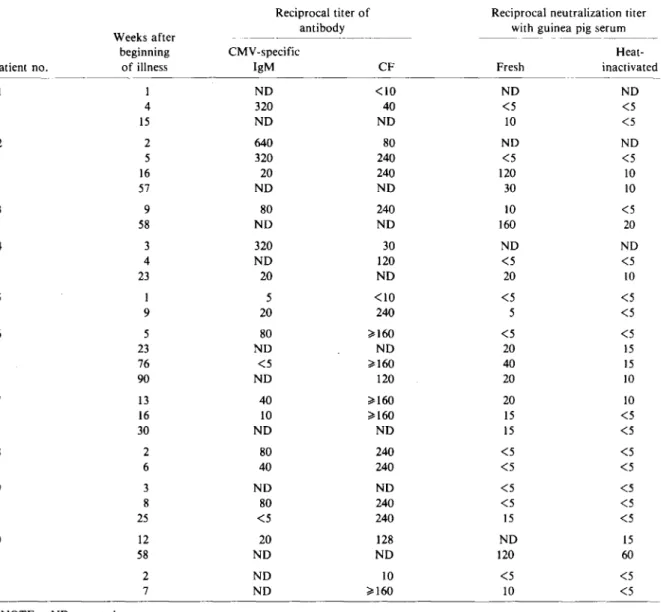
ence of neutralizing titer was observed when mix-ing periods of virus and serum were varied (30, 60, and 120 min) or when mixing was done at different temperatures (4, 22, and 37 C). Therefore mixing was done at 37 C for 30 min in all further experi-ments. Since reading the test after 15, rather 10, days of incubation changed in titers very little (on-ly, and that inconstant(on-ly, about half a dilution lower at 15 days), all tests were read at 10 days. Fresh guinea pig serum increased the titers up to l l-fold (mean, fourfold) (table 1). Several sera were positive only after addition of fresh guinea pig serum.
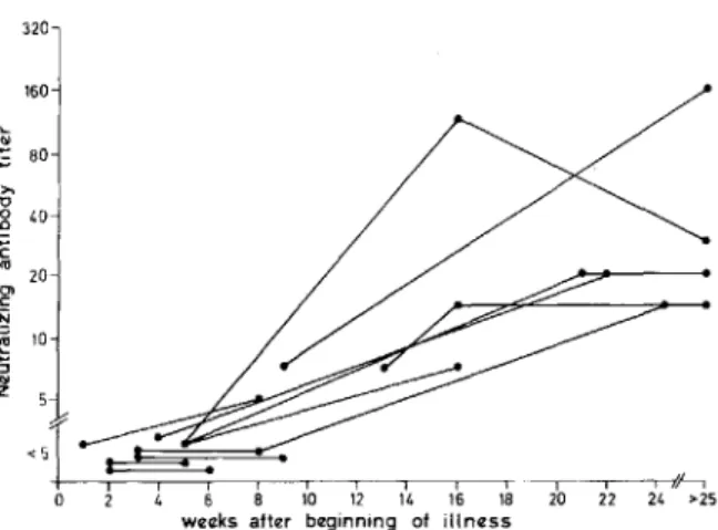
Patient sera. Sequential sera from 11 patients who appeared on clinical and virologic grounds to have primary CMV infection were examined. CMV was isolated from all except one patient, and all developed either CMV-specific fluorescent IgM antibody or a fourfold rise in CF antibody titer (tests performed by Dr. U. Krech). No serum con-tained detectable neutralizing antibodies earlier than seven weeks after the beginning of the illness. After 10 weeks, however, multiple sera from all patients were positive. Seroconversion was docu-mented in eight patients (table 1 and figure 1).
Ten sera from unpaid blood donors and 10 sera containing CF antibodies to HSV from patients from whom HSV was isolated were tested. Five of each group had positive titers of neutralizing an-tibody to CMV. Their values were compared with titers of CF antibody to CMV. No serum that lacked neutralizing antibody gave a positive CF test and vice versa.
Discussion
Using the microneutralization technique, we demonstrated the appearance of neutralizing an-tibodies in the sera of all patients suspected of having primary CMV infection, when tested 10 weeks or later after the onset of clinical symp-toms. However, none was positive before seven weeks. Late appearance of neutralizing, in con-trast to CF or fluorescent, antibodies has already been noted by Spencer and Andersen [11]. Al-though not tested in our laboratory, it is possible that early IgM antibodies have little neutralizing activity [12]. The clinical implication of this is not clear, but it is interesting to note that the clinical course as well as viral excretion in CMV infections is protracted. In our test system, addition of fresh guinea pig serum enhanced neutralizing antibody
104 Stalder and Ehrensberger
Table 1. Enhancement of serum neutralization titers by addition of fresh guinea pig serum in patients with
clinical-ly suspected primary infection with cytomegalovirus (CMV).
Reciprocal titer of Reciprocal neutralization titer Weeks after antibody with guinea pig serum
beginning CMV -specific Heat-Patient no. of illness IgM CF Fresh inactivated
1 NO <10 NO NO 4 320 40 <5 <5 15 NO NO 10 <5 2 2 640 80 NO NO 5 320 240 <5 <5 16 20 240 120 10 57 NO NO 30 10 3 9 80 240 10 <5 58 NO NO 160 20 4 3 320 30 NO NO 4 NO 120 <5 <5 23 20 NO 20 10 5 1 5 <10 <5 <5 9 20 240 5 <5 6 5 80 ~160 <5 <5 23 NO NO 20 15 76 <5 ~160 40 15 90 NO 120 20 10 7 13 40 ~160 20 10 16 10 ~160 15 <5 30 NO NO 15 <5 8 2 80 240 <5 <5 6 40 240 <5 <5 9 3 NO NO <5 <5 8 80 240 <5 <5 25 <5 240 15 <5 10 12 20 128 NO 15 58 NO NO 120 60 11 2 NO 10 <5 <5 7 NO ~160 10 <5
NOTE. NO = not done.
titers about fourfold, presumably due to the ac-tion of complement. Several sera became positive only after addition of fresh guinea pig serum. Its addition is therefore mandatory when this tech-nique is used.
Complement enhancement is not unique to neutralization of CMV but has also been noted with other viruses, especially those of the herpes group [13, 14]. However, complement enhance-ment of human sera has been found irregularly in CMV plaque-neutralization assays [5, 12, I5}. This observation is in contrast to those with animal sera, some of which are completely
depen-dent on complement for neutralizing activity [3, 12]. There was a complete correlation between the microneutralization test and the CF test in twenty control patients. No patient with a negative titer in one test had a positive one in the other.
This microneutralization technique [7] has several advantages over other neutralization tests; for example, transfer of virus-serum mixtures is avoided because the cells are added to the same well. Owing to the microtechnique, only small amounts of serum (0.025 ml for each serum dilu-tion), of virus, and of cells, are necessary. It is therefore easy to test multiple sera in the same
Microneutralization ojCMV 105
160 320
References
4. Waner, J. L., Budnick, J. E. Three-day assay for hu-man cytomegalovirus applicable to serum neutralization tests. Applied Microbiology 25:37-39, 1973.
5. Schmidt, N. J., Dennis, J., Lennette, E. H. Plaque re-duction neutralization test for human cytomegalovirus based upon enhanced uptake of neutral red by virus-infected cells. J. Clin. Microbiol. 4:61-66, 1976. 6. Pauls, F. P., Dowdle, W. R. A serologic study of
Herpesvirus hominis strains by microneutralization
tests. J. Immunol. 98:941-947, 1967.
7. Stalder, H., Oxman, M. N., Herrmann, K. L. Herpes simplex virus microneutralization: a simplification of the test. J. Infect. Dis. 131:423-430, 1975.
8. Karber, G. Beitrag zur kollektiven Behandlung pharma-kologischer Reihenversuche. Arch. Exp. Pathol. Phar-makol, 162:480-483, 1931.
9. Lennette, E. H. General principles underlying labora-tory diagnosis of viral and rickettsial infections. In
E. H. Lennette and N. J. Schmidt [ed.]. Diagnostic pro-cedures for viral and rickettsial infections. 4th ed. American Public Health Association, New York, 1969, p. 52-58.
10. Schmitz, H., Haas, R. Determination of different cy-tomegalovirus immunglobulins (IgA, IgG, IgM) by im-munofluorescence. Archiv fur gesamte Virusforschung 37:131-140, 1972.
11. Spencer, E. S., Andersen, H. K. The development of immunofluorescent antibodies as compared with complement-fixing and virus-neutralizing antibodies in human cytomegalovirus infection. Scand.J. Infect. Dis. 4:109-112, 1972.
12. Andersen, H. K. The influence of complement on cytomegalovirus neutralization by antibodies. Archiv fur gesamte Virusforschung 36:133-140, 1972. 13. Wallis, C., Melnick, J. L. Herpes virus neutralization:
the role of complement. J. Immunol. 107:1235-1242, 1971.
14. Schmidt, N. J., Lennette, E. H. Neutralizing antibody responses to varicella-zoster virus. Infec. Immun. 12: 606-613, 1975.
15. Minamishima, Y., Graham, B. J., Benyesh-Melnick, M. Neutralizing antibodies to cytomegaloviruses in nor-mal simian and human sera. Infec. Immun. 4:368-373, 1971.
16. Andersen, H. K. Studies of human cytomegalovirus strain variations by kinetic neutralization tests. Archiv fur gesamte Virusforschung 38:297-305, 1972. 17. Vonka, V., Benyesh-Melnick, M. Thermoinactivation of
human cytomegalovirus. J. Bacteriol. 91:221-226,1966. I#----'
24 >25
test, and furthermore, the test can be performed in multiple duplicates, eventually permitting subtyp-ing of CMV isolates [16]. Indeed, a similar test was originally used to detect subtypes of .HSV isolates [6]. Finally, the test can be performed at room temperature or at 37 C. The peculiar effect of preincubation of dilutions of virus at 4 C on reducing the subsequent development of CPE is unexplained. While it may simply be some reversi-ble physical phenomenon such as aggregation, it may also be related to the resistance of CMV to elevated temperatures which has been noted by Vonka and Benyesh-Melnick [17].
Figure 1. Development of neutralizing antibodies in patients with suspected primary cytomegalovirus infec-tion.
<5I~~
i i io 2 4 6 8 10 12 14 16 18
weeks after beginning of illness
,., u 2 40 C nl 20 01 C N ~ 10 "5... z ... ~ 80
1. Plummer, G., Benyesh-Melnick, M. A plaque reduc-tion neutralizareduc-tion test for human cytomegalovirus. Proc. Soc. Exp. BioI. Med. 117:145-150, 1964. 2. Andersen, H. K. Cytomegalovirus neutralization by
plaque reduction. Archiv fur gesamte Virusforschung 35:143-151, 1971.
3. Graham, B. J., Minamishima, Y., Dreesman, G. R., Haines, H. G., Benyesh-Melnick, M. Complement-requiring neutralizing antibodies in hyperimmune sera to human cytomegaloviruses. J. Immunol. 101:1618-1630, 1971.