HAL Id: hal-01954283
https://hal-univ-rennes1.archives-ouvertes.fr/hal-01954283
Submitted on 24 Jan 2019
HAL is a multi-disciplinary open access
archive for the deposit and dissemination of
sci-entific research documents, whether they are
pub-lished or not. The documents may come from
teaching and research institutions in France or
abroad, or from public or private research centers.
L’archive ouverte pluridisciplinaire HAL, est
destinée au dépôt et à la diffusion de documents
scientifiques de niveau recherche, publiés ou non,
émanant des établissements d’enseignement et de
recherche français ou étrangers, des laboratoires
publics ou privés.
Giulio Ruffini, Fabrice Wendling, Roser Sanchez-Todo, Emiliano Santarnecchi
To cite this version:
Giulio Ruffini, Fabrice Wendling, Roser Sanchez-Todo, Emiliano Santarnecchi. Targeting brain
net-works with multichannel transcranial current stimulation (tCS). Current Opinion in Biomedical
En-gineering, Elsevier, 2018, 8, pp.70-77. �10.1016/j.cobme.2018.11.001�. �hal-01954283�
M
AN
US
CR
IP
T
AC
CE
PT
ED
Targeting brain networks with multichannel transcranial current stimulation (tCS)
Giulio Ruffini
1,2, Fabrice Wendling
3, Roser Sanchez-Todo
1, Emiliano Santarnecchi
41
Neuroelectrics, 08035 Barcelona, Spain
2
Starlab Barcelona, 08035 Barcelona, Spain
3
LTSI, Univ-Rennes, INSERM, Rennes, France
4
Berenson-Allen Center for Non-Invasive Brain Stimulation, Beth Israel Medical Center, Harvard Medical School, Boston, MA, USA
Abstract
The brain is a complex, plastic, electrical network whose dysfunctions result in neurological
disorders. Multichannel transcranial electrical stimulation (tCS) is a non-invasive
neuromodulatory technique with the potential for network-oriented therapy. Challenges to
realizing this vision include the proper identification of involved networks in a patient-specific
context, a deeper understanding of the effects of stimulation on interconnected neuronal
populations - both immediate and plastic - and, based on these, developing strategies to
personalize brain stimulation interventions. For this reason, personalized hybrid biophysical and
physiological models of brain networks are poised to play a key role in the evolution of
network-oriented transcranial stimulation. We review some of the recent work in this emerging area of
research and provide an outlook for future modeling and experimental work, as well as for
developing its clinical applications in fields such as epilepsy.
Highlights
-
The human brain is a complex network, where dysfunction can ultimately be interpreted as
network dysfunction
-
Multichannel tCS offers a versatile, powerful non-invasive approach for network-oriented
therapy
-
Hybrid biophysical and physiological models of complex brain networks are crucial to
realizing the potential of tCS
-
Challenges include identifying relevant networks and modeling the effects of stimulation to
define therapeutic strategies
-
We discuss the application of multichannel network tCS in epilepsy, stroke and
Parkinson’s disease
1. INTRODUCTION
The brain is a complex, plastic, electrical network operating at multiple scales - neural
processing is essentially mediated by functional and structural networks. Over the past
decades, neuroscience has made significant advances in our understanding of brain function.
There is a growing body of evidence suggesting that large-scale networks underlie both
integration and differentiation processes which are fundamental for information processing in
the brain. For instance, putatively simple cognitive tasks such as object recognition have been
M
AN
US
CR
IP
T
AC
CE
PT
ED
shown to involve networks that include the bilateral occipital, the left temporal and the left/right
frontal regions [1]. Neuropsychiatric disorders ultimately result from network dysfunctions which
may arise from the abnormality in one or more isolated brain regions but produce alterations in
larger brain networks (see [2-4] and references therein).
In such a context, networks become the natural target of neuromodulatory interventions.
Advances in neuroimaging modalities such as positron emission tomography (PET), magneto-
and electroencephalography (EEG/MEG), functional magnetic resonance imaging (fMRI) and
diffusion tensor imaging (DTI) provide valuable tools for the identification of networks. For
instance, the ‘resting state’ paradigm is increasingly used to assess intrinsic brain activity and
brain connectivity using modalities such as fMRI, EEG or MEG [5]. Activity recorded during
spontaneous rest using fMRI can be decomposed into separate but integrated resting-state
networks (RSNs) [6,7] also known as “modules” [8] or “architectures” [9], with specific RSNs
reflecting the activity within sensory (e.g., visual, motor, auditory) and associative brain regions
related to high-order cognitive processes such as abstract reasoning, attention, language, and
memory. This organization, as captured via functional connectivity (FC) analysis of fMRI data
collected during resting-state (rs-fcMRI), is correlated with individual variability in several
cognitive functions and personality traits [10-13], with recent studies suggesting the possibility of
capturing individual brain uniqueness by means of finely tailored FC analysis [14]. A similar
approach can be used for the spatiotemporal decomposition of electrophysiological signals at
higher temporal resolution (~1ms) into so-called microstates [15,16]. These have been linked to
a variety of cognitive functions and pathologies [16-19]. While such approaches provide
stimulation targets at relatively high spatial resolution, currently used noninvasive brain
stimulation techniques - such as Transcranial Magnetic Stimulation (TMS) - cannot easily be
employed to simultaneously engage multiple network nodes or sub-networks. TMS network
manipulation based on the induction of spike-timing-dependent plasticity (STDP) has been
recently proposed [20,21], but requires relatively expensive hardware and can only be
performed in laboratory settings. Novel, safe, portable solutions for network-engagement are
needed.
Transcranial electrical current stimulation (tCS, sometimes also called tES), which includes both
direct and alternating current variants known as tDCS and tACS, is a non-invasive
sub-threshold neuromodulatory technique pioneered by Nitsche and Paulus [22]. Low intensity,
controlled currents (typically ~1 mA but <4 mA) are applied through scalp electrodes in repeated
20-40-minute sessions. This subtle but persistent modulation of neuronal activity is believed to
lead to plastic effects deriving from Hebbian mechanisms (see [23-25] and references therein).
That is, tCS induces concurrent and plastic effects from persistent (in time), mesoscale (in
space), weak electric fields acting on brain networks. Its clinical applications include
neuropathic chronic pain, major depression, stroke rehabilitation, addictive disorders and
epilepsy [26]. tCS is recognized for its applicability and safety [27].
The recent evolution of tCS has delivered multichannel systems using small electrodes much
like EEG. This advance comes with opportunities and challenges.
M
AN
US
CR
IP
T
AC
CE
PT
ED
2. MULTICHANNEL STIMULATION AND ITS OPTIMIZATION
In the past years, methods have been proposed to optimize multichannel tCS (see, e.g. [28])
proved that this problem is mathematically well-posed and showed that optimized electric fields
display significantly higher focality and, in general, a better alignment with the target vector than
those produced by standard bipolar electrode montages [29]. Based on work from Miranda et al.
[30] and Fox et al. [31], Ruffini et al. [32] proposed a method for optimization of multichannel
tCS (Stimweaver). Its main features are a focus on cortical excitability, the use of an interaction
mechanism inferred from prior in-vivo and in-vitro work (called the lambda-E model, [23]) that
places emphasis on the component of the electric field orthogonal to the cortical surface, MRI
driven finite element modeling of the electric fields produced by multichannel tCS, and a rapid
optimization method exploring number, current intensity and spatial location of electrodes. The
method requires as key inputs a specification of the target electric field on the cortex, a weight
map to prioritize target regions for the optimizer and other parameters such as the maximal
number of electrodes and currents allowed. Defining these maps is, of course, key and requires
a deep understanding of cortical function – including its network aspects.
This method has been employed by several research groups. For example, Fisher et al. [33]
explored whether the effects of tCS on a region can be enhanced by targeting its associated
network. In particular, a network associated with a local target on the left motor cortex (M1) was
defined using rs-fcMRI. In a cross-over study, fifteen healthy subjects were stimulated in several
conditions, including one with a bipolar montage targeting the seed (M1), another with an
eight-electrode montage targeting its associated resting state network, and a sham condition. Cortical
excitability of the left M1 was probed using TMS/MEPs, as in the pioneering work by Nitsche
and Paulus [22]. The authors observed that network-targeted tDCS led to a significant increase
in left M1 excitability over time compared to traditional tDCS.
Dagan et al. [34] recently studied the use of multichannel tDCS in Parkinson’s disease (PD) with
freezing of gait (FOG), one of its most disturbing and least understood symptoms. Several
hypotheses suggest that FOG is not only a motor problem but also partly the result of deficits in
executive function mediated by the dorsolateral prefrontal cortex (DLPFC)(see [34] and
references therein). Indeed, targeting the DLPFC with tDCS appears to positively affect
cognition, gait, and postural control in other populations. Because PD manifests strongly as a
motor disturbance phenomenon, including FOG, most studies in PD have also focused on M1,
reporting motor function and gait improvements with bipolar tDCS compared to sham
stimulation (see references in [34]). Dagan et al. [34] employed multichannel tCS optimized for
maximizing facilitation of both primary the motor cortex (M1) and the left dorsolateral prefrontal
cortex (DLPFC), and compared this to stimulation of M1 only and a Sham condition. Multitarget
stimulation of both areas provided a significant improvement over the other conditions.
In another recent example, research in disorders of consciousness has employed multichannel
network stimulation [35] sought to engage the external (frontoparietal) consciousness network in
severely brain-injured patients using a target map derived from rs-fMRI. Finally, in yet another
example with healthy subjects, attempts have been made to optimize multichannel solutions
M
AN
US
CR
IP
T
AC
CE
PT
ED
engaging cortical networks relevant for cognitive training, e.g., targeting flexibility or working
memory-related nodes while participants were undergoing executive function training [36].
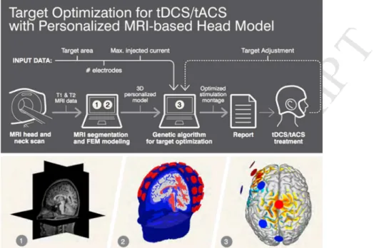
Figure 1. Workflow for the creation of a biophysical model and for model-driven tCS optimization.
Anatomical MRI data (1) is used to create a finite element biophysical model (FEM), and electrodes are placed using the 10-10 EEG system (2) (see Miranda et al. 2018 [37] for a review). A target specification is provided, together with the desired number of electrodes and maximal currents. The Stimweaver algorithm provides the solution (3, i.e., electrode positions and currents). The approach is applicable to tDCS, tACS and other tCS modalities [32].
3. tCS AND BRAIN NETWORKS: FROM BIOPHYSICS TO PHYSIOLOGY
If the critical features of pathological networks can be effectively captured in computational
models, they can be used for diagnosis and delivery of personalized therapeutic weak electric
fields (Figure 1).
Multiple studies in theoretical and computational neuroscience have
developed whole-brain network models [38-41] to explore the relationship between brain
function and its underlying connectivity. This increased interest in finding the origin of the
structure-function relationship has led to a newly developing field known as network
neuroscience (Bassett and Sporns, 2017) [42] that relies on graph theory to study the brain
across its multiple scales and complexities. Following earlier work by Merlet et al. [39],
Sanchez-Todo et al. [43] develop a method that allows for the use of a subject’s EEG and MRI
for the creation of a personalized whole brain model. The model is optimized to reproduce a
subject’s EEG and allows for virtual brain stimulation, and hence optimization. Earlier, Bansal et
al. [44], Spiegler et al. [45], and Muldoon et al. [46] proposed a similar approach. Although
“hybrid” models can produce physiologically-plausible EEG and simulate the generation of
realistic tCS electric fields (see Miranda et al. [37] in this issue, and references therein),
representing faithfully the effects of neuromodulation on brain plasticity remains an unresolved,
important challenge. We now know from experimental work that tCS can directly impact
M
AN
US
CR
IP
T
AC
CE
PT
ED
neuronal excitability and synaptic plasticity [47,48]. Marquez-Ruiz et al. [49], e.g., showed that
blocking adenosine A1 receptors prevents the long-term depression evoked in the
somatosensory cortex after cathodal tDCS in the rabbit. Based on molecular and functional
investigations (immunoblotting, immunofluorescence, and electrophysiological recordings),
Paciello et al. [50] provide novel evidence that anodal tDCS affects structural plasticity of the rat
auditory cortex in a paradigm of noise-induced hearing loss. Wischnewski et al. [51] also
reported that 20 Hz tACS can alter NMDA Receptor-Mediated plasticity in the human motor
cortex. A number of studies indicate that tCS can alter the release of neurotransmitters, typically
dopamine [52], glutamate and GABA [53]. Ultimately, electric field mediated effects translate
into short- or long-term changes in the network connectivity - and therapeutic effects. As the
precise mechanisms involved in tCS-induced plasticity changes still remain elusive, multiscale
computational models offer a unique framework to untangle them, allowing, for instance, to
distinguish effects occurring at presynaptic (membrane polarization of axon terminals,
neurotransmitter release) or postsynaptic (GABA or glutamate receptors) level.
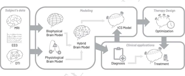
Figure 2. Workflow for the creation of hybrid models model-driven tCS optimization. DTI and
anatomical MRI data are combined to create a finite element biophysical model (FEM), which is then personalized using EEG and other data to reflect both biophysical and physiologic characteristics – from excitation/inhibition balance to plastic potential (long-term effects physiological model). The personalized hybrid brain model can be used to generate EEG and to simulate the effects of brain stimulation. As a result, personalized diagnosis and treatment can be applied, such as optimized stimulation protocols.
4. FUTURE APPLICATIONS
Epilepsy
Epilepsy is a devastating, chronic disease that severely affects the quality of life of 65 million
people worldwide (WHO, Fact Sheet on Epilepsy, 2012), 35% of whom do not respond to drugs.
Almost a third of patients (29%) are untreatable: in 19 million patients, drugs fail, and surgery is
not an option or has failed too. Treatment-resistant epilepsies represent not only a considerable
challenge for the health care system but also a tremendous burden at the individual, family, and
community levels [54]. They are characterized by an epileptogenic network (EN) interconnecting
distant brain areas located in one of the two hemispheres. There is a large body of evidence
M
AN
US
CR
IP
T
AC
CE
PT
ED
suggesting that patient-specific ENs [55] are responsible for the generation and spread of
seizures through synchronization processes that interconnect neuronal assemblies with altered
excitability [56]. Of note, some studies have tried to predict surgical outcome by removing EN
edges of the patient specific connectivity data in computational models of the subject’s brain
[57-61]. In this context, tCS can represent a valuable alternative to surgery [62], provided that
fundamental issues are addressed. First, epileptogenic networks are patient-specific. Therefore,
interventions must be “tailored” to each patient based on the accurate definition of target brain
areas and networks. Second, stimulation protocols must achieve a therapeutic effect through a
“network-aware” management of hyperexcitability - a hallmark of epileptogenic systems. Third,
therapeutic effects must be optimized in order to prevent the occurrence of seizures. A
protective and durable effect will certainly require a better understanding of the mechanisms of
action of weak electric fields on brain networks from short (minutes to hours) to long (days,
weeks) time scales.
Reaching subcortical targets via networks
Fox et al. [63] identified diseases treated with both non-invasive and deep brain stimulation
(DBS), listed the target sites thought to be most effective in each disease and tested the
hypothesis that these sites are nodes within a brain network as defined by rs-fcMRI. They found
that sites in which DBS was effective were functionally connected to sites where noninvasive
brain stimulation had been found to be effective in diseases including depression, PD,
obsessive-compulsive disorder, essential tremor, addiction, pain, minimally conscious state, and
Alzheimer’s disease. This suggests that rs-fcMRI may be useful for translating therapy across
stimulation modalities, optimizing treatment, and for the identification of new stimulation targets.
It also supports a more general network approach toward understanding and treating
neuropsychiatric disease, highlighting the therapeutic potential of targeted brain network
modulation. Examples of potential cortical and subcortical targets relevant for neuropsychiatric
conditions, as well as their rs-fcMRI map and corresponding multichannel optimization, are
shown in Figure 3 (see also Ruffini et al. [32] for further discussion on the use of these maps for
multichannel tCS optimization).
M
AN
US
CR
IP
T
AC
CE
PT
ED
Figure 3. Connectivity-based network targeting. (A) Cortical representation of rs-fMRI connectivity patterns of selected brain regions of clinical relevance in various neuropsychiatric conditions. The target regions are used as seeds, and their pattern of positively and negatively correlated regions in the brain are computed. Multichannel tCS can be optimized to enhance positive (“excitatory”) or negative (“inhibitory”) cortical nodes, inducing changes in their connectivity with the seed region and possibly modulating its spontaneous activity. (B) Examples of multichannel tCS solutions derived from Stimweaver [32] for targeting the supplementary motor area in patients with obsessive-compulsive disorder (OCD) and subgenual cortex in patients with depression.
Stroke
In another example, Otal et al. [64] proposed to identify networks affected by a stroke at the
individual level: location, extent, and pattern of functional network connectivity disruption should
be considered when determining the optimal tDCS intervention. Alstott et al. [65] did a related in
silico study where network edges were removed to investigate the effect of such perturbations
on simulated brain activity. See an extended review of Aerts et al. [66] regarding computational
lesion and empirical studies investigating brain network alterations in cancer, stroke and
traumatic injury patients. In the case of stroke, each lesion type displays a particular functional
and structural connectivity signature that determines the tDCS intervention goals. Lesion
topography is usually subcortical, with intracortical connectivity disruptions contributing strongly
M
AN
US
CR
IP
T
AC
CE
PT
ED
to behavioral deficits [67]. In general, we may consider three main approaches: a) targeting a
single region or node, b) targeting the single region indirectly via a network as described above,
or c) select a network or sub-network (i.e., multi-nodal) as the target. The latter may be
especially relevant given the correlation of connectivity disruption and symptoms. Depending on
the approach chosen, different optimization strategies can be envisioned. Tools based on
tractography can be used to assess damaged networks and devise therapeutic strategies [68].
5. CONCLUSIONS
Multichannel tCS provides a promising tool for targeting networks but is not yet used as a
standard treatment in any disease. This is related to several challenges and methodological
limitations: an overwhelming number of stimulation parameter combinations, empirical
parameter setting, an absence of a rational definition of targets and protocols, the qualitative
nature of results, unknown mechanisms of action, and an insufficient account for patient-specific
factors. A bottom-up, science-based mechanistic understanding of both the effects of tCS and
the desired cortical network changes is lacking. Research should aim to overcome this by
providing a better understanding mechanism of interaction - including both immediate and
longer-term plastic effects of electric fields in networks across scales. We also need to refine
our methods to better identify networks to be targeted and design the strategies for intervention
in each disease and patient. Finally, experiments should be carried out to investigate how
network interactions can best be leveraged by tCS, measuring the functional and structural
alterations induced by tCS using tools such as fMRI or DTI. Modeling in sufficient detail the
combined biophysics and physiology of tCS will be paramount for the design and interpretation
of studies and for their ultimate clinical translation.
Acknowledgments
This research was supported in part by the Future Emerging Technologies Open Luminous
project (H2020-FETOPEN-2014-2015-RIA under agreement No. 686764) as part of the
European Union’s Horizon 2020 research and training program 2014–2018. ES is partially
supported by Office of the Director of National Intelligence (ODNI), Intelligence Advanced
Research Projects Activity (IARPA), via 2014-13121700007. The views and conclusions
contained herein are those of the authors and should not be interpreted as necessarily
representing the official policies or endorsements, either expressed or implied, of the ODNI,
IARPA, or the U.S. Government. ES is supported by the Beth Israel Deaconess Medical Center
(BIDMC) via the Chief Academic Officer (CAO) Award 2017, and the Defense Advanced
Research Projects Agency (DARPA) via HR001117S0030. The content of this paper is solely
the responsibility of the authors and does not necessarily represent the official views of Harvard
University, and its affiliated academic health care centers.
DECLARATION OF INTEREST
Giulio Ruffini is a shareholder and works for Neuroelectrics, a company designing medical
devices for brain stimulation. Roser Sanchez-Todo is a researcher at Neuroelectrics.
REFERENCES
M
AN
US
CR
IP
T
AC
CE
PT
ED
1. Price C., Moore C., Humphreys G., Frackowiak R., Friston K. (1996). The neural regions sustaining object recognition and naming. Proceedings of the Royal Society of London Series B: Biological
Sciences 263: 1501-1507
2. Fox, Michael D., et al. (2012a). Measuring and manipulating brain connectivity with resting state functional connectivity magnetic resonance imaging (fcMRI) and transcranial magnetic stimulation (TMS). Neuroimage 62.4
3. Fox, M.D., Buckner, R.L., White, M.P., Greicius, M.D., Pascual-Leone, A. (2012b). Efficacy of transcranial magnetic stimulation targets for depression is related to intrinsic functional connectivity with the subgenual cingulate. Biol. Psychiatry 72.
4. Fornito A, Zalesky A, Breakspear M (2015). The connectomics of brain disorders, Nat Rev Neurosci. 16(3):159-72. doi: 10.1038/nrn3901.
5. Van Diessen E, Numan T, van Dellen E, van der Kooi A, Boersma M, Hofman D, et al. (2015). Opportunities and methodological challenges in EEG and MEG resting state functional brain network research, Clinical Neurophysiology, vol. 126, pp. 1468-1481.
6. Achard S and Bullmore E (2007). Efficiency and cost of economical brain functional networks. PLoS
Comput Biol 3, e17.
7. Sporns O (2011). The non-random brain: efficiency, economy, and complex dynamics. Front
Comput Neurosci 5, 5. https://doi.org/10.3389/fncom.2011.00005
8. Power JD, Cohen AL, Nelson SM, Wig GS, Barnes KA, Church JA, Vogel AC, Laumann TO, Miezin FM, Schlaggar BL, Petersen SE (2011). Functional network organization of the human brain. Neuron 72, 665–678. https://doi.org/10.1016/j.neuron.2011.09.006
9. Hearne, L.J., Cocchi, L., Zalesky, A., Mattingley, J.B. (2017). Reconfiguration of brain network architectures between resting state and complexity-dependent cognitive reasoning. J. Neurosci. 485–17. https://doi.org/10.1523/JNEUROSCI.0485-17.2017
10. Adelstein, J.S., Shehzad, Z., Mennes, M., Deyoung, C.G., Zuo, X.N., Kelly, C., Margulies, D.S., Bloomfield, A., Gray, J.R., Castellanos, F.X., Milham, M.P. (2011). Personality is reflected in the brain’s intrinsic functional architecture. PLoS One 6, e27633.
11. Corbetta, M. and Shulman, G.L. (2002). Control of goal-directed and stimulus-driven attention in the brain. Nat Rev Neurosci 3, 201–215. https://doi.org/10.1038/nrn755
12. Santarnecchi, E., Emmendorfer, A., Tadayon, S., Rossi, S., Rossi, A., Pascual-Leone, A. (2017a). Network connectivity correlates of variability in fluid intelligence performance. Intelligence 65, 35–47. https://doi.org/10.1016/j.intell.2017.10.002
13. Santarnecchi, E., Sprugnoli, G., Tatti, E., Mencarelli, L., Neri, F., Momi, D., Di Lorenzo, G., Pascual-Leone, A., Rossi, S., Rossi, A., (2018a). Brain functional connectivity correlates of coping styles.
M
AN
US
CR
IP
T
AC
CE
PT
ED
14. Finn, E.S., Shen, X., Scheinost, D., Rosenberg, M.D., Huang, J., Chun, M.M., Papademetris, X., Constable, R.T. (2015). Functional connectome fingerprinting: identifying individuals using patterns of brain connectivity. Nat Neurosci 18, 1664–1671. https://doi.org/10.1038/nn.4135
15. Khanna, A., Pascual-Leone, A., Michel, C.M., Farzan, F. (2015). Microstates in resting-state EEG: current status and future directions. Neurosci. Biobehav. Rev. 49, 105–113. https://doi.org/10.1016/j.neubiorev.2014.12.010
16. Lehmann, D., Strik, W.K., Henggeler, B., Koenig, T., Koukkou, M. (1998). Brain electric microstates and momentary conscious mind states as building blocks of spontaneous thinking: I. Visual imagery and abstract thoughts. Int. J. Psychophysiol. Off. J. Int. Organ. Psychophysiol. 29, 1–11.
17. Andreou, C., Faber, P.L., Leicht, G., Schoettle, D., Polomac, N., Hanganu-Opatz, I.L., Lehmann, D., Mulert, C. (2014). Resting-state connectivity in the prodromal phase of schizophrenia: insights from EEG microstates. Schizophr. Res. 152, 513–520. https://doi.org/10.1016/j.schres.2013.12.008 18. Nishida, K., Morishima, Y., Yoshimura, M., Isotani, T., Irisawa, S., Jann, K., Dierks, T., Strik, W.,
Kinoshita, T., Koenig, T. (2013). EEG microstates associated with salience and frontoparietal networks in frontotemporal dementia, schizophrenia and Alzheimer’s disease. Clin. Neurophysiol. 124, 1106–1114. https://doi.org/10.1016/j.clinph.2013.01.005
19. Santarnecchi, E., Khanna, A.R., Musaeus, C.S., Benwell, C.S.Y., Davila, P., Farzan, F., Matham, S., Pascual-Leone, A., Shafi, M.M., on behalf of Honeywell SHARP Team authors (2017b). EEG Microstate Correlates of Fluid Intelligence and Response to Cognitive Training. Brain Topogr. https://doi.org/10.1007/s10548-017-0565-z
20. Santarnecchi, E., Momi, D., Sprugnoli, G., Neri, F., Pascual-Leone, A., Rossi, A., Rossi, S. (2018b). Modulation of network-to-network connectivity via spike-timing-dependent noninvasive brain stimulation. Hum. Brain Mapp. https://doi.org/10.1002/hbm.24329
21. Veniero, D., Ponzo, V., Koch, G. (2013). Paired associative stimulation enforces the communication between interconnected areas. J Neurosci 33, 13773–13783. https://doi.org/10.1523/JNEUROSCI.1777-13.2013
22. M. A. Nitsche and W. Paulus (2000). Excitability changes induced in the human motor cortex by weak transcranial direct current stimulation. Journal of Physiology , 527.3, pp.633—639
23. Ruffini G., Wendling F., Merlet I., Molaee-Ardekani B., Mekonnen A., Salvador R., Soria-Frisch A., Grau C., Dunne S., Miranda P.C. (2013). Transcranial current brain stimulation (tCS): models and technologies, IEEE Trans Neural Syst Rehabil Eng. ;21(3):333-45.
24. Henrich-noack P., Sergeeva E. G. and Sabel B. A. (2017). Non-invasive electrical brain stimulation: from acute to late-stage treatment of central nervous system damage, 12(10). Neural Regeneration
Research. https://doi.org/10.4103/1673-5374.217322Nitsche
25. Karabanov, A., Ziemann, U., Hamada, M., George, M. S., Quartarone, A., Classen, J., et al. (2015). Brain Stimulation Consensus Paper: Probing Homeostatic Plasticity of Human Cortex With
Non-M
AN
US
CR
IP
T
AC
CE
PT
ED
invasive Transcranial Brain Stimulation. Brain Stimulation, 8(3), 442–454. https://doi.org/10.1016/j.brs.2015.01.404
26. Lefaucheur J.P., Antal A., Ayache S.S., Benninger D.H., Brunelin J., Cogiamanian F., Cotelli M., De Ridder D., Ferrucci R., Langguth B., Marangolo P., Mylius V., Nitsche M.A., Padberg F., Palm U., Poulet E., Priori A., Rossi S., Schecklmann M., Vanneste S., Ziemann U., Garcia-Larrea L., Paulus (2017). Evidence-based guidelines on the therapeutic use of transcranial direct current stimulation (tDCS), Clin Neurophysiol. ;128(1):56-92. doi: 10.1016/j.clinph.2016.10.087. Epub 2016 Oct 29. (**) This paper provides an up to date review of the clinical uses of tDCS, highlighting those applications
that show probable efficacy.
27. Antal A., Alekseichuk I., Bikson M., Brockmöller J., Brunoni A.R., Chen R., Cohen L.G., Dowthwaite G., Ellrich J., Flöel A., Fregni F., George M.S., Hamilton R., Haueisen J., Herrmann C.S., Hummel F.C., Lefaucheur J.P., Liebetanz D., Loo C.K., McCaig C.D., Miniussi C., Miranda P.C., Moliadze V., Nitsche M.A., Nowak R., Padberg F., Pascual-Leone A., Poppendieck W., Priori A., Rossi S., Rossini P.M., Rothwell J., Rueger M.A., Ruffini G., Schellhorn K., Siebner H.R., Ugawa Y., Wexler A., Ziemann U., Hallett M., Paulus W. (2017). Low intensity transcranial electric stimulation: Safety, ethical, legal regulatory and application guidelines, Clin Neurophysiol.;128(9):1774-1809. doi: 10.1016/j.clinph.2017.06.001
28. Dmochowski J. P., Datta A., Bikson M., Su Y., Parra L. C. (2011). Optimized multi-electrode stimulation increases focality and intensity at target. J. Neural Eng. 8, 046011.10.1088/1741-2560/8/4/046011
29. Wagner, S., Burger, M., and Wolters, C. H. (2016). An Optimization Approach for Well-Targeted Transcranial Direct Current Stimulation. SIAM Journal on Applied Mathematics, 76(6), 2154–2174. doi:10.1137/15m1026481
30. Miranda P. C., Mekonnen A., Salvador R., Ruffini G (2013). The electric field in the cortex during transcranial current stimulation. NeuroImage 70, 48–58.
31. Fox, Michael D., Buckner, Randy L., Liu, Hesheng, Chakravarty, M. Mallar, Lozano, Andres M., Pascual-Leone, Alvaro (2014b). Resting-State Networks Link Invasive and Noninvasive Brain Stimulation across Diverse Psychiatric and Neurological Diseases. Proc. Natl. Acad. Sci. USA 111 (41), E4367–E4375.
32. Ruffini G., Fox M. D., Ripolles O., Miranda P. C., and Pascual-Leone A. (2014). Optimization of multifocal transcranial current stimulation for weighted cortical pattern targeting from realistic modeling of electric fields, Neuroimage 89: 216-225.
33. Fisher, D. B., et al. (2017). Network-targeted non-invasive brain stimulation with multifocal tdcs. Brain
Stimulation: Basic, Translational, and Clinical Research in Neuromodulation 10.2
(**) This paper evaluates for the first time the potential of seed-based (rs-fcMRI) targeting and network stimulation using multichannel tCS. The authors find an enhanced effect of multichannel network vs. bipolar tDCS on motor cortex cortical excitability as measured by TMS/MEPs.
M
AN
US
CR
IP
T
AC
CE
PT
ED
34. Dagan M., Herman T., Harrison R., et al. (2018). Multitarget Transcranial Direct Current Stimulation for Freezing of Gait in Parkinson’s Disease. Movement disorders: official journal of the Movement
Disorder Society;33(4):642-646. doi:10.1002/mds.27300.
(**) This paper proposes a dual target approach to target networks relevant to freezing of gait and related outcomes. By targeting nodes involved in motor and cognitive tasks, the authors show it is possible to improve potential cognitively coupled motor tasks in freezing of gait. The authors also demonstrate the potential of an “active sham” concept using multichannel stimulation for better blinding.
35. Thibaut A., Martens G., and Laureys S. (2017). Multichannel tDCS of the frontoparietal network in patients with disorders of consciousness: A double blind sham controlled randomized clinical trial.
Brain Stimulation: Basic, Translational, and Clinical Research in Neuromodulation 10, no. 4
36. Brem, A.-K., Almquist, J.N.-F., Mansfield, K., Plessow, F., Sella, F., Santarnecchi, E., Orhan, U., McKanna, J., Pavel, M., Mathan, S., Yeung, N., Pascual-Leone, A., Kadosh, R.C., Honeywell SHARP Team authors, (2018). Modulating fluid intelligence performance through combined cognitive training and brain stimulation. Neuropsychologia.
https://doi.org/10.1016/j.neuropsychologia.2018.04.008
37. Miranda, P. C., M. A. Callejón-Leblic, R. Salvador, G. Ruffini, 2018. Realistic Modeling of Transcranial Current Stimulation: The Electric Field in the Brain. Current Opinion in Biomedical Engineering doi: https://doi.org/10.1016/j.cobme.2018.09.002
(*) A review of the state of the art in the modeling of electric fields in the brain, techniques, and validation. 38. Deco, G., Jirsa, V. K., & McIntosh, A. R. (2011). Emerging concepts for the dynamical organization of
resting-state activity in the brain. Nature Reviews Neuroscience, 12(1), 43–56.
39. Merlet, I., G. Birot, R. Salvador, B. Molaee-Ardekani, A. Mekonnen, A. Soria-Frish, G. Ruffini, P. C. Miranda & F. Wendling (2013). From oscillatory transcranial current stimulation to scalp EEG changes: a biophysical and physiological modeling study. PloS one 8(2):e57330 doi:10.1371/journal.pone.0057330.
40. Cabral, J., Luckhoo, H., Woolrich, M., Joensson, M., Mohseni, H., Baker, A., ... Deco, G. (2014). Exploring mechanisms of spontaneous functional connectivity in MEG: How delayed network interactions lead to structured amplitude envelopes of bandpass filtered oscillations. NeuroImage,
90, 423–435.
41. Bansal, K., Johan Nakuci, Sarah Feldt Muldoon (2018a). Personalized brain network models for assessing structure–function relationships, Current Opinion in Neurobiology, Volume 52, Pages 42-47
(*) This paper proposes the combination of structural data with neural mass models to study structure-function relationships in the human brain, with applications for therapy (surgery or brain stimulation). 42. Bassett, D. S., and Sporns, O. (2017). Network neuroscience. Nature Neuroscience, 20(3), 353–364.
M
AN
US
CR
IP
T
AC
CE
PT
ED
(*) The authors review emerging trends in network neuroscience and provide a path toward a better understanding of the brain as a multiscale networked system, and on how to leverage it.
43. Sanchez-Todo, R., Salvador, R., Santarnecchi, E., Wendling, F., Deco, G., Ruffini, G., (2018) Personalization of hybrid brain models from neuroimaging and electrophysiology data, BioRxiv
http://dx.doi.org/10.1101/461350.
(*) This methods paper shows, using real MRI and EEG data, how to integrate structural, anatomical and physiological data to create realistic brain models of the human brain, with applications in brain stimulation and basic neuroscience. Such hybrid models of biophysics and physiology can be used to model optimize tDCS on brain networks, for example.
44. Bansal K Medaglia J Bassett D Vettel J Muldoon S, (2018b). Data-driven brain network models differentiate variability across language tasks, PLOS Computational Biology, 018 vol: 14 (10) pp: e1006487
45. Spiegler A, Hansen ECA, Bernard C, McIntosh AR, Jirsa VK (2016). Selective Activation of Resting State Networks following Focal Stimulation in a Connectome-Based Network Model of the Human Brain. eNeuro, 3:1–17
46. Muldoon SF, Pasqualetti F, Gu S, Cieslak M, Grafton ST, Vettel JM, Bassett DS (2016) Stimulation Based Control of Dynamic Brain Networks. PLoS Comput Biol, 12:e1005076
(**) The authors performed in silico experiments in data-driven brain network models to relate patterns of activation due to targeted stimulation. Furthermore, they use their model to explore the relationship between cognitive systems and the underlying brain anatomy.
47. Modolo J, Denoyer Y, Fabrice W, Benquet P (2018). Physiological effects of low-magnitude electric
fields on brain activity: advances from in vitro, in vivo and in silico models, Current Opinion in Biomedical Engineering
(*) An up to date review on models for the interaction of weak electric fields with neurons and their larger scale effects.
48. Sánchez-León CA, Sánchez-López A, Ammann C, Cordones I, Carretero-Guillén A and Márquez-Ruiz J (2018). Exploring new transcranial electrical stimulation strategies in animal models for brain function modulation, Current Opinion in Biomedical Engineering
(*) Up to date review and outlook of research of tCS in animal models, with a discussion on emerging trends to improve focality and reach deeper targets.
49. Márquez-Ruiz J, Leal-Campanario R, Sánchez-Campusano R, Molaee-Ardekani B, Wendling F, Miranda P. C., Ruffini G., Gruart A, and Delgado-García J. M. (2012). Transcranial direct-current stimulation modulates synaptic mechanisms involved in associative learning in behaving rabbits,
PNAS | vol. 109 | no. 17
50. Paciello F, Podda MV, Rolesi R, Cocco S, Petrosini L, Troiani D, Fetoni AR, Paludetti G, Grassi C (2018). Anodal transcranial direct current stimulation affects auditory cortex plasticity in
normal-M
AN
US
CR
IP
T
AC
CE
PT
ED
hearing and noise-exposed rats. Brain Stimul.11(5):1008-1023. doi: 10.1016/j.brs.2018.05.017. Epub 2018 May 31.
51.
Wischnewski M, Engelhardt M, Salehinejad MA, Schutter DJLG, Kuo MF, Nitsche MA (2018). NMDA Receptor-Mediated Motor Cortex Plasticity After 20 Hz Transcranial Alternating Current Stimulation,Cereb Cortex. doi: 10.1093/cercor/bhy160.
52. Fonteneau C, Redoute J, Haesebaert F, Le Bars D, Costes N, Suaud-Chagny MF, Brunelin J. (2018). Frontal Transcranial Direct Current Stimulation Induces Dopamine Release in the Ventral Striatum in Human. Cereb Cortex. 28(7):2636-2646
53. Santana-Gómez CE, Alcántara-González D, Luna-Munguía H, Bañuelos-Cabrera I, Magdaleno-Madrigal V, Fernández-Mas R, Besio W, Rocha L (2015). Transcranial focal electrical stimulation reduces the convulsive expression and amino acid release in the hippocampus during pilocarpine-induced status epilepticus in rats, Epilepsy Behav. 49:33-9. doi: 10.1016/j.yebeh.2015.04.037. Epub 2015 May 23.
54. Pugliatti M, Beghi E, Forsgren L, Ekman M, Sobocki P. (2007). Estimating the cost of epilepsy in Europe: a review with economic modeling, Estimating the cost of epilepsy in Europe: a review with economic modeling, Epilepsia 48 (12):2224-33.
55. Bartolomei, F., S. Lagarde, F. Wendling, A. McGonigal, V. Jirsa, M. Guye and C. Benar, (2017). Defining epileptogenic networks: Contribution of SEEG and signal analysis. Epilepsia 58(7):1131-1147 doi:10.1111/epi.13791.
(**) This paper provides a historical overview of the emerging epileptogenic network concept, emphasizing the idea that “focal” epilepsies actually involve networks at varying scales, how to identify them, and the practical relevance of these findings to therapy.
56. Wendling, F., F. Bartolomei, F. Mina, C. Huneau & P. Benquet, (2012). Interictal spikes, fast ripples and seizures in partial epilepsies--combining multi-level computational models with experimental data. The European journal of neuroscience 36(2):2164-77 doi:10.1111/j.1460-9568.2012.08039.x. 57. Hutchings F, Han CE, Keller SS, Weber B, Taylor PN, Kaiser M (2015). Predicting Surgery Targets in
Temporal Lobe Epilepsy through Structural Connectome Based Simulations. PLoS Comput Biol, 11:e1004642–24
58. Khambhati AN, Davis KA, Lucas TH, Litt B, Bassett DS (2016). Virtual Cortical Resection Reveals Push-Pull Network Control Preceding Seizure Evolution. Neuron, 91:1170–1182
59. Goodfellow M, Rummel C, Abela E, Richardson MP, Schindler K, Terry JR (2016). Estimation of brain network ictogenicity predicts outcome from epilepsy surgery. Sci. Rep. 6:1–13.
60. Proix T, Bartolomei F, Guye M, Jirsa VK (2017). Individual brain structure and modelling predict seizure propagation. Brain, 140:641–654.
61. Sinha N, Dauwels J, Kaiser M, Cash SS, Brandon Westover M, Wang Y, Taylor PN (2017) Predicting neurosurgical outcomes in focal epilepsy patients using computational modelling. Brain, 140:319– 332.
M
AN
US
CR
IP
T
AC
CE
PT
ED
(*) In this work, authors predict surgial outcomes in epilepsy patients with high accuracy using personalized brain network models. They substitute the functional connectivity of the brain, derived from ECoG recordings of epileptic patients, for structural connectivity as a basis for the computational models.
62. Regner, G. G., Pereira, P., Leffa, D. T., de Oliveira, C., Vercelino, R., Fregni, F., & Torres, I. (2018). Preclinical to Clinical Translation of Studies of Transcranial Direct-Current Stimulation in the Treatment of Epilepsy: A Systematic Review. Frontiers in neuroscience, 12, 189. doi:10.3389/fnins.2018.00189 (*) A recent review of studies assessing the efficacy of tDCS in epilepsy.
63. Fox, MD, Randy L. Buckner, Hesheng Liu, M. Mallar Chakravarty, Andres M. Lozano, Alvaro Pascual-Leone (2014b). Linking invasive and noninvasive brain stimulation. Proceedings of the National
Academy of Sciences 111 (41) E4367-E4375; DOI: 10.1073/pnas.1405003111
64. Otal B, Dutta A, Foerster Á, Ripolles O, Kuceyeski A, Miranda PC, Edwards DJ, Ilić TV, Nitsche MA
and Ruffini G (2016). Opportunities for Guided Multichannel Non-invasive Transcranial Current Stimulation in Poststroke Rehabilitation. Front. Neurol. 7:21. doi: 10.3389/fneur.2016.00021
(*) This paper discusses potential opportunities for neuroimaging-guided tDCS-based rehabilitation strategies after stroke that could be personalized using brain networks.
65. Alstott J, Breakspear M, Hagmann P, Cammoun L, Sporns O (2009). Modeling the Impact of Lesions in the Human Brain. PLoS Comput Biol, 5:e1000408–12
66. Aerts H, Fias W, Caeyenberghs K, Marinazzo D (2016). Brain networks under attack: robustness properties and the impact of lesions. Brain, 139:3063–3083
67. Corbetta, M., Lenny Ramsey, Alicia Callejas, Antonello Baldassarre, Carl D. Hacker, Joshua S. Siegel, Serguei V. Astafiev, Jennifer Rengachary, Kristina Zinn, Catherine E. Lang, Lisa Tabor Connor, Robert Fucetola, Michael Strube, Alex R. Carter, and Gordon L. Shulman (2015). Common behavioral clusters and subcortical anatomy in stroke, Neuron. March 4; 85(5): 927–941. doi:10.1016/j.neuron.2015.02.027.
68. Kuceyeski, A., Navi, B. B., Kamel, H., Raj, A., Relkin, N., Toglia, J., Iadecola, C. and O'Dell, M. (2016). Structural connectome disruption at baseline predicts 6 months post stroke outcome.
Hum. Brain Mapp., 37: 2587-2601. doi:10.1002/hbm.23198
(**) Structural connectome disruption is estimated using the NeMo tool developed by the authors (which allows estimation of connectome disruption from MRI). The measures at baseline predict 6‐months post‐stroke outcome in various functional domains including cognition, motor function, and daily activities.