Cerebral Cortex November 2010;20:2531--2539 doi:10.1093/cercor/bhq001
Advance Access publication January 29, 2010
Time Scales of Auditory Habituation in the Amygdala and Cerebral Cortex
Isabella Mutschler
1,2, Birgit Wieckhorst
1,2, Oliver Speck
3, Andreas Schulze-Bonhage
4,5, Ju¨rgen Hennig
6, Erich Seifritz
7and
Tonio Ball
4,51
Department of Psychology, NCCR sesam, University of Basel, 4009 Basel, Switzerland,
2Department of Psychiatry, University of
Basel, 4025 Basel, Switzerland,
3Department of Biomedical Magnetic Resonance, Institute for Experimental Physics, Faculty of
Natural Sciences, University of Magdeburg, 39120 Magdeburg, Germany,
4Epilepsy Center, University Hospital Freiburg, 79106
Freiburg, Germany,
5Bernstein Center for Computational Neuroscience, University of Freiburg, 79104 Freiburg, Germany,
6MR-Physics, University Hospital Freiburg, 79106 Freiburg, Germany and
7Clinic for Affective Disorders and General Psychiatry,
Psychiatric Hospital Zurich, 8032 Zu¨rich, Switzerland
Address correspondence to Dr Isabella Mutschler, Department of Psychology, NCCR sesam, University of Basel, Birmannsgasse 8, 4009 Basel, Switzerland. Email: [email protected].
Habituation is a fundamental form of learning manifested by
a decrement of neuronal responses to repeated sensory stimulation.
In addition, habituation is also known to occur on the behavioral
level, manifested by reduced emotional reactions to repeatedly
presented affective stimuli. It is, however, not clear which brain
areas show a decline in activity during repeated sensory
stimulation on the same time scale as reduced valence and arousal
experience and whether these areas can be delineated from other
brain areas with habituation effects on faster or slower time
scales. These questions were addressed using functional magnetic
resonance imaging acquired during repeated stimulation with
piano melodies. The magnitude of functional responses in the
laterobasal amygdala and in related cortical areas and that of
valence and arousal ratings, given after each music presentation,
declined in parallel over the experiment. In contrast to this
long-term habituation (43 min), short-long-term decreases occurring within
seconds were found in the primary auditory cortex. Sustained
responses that remained throughout the whole investigated time
period were detected in the ventrolateral prefrontal cortex
extending to the dorsal part of the anterior insular cortex. These
findings identify an amygdalocortical network that forms the
potential basis of affective habituation in humans.
Keywords: amygdala, auditory perception, fMRI, habituation,
music-induced emotion, subregions
Introduction
Habituation is a fundamental form of learning manifested
by a stimulus-specific decrement of neuronal responses, that is,
to repeated sensory stimulation with the same or a similar
stimulus (Thompson and Spencer 1966; Dudai 2002).
Habitu-ation may serve the functional purpose of protecting the
organism from flooding with irrelevant sensory information by
allocating resources to new salient stimuli in the environment
(Siddle 1991). Repetition-related reductions in neural activity
have previously been reported at multiple time scales ranging
from milliseconds (Weiland et al. 2008), seconds (Fischer et al.
2003), minutes (Whalen et al. 1998) to weeks (Johnstone et al.
2005). A great interest in habituation in the human brain in
general and in the human amygdala—a key structure of
affective processing—in particular lies in the potential
impor-tance of disturbed habituation processes for the
pathophysiol-ogy of psychiatric disorders such as posttraumatic stress
disorder (Protopopescu et al. 2005; Shin et al. 2005).
Habituation especially to affective stimuli has also been
extensively researched on the behavioral level in the
frame-work of valence and arousal as 2 fundamental dimensions of
emotional experience (Russell 1980): repeated exposure to
pleasurable stimuli over a period of approximately 5 min
caused them to be judged as less emotionally valenced
(Leventhal et al. 2007) and subjective valence ratings
habitu-ated for pleasant but not for neutral sounds during repehabitu-ated
exposure close to 30 min (Martin-Soelch et al. 2006). Also
addressing habituation of arousal, unpleasant pictures were
found to be rated as less unpleasant, and both pleasant and
unpleasant pictures as less arousing after repetitions within
approximately half an hour (Codispoti et al. 2006). In summary,
these studies indicate a robust effect of affective habituation
that can be parameterized by decreased valence and arousal
ratings to emotional stimuli with repetitions.
Both animal studies (Sawa and Delgado 1963; Herry et al.
2007) and functional neuroimaging studies in humans suggest
that the amygdala plays an important role in habituation
processes (Breiter et al. 1996; Whalen et al. 1998; Phillips et al.
2001; Wright et al. 2001; Herry et al. 2007) and in the coding of
valence and arousal information (Anderson et al. 2003; Small
et al. 2003; Winston et al. 2005). The amygdala is not
a homogenous structure but composed of over 10 subnuclei
(Amunts et al. 2005). Recently, using functional magnetic
resonance imaging (fMRI) combined with a probabilistic atlas
system (Amunts et al. 2005), it was shown that during auditory
perception positive fMRI responses predominate in the
proba-bilistic defined laterobasal amygdala subregion (Ball et al. 2007).
In contrast to previous studies, the objectives of the present
study were to study the temporal dynamics of habituation in
parallel both on the behavioral and neuronal level, both in the
amygdala and its probabilistically defined subnuclei, and in
related cortical areas. To this aim, brain responses during
processing of repeatedly presented, similar auditory stimuli
were measured using blood oxygen level--dependent (BOLD)
fMRI. Subjects gave ratings of their valence and arousal
experience after each individual stimulus. This approach
allowed us to delineate the network of brain areas showing
a decline in activity that occurs on similar time scales as
reduced valence and arousal experience. Furthermore, the
present study aimed to differentiate this network from other
brain regions showing habituation responses on different time
scales (e.g., for the auditory cortex, see Seifritz et al. 2002).
To achieve these objectives, we analyzed brain responses
to auditory stimuli in order to identify brain areas showing
Ó The Author 2010. Published by Oxford University Press. All rights reserved.long-term decreases of the BOLD signal (habituation), increase
of the BOLD signal (sensitization), and sustained responses,
that is, responses showing neither habituation nor sensitization,
all within the whole investigated period of time (43 min).
Furthermore, short-term decreases within the duration of the
individual stimuli (i.e., within 24 s) were analyzed.
As auditory stimuli, piano melodies in 4 variations of harmony
and tempo (consonant-slow, consonant-fast, dissonant-slow,
and dissonant-fast) were presented to healthy subjects without
professional musical education during acquisition of BOLD
fMRI. The variations of harmony and tempo were applied to
induce different degrees of perceived valence and arousal.
Based on the existent literature as summarized above, we
hypothesized that, in particular, the amygdala might show
reduced fMRI responses in parallel to habituation of valence
and arousal ratings.
Materials and Methods
SubjectsTwenty right-handed healthy volunteers participated in the study. One participant was excluded from data analysis due to excessive head movements during scanning, resulting in a final sample of 19 subjects (11 females, 8 males, mean age=22.74 years, range=19--34 years). All subjects received regular musical education at school, which com-prised singing and the acquisition of elementary theoretical knowledge about music but no professional musical training. Handedness and lifetime music education were assessed before the fMRI experiment with questionnaires (Oldfield 1971; Litle and Zuckerman 1985); the findings are summarized in the section ‘‘Ratings and Questionnaire Data.’’ All participants had normal vision, no history of psychiatric or neurological diseases, and no hearing impairments. Only subjects free of any medication were included. The study was approved by the ethics committee of the University of Freiburg, Germany. Before participation, subjects signed written informed consent. The participants received a modest monetary compensation for participation.
Stimuli
For auditory stimulation, 40 piano pieces, each of 24-s duration, were presented during fMRI data acquisition. To create the 40 piano pieces, 10 melodies were selected. All melodies were taken from the major--minor tonal music, and the mean tempo was for all melodies 120 beats per minute, determined using the MIDI toolbox by Toiviainen and Eerola (the MIDI toolbox can be freely accessed through: https://www.jyu.fi/ hum/laitokset/musiikki/en/research/coe/materials/miditoolbox/).
The tempo and the harmony of the 10 selected piano melodies (see Table 1 in the Supplementary Material) were varied, creating 4 versions of each tune: consonant-fast, consonant-slow, dissonant-fast, and dissonant-slow. Tempi for the fast and slow versions were 156 and 84 beats per minute, respectively. These values were selected because they represented fastest and slowest tempi, respectively, that still sounded natural to one professional musician and 5 nonmusicians. The consonant stimuli were the original tunes, whereas the dissonant stimuli were electronically manipulated counterparts of the original tunes created by shifting the melody of the original excerpt but not the accompanying chords, by half tone below the original pitch. Thus, the dissonant and consonant and the fast and slow versions of a tune had the same rhythmic structure. All stimuli were processed using Cubase VST/32 R.5 (Steinberg Media Technologies GmbH, Hamburg, Germany). The variations of harmony and tempo were applied to induce different degrees of perceived valence and arousal. Finally, all sound files were transformed into wave files for stimulation in the scanner using WaveLab 4.0 (Steinberg, Media Technologies GmbH Hamburg). Importantly, the average sound pressure level of all music pieces was equalized using Steinberg WaveLab 4.0.
Moreover, to investigate the network of brain areas underlying habituation during the perception of auditory stimuli, we had the aim to select unknown piano melodies to avoid confounding differences in
the level of familiarity. We assessed familiarity by subjects’ ratings after scanning (ranging from 0=not familiar to 3=familiar). The mean value for familiarity ratings of the pleasant stimuli was 0.79 (
±
0.86), and the corresponding value for the unpleasant melodies was 0.74 (±
0.81, see also ‘‘Results section’’), showing that the selected musical pieces were relatively unfamiliar to the subjects.Experimental Setup and Procedure
Subjects were asked to complete the state anxiety inventory (STAI-S) in the scanner by using an magnetic resonance (MR) imaging compatible mouse (Laux et al. 1981). This questionnaire was applied to evaluate the subjects’ anxiety level. The mouse allowed subjects to move a white box to the left or right along a visually presented scale by pressing the corresponding mouse buttons with the right hand. During this procedure, the same echo planar imaging (EPI) sequences were run as also later during the fMRI experiment to generate the same level of scanner noise, as scanner noise is one factor likely to contribute to subjects’ anxiety in the scanner.
Afterward, the actual fMRI experiment was conducted in a ‘‘block design.’’ The 40 piano melodies were presented in a random order, using in-house developed presentation software, via MR-compatible headphones (NordicNeuroLab, Bergen, Norway). Subjects viewed a fixation cross during the experiment and were instructed to listen attentively to the music and to avoid any overt movement. Each melody was preceded by a written instruction presented on the screen (‘‘music starts’’), and each melody lasted 24 s. An evaluation period followed each melody presentation. Within this period, participants were asked to rate the preceding melody on a 7-point self-assessment bipolar scale along the dimensions valence (ranging from–3=very unpleasant to 3=
very pleasant) and arousal (ranging from–3=very calming to 3=very arousing). Each of the 2 rating periods lasted 6 s. Subject communi-cated their decisions by using a scanner-compatible mouse that allowed them to move a white box on the visually presented scale leftwards or rightwards by pressing the corresponding mouse button with their right hand. The evaluation was followed by a resting period of 11-s duration. The experiment had 40 runs (each consisting of melody presentation, evaluation, and rest), and the total scanning time for the experiment was 43 min. The experimental procedure is summarized in Figure 1 in the Supplementary Material.
Data Acquisition
Functional and structural images were acquired on a 3-T scanner (Siemens Magnetom Trio, Erlangen, Germany). Image acquisition started with localizing the brain, a reference scan for the distortion correction, and the anatomical scans of the brain that were obtained using a magnetization-prepared rapid-acquired gradient echoes (MPRAGE) sequence of 7-min duration. Subsequently, the fMRI experiment was conducted. Structural T1-weighted images were obtained using a MPRAGE sequence (resolution: 1-mm isotropic, matrix: 256 3 256 3 160, time repetition [TR]: 2200 ms, time to inversion: 1000 ms, 12° flip angle). Functional images were obtained using a multislice gradient EPI method. Each volume consisted of 44 sagittal slices (resolution: 3-mm slice thickness, matrix: 64364, field of view: 1923192 mm, volume TR: 3000 ms, time echo: 30 ms, 90° flip angle). The sagittal slice orientation resulted in significantly lower acoustic noise generated by the imaging gradients, enabling a better auditory perception. In addition, this orientation in combination with the slice thickness of 3 mm reduced the signal loss in the amygdala region to give more reliable detection of responses. An accurate registration of the functional and structural images was enabled by correction of the functional image data for geometric distortions (Zaitsev et al. 2004). The distortion field was derived from the local point spread function in each voxel as determined in a 1-min reference scan. Prior to distortion correction, the data were motion corrected by image realignment with the reference scan. A representative example of EPI data after distortion correction is shown in Ball et al. (2007) demonstrating good EPI signal quality in the amygdala region. Preprocessing and Statistical fMRI Analysis
Motion and distortion correction were performed online during the reconstruction process (see above). Preprocessing consisted of
normalization and smoothing. All functional images were normalized into standard stereotaxic space of the Montreal Neurological Institute (MNI) template. Subsequently, the images were smoothed using a 6-mm full width at half maximum Gaussian kernel to minimize the effects of individual variations in anatomy and to improve the signal-to-noise ratio. The timing information of the piano melodies and the evaluation periods was each modeled with a boxcar function convolved with a canonical hemodynamic response function. A high-pass filter with a cutoff of 1/128 Hz was applied before parameter estimation. We performed preprocessing and data analysis using SPM5 (Wellcome Department of Cognitive Neurology, London, UK). On the single-subject level, contrast images of music perception >baseline (i.e., time
periods during which subjects passively viewed the fixation cross without stimulus presentation, for further details regarding the experiment, see also Fig. 1 in the Supplementary Material) were calculated for all subjects. Fast habituation was analyzed on the single-subject level by comparing the first half with the second half of the music presentation (i.e., the first 12 s with the second 12 s). Long-term habituation and sensitization were analyzed using a regressor modeling linearly changes in music-related response amplitudes over the whole time of the experiment (43 min). Moreover, differential habituation patterns in response to the 4 music conditions (consonant-slow, consonant-fast, dissonant-slow, and dissonant-fast) were analyzed on the single-subject level by comparing fast with slow piano melodies (collapsing consonant and dissonant melodies) and consonant with dissonant melodies (collapsing fast and slow melodies).
Subsequently, group-level statistics were computed to reveal signif-icant responses over the total group of the 19 subjects in the contrast music perception>baseline. The results are reported for the t-test at P<
0.05, false discovery rate (FDR) corrected, cluster size >10 voxels. For anatomical assignments, we used probabilistic anatomical maps (Toga et al. 2006). The results of the fast and long-term habituation were analyzed using a region of interest (ROI) model defined by the amygdala taken from the probabilistic anatomical map to the human amygdala (Amunts et al. 2005) and the activated regions in the contrast music perception > baseline. The results of the habituation analyses are reported at P<0.005 level (t-test, uncorrected, cluster size>50 voxels).
To test for differences in the speed of long-term habituation, in particular, in the right versus left amygdala, correlation coefficients of BOLD response amplitude with time were determined at both the right and left amygdala long-term habituation peaks (for further details, see Results Section) for each subject, and correlation coefficients from the right versus left amygdala were then compared using a sign test. In addition, sensitization was analyzed by assessing the increase in the BOLD signal over the whole time of the experiment at P <0.05, FDR-corrected, cluster size >10 voxels for whole-brain analysis and for the ROI (amygdala+regions activated in the music>baseline contrast) at P<0.005 (t-test, uncorrected, cluster size>50 voxels).
Furthermore, to assess brain areas showing sustained responses, that is, showing neither long-term (over the experiment’s 43 min-duration) nor short-term habituation effects, we masked the results of the contrasts music perception > baseline with regions showing no
significant effect neither in the short-term nor in the long-term habituation contrasts at P>0.3. Results are thus reported at the P>0.3
level (t-test, uncorrected, cluster size >50 voxels). Finally, to analyze the impact of subjects’ anxiety level and music education on BOLD effects, the scores of the questionnaires STAI-S and music education were correlated with the contrast music versus baseline, habituation (short- and long-term), sensitization, and sustained responses.
Analysis of Ratings and Questionnaire Data
Normal distribution of the questionnaire data (STAI-S and maximum lifetime music education) and subjects’ ratings (the valence and arousal ratings which were given by the subjects in the scanner after each stimulus presentation and the global familiarity ratings after scanning) were tested with a Kolmogorov--Smirnov test and analyzed using SPSS (Version 11.0, Mac OS X Version) and Matlab (Version 7.0.4, MathWorks, Natick, MA). Ratings of valence and arousal were analyzed for habituation effects by calculating the correlation coefficients between the time course of the experiment and the magnitude of both valence and arousal ratings. For this analysis, the scores of the 2 bipolar dimensions valence
(ranging from–3 = very unpleasant to 3= very pleasant) and arousal (ranging from–3=very calming to 3=very arousing) were transformed into absolute scores (ranging from 0 to 3, with 0 corresponding to neutral), and subsequently, correlations of these values with time were tested using Spearman’s rank correlation coefficient.
Results
Ratings and Questionnaire Data
The ratings of valence, arousal, and the global familiarity rating
as well as the questionnaire data (STAI-S and music education)
were Gaussian distributed (Kolmogorov--Smirnov test, P
<0.05). Total scores for the STAI-S of the 19 subjects of the fMRI
study ranged from 29 to 61, with a mean of 38.05 and standard
deviation of
±
8.5. Maximum lifetime music education had
a mean of 7.97
±
4.65 years. All subjects were right-handed
according to the Edinburgh handedness questionnaire
(Old-field 1971): mean
=84.95%, range
=75--100%. After scanning,
subjects were asked to rate globally the familiarity (ranging
from 0
=not familiar to 3
=familiar) of the pleasant and for the
unpleasant melodies. Familiarity ratings for the pleasant and
unpleasant melodies were identical in all cases. The mean
familiarity ratings were 0.74 (
±
0.81), indicating that musical
pieces were relatively unfamiliar to the subjects. The
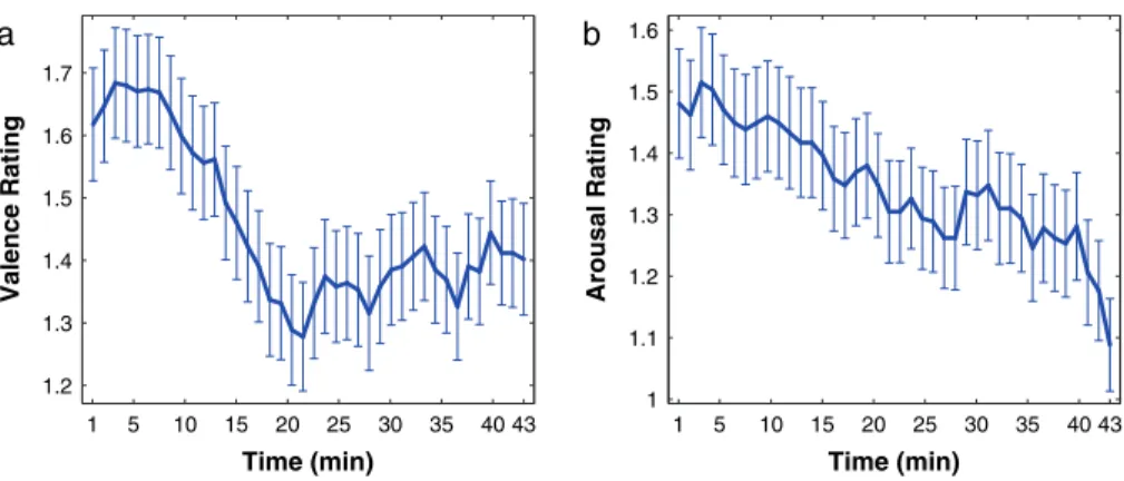
magni-tude both of valence and arousal ratings showed a significant
negative correlation with time over the course of the
ex-periment (P
=0.0086 for valence and P
=0.0053 for arousal
ratings). The time course of the magnitude of valence and
arousal ratings is illustrated in Figure 1, showing a clear decline
of magnitude in both cases. Figure 1 shows that valence ratings
declined within the first 20 min, whereas arousal ratings
decreased over the whole time of the experiment (43 min).
Functional Imaging Data
Figure 2 shows the brain areas activated in the 19 right-handed
subjects during the perception of piano melodies. Significant
responses of voxels at P
<0.05 (FDR-corrected, cluster size
>10
voxels) for the contrast music perception
>baseline were
located in the right and left primary and secondary auditory
cortex. Responses were also found in the insular cortex (left)
and in the region of the temporal poles (bilateral). In addition,
increased BOLD signal was found in the inferior frontal cortex
including Broca’s area and the right hemisphere homologue to
Broca’s area (area 44/45) extending to the right and left
ventrolateral prefrontal cortex, the supplementary motor area
(SMA), and in the cerebellum (bilateral). Responses were also
found in the right and left amygdala, in the right hippocampus,
and in the left caudate nucleus. Brain areas with significant
responses are summarized in Supplementary Table 2.
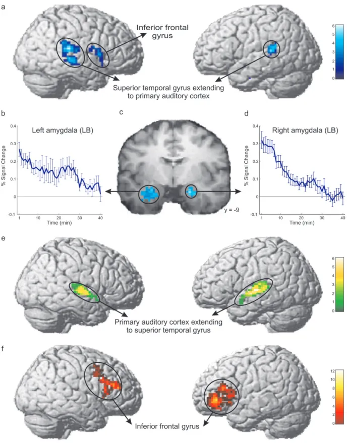
Significant long-term habituation occurring within 43 min,
that is, the whole duration of the experiment, was found both in
the right and left amygdala. The habituation peaks were located
in the probabilistically defined laterobasal amygdala subregion
(Fig. 3b--d). Habituation was more rapid in the right than in the
left laterobasal amygdala (sign test, P
<0.05). Moreover,
long-term habituation of brain responses was also found in the right
and left superior temporal gyrus extending to the primary
auditory cortex, in the right inferior frontal cortex (including
Broca’s homologue/area 44), and in the left hippocampus region
(see Fig. 3a). The results of the long-term habituation are
summarized in Tables 3 and 4 in the Supplementary Material. No
differential long-term habituation by comparing fast with slow
piano melodies (collapsing consonant and dissonant melodies)
as well as by comparing consonant with dissonant melodies
(collapsing fast and slow melodies) were found.
Significant short-term habituation occurring within 24 s, that
is, the duration of the individual piano melodies, was found
in the probabilistically defined primary auditory cortex
extending to the superior temporal gyrus (Rademacher et al.
2001; see Fig. 3e). Peak MNI coordinates and anatomical
assignment using the probabilistic anatomical map of the
primary auditory cortex are summarized in Table 5 in the
Supplementary Material. There was no significant sensitization,
that is, no BOLD response increase, over time course of the
experiment in any brain region.
Sustained brain responses during the whole time of the
experiment, that is, responses showing neither significant fast
nor long-term habituation in response to piano music
perception, were found in the left ventrolateral prefrontal
cortex extending into the anterior insula and Broca’s area (area
44/45) and in its homologue on the right hemisphere (area 45)
at P
>0.3 level (t-test, uncorrected, cluster size
>50 voxels;
see Fig. 3f and Table 6 in the Supplementary Material).
Finally, we found positive correlations with the STAI-S and
the individual contrast music perception versus baseline in the
left parietal lobule and left area 2 (P
<0.05, FDR-corrected,
cluster size
>10 voxels, Table 7 in the Supplementary Material)
and positive correlations of the lifetime music education scores
Figure 2. Brain areas with significant responses during the perception of piano music. Areas with significant responses (P \ 0.05, FDR-corrected, cluster size [ 10 voxels) are superimposed on the cortical surface of a template brain (a and b) and on the coronal (c and d) slices of the mean structural scan of the 19 subjects investigated in the fMRI experiment. The results reveal bilateral music perception related responses in the auditory cortex, in the inferior frontal gyrus (IFG, including Broca’s area/Broca’s homologue and the ventrolateral prefrontal cortex), in the insular cortex, and in the cerebellum. In addition, there were significant bilateral responses in the amygdala and in the supplementary motor area. For coordinates of response peaks and anatomical assignment using probabilistic cytoarchitectonic maps, see Supplementary Table 2.1 5 10 15 20 25 30 35 40 43 1.2 1.3 1.4 1.5 1.6 1.7 Time (min) Valence Rating
a
1 5 10 15 20 25 30 35 40 43 1 1.1 1.2 1.3 1.4 1.5 1.6 Time (min) Arousal Ratingb
Figure 1. Valence and arousal ratings during fMRI experiment. Subjects gave ratings of their valence and arousal experience after each melody presentation. The amplitude of both valence (a) and arousal ratings (b) induced by the musical pieces decreased during the experiment, that is, they tended toward neutral. The blue lines show the mean ratings during the experiment (43 min). Error bars indicate the standard error of the mean from the average across subjects.
Figure 3. (a) Long-term habituation (occurring within 43 min) was observed in the right and left nonprimary auditory cortex and in the right inferior frontal cortex (including the right hemisphere Broca’s homologue). (b--d) Significant long-term habituation was also observed in the probabilistically defined laterobasal amygdala subregions. The found decrement was more rapid in the right (d) than in the left laterobasal amygdala region (b). The results are projected upon a coronal slice of the mean structural scan of the 19 subjects investigated in the fMRI experiment (c). The blue lines (b and d) show the mean decrease of the music-related BOLD responses during the experiment (43 min). Error bars indicate the standard error of the mean of the average across subjects. (e) Significant short-term habituation (occurring within 24 s) was observed in the primary auditory cortex extending to superior temporal gyrus (STG; P \ 0.005 uncorrected, cluster size [ 50 voxels). The results are displayed on the MNI single subject brain and assigned using the probabilistic anatomical map of the primary auditory cortex (Rademacher et al. 2001). ROIs were the activated brain areas for the contrast music perception [ baseline (P \ 0.005, uncorrected, cluster size [ 50 voxels). (f) Sustained response with neither short- nor long-term habituation nor sensitization in response to music was found in the left ventrolateral prefrontal cortex extending into the anterior insula and Broca’s area (area 44/45) and in its homologue on the right hemisphere (area 45). Statistical parametric maps were rendered on the lateral surface of a representative 3D brain volume (P [ 0.3, cluster size [ 50 voxels).
and the individual short-term habituation in the right superior
temporal gyrus (P
<0.005, uncorrected, cluster size
>50
voxels, masked with the contrast music perception
>baseline).
Discussion
In the present study, we have investigated the neural basis
of affect dynamics in the human brain using an integrated
approach, tracing temporal changes both in brain responses
using BOLD sensitive fMRI and in affective experience using
immediate rating of each presented stimulus within the fMRI
scanner. We find a decline in the stimulus induced fMRI
BOLD signal with repeated auditory stimulation in the
probabi-listically defined laterobasal amygdala occurring on a time scale
of minutes (i.e., within the total 43 min of the investigated
period of time). This slow decline was paralleled by a decrease of
the amplitude of valence and arousal ratings of the music pieces.
Furthermore, the nonprimary auditory cortex (the superior
temporal gyrus), the right inferior frontal gyrus including
Broca’s area right hemisphere homologue (area 44), and the
hippocampus region demonstrated habituation on the same,
slow time scale. In contrast, short-term decreases occurring
within seconds predominated in the primary auditory cortex
and extended to the superior temporal gyrus. Sustained
responses throughout the whole investigated period of time
were detected in the ventrolateral prefrontal cortex, in the
dorsal part of the anterior insular cortex, and in Broca’s area
(area 44/45) and its homologue on the right hemisphere (area
45). Together, these findings demonstrate that different time
scales of habituation during auditory perception coexisting in
the human brain and indicate an amygdalocortical network
underlying affective habituation to music. The integrated,
neuroimaging-behavioral approach of the present study may in
the future be valuable not only to study affect dynamics in
healthy subjects but also its possible disturbances in
neuropsy-chiatric disorders.
Consistent with previous neuroimaging studies on music
perception in individuals without professional music education,
the current investigation identified music-related responses in
areas including the auditory cortices, the insular cortex, the left
and right inferior frontal cortex including Broca’s area and
Broca’s area right hemisphere homologue (area 44/45), the
ventrolateral prefrontal cortex, the amygdala, and the
hippo-campus. In addition, the cerebellum, the SMA, and the caudate
nucleus were also found to be activated (Blood and Zatorre
2001; Koelsch et al. 2006; Zatorre et al. 2007). The amygdala has
repeatedly shown to be responsive to auditory stimuli-like
human vocalization (Sander and Scheich 2001, 2005; Seifritz
et al. 2003) and music (Blood and Zatorre 2001; Koelsch et al.
2006; Ball et al. 2007; Mutschler et al. 2008). The cerebellum, the
SMA, and the caudate nucleus have been implicated in musical
rhythm perception (Chen et al. 2008). Alternatively, the SMA
activity observed in the present study might also be related to
suppression of movement execution (Rizzolatti et al. 1990; Ball
et al. 1999) because listening to music might induce the desire
to move (e.g., to tap the rhythm), which subjects had to inhibit
during the experiment because they received the explicit
instruction to avoid any movement.
Further, Broca’s area and its right hemisphere homologue
(area 44 and 45) have been reported as being involved in music
perception (Tillmann et al. 2003; Koelsch et al. 2005).
Activation in Broca’s area during music perception has been
related to music-syntactic processing (Koelsch 2005). The
music-related responses that we find in the ventrolateral
prefrontal cortex are consistent with animal studies showing
that neurons in the ventrolateral prefrontal cortex are selective
for complex sounds (Romanski and Goldman-Rakic 2002;
Romanski 2007). However, it is still under debate to which
extent monkey’s ventrolateral prefrontal cortex is homologue
to human ventrolateral prefrontal cortex or rather to Broca’s
area (Petrides and Pandya 2002). To our knowledge, the
present study is the first to find music perception related
responses in the human ventrolateral prefrontal cortex,
specifically in a region anterior to Broca’s area located on the
lateral convexity of the frontal lobe.
The responses we observed in the left anterior insular cortex
are in line with a recent meta-analysis of brain imaging studies
showing that the dorsal anterior part of the anterior insula is
reproducibly involved in auditory processing such as in the
perception of vocalizations and music (Mutschler et al. 2009).
After having delineated this highly plausible network of
music-related brain areas (see Fig. 2), we have investigated the
dynamics of the BOLD responses in these areas on different
scales of time.
We find significant long-term habituation effects bilaterally
localized in the amygdala, in the nonprimary auditory cortex
extending to primary auditory cortex, in the right hemisphere
Broca’s homologue (area 44), and in the hippocampus region
(see Fig. 3a), suggesting that these brain areas might constitute
a functional network. These hypotheses are supported by
anat-omical studies showing that both the hippocampus and the
nonprimary auditory cortex are anatomically connected to the
amygdala (McDonald 1998), and there are temporal--frontal
projections to Broca’s homologue (Glasser and Rilling 2008).
According to a widely applied model of amygdala function,
a short latency thalamic pathway supplies the amygdala with
a crudely analyzed version of sensory inputs allowing for a fast
response, while a long latency cortical pathway provides an
elaborately processed version of the incoming information to
the amygdala (LeDoux 2000). The analysis of the auditory input
taking place in Broca’s area, which has been described being
related to music syntax processing (Tillmann et al. 2003)
together with the superior temporal region, which is also
assumed to be involved in the processing of complex sounds
(Rauschecker and Scott 2009), likely belongs to the cortical
pathway feeding highly processed auditory information to the
amygdala complex.
Amygdala habituation in response to complex auditory
stimuli might reflect the fact that the sensory stimuli are no
longer of relevance for the individuals because the amygdala
is thought to be essential in evaluating stimulus salience and
in initiating behavioral responses (Sander et al. 2003). This
assumption is supported by our data: valence and arousal
ratings induced by the musical pieces decreased during the
experiment (i.e., they tended towards neutral). In particular,
arousal ratings decreased over the whole time of the
experi-ment (43 min) and valence ratings declined within the first
20 min. Our behavioral findings support previous results
showing that affective reactions decline over time in response
to repeated sensory exposure (Dijksterhuis and Smith 2002).
Moreover, our findings indicate a neural basis for these
habituation effects of emotional ratings: We show that ratings
of valence and arousal declined on a similar time scale as BOLD
responses of the amygdala, which is thought to encode an
integrated representation of valence and arousal of sensory
stimuli (Winston et al. 2005). In the present study, long-term
habituation patterns were modeled by a linear decrease both in
BOLD response amplitude. The choice of this linear approach
is supported by the findings of Leventhal et al. (2007),
specifically demonstrating a linear decrease in affect responses
to repeated exposure of pleasurable stimuli.
The peaks of long-term habituation effects were bilaterally
localized in the probabilistically defined laterobasal amygdala.
This result is in line with previous animal research: in mice,
habituation of single-cell activity in responses to sounds was
found within the laterobasal amygdala (Herry et al. 2007).
Furthermore, studies in various animal species consistently
show that the majority of sensory, including auditory, afferents
project to the laterobasal amygdala subregion (Bordi and
LeDoux 1992; McDonald 1998, 2003). The more rapid
habituation rate for the right in comparison to the left
amygdala (Phillips et al. 2001; Wright et al. 2001) might also
explain why across studies, the left amygdala is more often
found to be activated than the right amygdala (Baas et al. 2004;
Ball et al. 2009). The finding that the left amygdala
demon-strates slower habituation across various sensory modalities is
in agreement with the suggestion by Hardee et al. (2008) that
the left amygdala is involved in stimuli processing in a more
elaborative way than the right amygdala and might therefore
remain longer involved.
Parallel to the network demonstrating habituation on a time
scale of minutes, there were no significant increases
(sensitiza-tion) in music-related responses on the same time scale
detectable in the present study. Two different brain mechanisms
mediating affective habituation have been previously proposed:
The first mechanism assumes that the emotional network
including the amygdala may be extrinsically suppressed by
another brain system showing gradually increasing activity
(Feinstein et al. 2002). Alternatively, the second mechanism
proposes that the emotional circuits including the amygdala
show intrinsic habituation without being extrinsically
sup-pressed by another brain system showing increased activation
(Hatta et al. 2006). The present results are in favor of the second
hypothesis, that is, they suggest that emotional habituation is
not dependent on increased responses in more cognitive brain
regions suppressing the emotional system. However, particular
care has to be taken in interpretation of negative findings. The
fact that we do not find any sign of neuronal sensitization may be
due to the limitations of the fMRI method (Logothetis 2008) or
detection of (weak) sensitization effects might require a larger
sample of subjects to be investigated. Therefore, further studies
using fMRI but also other, for example, electrophysiological
methods will be required for evaluating the 2 proposed
mechanisms of affective habituation.
In contrast to the long-term decreases observed in the
nonprimary auditory cortex, we find short-term decreases of the
BOLD signal in response to music in the probabilistically defined
primary auditory cortex extending to the superior temporal
gyrus (see Fig. 3e). Neural habituation effects within seconds
have been obtained for pure tones in the primary auditory
cortex of anesthetized cats (Ulanovsky et al. 2003, 2004) and
awake monkeys (Micheyl et al. 2005). In terms of the underlying
mechanisms, it is unlikely that the observed short-time
decreases in the primary auditory cortex can be fully explained
by sensory adaptation, which is defined as the decrease in
a sensory response over time in the presence of a constant
stimulus (Eatock 2000), in particular as hair cells in the inner ear
exhibit sensory adaptation that occurs on a much shorter time
scale, that is, within milliseconds, than our decreases during
music presentation that occurred within seconds (Eatock 2000).
Interestingly, subjects with a higher lifetime music education
score demonstrated faster short-term habituation in the superior
temporal gyrus. Musical training has been shown to induce
functional and anatomical changes in the human brain (Munte
et al. 2002; Fujioka et al. 2006). The present findings indicate
that changes in auditory habituation, in particular, in fast
habituation processes in primary auditory and surrounding
areas, are part of the changes induced by long-term musical
training. These habituation changes in subjects with more music
education may have a functional significance for the musical
skills that are acquired through musical training.
Studies in monkeys and humans indicate a hierarchical
organization of the cortical auditory system: the primary auditory
cortex—or auditory core area—represents the first cortical stage
of sound processing. The secondary or belt areas that surround
the primary auditory cortex are thought to be especially involved
in processing of complex sounds with more temporal structure
and a broader frequency spectrum than simple single-frequency
tones (Rauschecker and Scott 2009). The belt areas and the
adjacent regions of the superior temporal gyrus demonstrate
stronger fMRI responses to band-pass noise compared with pure
tones (Seifritz et al. 2006) and have been implicated in the
processing of complex sounds such as speech (Fecteau et al.
2005) and animal vocalizations (Altmann et al. 2007).
In the present study, we investigated response to a highly
complex and varied auditory stimulus, that is, excerpts of
classical piano music in different variation of harmony and
tempo. Previously, Seifritz et al. (2002) investigated the spatial
and temporal patterns of neural processing in the human
auditory cortex using less complex sounds, finding that the
contribution of a sustained responses component became less
predominant as one moves from the primary auditory cortex,
or core area, to the surrounding belt area of the auditory
cortex, while the opposite was found for a transient response
component at stimulus onset. Together with our present study,
these findings indicate that the temporal patterns of
habitua-tion processes in the human brain substantially depend on
stimulus complexity. The predominance of a transient response
to simple tones in the auditory belt area (Seifritz et al. 2002)
and of a slowly habituating response to complex sounds in the
belt and adjacent areas (present study) might thus reflect
a preference for complex sound processing. In summary, the
present findings are in agreement with the idea of
hierarchi-cally auditory processing in the cortical auditory system
(Rauschecker and Scott 2009).
In the present study, we also find sustained responses, that is,
responses with neither short nor long-term habituation nor
sensitization throughout the whole investigated period of time
(43 min). Such sustained responses were found in Broca’s area
(area 44/45) and its homologue on the right hemisphere (area
45), in the left ventrolateral prefrontal cortex, and in the left
anterior insula (see Fig. 3f). Because subjects had to rate each
melody after presentation throughout the whole experiment,
the sustained responses in the insula might reflect task-set
processing (Dosenbach et al. 2006) related to the rating task.
The ventrolateral prefrontal responses might be explained by
the working memory demands of the rating task (Arnott et al.
2005), which remained constant throughout the experiment.
Interestingly, a recent study in macaque monkeys showed that
neurons in the prefrontal cortex demonstrated only weak
size-contingent repetition effects (Verhoef et al. 2008).
In summary, the present study delineates a neural basis for the
habituation effects of emotional experience in humans. We
show that ratings of valence and arousal declined on a similar,
slow time scale as habituation of responses of the laterobasal
amygdala. Our results demonstrate a temporal specificity of
BOLD fMRI to disentangle habituation processes on different
scales of time. Electrophysiologically, however, habituation
effects have been shown after even shorter stimulus repetition
times (Fischer et al. 2003; Weiland et al. 2008). The results of the
present study demonstrate that different time scales of
habituation coexist during the perception of music in an
amygdalocortical network that might support a hierarchical
model of complex auditory processing. From a psychological
perspective, it has been assumed that the mechanisms
un-derlying habituation have a functional purpose by protecting the
organism from flooding with irrelevant sensory information by
allocating resources to new salient stimuli in the environment
(Siddle 1991). It has been hypothesized that it is maladaptive not
to habituate (Dijksterhuis and Smith 2002) as, for example, in
patients with anxiety disorders (Protopopescu et al. 2005; Shin
et al. 2005). Future studies could therefore utilize stimuli with
varying complexity/biological relevance in order to investigate
adaptive and disturbed habituation processes as they have been
proposed to be critical in psychiatric disorders. Employing such
habituation paradigms in an integrated neuroimaging-behavioral
fashion as in the present study may be a valuable approach for
such future investigations of the neuronal mechanisms of affect
dynamics in the human brain.
Funding
Swiss National Science Foundation (grant 51A240-104890); and
the VolkswagenStiftung (grant I/83 078) within the European
Platform.
Supplementary Material
Supplementary material can be found at: http://www.cercor .oxfordjournals.org/.
Notes
The authors thank Manuela Keckeis from the MR Physics, Freiburg for support during the fMRI experiments, Prof. Wilfried Gruhn from the University of Music in Freiburg and Christoph Kaller from the Department of Neurology, Freiburg for help regarding music selection. Furthermore, we thank the ‘‘YAMAHA Stiftung 100 Jahre e.V.’’ for providing the MR-compatible headphones. Conflict of Interest : None declared.
References
Altmann CF, Doehrmann O, Kaiser J. 2007. Selectivity for animal vocalizations in the human auditory cortex. Cereb Cortex. 17(11): 2601--2608.
Amunts K, Kedo O, Kindler M, Pieperhoff P, Mohlberg H, Shah NJ, Habel U, Schneider F, Zilles K. 2005. Cytoarchitectonic mapping of the human amygdala, hippocampal region and entorhinal cortex: intersubject variability and probability maps. Anat Embryol (Berl). 210(5--6):343--352.
Anderson AK, Christoff K, Stappen I, Panitz D, Ghahremani DG, Glover G, Gabrieli JD, Sobel N. 2003. Dissociated neural representa-tions of intensity and valence in human olfaction. Nat Neurosci. 6(2):196--202.
Arnott SR, Grady CL, Hevenor SJ, Graham S, Alain C. 2005. The functional organization of auditory working memory as revealed by fMRI. J Cogn Neurosci. 17(5):819--831.
Baas D, Aleman A, Kahn RS. 2004. Lateralization of amygdala activation: a systematic review of functional neuroimaging studies. Brain Res Brain Res Rev. 45(2):96--103.
Ball T, Derix J, Wentlandt J, Wieckhorst B, Speck O, Schulze-Bonhage A, Mutschler I. 2009. Anatomical specificity of functional amygdala imaging of responses to stimuli with positive and negative emotional valence. J Neurosci Methods. 180(1):57--70.
Ball T, Rahm B, Eickhoff SB, Schulze-Bonhage A, Speck O, Mutschler I. 2007. Response properties of human amygdala subregions: evidence based on functional MRI combined with probabilistic anatomical maps. PLoS One. 2:e307.
Ball T, Schreiber A, Feige B, Wagner M, Lucking CH, Kristeva-Feige R. 1999. The role of higher-order motor areas in voluntary movement as revealed by high-resolution EEG and fMRI. Neuroimage. 10(6): 682--694.
Blood AJ, Zatorre RJ. 2001. Intensely pleasurable responses to music correlate with activity in brain regions implicated in reward and emotion. Proc Natl Acad Sci U S A. 98(20):11818--11823.
Bordi F, LeDoux J. 1992. Sensory tuning beyond the sensory system: an initial analysis of auditory response properties of neurons in the lateral amygdaloid nucleus and overlying areas of the striatum. J Neurosci. 12(7):2493--2503.
Breiter HC, Etcoff NL, Whalen PJ, Kennedy WA, Rauch SL, Buckner RL, Strauss MM, Hyman SE, Rosen BR. 1996. Response and habituation of the human amygdala during visual processing of facial expression. Neuron. 17(5):875--887.
Chen JL, Penhune VB, Zatorre RJ. 2008. Listening to musical rhythms recruits motor regions of the brain. Cereb Cortex. 18(12):2844--2854.
Codispoti M, Ferrari V, Bradley MM. 2006. Repetitive picture process-ing: autonomic and cortical correlates. Brain Res. 1068(1):213--220. Dijksterhuis A, Smith PK. 2002. Affective habituation: subliminal exposure to extreme stimuli decreases their extremity. Emotion. 2(3):203--214.
Dosenbach NU, Visscher KM, Palmer ED, Miezin FM, Wenger KK, Kang HC, Burgund ED, Grimes AL, Schlaggar BL, Petersen SE. 2006. A core system for the implementation of task sets. Neuron. 50(5):799--812.
Dudai Y. 2002. Memory from A to Z. New York: Oxford University Press. Eatock RA. 2000. Adaptation in hair cells. Annu Rev Neurosci.
23:285--314.
Fecteau S, Armony JL, Joanette Y, Belin P. 2005. Sensitivity to voice in human prefrontal cortex. J Neurophysiol. 94(3):2251--2254. Feinstein JS, Goldin PR, Stein MB, Brown GG, Paulus MP. 2002.
Habituation of attentional networks during emotion processing. Neuroreport. 13(10):1255--1258.
Fischer H, Wright CI, Whalen PJ, McInerney SC, Shin LM, Rauch SL. 2003. Brain habituation during repeated exposure to fearful and neutral faces: a functional MRI study. Brain Res Bull. 59(5):387--392. Fujioka T, Ross B, Kakigi R, Pantev C, Trainor LJ. 2006. One year of musical training affects development of auditory cortical-evoked fields in young children. Brain. 129:2593--2608.
Glasser MF, Rilling JK. 2008. DTI tractography of the human brain’s language pathways. Cereb Cortex. 18(11):2471--2482.
Hardee JE, Thompson JC, Puce A. 2008. The left amygdala knows fear: laterality in the amygdala response to fearful eyes. Soc Cogn Affect Neurosci. 3(1):47--54.
Hatta N, Nishikawa T, Ikejiri Y, Tokunaga H, Masaki Y, Uema T, Kazui H, Doronbekov T, Ogino A, Miyoshi N, et al. 2006. Neural substrates of emotional habituation: a PET study using film stimuli. Psychiatry Clin Neurosci. 60:40--45.
Herry C, Bach DR, Esposito F, Di Salle F, Perrig WJ, Scheffler K, Luthi A, Seifritz E. 2007. Processing of temporal unpredictability in human and animal amygdala. J Neurosci. 27(22):5958--5966.
Johnstone T, Somerville LH, Alexander AL, Oakes TR, Davidson RJ, Kalin NH, Whalen PJ. 2005. Stability of amygdala BOLD response to fearful faces over multiple scan sessions. Neuroimage. 25(4): 1112--1123.
Koelsch S. 2005. Neural substrates of processing syntax and semantics in music. Curr Opin Neurobiol. 15(2):207--212.
Koelsch S, Fritz T, Schulze K, Alsop D, Schlaug G. 2005. Adults and children processing music: an fMRI study. Neuroimage. 25(4):1068--1076. Koelsch S, Fritz T, von Cramon Dy, Muller K, Friederici AD. 2006.
Investigating emotion with music: an fMRI study. Hum Brain Mapp. 27(3):239--250.
Laux L, Glanzmann P, Schaffner P, Spielberger CD. 1981. Das State-Trait-Angstinventar (STAI). Weinheim (Germany): Beltz Verlag. LeDoux JE. 2000. Emotion circuits in the brain. Annu Rev Neurosci.
23:155--184.
Leventhal AM, Martin RL, Seals RW, Tapia E, Rehm LP. 2007. Investigating the dynamics of affect: psychological mechanisms of affective habituation to pleasurable stimuli. Motiv Emot. 31(2):145--157. Litle P, Zuckerman M. 1985. Sensation seeking and music performance.
Pers Individ Dif. 7:575--577.
Logothetis NK. 2008. What we can do and what we cannot do with fMRI. Nature. 453(7197):869--878.
Martin-Soelch C, Stocklin M, Dammann G, Opwis K, Seifritz E. 2006. Anxiety trait modulates psychophysiological reactions, but not habituation processes related to affective auditory stimuli. Int J Psychophysiol. 61(2):87--97.
McDonald AJ. 1998. Cortical pathways to the mammalian amygdala. Prog Neurobiol. 55(3):257--332.
McDonald AJ. 2003. Is there an amygdala and how far does it extend? An anatomical perspective. Ann N Y Acad Sci. 985:1--21.
Micheyl C, Tian B, Carlyon RP, Rauschecker JP. 2005. Perceptual organization of tone sequences in the auditory cortex of awake macaques. Neuron. 48(1):139--148.
Munte TF, Altenmuller E, Jancke L. 2002. The musician’s brain as a model of neuroplasticity. Nat Rev Neurosci. 3(6):473--478. Mutschler I, Wieckhorst B, Kowalevski S, Wentlandt J, Derix J,
Schulze-Bonhage A, Ball T. 2009. Functional organization of the human anterior insular cortex. Neurosci Lett. 457(2):66--70.
Mutschler I, Wieckhorst B, Schulze-Bonhage A, Hennig J, Speck O, Seifritz E, Ball T. 2008. Probabilistic assignment of brain responses to the human amygdala and its subregions using high resolution functional MRI. IFMBE Proc. 22:807--810.
Oldfield RC. 1971. The assessment and analysis of handedness: the Edinburgh inventory. Neuropsychologia. 9(1):97--113.
Petrides M, Pandya DN. 2002. Comparative cytoarchitectonic analysis of the human and the macaque ventrolateral prefrontal cortex and corticocortical connection patterns in the monkey. Eur J Neurosci. 16(2):291--310.
Phillips ML, Medford N, Young AW, Williams L, Williams SC, Bullmore ET, Gray JA, Brammer MJ. 2001. Time courses of left and right amygdalar responses to fearful facial expressions. Hum Brain Mapp. 12(4):193--202.
Protopopescu X, Pan H, Tuescher O, Cloitre M, Goldstein M, Engelien W, Epstein J, Yang Y, Gorman J, LeDoux J, et al. 2005. Differential time courses and specificity of amygdala activity in posttraumatic stress disorder subjects and normal control subjects. Biol Psychiatry. 57(5):464--473.
Rademacher J, Morosan P, Schormann T, Schleicher A, Werner C, Freund HJ, Zilles K. 2001. Probabilistic mapping and volume measurement of human primary auditory cortex. Neuroimage. 13(4):669--683.
Rauschecker JP, Scott SK. 2009. Maps and streams in the auditory cortex: nonhuman primates illuminate human speech processing. Nat Neurosci. 12(6):718--724.
Rizzolatti G, Gentilucci M, Camarda RM, Gallese V, Luppino G, Matelli M, Fogassi L. 1990. Neurons related to reaching-grasping arm movements in the rostral part of area 6 (area 6a beta). Exp Brain Res. 82(2):337--350.
Romanski LM. 2007. Representation and integration of auditory and visual stimuli in the primate ventral lateral prefrontal cortex. Cereb Cortex. 17(Suppl 1):i61--i69.
Romanski LM, Goldman-Rakic PS. 2002. An auditory domain in primate prefrontal cortex. Nat Neurosci. 5(1):15--16.
Russell JA. 1980. A circumplex model of affect. J Pers Soc Psychol. 39:1161--1178.
Sander D, Grafman J, Zalla T. 2003. The human amygdala: an evolved system for relevance detection. Rev Neurosci. 14(4):303--316. Sander K, Scheich H. 2001. Auditory perception of laughing and crying
activates human amygdala regardless of attentional state. Brain Res Cogn Brain Res. 12(2):181--198.
Sander K, Scheich H. 2005. Left auditory cortex and amygdala, but right insula dominance for human laughing and crying. J Cogn Neurosci. 17(10):1519--1531.
Sawa M, Delgado JM. 1963. Amygdala unitary activity in the un-restrained cat. Electroencephalogr Clin Neurophysiol. 15:637--650. Seifritz E, Di Salle F, Esposito F, Herdener M, Neuhoff JG, Scheffler K.
2006. Enhancing BOLD response in the auditory system by neurophysiologically tuned fMRI sequence. Neuroimage. 29(3): 1013--1022.
Seifritz E, Esposito F, Hennel F, Mustovic H, Neuhoff JG, Bilecen D, Tedeschi G, Scheffler K, Di Salle F. 2002. Spatiotemporal pattern of neural processing in the human auditory cortex. Science. 297(5587):1706--1708.
Seifritz E, Esposito F, Neuhoff JG, Luthi A, Mustovic H, Dammann G, von Bardeleben U, Radue EW, Cirillo S, Tedeschi G, et al. 2003. Differential sex-independent amygdala response to infant crying and laughing in parents versus nonparents. Biol Psychiatry. 54(12):1367--1375.
Shin LM, Wright CI, Cannistraro PA, Wedig MM, McMullin K, Martis B, Macklin ML, Lasko NB, Cavanagh SR, Krangel TS, et al. 2005. A functional magnetic resonance imaging study of amygdala and medial prefrontal cortex responses to overtly presented fearful faces in posttraumatic stress disorder. Arch Gen Psychiatry. 62(3):273--281.
Siddle DA. 1991. Orienting, habituation, and resource allocation: an associative analysis. Psychophysiology. 28(3):245--259.
Small DM, Gregory MD, Mak YE, Gitelman D, Mesulam MM, Parrish T. 2003. Dissociation of neural representation of intensity and affective valuation in human gustation. Neuron. 39(4):701--711.
Thompson RF, Spencer WA. 1966. Habituation: a model phenomenon for the study of neuronal substrates of behavior. Psychol Rev. 73(1):16--43.
Tillmann B, Janata P, Bharucha JJ. 2003. Activation of the inferior frontal cortex in musical priming. Brain Res Cogn Brain Res. 16(2):145--161. Toga AW, Thompson PM, Mori S, Amunts K, Zilles K. 2006. Towards multimodal atlases of the human brain. Nat Rev Neurosci. 7(12):952--966.
Ulanovsky N, Las L, Farkas D, Nelken I. 2004. Multiple time scales of adaptation in auditory cortex neurons. J Neurosci. 24(46): 10440--10453.
Ulanovsky N, Las L, Nelken I. 2003. Processing of low-probability sounds by cortical neurons. Nat Neurosci. 6(4):391--398.
Verhoef BE, Kayaert G, Franko E, Vangeneugden J, Vogels R. 2008. Stimulus similarity-contingent neural adaptation can be time and cortical area dependent. J Neurosci. 28(42):10631--10640. Weiland BJ, Boutros NN, Moran JM, Tepley N, Bowyer SM. 2008.
Evidence for a frontal cortex role in both auditory and somatosen-sory habituation: a MEG study. Neuroimage. 42(2):827--835. Whalen PJ, Rauch SL, Etcoff NL, McInerney SC, Lee MB, Jenike MA.
1998. Masked presentations of emotional facial expressions modulate amygdala activity without explicit knowledge. J Neurosci. 18(1):411--418.
Winston JS, Gottfried JA, Kilner JM, Dolan RJ. 2005. Integrated neural representations of odor intensity and affective valence in human amygdala. J Neurosci. 25(39):8903--8907.
Wright CI, Fischer H, Whalen PJ, McInerney SC, Shin LM, Rauch SL. 2001. Differential prefrontal cortex and amygdala habituation to repeatedly presented emotional stimuli. Neuroreport. 12(2): 379--383.
Zaitsev M, Hennig J, Speck O. 2004. Point spread function mapping with parallel imaging techniques and high acceleration factors: fast, robust, and flexible method for echo-planar imaging distortion correction. Magn Reson Med. 52(5):1156--1166.
Zatorre RJ, Chen JL, Penhune VB. 2007. When the brain plays music: auditory-motor interactions in music perception and production. Nat Rev Neurosci. 8(7):547--558.