Complications in the native lung after single lung transplantation
q
Federico Venuta
a,*, Annette Boehler
b, Erino A. Rendina
a, Tiziano De Giacomo
a, Rudolf Speich
b,
Ralph Schmid
b, Giorgio Funo Coloni
a, Walter Weder
baDepartment of Thoracic Surgery, University of Rome La Sapienza, Cattedra di Chirurgia Toracica, Policlinico Umberto I, 00100 Rome, Italy bDepartment of Thoracic Surgery and Pulmonology, University of Zurich, Zurich, Switzerland
Received 5 October 1998; received in revised form 23 March 1999; accepted 7 April 1999
Abstract
Objectives: Single lung transplantation is a viable option for patients with end-stage pulmonary disease; despite encouraging results, we observed serious complications arising in the native lung. We retrospectively reviewed 36 single lung transplants to evaluate the incidence of complications arising in the native lung, their treatment and outcome. Methods: Between 1991 and 1997, 35 patients received 36 single lung transplants for emphysema (16), pulmonary ®brosis (14), lymphangioleiomyomatosis (4), primary pulmonary hypertension (1) and bronch-iolitis obliterans (1). The clinical records were reviewed and the complications related to the native lung were divided into early (up to 6 weeks after the transplant) and late complications. Results: Nineteen complications occurred in 18 patients (50%), leading to death in nine (25%). Early complications (within 6 weeks from the transplant) were bacterial pneumonia (1), overin¯ation (3), retention of secretions with bronchial obstruction and atelectasis (1), hemothorax (1), pneumothorax (1) and invasive aspergillosis (3); one patient showed active tuberculosis at the time of transplantation. Two patients developed bacterial pneumonia and invasive aspergillosis leading to sepsis and death. The other complications were treated with separate lung ventilation (1), bronchoscopic clearance (1), chest tube drainage (1) and wedge resection and pleurodesis (mechanical) by VATS (1). One patient with hyperin¯ation of the native lung eventually required pneumonectomy and died of sepsis. The patient with active tuberculosis is alive and well after 9 months of medical treatment. Late complications were recurrent pneumothorax (4), progressive overin¯ation with functional deterioration (2), aspergillosis (1) and pulmonary nocardiosis (1). Recurrent pneumothorax was treated with chest tube drainage alone (1), thoracoscopic wedge resection and/or pleurodesis (2) and pneumonectomy (1); hyperin¯ation was treated with thoracoscopic lung volume reduction in both cases; both patients with late infectious complications died. Conclusions: After single lung transplantation, the native lung can be the source of serious problems. Early and late infectious complications generally result in a fatal outcome; the other complications can be successfully treated in most cases, even if surgery is required. q 1999 Elsevier Science B.V. All rights reserved.
Keywords: Lung transplantation; Native lung; Complications
1. Introduction
Lung transplantation is a viable option for patients with end-stage pulmonary parenchymal and vascular diseases. Single lung transplantation (SLT) is considered the ideal procedure for patients over the age of 50 years affected by respiratory failure without septic lung disease [1]. Pulmon-ary ®brosis (idiopathic and secondPulmon-ary to other conditions), emphysema (in older patients without bullae), primary pulmonary hypertension and Eisenmenger's Syndrome (according to the policy of each institution) are the most frequent indications; other more rare disorders like
lymphangioleiomyomatosis (LAM) [2] represent an indica-tion in selected cases. SLT allows an ef®cient use of a limited supply of donor organs yielding a satisfactory pulmonary function with a relatively reduced surgical trauma; for this reason it has been increasingly used despite the potential disadvantage of a diseased native lung in place. However, even if the results are encouraging with a 3-year survival of 50±60% [3], serious complications may still occur. The native lung could be the source of early and late problems related to the progression of the underlying disease and its ensuing complications. The susceptibility of the native lung to develop complications may affect the outcome of the transplant; this is an additional factor to be considered when choosing the appropriate procedure for each patient.
We retrospectively reviewed a combined series of SLT performed at two institutions in order to evaluate the
inci-1010-7940/99/$ - see front matter q 1999 Elsevier Science B.V. All rights reserved. PII: S1010-7940(99)00141-4
qPresented at the 12th Annual Meeting of the European Association for
Cardio-thoracic Surgery, Brussels, Belgium, September 20±23, 1998. * Corresponding author. Tel.: 1 39-6-4461971; fax: 1 39-6-49970735. E-mail address: [email protected] (F. Venuta)
dence of complications arising in the native lung and their outcome.
2. Methods
Between 1991 and 1997, 35 patients (nine males and 26 females; mean age: 36 years; range between 14 and 56 years) received 36 single lung transplants (23 right and 13 left) at two different institutions. All patients had end-stage lung disease requiring supplemental oxygen and ful®lled the standard criteria for inclusion in the waiting list for lung transplantation. The indications were emphysema (44.4%), idiopathic pulmonary ®brosis (36.1%), lymphangioleio-myomatosis (11%), ®brosis secondary to
chemo-radiother-apy for Hodgkin's disease (2.8%), primary pulmonary hypertension (2.8%) and obliterative bronchiolitis (2.8%). Preoperative work-up included clinical evaluation, sputum examination, ®beroptic bronchoscopy with bronchoalveolar lavage in selected cases and CT scan to exclude any source of potential infectious complications contraindicating SLT. All the charts were retrospectively reviewed with particular attention to preoperative conditions favouring the onset of possible complications in the native lung (stable infectious colonization, presence of blebs or bullae, history of pneu-mothorax, diabetes mellitus and steroid administration). The protocol of immunosuppression included ALG (for induc-tion), cyclosporin, azathioprine and prednisone (starting with 0.5±1 mg/kg per day and tapered to 0.1±0.2 mg/kg per day after 6±8 months). Postoperative follow-up included sequential radiological, functional and bronchoscopic work-up at predetermined intervals (at least after 1, 3, 6 and 12 months), or when required. All the complications and their outcomes were recorded with speci®c reference to any problem coming from the native lung. Early (within 6 weeks from operation) and late complications were reviewed separately. Particular attention was given to the type and number of treatments required (medical treatment, thoracoscopic procedures, surgical treatment). Survival was calculated according to the Kaplan±Meier method [4]. The Wilcoxon test was used to compare survival curves. 3. Results
The native lung was a source of 19 signi®cant complica-tions occurring in the native lung in 18 patients (50%) lead-ing to death in nine cases. Early complications were present in 11 cases (Table 1): bacterial pneumonia (9.1%), overin-¯ation of the native lung after SLT for emphysema (27.3%), retention of secretions with bronchial obstruction and atelectasis (9.1%), hemothorax (9.1%), pneumothorax (9.1%) (Fig. 1) and invasive aspergillosis (27.3%); one patient was found to have active tuberculosis (9.1%) at
Table 1
Early complications arising in the native lung after single lung transplantation. COPD, chronic obstructive pulmonary disease; IPF, idiopathic pulmonary ®brosis; LAM, lymphangioleiomyomatosis; PPH, primary pulmonary hypertension; TB, Tuberculosis, P.O., postoperatively; CMV, Cytomegalovirus; MOF, multiple organ failure; VATS, video assisted thoracoscopic surgery.
Diagnosis Age Complications Time of onset Therapy Outcome Cause of death
COPD 51 Pneumothorax 2nd day VATS Alive (5 years)
LAM 48 Invasive aspergillosis 4th week Medical/pneumonectomy Death (1 month) Fungal sepsis
IPF 54 Invasive aspergillosis 6th week Medical Death (1 month) Fungal sepsis/CMV
COPD 44 Overin¯ation Immediate P.O. Observation Alive (4 years)
IPF 47 Invasive aspergillosis 1 week Medical Death (1 month) Fungal sepsis
COPD 56 Overin¯ation/pneumothorax 2nd day Pneumonectomy Death (1 month) Sepsis
PPH 51 TB Immediate P.O. Medical Alive (5 years)
IPF 38 Pneumonia 2 weeks Medical Death (1 month) Sepsis/CMV
COPD 44 Secretion retention 2±3weeks Repeated FOB Alive (5 years)
IPF 33 Hemothorax 2 weeks Chest drainage Alive (2 years)
COPD 52 Overin¯ation 2±3 days Separate lung ventilation Death (2 months) Infection/MOF
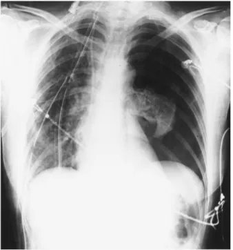
Fig. 1. Left pneumothorax after right single lung transplantation for emphysema.
the time of transplantation diagnosed in the explanted lung. Infectious complications were thus present in ®ve cases; two of these patients died within 2 months from bacterial pneu-monia (due to Kiebsiella pneupneu-moniae) and invasive pulmonary aspergillosis. None of these patients presented either bronchiectasis or preoperative colonization by the agent causing the infectious complication or other resistant strains. None of the patients had diabetes before the trans-plant or assumed steroids at doses higher than 0.2 mg/kg per day; one patient developed diabetes after the operation. The immunosuppressive protocol was the same for all patients. Fungal infections were not related to intensive care unit outbreak. The non-infectious complications (6) were treated with separate lung ventilation (n 1), repeated airway cleaning with the ®beroptic bronchoscope (n 1), chest tube drainage (n 1) and video-assisted thoracoscopic wedge resection and pleurodesis (mechanical) (n 1). One patient with persistent hyperin¯ation underwent pneu-monectomy and died of sepsis 2 months later. None of the patients developing early overin¯ation had bullae in the native lung. The patient found to have active tuberculosis at the time of transplantation received medical treatment for
9 months (Myambutol, Rifampicin and INH) and is alive and well 5 years after the transplant.
Late complications (Table 2) were recurrent pneu-mothorax (n 4), progressive overin¯ation with functional deterioration after SLT for emphysema (n 2), aspergillo-sis (n 1) (Fig. 2) and pulmonary nocardioaspergillo-sis (n 1) (Fig. 3). Recurrent pneumothorax occurred after a mean of 21 months (range 12±24 months) from the operation and was treated with chest tube drainage in one patient and thoraco-scopic wedge resection and pleurodesis (talc and mechan-ical) in two cases; in one patient pneumonectomy was performed after a previous attempt to solve the problem with minimally invasive procedures; the patient is alive and well. Hyperin¯ation of the native lung with functional deterioration and dislocation of the mediastinum and hemi-diaphragm occurred in two patients, respectively 2 and 3 years after the transplant. The possible role of the trans-planted lung in the functional derangement was extensively investigated [5] and obliterative bronchiolitis was excluded with repeated trans-bronchial lung biopsies. Hyperin¯ation was successfully treated with thoracoscopic lung volume reduction and both patients are alive and well, respectively 2 and 5 years after the procedure. The two patients with late infectious complications (one patient developed insulin dependent diabetes after transplantation) died; one patient with pulmonary nocardiosis presented a systemic spread of the disease and died of sepsis 2 years after transplantation; one patient developed systemic aspergillosis and died 14 months after the transplant. Two-year survival for patients with early complications and patients without complications in the native lung is shown in Fig. 4 (P 0:04). Three-year survival for patients with late complications was 75%. 4. Discussion
Single lung transplantation (SLT) offers an effective option for the treatment of several pulmonary diseases [6± 8]. Generally, the indications for SLT and DLT overlap in patients without septic disorders and aged less than 45±50 years. The limitation of suitable donors requires a better use of the resources, and splitting of the double lung block
Table 2
Late complications arising in the native lung after single lung transplantation. LAM, lymphangioleiomyomatosis; IPF, idiopathic pulmonary ®brosis; COPD, chronic obstructive pulmonary disease; PF, pulmonary ®brosis; VATS, video assisted thoracoscopic surgery; LVRS, lung volume reduction surgery; OB, obliterative bronchiolitis.
Diagnosis Age Complications Time of onset Therapy Outcome Cause of death
LAM 36 Pneumothorax 2 years Chest Tube Alive (5 years)
LAM 33 Pneumothorax 2 years VATS Alive (3 years)
IPF 46 Pneumothorax 2 years Pneumonectomy Alive (3 years)
COPD 56 Overin¯ation 2 years VATS-LVRS Alive (3 years)
IPF 42 Pneumothorax 2 years VATS Death (7 years) OB
COPD 44 Overin¯ation 3 years VATS-LVRS Alive (5 years)
COPD 56 Invasive Aspergillosis 13 months Medical Death (14 months) Fungal sepsis
PF POST CHEMO 14 Nocardia 2 years Medical Death (2 years) Fungal sepsis
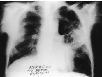
Fig. 2. Pulmonary aspergillosis in the native lung after right single lung transplantation for emphysema.
between two recipients (twinning procedure) [9] has become an effective approach to improve organ sharing. However, after SLT, the contralateral native lung remains a potential source of complications that may impair both early and late outcome [10±13]. For this reason, the impact of the remaining lung on morbidity and mortality following SLT may affect the choice of the type of transplant, espe-cially in patients in whom either SLT and DLT can be performed.
Most of the previous reports emphasize the incidence of ventilation/perfusion (V/Q) mismatch, early and late hyper-in¯ation and even sepsis after SLT for emphysema [5,12,14]; early hyperin¯ation and V/Q mismatch may be a dreadful complication; it can be prevented by careful selection of patients and donors: patients with bullous disease and repeated infections due to bacterial colonization should be excluded from SLT. Patients with extremely
severe preoperative airway obstruction, air trapping and relative pulmonary hypertension seem to be at the greatest risk of developing hyperin¯ation [15]; however, in our series, none of the patients developing this complication showed these preoperative risk factors. The use of marginal donors should be avoided; in fact, a well functioning trans-planted lung, larger than the predicted total lung capacity of the recipient, can prevent severe hyperin¯ation of the native lung. Right SLT for emphysema should be preferred, since the mild hyperin¯ation of the native lung easily displaces the left hemidiaphragm downward, without mediastinal shifting.
Late hyperin¯ation of the native lung requires different considerations; it may be related to the decreased compli-ance of the graft (obliterative bronchiolitis ± BOS, infection, rejection) or to the progression of the underlying chronic obstructive pulmonary disease in the native lung itself. Infectious complications should be excluded and the possi-ble development of obliterative bronchiolitis should be ruled out. In the absence of histologic con®rmation, the diagnosis of BOS should be presumed. If augmented immu-nosuppression does not result in functional improvement and hyperin¯ation of the native lung persists, the latter could play a role in the development of dyspnoea. However, preoperative work-up may not allow a precise assessment of the amount of dysfunction related to the progressive compression of the graft by the native lung and the possible coexistence of BOS. If marked functional improvement follows unilateral volume reduction, the diagnosis of BOS may be left out. When hyperin¯ation is related to the presence of large bullae, bullectomy is well established to relieve compression [16]. More recently, attention has been focused on the potential bene®ts of surgical resection of areas of diffusely emphysematous lung parenchyma to reduce hyperin¯ation and improve dyspnoea [17,18]. Bullectomy and unilateral volume reduction surgery of the native lung after SLT for emphysema have been reported [5,19,20]; both thoracotomy and thoracoscopy were performed and the results were satisfactory with both approaches.
The underlying lung disease in the native lung often predisposes to pneumothorax (e.g. in patients with emphy-sema, pulmonary ®brosis and LAM). Mechanical ventila-tion in the early postoperative period may favour its onset; however, pneumothorax may also present later, even several years after the transplant, in line with the natural history of the underlying disease. Thoracoscopic pleurodesis is the therapeutic gold standard and results are satisfactory in most of the cases [12,21].
Infectious complications in the native lung [11,22,23] often result in a fatal outcome. Despite sputum examination and ®beroptic bronchoscopy during preoperative work-up in selected cases, infectious complications in the native lung are not uncommon. The persistence of bacterial coloniza-tion even after appropriate medical treatment may support the indication for double lung transplantation, especially in
Fig. 3. Nocardia arising in the native lung after left single lung transplanta-tion for pulmonary ®brosis.
Fig. 4. Survival of patients with early complications arising in the native lung and patients without complications (P 0:04).
patients with pulmonary ®brosis. This careful assessment is intended to reduce the chance of infectious complications arising in the native lung and spreading to the graft after the onset of immunosuppression. None of our patients presented infectious problems at preoperative evaluation; however, 75% of patients presenting early infectious complications (bacterial and fungal) had pulmonary ®brosis and received azathioprine and steroids for a long time before the operation and this may have contributed to the increased incidence of this complication. Radiological evaluation of the native lung may be dif®cult: patients with pulmonary ®brosis show an increased density of the native lung on chest x-ray, and early diagnosis of pneumonia may be dif®-cult or even impossible. Computed tomography is manda-tory for con®rmation in suspected cases. In our experience, diffuse spreading of the infection was always fatal. The predisposition to infections and their early spreading may be due to the decreased mucociliary clearance, altered sputum characteristics and, at times, chronic bacterial colo-nization [22] and diabetes.
Postmortem ®ndings [24] after SLT showed the presence of a signi®cant number of pathologic conditions, in addition to the native disease. Most of these ®ndings are related to infectious complications, but also lymphoproliferative disorders and lung cancer have been described, probably in relation to the smoking history of the patient. We recog-nize that our incidence of complications arising in the native lung is higher than that reported by other groups; this may be in part related to the fact that most of these complications occurred in the early period of our lung transplantation program. In recent times, DLT have been performed more often in our lung transplant program to reduce the incidence of problems related to the native lung both in the early and late course after the operation.
In conclusion, after single lung transplantation, the native contralateral lung may be the source of serious problems. Infectious complications often resulted in a fatal outcome, despite appropriate medical treatment. Non-infectious complications were successfully treated in most of the cases, also when required a surgical approach. Double lung transplantation should be considered more often, espe-cially for young patients, in order to decrease the rate of complications arising in the native lung and improve func-tional outcome, in particular when BOS occurs.
References
[1] Trulock, E.P. Recipient selection. In: Patterson, G.A. Cooper, J.D., editors. Lung transplantation. Chest Surg Clin North Am 1993;3:1-8. [2] Boehler A, Speich R, Russi EW, Weder W. Lung transplantation for lymphangioleiomyomatosis. New Engl J Med 1996;335:1275±1280. [3] Hosenpund JD, Bennett LE, Keck BM, Fiol B, Bonceck HM, Novick RJ. The registry of the International Society for Heart and Lung Transplantation. Fifteenth Of®cial Report 1998;17:656±668.
[4] Kaplan EL, Meier P. Nonparametric estimation from incomplete observations. J Am Stat Ass 1958;53:457±481.
[5] Venuta F, De Giacomo T, Rendina LA, Della Rocca G, Flaishman I, Guarino E, Ricci C. Thoracoscopic volume reduction of the native lung after single lung transplantation for emphysema. Am J Resp Crit Care Med 1997;156:292±293.
[6] Levy RD, Ernst P, Levine SM. Exercise performance after lung trans-plantation. J Heart Lung Transpl 1993;12:27±33.
[7] Williams TJ, Patterson GA, McClean PA, Zamel N, Maurer JR. Maxi-mal exercise testing in single and double lung transplant recipients. Am Rev Resp Dis 1992;145:101±105.
[8] Miyoshi S, Trulock EP, Schaefers HJ, Hsieh CH, Patterson GA, Cooper JD. Cardiopulmonary exercise testing after single and double lung transplantation. Chest 1990;97:1130±1136.
[9] Sommers KE, Grif®th BP, Hardesty RL, Keenan JK. Early lung allo-graft function in twin recipients from the same donors: risk factor analysis. Ann Thorac Surg 1996;62:784±790.
[10] Frost AL, Keller CA, Noon GP, Short UD, Cagle PT. The Multiorgan Transplant Group. Outcome of the native lung after single lung trans-plant. Chest 1995;107:981±984.
[11] Horvath J, Dummer S, Loyd J, Walker B, Merrill WH, Frist WH. Infection in the transplanted and native lung after single lung trans-plantation. Chest 1993;104:681±685.
[12] Speziali G, McDougall JC, Midthun DF, Peters SG, Scott JP, Daly RC, McGregor CG. Native lung complications after single lung trans-plantation for emphysema. Transpl Int 1997;10:113±115.
[13] Venuta F, Rendina EA, De Giacomo T, Ciriaco P, Della Rocca G, Ricci C. Thoracoscopic treatment of recurrent contralateral pneu-mothorax after single lung transplantation. J Heart Lung Transplant 1994;13:555±557.
[14] Koerner SK, Vefth FJ. Ventilation-perfusion relationship between transplanted and emphysematous lungs. Vasc Surg 1974;8:283±297. [15] Yonan NA, El-Gamel A, Egan J, Kakadellis J, Rahaman A, Deiraniya AK. Single lung transplantation for emphysema: predictors of native lung hyperin¯ation. J Heart Lung Transplant 1998;17:192±201. [16] Kuno R, Kanter KR, Torres WE, Lawrance EC. Single lung
trans-plantation followed by contralateral bullectomy for bullous emphy-sema. J Heart Lung Transplant 1996;15:389±394.
[17] Cooper JD, Trulock EP, Trianta®llou AN, Patterson GA, Pohl MS, Deloney PA, Sundaresan RS, Roper CL. Bilateral pneumectomy (volume reduction) for chronic obstructive pulmonary disease. J Thorac Cardiovasc Surg 1995;109:106±119.
[18] Russi EW, Stammberger U, Weder W. Lung volume reduction surgery for emphysema. Eur Resp J 1997;10:208±218.
[19] Le Pimpec-Barthes F, Debrasse D, Cuenod CA, Gandjbakheh I, Riquet M. Late contralateral lobectomy after single lung transplanta-tion for emphysema. Ann Thorac Surg 1996;61:231±234.
[20] Kapelanski DP, Anderson MB, Kriett JM, Colt HG, Smith CM, Mateos M, Jamieson SW. Volume reduction of the native lung after single lung transplantation for emphysema. J Thorac Cardiovasc Surg 1996;111:898±899.
[21] Waller DA, Conacher ID, Dark JH. Videothoracoscopic pleurectomy after contralateral single -lung transplantation. Ann Thorac Surg 1994;57:1021±1023.
[22] Calquhoun IW, Gascoigne AD, Gould K, Corris PA, Dark JH. Native pulmonary sepsis after single lung transplantation. Transplantation 1991;52:931±933.
[23] McDougall JC, Vigneswaran WT, Peters SG, Marshall WT, McGre-gor CG. Fungal infection of the contralateral native lung after single lung transplantation. Ann Thorac Surg 1993;56:176±178.
[24] Husain AN, Siddiqui MT, Reddy VB, Yeldandi V, Montoya A, Garr-ity ER. Postmortem ®ndings in lung transplant recipients. Mod Pathol 1996;9(7):752±761.