Chemosensing Strategies
Utilizing the Novel Sulfonamidohydroxyquinoline Amino Acid Sox by
Melissa Dawn Shults B.S. Chemistry (2000) University of California, San Diego
Submitted to the Department of Chemistry
in Partial Fulfillment of the Requirements for the Degree of Doctor of Philosophy in Organic Chemistry
at the
Massachusetts Institute of Technology June 2005
© 2005 Massachusetts Institute of Technology All rights reserved
I MASSACUSES INSE OF TECHNOLOGY JUN 212005
LIBRARIES
Signature of Author: / , -E-
.-Department of Chemistry May 5, 2005 Certified by: Barbara Imperiali Class of 1922 Professor of Chemistry and Professor of Biology Thesis Supervisor Accepted By:
Robert W. Field Chairman, Departmental Committee on Graduate Students
This doctoral thesis has been examined by a committee of the Department of Chemistry as follows: -77--Timothy MA ager Professor of Chenistry Chair Barbara Imperiali Class of 1922 Professor of Chemistry and Professor of Biology Thesis Supervisor
v
a
Z
/ iujas . Lauffenburger
Whitaker Professor of Biological Engineering, Professor of Chemical Engineering and Biology
, _, we
P -
tepheSJ. LippardArthur Amos Noyes Professor of Chemistry
Chemosensing Strategies
Utilizing the Novel Sulfonamidohydroxyquinoline Amino Acid Sox by
Melissa Dawn Shults
Submitted to the Department of Chemistry on May 5, 2005 in partial fulfillment of the requirements for the Degree of Doctor of Philosophy
in Organic Chemistry ABSTRACT
Modular peptide-based fluorescent chemosensors utilizing the chelation-sensitive fluorophore 8-hydroxy-5-(N,N-dimethylsulfonamido)-2-methylquinoline are powerful tools for sensing Zn2+ and for sensing protein kinase activity. This signaling component is prepared as the
protected amino acid derivative Fmoc-Sox-OH, and integrated into peptide sequences.
Selective and tunable chemosensors for Zn2+ can afford qualitative and quantitative
information about the presence, distribution and concentration of this biologically-important metal ion. Nineteen synthetic peptides incorporating Sox exhibit a range of affinities for Zn2+
through variation of the type and number of Zn2+ ligands, ligand arrangement and the -turn
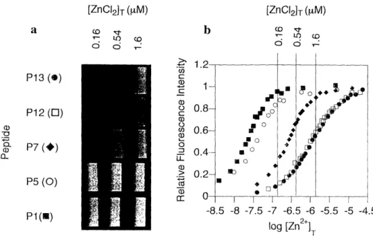
sequence that acts as a preorganization element between the ligands. The binding stoichiometry and fluorescence response to pH changes and various relevant competing metal ions was carefully characterized. Eleven of these sequences form only 1:1 complexes with Zn2+ and their affinities range from 10 nM to nearly 1 tM. When used in concert, the relative intensities of different chemosensor readouts can provide Zn2+concentration information in a valuable range.
This modular scaffold is useful for ratiometric sensing when an additional fluorophore is incorporated in the peptide sequence.
New methods to quantify protein kinase activities are critical for understanding biological regulatory pathways. Fluorescent chemosensors of protein kinase activity utilizing Sox and physiological Mg2+concentrations report phosphorylation with dramatic fluorescence changes in a continuous, high-throughput sensing format. The chemosensor comprises a small sensing module, containing Sox and a S-turn sequence, appended to an optimized peptide substrate for the target kinase. The Mg2+-binding affinity of the product phosphopeptide is much greater than the substrate peptide, which results in a large fluorescence increase upon phosphorylation. Notably, the reactivity of substrates is not affected by introduction of the sensing module on either side of the serine, threonine or tyrosine to be phosphorylated. Further, a homogeneous kinase assay utilizing these probes was developed that was reproducible, linear and highly preferential for monitoring changes in cellular activity of the target kinase in unfractionated cell lysates. These kinase chemosensors are powerful tools for studying the activity of recombinant kinases in vitro and endogenous kinases ex vivo.
Thesis Supervisor: Barbara Imperiali
Acknowledgements
First and foremost, I would like to thank my advisor, Professor Barbara Imperiali, for her support, both professional and personal, as well as encouragement over the last five years. Under her guidance, have received an excellent graduate education. Barbara, your creativity, enthusiasm for science, and teaching ability have been truly inspiring. I am grateful for your ideas and broad vision that improved my research. I sincerely appreciate your trust and the wealth of opportunities to learn, even when I did not fully realize it. The breadth of your knowledge and interests has generated a lab environment with quite diverse projects, from which I have learned a great deal. I am especially grateful for your selection of talented, helpful and diverse scientists. Also, thank you for entrusting me with your old iBook to get me through this thesis.
I would like to thank the members of my thesis committee for their support during my time at MIT: Professor Doug Lauffenburger, Professor Tim Swager and Professor Steve Lippard. Doug has been an incredible champion of my work and a real pleasure with which to collaborate. ][ admire his positive attitude, creativity and scientific curiosity. I truly admire Tim's ideas and appreciate his willingness to attend my thesis defense from England via iChat, and will always remember our "virtual handshake". Tim, thank you for your contacts that enabled my interview for my new position at Illumina, Inc. Steve, thank you for your thoughtful comments and careful reading of this thesis.
My co-workers in the Imperiali lab have provided a wonderful work environment and on many occasions have offered much-appreciated scientific advice and instruction. They made the lab a friendly, supportive and fun place to spend a significant portion of my life. In addition, I am grateful to all of them for sharing their time and feedback on many practice talks. I owe a special thank you to Dora Carrico, Elvedin Lukovic, and Bianca Sculimbrene for each proofreading portions of this thesis.
Most importantly, Debbie Rothman and Beth Vogel have been truly wonderful friends. Debbie, I have really enjoyed many good talks with you about anything and everything, at lunch, in the thesis-writing "cave", and outside the lab. I am so grateful to have shared many experiences, conferences, and opinions with you (and even a hood!)! Your support and listening ear when I was stressed and freaking out have meant so much to me. I continue to be inspired by
your organization and sense of humor, both of which were helpful for me as I was plowing through this thesis. You are a fantastic friend and I will miss you a lot. Beth, the balance that you have attained and your positive attitude are an inspiration to me and I thank you for helping me to see many things from a different perspective. Thank you for showing me all the best things about New England, and especially fruit picking! Thank you also for motivating me to go to the gym and for being a fantastic workout partner. I have really enjoyed our talks and will really miss you.
I am incredibly grateful to my first co-worker in the lab, Dr. Dierdre Pearce, for teaching me so much and helping me to adjust to graduate school. Dierdre, I really appreciated your scientific inspiration and moral support for the Zn2+-sensing project. I thank Kathy Franz and Kevin McDonnell for a lot of very helpful research advice early in my PhD. I am also grateful to Dr. K. Jebrell Glover for spending a significant portion of his own time and energy to provide advice, to answer my questions, and to help me with biochemistry and computers. Jebrell, I admire your philosophy about research and have enjoyed speaking about research, music and politics with you. I owe many thanks to Elizabeth Fong for taking care of tons of little things for me and explaining many administrative things throughout my graduate career. Elizabeth, I have enjoyed our many chats, and thank you for sharing you love of plants and some clippings with me! To my co-workers on the kinase sensing project - Elvedin Lukovic and Dora Carrico - I cannot think of two better people to carry on the project and I look forward to many exciting results in the future. Elvedin, thank you for always listening to my research frustrations and for many fun dancing memories. Dora, thank you for checking up on me during the thesis writing, for your openness and for good talks - I will miss a chance to get to know you better. Mayssam Ali, thank you for your friendship, advice, and especially encouragement during the job search. I will also be forever indebted for the suggestion to use phosphoserine in a Zn2 +-sensing peptide! Maria Ufret, I am thankful for your continued friendship, for sharing mass spec duties, and for great lunches out in the sunshine! I thank Carlos Bosques for his support, for joking around with me, and for lots of loud salsa music and singing. Bianca Sculimbrene, thank you for helping to keep me sane through the job search and thesis writing, I really appreciate the moral support and that you shared your experiences with me. Langdon Martin, thank you for always keeping me laughing. I really admire your creativity and attitude. The Sox kinase assay will always be
"CHAMP" to me. I continue to be inspired by the dedication to science of my classmates Eranthie Weerapana and Seungjib Choi; I wish both of you many future successes. Christina Carrigan, your enthusiasm and efficiency was incredibly inspiring and I very much enjoyed my first bike ride out to Walden Pond with you. I thank Eugenio Vazquez for his smile and for help both with research and with computers. I have enjoyed many great lunchtime conversations with Debby Pheasant and appreciated her cheerfulness in the instrument room when experiments were going awry. Mary O'Reilly, thank you for listening to my random outbursts at my desk - I enjoyed many talks with you and wish you the best in your post-doc. I thank Jennifer Ottesen for her enthusiasm in giving me a crash course in managing the lab Mac computers. Galen Loving, I wish you much luck with the mass spec - you are already doing a fantastic job with the e-mails. I thank Guofeng Zhang for entertaining Costco trips and thought-provoking debates. I thank Mark Nitz for sharing his knowledge with me and for research advice. I thank Christian Hackenburger for lots of chocolate from Germany and for the humorous pictures of the Governator. I thank Soonsil Hyun for being a good hoodmate and for teaching me some things about her project. I also thank Rob Dempski and Harm Brummerhop for answering many questions when I arrived. I have enjoyed the company of the undergraduates in the lab: Ryu Yoshida, Mike Torrice, Rebecca Maglathlin and Jose Class. I wish the best to several people who recently joined and whom I have not been able to get to know: Mark Chen, Andrew Dutton, Matthieu Sainlos and Nelson Olivier.
My collaboration with Kevin Janes benefited greatly from his work ethic, experimental consistency and wealth of knowledge. Kevin, thank you for being organized, prompt and enthusiastic and for your careful editing of our manuscript. I feel lucky to have been a part of
such a rewarding and successful collaboration.
To the "hotbed of [Red] Sox fandom that is the Imperiali lab" (quote courtesy of Jenny Yang) - Beth, Debbie, Bianca and Langdon - I have enjoyed sharing my obsession with the World Champion Boston Red Sox with you all. I will always remember the games, our signs, the player of the week board and the many fun conversations. I would also like to thank the players, especially Gabe Kapler, Curt Schilling and Manny Ramirez, for inspiration.
I thank the building 18 renovations for providing an opportunity for character-building and, in retrospect, some good laughs.
My previous mentors and chemistry teachers who have inspired me and sparked my interest in chemistry deserve special mention: my high school chemistry teacher, Dawn Gamberale, and my undergraduate research advisor at UCSD, Professor Jay S. Siegel.
Many wonderful friends have supported me through busy and often stressful times. I owe special thanks to my awesome roommates, Laurel Ng and Ping Xu. You have both shared my love of good food and I have enjoyed many dinners, good conversations and fun parties with you. Ping, I am also grateful for the Perl script to reformat my fluorescence plate reader data that saved me a great deal of time. To my dear friends Julie Garg and Rachana Sidhu, I treasure your fiiendship.
To Jey - your support, love, and friendship means so much to me. Thank you for moving to Boston to see me through the end of my PhD, for always making me laugh, for helping me see things in a different light and for helping me to kick back every once in awhile. I love you.
Most importantly, to Mom, Dad and Amanda - words cannot describe how much your support, encouragement, unconditional love, and friendship throughout my life and especially now has meant to me. Mom and Dad, you have encouraged me to do what I love and have never pressured me - I am so grateful. Mom, thank you for your wisdom and for always being there when I need to talk. Dad, thank you for your incredible sense of humor and for much computer assistance. Amanda, thank you for sharing so much of your exciting life with me and yet always finding time to hear about mine. I am so lucky to have such a wonderful sister and best friend. You are the best family a girl could wish for and I am looking forward to being back home again soon. Mom, Dad and Amanda, I love you all very much and I dedicate this thesis to you.
Table of Contents Abstract ... 3 Acknlowledgements ... 4 Table of Contents ... 8 List of Figures ... 10 List of Schemes ... 12 List of Tables ... 13 List of Abbreviations ... 14 Chapter 1 Introduction ... 16
Introduction to Biological Signaling ... 17
Properties of an Ideal Chemosensor ... 18
Advantages of Peptide-based Sensors ... 20
Metal-chelating Peptides ... 20
Dissertation Objective ... 22
References... 23
Chapter 2 Synthesis, Characterization and Fluorescence Properties of (S)-2-Amino-N-(9- fluorenylmethyloxycarbonyl)-3-(8-hydroxy-5-(N,N-dimethylsulfonamido)quinoline-2-yl) Propionic Acid (Fmoc-Sox-OH) ... 26
Introduction... 27
Results and Discussion ... 29
Synthesis of Fmoc-Sox-OH ... 29
Spectroscopic Properties of Sox ... 31
Conclusion...33
Acknowledgements... 34
Experimental...34
References...39
Chapter 3 Modular and Tunable Chemosensing Scaffold for Divalent Zinc ... 42
Introduction... 43
Results and Discussion...50
Intensity-based Peptide Sensor Design and Synthesis ... 51
Absorbance and Fluorescence Properties ... 52
Binding Stoichiometry ... 53
Binding Trends ... 57
Implications for Further Tuning ... 57
Influence of pH on Chemosensing Peptides ... 58
Metal Competition Studies ... 59
Probes of Zn2+Concentration ... 61
Investigation of Sox-based Emission and Excitation Ratiometric Zn2+Sensors ... 62
Conclusion... 69
Future Directions ... 70
Experimental... 70
References ... 77
Chapter 4 Versatile and Sensitive Fluorescent Chemosensors of Protein Kinase Activity: Design and Optimization ... 81
Introduction... 82
Results...92
Substrate Design and Synthesis ... 92
Fluorescence Properties ... 93
Sensing Mechanism ... 93
Magnitude of the Fluorescence Increase ... 94
Fluorescence Increase Quantifies Product Formation ... 98
Determination of Kinetic Parameters for Chemosensors ... 101
Influence of Relevant Metal Ions on Kinase Sensing Ability ... 103
Application to High-Throughput Kinase Inhibitor Screening ... 104
Discussion ... 105 Conclusion ... 108 Future Directions ... 108 Acknowledgements... 109 Experimental ... 110 References ... 116
Chapter 5 A Multiplexed Homogeneous Fluorescence-Based Assay For Protein Kinase Activity In Cell Lysates...122
Introduction... 123
Results... 126
Chemosensor Properties ... 126
Development of an Akt-S 1 Kinase Activity Assay ... 126
Validation of the Akt-S1 Kinase Assay ... 129
Development of MK2-S1 and PKA-S3 Kinase Activity Assays ... 134
Validation of the MK2-S1 Kinase Activity Assay ... 134
Validation of the PKA-S3 Kinase Activity Assay ... 138
The Multiplex Akt-MK2-PKA Kinase Assay ... 140
Discussion ... 141 Conclusion ... 142 Future Directions ... 143 Acknowledgements... 144 Experimental ... 144 References... 150
List of Figures Chapter 1 Figure 1-1. Figure 1-2. Figure 1-3. Chapter 2 Figure 2-1. Figure 2-2. Figure 2-3. Chapter 3 Figure 3-1. Figure 3-2. Figure 3-3. Figure 3-4. Figure 3-5. Figure 3-6. Figure 3-7. Figure 3-8. Figure 3-9. Figure 3-10. Figure 3-11. Figure 3-12. Figure 3-13. Figure 3-14. Figure 3-15. Figure 3-16. Figure 3-17. Figure 3-18. Figure 3-19.
Properties of an ideal chemical sensor ... 19
Structure of a -turn ... 21
Metal-binding framework for fluorescent chemosensors containing the Sox amino acid ... 23
Chemical structures of 8-hydroxyquinoline, 2-methyl-8-hydroxyquinoline, and 8-hydroxy-5-(N,N-dimethylsulfonamido)-2-methylquinoline ... 28
UV-Vis absorbance spectra of a representative peptide and peptide-Zn2 + complex ... 32
Excitation and emission spectra of representative peptide complexes with Zn 2 + , Mg2 + , Ca2 + and Cd2+... 33
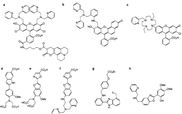
Structures of existing small-molecule Zn2+chemosensors ... 44
Schematic depictions of peptide and protein intensity-based Zn2+ chemosensors ... 45
Structures of small-molecule ratiometric Zn2+probes ... 47
Peptide- and protein-based Zn2+ratiometric chemosensors ... 48
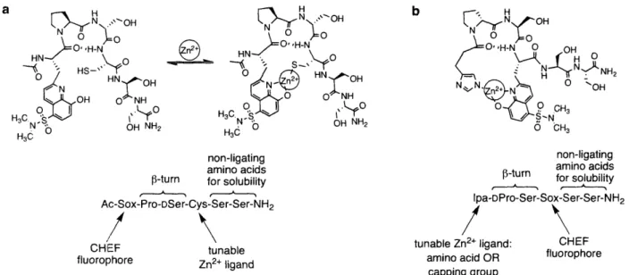
Schematic representation of modular Sox-based Zn2+chemosensors ... 50
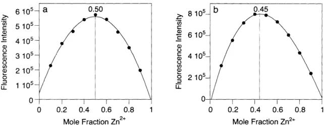
Job plots for formation of a 1:1 complex or mixed complexes ... 54
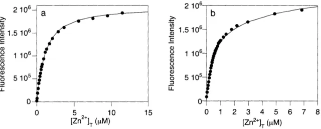
Scatchard plots for formation of a 1:1 complex or mixed complexes ... 54
Complexation model that best fits the titration data for a 1:1 complex or mixed complexes ... 55
Behavior of the maximum emission wavelength during a titration for a 1:1 complex and mixed complexes ... 55
pH profiles of representative peptide and peptide-Zn2+complexes ... 58
Metal ion competition plots for representative peptide and peptide-Zn2+ complexes, based on donor ligands ... 61
Visual representation of the range of Zn2+ affinities of five chemosensing peptides ... 62
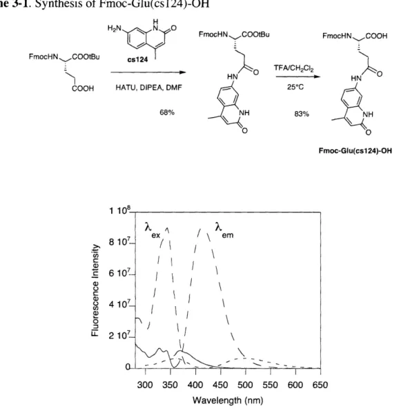
Schematic depictions of Sox-containing ratiometric chemosensors ... 63
Excitation and emission spectra for cs124, Fmoc-Glu(cs 124)-OH, and the P 10-Zn2+complex ... 64
Evidence of FRET with emission ratiometric chemosensor MDS32 ... 66
Effect of increasing concentrations of Zn2+ on fluorescence spectra of the emission ratiometric chemosensors MDS32, MDS33, and MDS34 ... 67
Representative curve fit of the emission ratio for calculation of KD for MDS33 ... 67
Structure of BodipyR6G succinimidyl ester ... 68
Effect of increasing concentrations of Zn2+on fluorescence excitation spectra MDS35 and MDS36 ... 69
4-1. 4-2. 4-3. Figure 4-4. Figure 4-5. Figure 4-6. Figure 4-7. Figure Figure Figure 4-8. 4-9. 4-10. Figure 4-11.
The reaction catalyzed by protein kinases ... 82 Examples of discontinuous assays of protein kinase activity ... 86 Fluorescence response generation for continuous assays of protein kinase activity ... 90 Design of fluorescent Sox-based chemosensors of protein kinase activity... 93 Origin of the difference in fluorescence intensity between substrate peptide and product phosphopeptide ... 94 Mg2+titration curves for MK2-P1, Akt-P1 and PKC-P 1 ... 97 Confirmation of product formation and calculation of kinetic parameters from continuous fluorescence response of peptide chemosensor during kinase reaction ... 100 Cross-reactivity of select peptide substrates with PKCQ and PKA ... 103 Effect of physiological metal ions on kinase chemosensors ... 104 Influence of varying concentrations BIM I, BIM IV, and BIM V on PKCQ activity ... 105 Optimized Sox-based kinase chemosensor design for serine/threonine
phosphorylation ... 107 Chapter 5 Figure 5-1. Figure 5-2. Figure 5-3. Figure 5-4. Figure 5-5. Figure 5-6. Figure 5-7. Figure 5-8. Figure 5-9. Figure 5-10. Figure 5-11. Figure 5-12. Figure 5-13. Figure 5-14.
Comparison of Akt-S1 kinase assay sensitivity at 10 jtM and 1 mM ATP... 127 Effect of kinase inhibitors GF109203X, PKC inhibitor peptide, PKItide and calmidazolium on recombinant Aktl , MK2, PKA and PKC ... 128 Linearity of the Akt-S 1 kinase activity assay and comparison with an
established radioactivity assay ... 130 Quenched-point Akt-S 1 fluorescence assays with immunopurified Akt from insulin-stimulated lysates ... 131 Inhibition and immunodepletion of Akt-S 1 kinase activity ... 133 Comparison of Akt-S 1 kinase activity in CHO cell lysates with a radioactive assay ... 133 Linearity of the MK2-S 1 kinase activity assay and comparison with an
established radioactivity assay ... 135 Inhibition and immunodepletion of MK2-S 1 kinase activity ... 136 Quenched-point fluorescence assays with immunopurified MK2 from NaCl-stimulated lysates ... 137 Comparison of MK2-S 1 kinase activity in CHO cell lysates with a radioactive assay ... 137 Linearity of the PKA-S3 kinase activity assay ... 138 Dose-response curves for H89 and PKItide inhibition of endogenous PKA in forskolin-treated HT-29 lysates recombinant PKA ... 139 The response of the multiplex Akt-MK2-PKA kinase assay to stimulation by EGF and insulin ... 140 Preliminary PKC-S 1 kinase activity assay results ... 143
Chapter 4
Figure Figure Figure
List of Schemes
Chapter 2 Scheme 2-1.
Scheme 2-2.
Synthesis of Fmoc-Sox-OH ... 30 Derivatization of Sox with Marfey's reagent ... 31
Chapter 3
List of Tables Chapter 1 Table 1-1. Chapter 2 Table 2-1. Chapter 3 Table 3-1. Table 3-2. Table 3-3. Table 3-4. Table 3-5. Table 3-6. Chapter 4 Table 4-1. Table 4-2. Table 4-3. Table 4-4. Table 4-5. Chapter 5 Table 5-1.
{ and tp angles of Type II and II' p3-tumrns and representative sequences ... 21
Fluorescence properties of various Sox-metal ion complexes ... 33
Zn2+chemosensor peptide sequences and dissociation constants ... 51
Extinction coefficient and quantum yield values for representative peptides... 52
Dissociation constants for 1:1 Zn2+:peptide complexes ... 56
pKa's for chemosensor-Zn2+complexes ... 59
Sequences of emission ratiometric Zn2+chemosensors ... 65
Sequences of excitation ratiometric Zn2+chemosensors ... 68
Properties of individual continuous fluorescent chemosensors of protein kinase activity ... 89
Peptide substrate sequences and fluorescence increases with 10 mM Mg2+... 96
Apparent dissociation constants for select phosphopeptide-Mg2+ complexes... 98
HPLC and ESI-MS verification and quantification of product formation observed by fluorescence ... 100
Kinetic parameters for select kinase chemosensors ... 102
List of Abbreviations
Standard 3-letter and 1-letter codes are used for the 20 natural amino acids
D preceding the amino acid code indicates D-chirality
Abl Abelson tyrosine kinase
Ac acetyl
ADP adenosine-5'-diphosphate
AFP Aequoria victoria fluorescent protein
ATP adenosine-5'-triphosphate
B3SA bovine serum albumin
Bzl benzyl
cAMP adenosine cyclic 3'-5'-phosphate CHEF chelation-enhanced fluorescence
Ches 2-(cyclohexylamino)ethanesulfonic acid
cpAFP circularly-permuted AFP
DCM dichloromethane
DIPEA N,N-diisopropylethylamine
DMF N,N-dimethylformamide
DMSO dimethylsulfoxide
DTT dithiothreitol
c extinction coefficient or molar absorptivity
EDT ethanedithiol
EDTA ethylenediaminetetracetic acid
EGTA ethylene glycol-bis-(2-aminoethylether)-N,N,N',N'-tetraacetic acid ELISA enzyme-linked immunosorbent assay
ESI-MS electrospray ionization mass spectrometry
4)i quantum yield
FDAA Marfey's reagent or 1-fluoro-2,4-dinitrophenyl-5-L-alanineamide
Fmoc fluoren-9-ylmethyloxycarbonyl
F]P fluorescence polarization
FRET fluorescence resonance energy transfer
HATU O-(7-azabenzotriazol- 1 -yl)- 1,1 ,3,3-tetramethyluronium hexafluorophosphate
HBrTU 2-( lH-benzotriazole- 1 -yl)- 1,1 ,3,3-tetramethyluronium hexafluorophosphate
Hepes N-(2-hydroxyethyl)piperazine-N'-ethanesulfonic acid
HOBt N-hydroxybenzotriazole
HPLC high-performance liquid chromatography HRMS high-resolution mass spectrometry ICso concentration that gives 50% inhibition Ipa 3-(imidazol-4-yl)propionic acid
Kr) dissociation constant
KM Michaelis constant
?ex: excitation wavelength
MeCN acetonitrile
Mes 2-morpholinoethanesulfonic acid
MK2 mitogen-activated protein kinase-activated protein kinase-2 NBD 7-nitrobenz-2-oxa- 1,3-diazole
NBS N-bromosuccinimide
NMPI' N-methylpyrrolidinone
NMR nuclear magnetic resonance
C)xn 8-hydroxyquinoline or oxine
PBS phosphate-buffered saline
Pen penacillamine or 3-dimethylcysteine PET photoinduced electron transfer
PKA cAMP-dependent protein kinase
PKC protein kinase C
pSer phosphoserine
pThr phosphothreonine
pTyr phosphotyrosine
Py3BOP benzotriazole- 1 -yl-oxy-tris-pyrrolidino-phosphonium hexafluorophosphate
QAA quantitative amino acid analysis
RP-HPLC reverse-phase HPLC
s.e.m. standard error
SPPS solid-phase peptide synthesis std. dev. standard deviation
TBS tributylsilyl
TBDMS tert-butyldimethylsilyl T:BDPS tert-butyldiphenylsilyl
tBu tert-butyl
TFA trifluoroacetic acid
TIS triisopropylsilane
TMS tetramethylsilane
tR retention time
TLC thin layer chromatography
Tris tris(hydroxymethyl)aminomethane
tYV--Vis ultraviolet-visible
Xaa used to denote any amino acid
Vinax maximum velocity
Chapter 1
Introduction to Biological Signaling
Signaling processes in biological systems are extremely complex. Within an individual cell, signaling events are carefully orchestrated in location and time by diverse cellular species, including metal ions, small molecules, enzymes, and multi-protein complexes. Individual signaling events within signaling cascades are vital for cellular function; for example, they mediate the cell cycle. Signaling cascades also transduce extracellular signals, such as growth factor presence, into a myriad of cellular responses. These signaling pathways afford signal amplification and implement a variety of control mechanisms to tightly regulate changes in cellular function. Due to their complexity, understanding the spatial and temporal roles of each signaling species remains a significant challenge in biology.
Many biological, biochemical and physical approaches have provided a wealth of information regarding the high specificity of cellular signaling. Genes can be knocked out to determine their function, and certain species may be studied by immunoblot analysis or reconstitution in vitro. Further, metal ions can be detected by methods such as atomic absorption spectroscopy. However, these methods all lack the critical element of real-time analysis. Genetic methods require several days or weeks to perform,' and other methods either provide total concentration measurements that are often not relevant to signaling or alternatively remove the components from their native environment.2
To study species in their native cellular environment on a significant signaling time-scale, chemically-driven strategies that allow for spatial and temporal resolution are finding widespread use."3'4 These chemical strategies provide a means to perturb cellular signaling to observe
downstream effects. Certain small molecules can modulate of cellular function"s and caged bioactive species allow controlled release of function.6'7 In addition, chemosensors report the location and activity of signaling species, such metal ions,8 small molecules,4 or enzymes.9
Specifically, chemosensors are an attractive approach for studying signaling because they generate an electrically or spectroscopically measurable signal upon binding or modification. Chemosensors may be of biotic or abiotic origin. Biotic chemosensors can be produced exclusively via genetic methods and are expressed and used within cells; for example, sensors based on the Aequoria victoria fluorescent proteins (AFPs). These endogenous probes have the advantage that introduction into the cell is not disruptive and that chemosensor size is not
prohibitive. Abiotic probes are prepared ex vivo and may be introduced into living cells via mnicroinjection, transfection, or through utilization of protein transduction domains. These exogenous chemosensors include both small molecules and peptides; they provide unique and versatile sensing approaches because nonnatural elements may be incorporated. At the current time, abiotic and biotic probes complement each other; while the endogenous probes are large and limited in their composition, exogenous probes can be difficult to introduce to cells. An exciting combination of the power of both biotic and abiotic approaches has been achieved by site-specifically directing an exogenous label to an endogenous species. 10-12
The chemosensing strategies described in this dissertation are designed to monitor two very different signaling species in biological systems: divalent zinc cations and protein kinase enzymes. The individual chapters will provide detailed descriptions of work in these fields while this introductory chapter will provide a general background to chemosensing and the general
strategy utilized herein.
Properties of an Ideal Chemosensor
The ultimate goal is to study physiological processes in their native environment in a noninvasive manner. Fluorescence detection is ideal since it is noninvasive and exhibits a low background signal. The properties of an ideal fluorescent chemosensor have been discussed in depth elsewhere (refs. 2,3,13,14), and are summarized in Figure 1-1. Notably, chemosensors should rapidly bind the species that is relevant for signaling with good selectivity and signal sensitivity (Figure 1-1la-d). Additionally, the affinity for the target analyte should approximate the expected concentration; this condition prevents disruption of normal cellular signaling processes and allows detection of increases and decreases of the analyte (Figure 1-le). The chemosensor should be stable to all aspects of the sensing environment (Figure 1-lf) and have appropriate fluorescence properties (Figure 1-1 g). Ideally, a readout independent of chemosensor concentration is desirable; chemosensor concentration can be difficult to determine within a cell (Figure 1-1h). Finally, the chemosensor should be easily introduced and distributed uniformly within a cell (Figure 1-1 i).
A Detects onlyfree or active species (~ ~~Boundf
(
s__) analyte0
0
- ~ Inactive Active/free analyte analyteF Chemically-, photophysically- and pH-stable
C
chemical environment B Selectivity A~ ·A
I' (#
®E3
relevant competitors C Rapid kinetics (Z +*4--G Appropriate kex and Xem
c(:
kIex < 340 nmcell I
>.em > 500 nm to avoid cellular
autofluorescence
H Readout independent of [sensor]
H Sensitivity
0
0
I
·
0
large signal change
(#
0
I % WavI Wavelemgt Wavelength - bound -- unbound I Noninvasive introduction'~)]
uniform distribution cellE Affinity near expected analyte concentration
0
(#
0.
normal signaling processes
4-- and binding partners
0
Figure 1-1. Properties of an ideal chemical sensor.
Al_'
,n pH>< > t,
Advantages of Peptide-Based Chemosensors
A chemosensor built from a modular peptide scaffold offers many advantages. The modular architecture allows incorporation of both natural and nonnatural amino acids via combinatorial or rational design approaches, made facile via solid-phase peptide synthesis. Optimization and tuning can be easily achieved by altering the peptide sequence. A number of fluorescence-based sensing mechanisms have been implemented with peptide-based chemosensors in the Imperiali laboratory: chelation-enhanced fluorescence,15-8 environment-sensitive fluorescence,9 -22 fluorescence quenching,23'24 fluorescence resonance energy transfer (FRET),2 5 and in some cases, metal-based fluorescence.2 6 27Incorporation of other functionalities is also feasible via amide bond formation, e.g. an additional fluorophore for an internal standard or a cellular internalization sequence28-31 to transport the chemosensors into cells. In addition, the chemosensors could be attached to a solid support for diagnostic applications.
Metal-Chelating Peptides
Both sensing strategies described in this dissertation were inspired by previous work in this laboratory on the design of metal-binding peptides, many of which contained novel nonnatural chelating amino acids. In all cases, the -turn motif (Figure 1-2), which causes a directionality change in a peptide sequence, has been used as a template for metal coordination between sets of natural or nonnatural amino acid ligands. p3-turn motifs are classified into many different types, defined by the and p dihedral angles of the i+1 and i+2 residues. Many of these conformations promote hydrogen-bond formation between the carbonyl of the i residue and the amide proton of the i+3 residue. In particular, optimized type II and II' p3-turns contain a heterochiral sequence in the two core residues (one D-amino acid and one L-amino acid) and are defined by the dihedral angles listed in Table 1-1 .32
i_~~~~~-
1
;~~~-i')
i F
D
:3 i+3
Figure 1-2. Structure of a 1-turn
Table 1-1. and p dihedral angles and examples of Type II and II' 3-turns
Turn C, p, + Cb+2 tP,+2 Literature examples
II -60 120 80 0 Pro-DSer, Pro-Gly
II' 60 -120 -80 0 DPro-Ser, Ser-DPro
Previous researchers in the Imperiali laboratory have defined reverse-turn motifs in short peptides sequences in aqueous solution and applied this knowledge to preorganize a metal-binding site. When a peptide sequence with significant type II -turn propensity, Ac-Val-Pro-i)Ser-Phe-NH2,3 3 was flanked by metal-binding histidine or carboxylate ligands, chelation of Zn2 +
and Co2 + was effectively promoted. The turn sequence was essential for metal-binding, as no metal-binding was observed when both i+1 and i+2 were glycine residues.3 4 Further work involved replacing natural histidine and aspartate residues with nonnatural phenanthroline metal-chelating residues to alter the affinity of the peptide for Zn2 + and Co2 +. In addition, the use of
sequences with a weaker propensity for type II 13-turn structure, such as Thr-Pro-DAla-Val, could be incorporated to weaken the affinity for the metal ion. Thus, the affinity could be tuned over 5 orders of magnitude through modulation of both the turn sequence and the ligating residues.35 Since these seminal investigations, the 3-turn has been used as an important structural element in the dtie novo design of independently-folded proteins36'37and in metal-coordinating peptides5' 1 6. In
this dissertation, the 3-turn sequence has been optimized for metal binding.
Dissertation Objective
Herein two types of modular peptide-based fluorescent chemosensors will be reported: tunable Zn2+chemosensors and versatile probes of protein kinase activity. Both of these sensing
strategies exploit a chelation-enhanced fluorescence (CHEF) sensing mechanism with an optimized two-residue 3-turn sequence and the novel sulfonamidohydroxyquinoline amino acid Sox (Figure 1-3). This amino acid contains a derivative of the bidentate chelate fluorophore 8-hydroxyquinoline (Oxn) as the side chain and is named for the fluorophore unit sulfonamido-oxine. Oxn derivatives undergo an increase in fluorescence quantum yield upon metal binding to enable detection of certain metal ions. Additional background on the choice of fluorophore can be found in Chapter 2. The synthesis of the Sox amino acid will be presented in Chapter 2, and employs an asymmetric alkylation as the key synthetic step. Chapter 2 also provides relevant spectroscopic properties of the fluorophore.
A family of intensity-based Zn2+ chemosensors for use in monitoring a range of Zn2+
concentrations will be described in Chapter 3. This family of smaller, improved chemosensors for Zn2+ was inspired by previous metallopeptide designs. The optimized 2-residue 3-turn sequence provided a smaller chemosensor with improved metal-binding properties. The binding stoichiometry, as well as the influence of other metal ions and pH, was carefully characterized for each chemosensor. Chapter 3 highlights the ability to design probes whose affinities have been tuned over two orders of magnitude with a modular peptide scaffold. The use of these chemosensors in concert for monitoring Zn2 + concentration will be illustrated. An extension of this peptide-based sensing platform to design ratiometric peptide-based Zn2+ chemosensors is also investigated.. These chemosensors employ an additional fluorophore to provide internal calibration, and include both excitation and emission ratiometric chemosensors.
The Zn2+ chemosensors inspired a novel approach to use the Sox amino acid, a 3-turn motif and the biologically available Mg2+ion to sense phosphorylation of protein kinase peptide substrates. This versatile strategy, introduced in Chapter 4, allows detection of serine, threonine and tyrosine phosphorylation. These probes are the most sensitive for assessing kinase activities reported to date. The design involves an optimized peptide substrate for the target kinase, with a small sensing module appended, containing Sox and a 3-turn. This module may be placed either N- or C-terminal to the residue to be phosphorylated. The probes are extremely effective due to
the large signal change upon phosphorylation and good reactivity with the target kinase. Notably, the reactivity of substrates is not affected by introduction of the sensing motif. High-throughput applications are introduced and further described in the following chapter. Chapter 5 details one valuable application of these probes in a multiplex high-throughput assay of kinase activity in unfractionated cell lysates. These kinase chemosensors are powerful tools for studying the activity of recombinant kinases in vitro and endogenous kinases ex vivo.
Figure 1-3. The metal-binding framework for fluorescent chemosensors containing the amino
acid Sox, a two-residue 0-turn motif and an additional ligand for the metal ion. Inset: chemical structure of Sox.
References
(1) Shogren-Knaak, M. A.; Alaimo, P. J.; Shokat, K. M. Recent advances in chemical approaches to the study of biological systems. Annu. Rev. Cell Dev. Biol. 2001, 17, 405-433.
(2) Tsien, R. Y. Fluorescent and photochemical probes of dynamic biochemical signals inside living cells. In Fluorescent Chemosensors for Ion and Molecule Recognition; Czarnik, A. W., Ed.; American Chemical Society: Washington, DC, 1993, p 130-146. (3) Czarnik, A. W. Desperately seeking sensors. Chem. Biol. 1995, 2, 423-428.
(4) Zhang, J.; Campbell, R. E.; Ting, A. Y.; Tsien, R. Y. Creating new fluorescent probes for cell biology. Nat. Rev. Mol. Cell. Biol. 2002, 3, 906-918.
(:5) Schreiber, S. L. The small-molecule approach to biology. Chem. Eng. News 2003, 81, 51-61.
(6) Shigeri, Y., Tatsu, Y., Yumoto, N. Synthesis and application of caged peptides and proteins. Pharmacol. Ther. 2001, 91, 85-92.
(7) Marriott, G., Roy, P., Jacobson, K. Preparation and light-directed activation of caged proteins. Methods Enzymol. 2003, 360, 274-288.
SON(CH) 2 l
(8) Kikuchi, K.; Komatsu, K.; Nagano, T. Zinc sensing for cellular application. Curr. Opin.
Chem. Biol. 2004, 8, 182-191.
(9) Baruch, A.; Jeffery, D. A.; Bogyo, M. Enzyme activity - it's all about image. Trends Cell
Biol. 2004, 14, 29-35.
(10) Chen, I.; Ting, A. Y. Site-specific labeling of proteins with small molecules in live cells.
Curr. Opin. Biotechnol. 2005, 16, 35-40.
(11) Miller, L. W.; Cornish, V. W. Selective chemical labeling of proteins in living cells.
Curr. Opin. Chem. Biol. 2005, 9, 56-61.
(1 2) Jessani, N.; Cravatt, B. F. The development and application of methods for activity-based protein profiling. Curr. Opin. Chem. Biol. 2004, 8, 54-59.
(13) Lakowicz, J. R. Principles of Fluorescence Spectroscopy; 2nd ed.; Kluwer Academic/Plenum Publishers: New York, 1999.
(14) Czarnick, A. W. Supramolecular chemistry, fluorescence and sensing. In Fluorescent
Chemosensors for Ion and Molecule Recognition; Czarnick, A. W., Ed.; American
Chemical Society: Washington, DC, 1993, p 1-9.
(']15) Walkup, G. K.; Imperiali, B. Stereoselective synthesis of fluorescent ac-amino acids
containing oxine (8-hydroxyquinoline) and their peptide incorporation in chemosensors for divalent zinc. J. Org. Chem. 1998, 63, 6727-6731.
( 6) Jotterand, N.; Pearce, D. A.; Imperiali, B. Asymmetric synthesis of a new
8-hydroxyquinoline-derived ac-amino acid and its incorporation in a peptidyl sensor for divalent zinc. J. Org. Chem. 2001, 66, 3224-3228.
(17) Shults, M. D.; Pearce, D. A.; Imperiali, B. Modular and tunable chemosensing scaffold for divalent zinc. J. Am. Chem. Soc. 2003, 125, 10591-10597.
(18) Shults, M. D.; Imperiali, B. Versatile fluorescence probes of protein kinase activity. J.
Am. Chem. Soc. 2003, 125, 14248-14249.
(19) Walkup, G. K.; Imperiali, B. Design and evaluation of a peptidyl fluorescent chemosensor for divalent zinc. J. Am. Chem. Soc. 1996, 118, 3053-3054.
(20) Walkup, (G. K.; Imperiali, B. Fluorescent chemosensors for divalent zinc based on zinc
finger domains. enhanced oxidative stability, metal binding affinity, and structural and functional characterization. J. Am. Chem. Soc. 1997, 119, 3443-3450.
(21) Vazquez, M. E.; Nitz, M.; Stehn, J.; Yaffe, M. B.; Imperiali, B. Fluorescent caged phosphoserine peptides as probes to investigate phosphorylation-dependent protein associations. J. Am. Chem. Soc. 2003, 125, 10150-10151.
(22) Vazquez, M. E.; Blanco, J. B.; Imperiali, B. Photophysics and biological applications of the environment-sensitive fluorophore 6-N,N-dimethylamino-2,3-naphthalimide. J. Am.
Chem. Soc. 2005, 127, 1300-1306.
(23) Torrado, A.; Imperiali, B. New synthetic amino acids for the design and synthesis of peptide-based metal ion sensors. J. Org. Chem. 1996, 61, 8940-8948.
(24) Torrado, A.; Walkup, G. K.; Imperiali, B. Exploiting polypeptide motifs for the design of selective Cu(II) ion sensors. J. Am. Chem. Soc. 1998, 120, 609-610.
(25) Pearce, D. A.; Walkup, G. K.; Imperiali, B. Peptidyl chemosensors incorporating a FRET mechanism for detection of Ni(II). Bioorg. Med. Chem. Lett. 1998, 8, 1963-1968.
(26) Franz, K. J.; Nitz, M.; Imperiali, B. Lanthanide-binding tags as versatile coexpression probes. ChemBioChem 2003, 4, 265-271.
(27) Nitz, M.; Franz, K. J.; Maglathlin, R. L.; Imperiali, B. A powerful combinatorial screen to identify high-affinity terbium(III)-binding peptides. ChemBioChem 2003, 4, 272-6. (28) Wadia, J. S.; Dowdy, S. F. Protein transduction technology. Curr. Opin. Biotechnol.
2002, 13, 52-56.
(29) Kabouridis, P. S. Biological applications of protein transduction technology. Trends
Biotechnol. 2003, 21, 498-503.
(30) Zorko, M.; Langel, U. Cell-penetrating peptides: mechanism and kinetics of cargo delivery. Adv. Drug Deliv. Rev. 2005, 57, 529-545.
(31) Dunican, D. J.; Doherty, P. Designing cell-permeant phosphopeptides to modulate intracellular signaling pathways. Biopolymers 2001, 60, 45-60.
(32) Rose, G. D.; Gierasch, L. M.; Smith, J. A. Turns in peptides and proteins. Adv. Protein
Chem. 1985, 37, 1-109.
(33) Imperiali, B.; Fisher, S. L.; Moats, R. A.; Prins, T. J. A conformational study of peptides with the general structure Ac-L-Xaa-Pro-D-Xaa-L-Xaa-NH2: Spectroscopic evidence for
a peptide with significant -turn character in water and in dimethyl sulfoxide. J. Am.
Chem. Soc. 1992, 114, 3182-3188.
(34) Imperiali, B.; Kapoor, T. M. The reverse turn as a template for metal coordination.
Tetrahedron 1993, 49, 3501-3510.
(35) Cheng, R. P.; Fisher, S. L.; Imperiali, B. Metallopeptide design: tuning the metal cation affinities with unnatural amino acids and peptide secondary structure. J. Am. Chem. Soc.
1996, 118, 11349-11356.
(36) Struthers, M. D.; Cheng, R. P.; Imperiali, B. Design of a monomeric 23-residue polypeptide with defined tertiary structure. Science 1996, 271, 342-345.
(37) Struthers, M. D.; Cheng, R. P.; Imperiali, B. Economy in protein design: evolution of a metal-independent I3Ipc motif based on the zinc finger domains. J. Am. Chem. Soc. 1996,
Chapter 2:
Synthesis, Characterization and Fluorescence Properties of (S)-2-Amino-N-(9-fluorenylmethyloxycarbonyl)-3-(8-hydroxy-5-(N,N-dimethylsulfonamido)quinoline-2-yl)
Propionic Acid (Fmoc-Sox-OH)
A significant portion of the work described in this chapter has been published in:
Shults, M. D.; Pearce, D. A.; Imperiali, B. Modular and tunable chemosensing scaffold for divalent zinc. J. Am. Chem. Soc. 2003, 125, 10591-10597.
Introduction
Fluorescence chemosensors utilizing CHEF to signal metal ion binding with a fluorescence signal consist of receptor and fluorophore domains; typically, these domains are separated by a spacer and can be quite large.' Analyte binding to the receptor is signaled to the attached fluorophore by a variety of mechanisms, reviewed in reference 2. A few CHEF fluorophores have receptor and fluorophore moieties that coexist; one example of such a fluorophore was introduced in Chapter 1, 8-hydroxyquinoline (Oxn, Figure 2-1). Oxn has been substantially used in analytical chemistry for metal ion detection.3 4 Its small size and low susceptibility to photobleaching are desirable for sensing applications.
Oxn is weakly fluorescent in aqueous solution, but strongly fluorescent in the presence of certain metal ions. There are two theories regarding the origin of this large fluorescence change. The first is that the lowest electronic excited state of unbound Oxn (n -> J* transition) is non-fluorescent due to rapid intersystem crossing. Metal chelation modulates the lowest energy excited state to the Jr -> r* transition, which does not undergo intersystem crossing and thus is fluorescent.3 Since intersystem crossing in unbound Oxn has been difficult to verify, an alternate theory is that photo-induced proton transfer from the phenolic hydroxyl to the quinoline nitrogen in the excited state of unbound Oxn results in fluorescence quenching.5 6 Metal complexation results in deprotonation of the phenolic hydroxyl (due to a significantly lowered pKa), thereby disrupting the quenching mechanism.7
The bidentate chelate moiety of Oxn is capable of binding many metal ions, with varying affinities. For example, the affinity of Oxn for Zn2+is 4 orders of magnitude greater that that for Mg+.8Metal ions with filled valencies exhibit fluorescent complexes with Oxn, including Mg2+, Ca2+, Al3 +, Zn2 +, and Cd2+;9 the emission wavelengths and quantum yields of these complexes vary moderately depending on the metal ion.9'10 Complexes of Oxn with paramagnetic ions or "heavy atoms" are nonfluorescent because these ions perturb the electron spins of the ligand to favor intersystem crossing.3
A number of substituted derivatives of Oxn have been prepared with a range of fluorescence properties, in this laboratory" as well as in the Anzenbacker laboratory'2. Chemical substitutions on the Oxn core dramatically alter the excitation wavelength, emission wavelength,
8-hydroxy-5-(N,N-dimethylsulfonamido)-2-methylquinoline (1, Figure 2-1) is 150 times "brighter" than that of the parent fluorophore, 2-methyl-8-hydroxyquinoline (2-Me-Oxn, Figure 2-1), when compared at their respective fluorescence emission maxima." Brightness is a product of the extinction coefficient (), which measures the probability of the electronic transition, and the quantum yield (P), which measures how efficiently photons absorbed are translated into photons emitted. Individual values for the Zn2+complexes of 2-Me-Oxn and 1 in aqueous solution are mx = 2290
lV-i cm', cI = 0.004 and ma = 5560 M-' cm-', PI = 0.24, respectively. A building block
containing the sulfonamidohydroxyquinoline moiety would allow for unlimited placement within any peptide sequence. Indeed, less sensitive derivatives of Oxn have been successfully incorporated into peptide-based Zn2+chemosensors in the Imperiali laboratory.3'4
SO2NMe2
OH OH OH
Oxn 2-Me-Oxn 1
Figure 2-1. Chemical structures of 8-hydroxyquinoline (Oxn), 2-methyl-8-hydroxyquinoline
(2-Me-Oxn) and 8-hydroxy-5-(N,N-dimethylsulfonamido)-2-methylquinoline (1).
This chapter reports the integration of the enhanced CHEF fluorophore 8-hydroxy-5-(N,N-dimethylsulfonamido)-2-methylquinoline into the novel amino acid, Sox. The synthesis of optically pure nonnatural amino acids has been achieved by both asymmetric synthesis 15 16 and
enantiomeric resolution.7 8 Asymmetric synthetic methods typically require fewer synthetic steps and result in a higher yield of the desired enantiomer. In particular, the Corey adaptation 19.20 of the O'Donnell asymmetric alkylation method s has been shown to give outstanding yields of target enantiomers, and thus was chosen for the synthesis of Sox.
The general spectroscopic properties of Sox pertinent to the sensing strategies presented in this dissertation will also be investigated. Future chapters will describe the successful application of the Sox fluorophore for Zn2+ sensing and for phosphorylation sensing in concert
Results and Discussion: Synthesis of Fmoc-Sox-OH
The synthesis of the protected amino acid is outlined in Scheme 2-1. Two modifications to the previously reported" synthesis of 1 from 8-hydroxyquinaldine resulted in an improved yield of sulfonamide 1. First, dilution of the reaction mixture by 4-fold prevented the formation of a side-product with a mass corresponding to a dimer (m/z = 488). Second, restriction of the amount of dimethylamine base to 5 equivalents prevented the formation of a side product (m/z = 294) where the phenolic hydroxyl group was replaced by dimethylamine. This side reaction presumably proceeds through the semiquinone tautomer under highly basic conditions. The phenolic hydroxyl group of 1 was then protected as the tert-butyldiphenylsilyl ether. Smaller silyl ether protecting groups such as TBS and TBDMS were found to be significantly more labile
on this compound (N. Jotterand, unpublished results).
Installation of a bromide on the 2-methyl group of the quinoline ring was desired to provide an appropriate precursor for the asymmetric alkylation. Conversion to the bromide in one step via a radical bromination was not successful because 2 was not soluble solvents compatible with this reaction (benzene or carbon tetrachloride). To circumvent this difficulty, a three-step procedure was performed without intermediate purification and included oxidation to the aldehyde 3 with selenium dioxide, reduction to the alcohol 4 with sodium borohydride, and bromination with N-bromosuccinimide and triphenylphosphine. Intermediate purification was not performed due to the instability of the aldehyde and alcohol on silica gel. Purification of bromide 5 was accomplished via chromatography on florisil. NMR analysis revealed that the purified bromide contains 15% of 1, brought through from the selenium dioxide oxidation step. This impurity does not affect future reactions and can be washed away during peptide synthesis.
Synthesis of the Sox amino acid from 5 was performed by the Corey adaptation'9 20 of the O'Donnell asymmetric alkylation method,5 utilizing (8S,9R)-O-(9)-allyl-N-9-anthracenyl methylcinchonidium bromide as the phase-transfer catalyst. Since the benzophenone imine and tert-butyldiphenylsilyl ether are relatively acid labile, the adduct from the alkylation reaction was not purified by silica gel chromatography but instead hydrolyzed immediately upon complete formation in refluxing 6 M hydrochloric acid. The product, Sox, was isolated as the hydrochloride salt and used without further purification.
A pure sample of this amino acid was derivatized with Marfey's reagent (1-fluoro-2,4-dinitrophenyl-5-L-alanine amide)21 to determine that the reaction gave 96% e.e. in favor of the L-enantiomer. Preference for L-enantiomer formation with the catalyst O-(9)-allyl-N-9-anthracenyl mnethylcinchonidium bromide is well-established over a wide range of electrophiles.2° Marfey's
reagent derivatized the phenolic hydroxyl group in addition to the free amine (Scheme 2-2). Because bis-derivatization began prior to complete consumption of the free amino acid, rigorous e.e. determination required that bis-derivatization to proceed to completion. At this point, it was confirmed that integration of signals from mono-derivatized peaks after 24 hours gave a reasonable estimate of the e.e. for analysis of additional samples.
For subsequent peptide synthesis, the free amine was protected with the 9-fluorenyl-methoxycarbonyl (Fmoc) group by treatment with 9-fluorenylmethyl succinimidyl carbonate (Fmoc-OSu) to give Fmoc-Sox-OH in 70% yield over 3 steps by NMR. For the synthesis of peptides throughout the remainder of this dissertation, Fmoc-Sox-OH is used without further purification in standard Fmoc solid-phase peptide synthesis (SPPS). The small amount of peptide containing D-Sox is separated from the desired peptide during HPLC purification.
Scheme 2-1. Synthesis of Fmoc-Sox-OH
SO2NMe2 TBDPSiCI SO2NMe2 SO2NMe2
'midazole SeQ2 ¢;S> 57°O - I~~~~ S e O 2 '[ > O 57% OH OTBDPS OTBDPS 1 2 3 NaBH4 SO2NMe2 N N NBS, PPh3 OH OTBDPS 4 SO2NMe2 N[~ Br OTBDPS 5 49% over 3 steps + 'O0N + N jt, OtBu SO2NMe2 SO2NMe2 0 N HO FmocOSu HO :N_ -Ou 2) 6N HCI 0 NaHCO3 NO
OtBu > N
/ / \~~OH NH3CI OH NHFmoc
N Sox-HCI Fmoc-Sox-OH
TBDPSO SO2NMe2 96% e.e. 70% over 3 steps
1) CsOH,
Br-
,
Scheme 2-2. Derivatization of Sox with Marfey's reagent. 02N- NO 2 O. - H3 F N NH2 SO2NMe2 H O ,'- NH2
N "'''' COOH NaHCO3, (CH3)2CO, H20
OH
Spectroscopic Prioperties of Sox
Both UV-Vis and fluorescence spectroscopy were used to characterize Sox. Due to limited solubility of the amino acid, fluorescence properties were assessed with peptides containing Sox. The specific sequences used to obtain spectra are not particularly relevant to the data presented in this chapter but have been noted in the figure legends; their synthesis is also reported in subsequent chapters.
A UV-Vis spectrum of the free Sox ligand shows maximum absorbance bands at 246 nm and 316 nm (Figure 2-2). Upon binding of a variety of metal ions, these bands shift to 260 nm and 360 nm (Figure 2-2). The fact that the same shift is observed with increasing pH indicates that deprotonation of the hydroxyl group results in the observed change in electronic properties upon metal ion binding. The intensity of the 360 nm band does not depend on the type of metal ion but is sensitive to solvent.
U) C, -0 .0 250 300 350 400 450 Wavelength (nm)
Figure 2-2. UV-Vis absorbance spectra of a peptide containing Sox
(Ac-Sox-Val-Pro-DSer-Phe-Glu-Ser-Ser-NH 2, 4 tM) in absence (- -) and presence (-) of Zn2+ (80 tM) in 50 mM Hepes
(pH 7.0), 150 mM NaCl.
The excitation and emission spectra for the fluorescent complexes of Sox with Zn2 +, Cd2+,
Mg2+, Ca2+ were investigated (Figure 2-3). Saturating concentrations of these metal ions were
used to ensure that the 1:1 complex was observed. It is immediately apparent that the fluorescence intensities differ significantly depending on the metal ion. The maximum excitation wavelengths all occur at 360 nm, but there are slight differences in the maximum emission wavelength (Table 2-1). These differences allow some discrimination between metal ions based on the fluorescence emission spectra.
The separation between the excitation and emission wavelengths (the Stokes' shift) for all complexes is quite large. This large Stokes' shift results in low background at the emission wavelength and allows many different excitation and emission wavelengths to be used for sensing applications.
b l 51 41- 41 co Ca) 4 11 C)314 C ax CD, a 2 1
011
'-'- 11 280 320 360 400 440 480 520 560 600 Wavelength (nm)Figure 2-3. Excitation and emission spectra of a peptide containing Sox
(Ac-Sox-Pro-DSer-Glu-Ser-Ser-NH2, 10 tM) in the presence of Zn2+(-, 10 tM), Mg2+(- -, 50 mM), Ca"+(- -, 400
:rrM), and Cd2 +(..--, 10 M) in 50 mM Hepes (pH 7.0), 150 mM NaCl.
Table 2-1. Fluorescence properties of various Sox-metal ion complexes.a
Metal Ion Xexmax (nm) ,emmax (nm) Relative Intensity
Zn2+ 360 500 1
Mg2 + 360 485 2
Ca2+ 360 483 1.5
Cd2+ 360 503 1.2
a Data were obtained from spectra in Figure 3.
Conclusion:
The Sox amino acid has been synthesized in good yield and enantiomeric excess for based sensing applications. This work provides a chemical building block for peptide-based chemosensor construction that can be utilized in laboratories that are not equipped to do chemical manipulations. The spectroscopic properties of peptides containing Sox are influenced by the type of bound metal ion. While absorption and excitation spectra are identical, the fluorescence emission spectra vary in intensity and maximum wavelength. This understanding of the fluorescence properties of Sox will be useful for chemosensing applications.
Acknowledgements
I would like to thank Nathalie Jotterand for early procedures towards the synthesis of Fmoc-Sox-OH and for providing the catalyst used for the asymmetric alkylation. I also thank Dierdre Pearce for many helpful discussion. I am gratefull to Mayssam Ali and Kevin McDonnell for solving the mystery regarding the side-product of formation of 1 (m/z = 294). Research grants were provided by the NSF (CHE-9996335) and an NSF graduate research fellowship. The NMR spectrometers at MIT were provided by a grant from the NSF (DBI-9729592 and CHE-9808061) and NIH (1S O10RR13886-01).
Experimental
General Synthetic Procedures and Materials
All starting materials, solvents and reagents except N-bromosuccinimide (NBS) from commercial suppliers were used without further purification. The NBS (Aldrich) was recrystallized from 10 times its weight in water to form white flakes.22 Dry dioxane and N,N-dimethylformamide (DMF) were purchased from Aldrich. Selenium dioxide (98%+, less than a year old from Aldrich) and was a fine off-white powder (bright white or chunky solid is not effective). O-(9)-Allyl-N-9-anthracenylmethylcinchonidium bromide was prepared according to the method described by Corey.9 Dry dichloromethane was distilled from calcium hydride. Dry tetrahydrofuran was distilled from sodium/benzophenone. Glassware for anhydrous reactions was oven-dried overnight, and cooled under nitrogen. Water or air-sensitive reactions were carried out under a positive pressure of nitrogen. Glass-backed thin layer chromatography plates were obtained from EM Science (silica gel 60 F254, 0.25 micron thickness) and visualized by UV
or for free amines, 0.2% ninhydrin solution followed by heat. Flash column chromatography was performed on 230-400 mesh silica gel according to the Still procedure.2 3 Chemical shifts are reported relative to a value of 0 ppm for TMS or 0 ppm for 3-(trimethylsilyl)-1-propane-sulfonic
Instrumentation
NMR Spectrometers: Varian 500 MHz instrument.
Iligh-Performance Liquid Chromatography (HPLC): Waters 400 and 600 systems (solvent A =
water, 0.1% v/v TFA; solvent B = MeCN, 0.1% v/v TFA). Columns used: C18 analytical,
Beckman U]trasphere ODS, 5 [tm, 150 x 4.6 mm; C8 preparatory, YMC-Pack Pro, 5 [tm,
250 x 20 mmn.
5-(N,N-Dimethyl)sulfonamido-8-hydroxy-2-methylquinoline (1):
A solution of dirnethylamine in tetrahydrofuran (2 M, 7 ml, 14 mmol), was added under nitrogen to tetrahydrofuran (300 ml). The 8-hydroxy-2-methylquinoline-5-sulfonyl chloride (665 mg, 2.58 mmol) was added in small portions over 3 hours (about 15 mg per addition every 4 minutes). The solution was stirred an additional 10 minutes, and the solvent was removed by rotary evaporation. Excess dimethylamine was removed by re-dissolving the sticky solid in dichloromethane and removing by rotary evaporation (3 x 50 ml). The slightly pink-orange solid obtained was purified by flash chromatography (silica gel, ethylacetate/hexane, 1:2) to yield a white solid (626 mg, 91%). TLC Rf = 0.38 (silica, 2:1 hexanes/ethyl acetate) H NMR, ESI-MS and other data is consistent with that obtained by Pearce et. al."'
8-tert-Butyldiphenylsilyloxy-5-(N,N-dimethyl)sulfonamido-2-methylquinoline (2):
8-Hydroxyquinaldine-5-(N,N-dimethyl)sulfonamide (515 mg, 1.93 mmol), imidazole (141 mg, 2.07 mmol), dry DMF (3 ml) and tert-butyldiphenylsilyl chloride (0.55 ml, 2.11 mmol) were sequentially added to a dry flask under nitrogen and the mixture was stirred for 2 hours. The solution was diluted with ethyl acetate (250 ml), washed with saturated ammonium chloride solution (50 ml), brine (2 x 50 ml) and dried (MgSO4). The solvent was removed by rotary
evaporation and the white solid was purified by flash chromatography (silica gel, 9:1 hexanes/ethyl acetate) to yield a white solid (0.55 g, 57%). The silica gel was pretreated by stirring with acetone for 15 min. to decrease product decomposition on the column. m.p. 157.5-158°C. TLC Rf = 0.10 (silica, hexanes/ethyl acetate, 9:1), Rf = 0.32 (silica,
hexanes/ethylacetate, 4:1) H NMR (500 MHz, CDC13) 8.78 (d, J = 8.5 Hz, H), 7.96 (d, J
= 7.5 Hz, 4 H), 7.17 (d, J = 8.5 Hz, 1 H), 7.13 (d, J = 8.0 Hz, 1 H), 2.70 (s, 6 H), 2.17 (s, 3 H), 1.15 (s, 9H). C NMR (125 MHz, CHC13) 6 157.6, 156.8, 140.6, 134.8, 134.0, 133.1, 131.1,
]29.4, 127.4, 124.5, 123.5, 123.4, 115.0, 37.4, 26.6, 23.7, 20.2. HRMS (+ESI-MS): Calcd for C28H33N203SSi [M+H]+505.1976, Found 505.1973.
8-tert-Butyldiphenylsilyloxy-5-(N,N-dimethyl)sulfonamido-2-formylquinoline (3):
8-tert-Butyldiphenylsilyloxy-2-methyl-5-(N,N-dimethyl)sulfonamidoquinoline (0.92g, 1.83 mmol) was added to a dry flask under nitrogen, followed by dry dioxane (13.3 ml) and molecular sieves (4 A, 600 mg). Selenium dioxide (250 mg, 2.25 mmol) was added and the reaction was stirred at 95°C for 17 hours and then cooled to room temperature. The reaction mixture was filtered through celite to remove the black residue and molecular sieves, and the celite was washed with dioxane ( 30 ml, until no chromophore was detected by UV when spotting on silica gel TLC plates). The dioxane was removed by rotary evaporation and the yellow oil was re-dissolved in ethyl acetate (900 ml), washed with brine (100 ml), water (100 ml), saturated potassium carbonate solution (100ml) and dried (MgSO4). Rotary evaporation of the solvent
yielded a sticky yellow oil (932 mg, 95%) which was used in the next step without further purification. TLC Rf = 0.50 (silica, 2:1 hexanes/ethyl acetate). H NMR (500 MHz, CDCl3) 9.58 (d, J = 0.5 Hz, 1 H), 9.10 (dd, J = 9 Hz, 1 H), 8.14 (d, J = 8.5 Hz, 1 H), 7.95 (d, J = 9.0 Hz,
11H), 7.70-7.76 (, 4 H), 7.29-7.46 (m, 6 H), 7.17 (d, J = 8.5 Hz, 1 H), 2.74 (s, 6 H), 1.21 (s, 9 H).
8-tert-Butyldiphenylsilyloxy-5-(N,N-dimethyl)sulfonamido-2-(hydroxymethyl)quinoline (4):
Sodium borohydride (70 mg, 1.80 mmol) was dissolved in absolute ethanol (15 ml) and cooled to 0°C. To this solution was added dropwise a solution of the crude aldehyde (932 mg, 1.80 mmol) in dry dichloromethane (30 ml). The reaction mixture was stirred for 15 minutes, then diluted with diethyl ether (800ml), washed with saturated ammonium chloride (200 ml), water (2 x 100 ml), brine (100 ml), and dried (MgSO4). The solvent was removed by rotary evaporation
to yield a pale yellow sticky solid (804 mg, 86%). This material was used in the subsequent step without purification. TLC Rf = 0.14-0.37 (silica, ethyl acetate). H NMR (300 MHz, CDC13)
8.98 (d, J = 8.7 Hz, 1 H), 7.95 (d, J = 8.1 Hz, 1 H), 7.70-7.77 (m, 4 H), 7.27-7.44 (m, 7 H), 7.05 (d, J = 8.1 Hz, H), 4.67 (s, 2 H), 2.72 (s, 6 H), 1.19 (s, 9 H).
2-Bromomethyl-8-tert-butyldiphenylsilyloxy-5-(N,N-dimethyl)sulfonamidoquinoline (5):
The alcohol from the previous reaction (804 mg, 1.54 mmol) was dried over P20 5overnight and
then was dissolved in dry dichloromethane (8 ml) under nitrogen. The solution was cooled to 0°C and then N-.bromosuccinimide (275 mg, 1.54 mmol) and triphenylphosphine (445 mg, 1.69 nimol) were added to the reaction. The reaction was stirred at 0°C for 4 hours, after which it was diluted with diethyl ether (1200 ml), washed with water (150 ml), brine (100 ml) and dried (MgSO4). The solvent was removed by rotary evaporation to give a white solid (1.32 g), which
was purified by flash chromatography (florisil, 100-200 mesh, 9:1 hexanes/ethyl acetate) to yield a clear oil (538 mg, 49% over 3 steps). This sample contains a 10% impurity, which was not present in its first fractions off the column. These first fractions (also a clear oil) were used for characterization. TLC Rf = 0.10 (silica, hexanes/ethyl acetate, 9:1), Rf = 0.31 (silica, hexanes/ethylacetate, 4:1) H NMR (500 MHz, CDC13) 8.93 (d, J = 8.9 Hz, 1 H), 8.02 (d, J =
8.2 Hz, 1 H), 7.73 (dd, J = 7.9, 1.2 Hz, 4 H), 7.51 (d, J = 9.1 Hz, H), 7.37 (tt, J = 7.3, 2.4 Hz, 2 Ht), 7.30 (t, J = 7.3 Hz, 4 H), 7.16 (d, J = 8.5 Hz, 1 H), 4.12 (s, 2 H), 2.72 (s, 6 H), 1.17 (s, 9 H).
1 3C NMR (125 MHz, CDC1
3) 6 157.4, 155.6, 140.5, 135.1, 134.8, 133.6, 132.6, 129.9, 127.8,
125.6, 124.0, 122.9, 116.0, 37.5, 32.9, 26.8, 20.4. HRMS (+ESI-MS): Calcd for C,8H3,BrN203SSi [M+H]+ 583.1083, Found 583.1059.
(S)-2-Amino-3-(8-hydroxy-5-(N,N-dimethylsulfonamido)quinoline-2-yl)propionic acid
hydrochloride salt (Sox HCl): To a dry flask was added N-(diphenylmethylene)glycine tert-butyl
ester (141 mg, 0.477 mmol), (8S, 9R)-O-(9)-allyl-N-9-anthracenyl methylcinchonidium bromide (29 mg, 0.047 rnmmol), and cesium hydroxide monohydrate (800 mg, 4.76 mmol). Dry dichloromethane (12 ml) was added and the solution was stirred vigorously. The flask was cooled to -78°C, whereafter a solution of the bromide (300 mg, 0.514 mmol) was added dropwise. The solution was kept at -78°C for 20 hours, and -55°C until the imine was consumed by TLC (31 hours). The suspension was poured into diethyl ether (300 ml), then washed with water (50 ml), brine (40 ml), and dried (MgSO4). The solvent was removed by