Active mitral ring for post-surgical remote correction of residual
mitral regurgitation on the beating heart
†
Piergiorgio Tozzi
a,*, Didier Locca
b, Fabrizio Gronchi
c, Daniel Hayoz
d, Enrico Ferrari
a,
Ludwig K. von Segesser
aand Roger Hullin
ba
Department of Cardiovascular Surgery, Centre Hospitalier Universitaire Vaudois, Lausanne, Switzerland
b Department of Cardiology, Centre Hospitalier Universitaire Vaudois, Lausanne, Switzerland
c
Department of Anesthesiology, Centre Hospitalier Universitaire Vaudois, Lausanne, Switzerland
d Department of Internal Medicine, Fribourg Hospital, Fribourg, Switzerland
* Corresponding author. Chirurgie Cardio vasculaire, Centre Hospitalier Universitaire Vaudois, Rue du Bugnon 46, 1011 Lausanne, Switzerland. Tel: +41-21-3142308; fax: +41-21-3142279; e-mail: [email protected], [email protected] (P. Tozzi).
Received 29 August 2013; received in revised form 23 November 2012; accepted 7 December 2012
Abstract
OBJECTIVES: Residual mitral regurgitation after valve repair worsens patients’ clinical outcome. Postimplant adjustable mitral rings potentially address this issue, allowing the reshaping of the annulus on the beating heart under echocardiography control. We devel-oped an original mitral ring allowing valve geometry remodelling after the implantation and designed an animal study to assess device effectiveness in correcting residual mitral regurgitation.
METHODS: The device consists of two concentric rings: one internal andflexible, sutured to the mitral annulus and a second external and rigid. A third conic element slides between the two rings, modifying the shape of theflexible ring. This sliding element is remotely activated with a rotating tool. Animal model: in adult swine, under cardio pulmonary bypass and cardiac arrest, we shortened the primary chordae of P2 segment to reproduce Type III regurgitation and implanted the active ring. We used intracardiac ultrasound to assess mitral regurgitation and the efficacy of the active ring to correct it.
RESULTS: Severe mitral regurgitation (3+ and 4+) was induced in eight animals, 54 ± 6 kg in weight. Vena contracta width decreased from 0.8 ± 0.2 to 0.1 cm; proximal isovelocity surface area radius decreased from 0.8 ± 0.2 to 0.1 cm and effective regurgitant orifice area decreased from 0.50 ± 0.1 to 0.1 ± 0.1 cm2. Six animals had a reversal of systolic pulmonary flow that normalized following the activation of the device. All corrections were reversible.
CONCLUSIONS: Postimplant adjustable mitral ring corrects severe mitral regurgitation through the reversible modification of the annulus geometry on the beating heart. It addresses the frequent and morbid issue of recurrent mitral valve regurgitation.
Keywords:Mitral regurgitation• Mitral valve repair • Valvuloplasty • Echocardiography
INTRODUCTION
The annuloplasty ring developed by Carpentier [1] in the late 1960s is one of the key elements of mitral valve repair and, since its description in 1983, this technique has contributed to the successful treatment of millions of patients suffering from mitral regurgitation (MR). Despite the recent development of less-invasive procedures such as MitraClips [2], surgical repair is still the gold standard treatment for MR [3]. Because the surgical tech-nique is technically demanding, residual MR is often detectable at the end of the procedure and affects the surgeon’s decision to redo the repair or eventually replace the valve: trivial (1+) and mild (2+) residual MR are usually tolerated, because the mortality and morbidity associated with another cardiac arrest seem to exceed the clinical benefit; moderate (3+) and severe
(4+) residual MR usually led to valve replacement. Every cardiac surgeon has experienced this stressful situation at least once in his career, but its incidence is not clearly described in the literature and depends on aetiology. Some authors reported that, in 5–11% of cases, the postoperative echocardiography identifies residual MR that requires immediate surgical intervention [4, 5]. Other studies reported an incidence of residual MR of 30% in patients treated for ischaemic MR (IMR) with undersized ring annuloplasty [6]. The clinical impact of less-than-moderate residual MR after repair is difficult to quantify as well, and only prospective studies on large cohorts of patients would allow stratifying the risk in patients suffering from cardiovascular and other diseases. However, residual 1+ and 2+ MR are clearly associated with a higher reoperation rate for recurrent MR [7]. There are several causes leading to failed repair, however, they usually share the same pathophysiology: inadequate coaptation of the two leaflets during systole, which means <2 mm of leaflet overlap.
†Presented at the 26th Annual Meeting of the European Association for
Cardio-Thoracic Surgery, Barcelona, Spain, 27–31 October 2012.
© The Author 2013. Published by Oxford University Press on behalf of the European Association for Cardio-Thoracic Surgery. All rights reserved.
European Journal of Cardio-Thoracic Surgery 44 (2013) 370–374
ORIGINAL ARTICLE
Theoretically, any device allowing leaflet coaptation surface in-crease should dein-crease residual MR.
Postimplant adjustable mitral rings potentially address the problem of residual MR after repair, allowing the reshaping of the annulus on the beating heart, particularly reducing the septo-lateral diameter under echocardiography guidance. Such devices would help to improve clinical results and probably avoid reoperation.
We developed an original mitral ring that is able to remodel valve geometry after its implantation and designed an animal study to assess device effectiveness in correcting residual MR.
MATERIALS AND METHODS
Device description
The Mitralflex ring device (Kephalios SA, Paris, France) consists of two concentric rings: one internal andflexible, sutured to the mitral annulus and a second, external and rigid. A third conic element slides between the two rings, modifying the shape of
the flexible ring. The three components receive a Dacron
coating like the current mitral rings. This sliding element is re-motely activated with a rotating tool that is eventually positioned under the skin, the same as a pacemaker. The device is currently available in sizes 28–34 mm (Fig.1).
Animal model
‘Animal model’ in adult swine equipped with a central venous line, arterial line and electro cardiogram, under general anaesthesia, the heart was exposed through left thoracotomy. A 19F Biomedicus cannula was inserted into the ascending aorta, and a Smart cannula was inserted into the right atrium trough the right jugular vein. Cardio pulmonary bypass (CPB) was started and, once the theoret-ical fullflow was reached, the ascending aorta was cross-clamped and cardiac arrest was induced, injecting crystalloid cardioplegia into the ascending aorta. Through left atriotomy, the mitral valve was exposed. Severe MR was created, shortening the primary
chordae of the P2 segment. The shortening was achieved folding the chordae with metallic clips (Fig.2). This procedure reproduces Type III MR according to Carpentier’s classification of MR [8]. The Mitralflex ring was then implanted according to the intertrigonal distance and using Carpentier’s technique [1] (Fig. 3). Eight 2/0 Ticron sutures were used tofix the ring to the mitral annulus. The left atrium was closed with running suture, leaving the cable pier-cing the atrial wall at the level of atriotomy. Cross-clamp was released and the heart defibrillated, if necessary. After weaning the CPB, we used intracardiac ultrasound (Acuson Sequoia 256) inserted into the right atrium to assess iatrogenic MR (starting point). The third element was then displaced in order to decrease regurgitation as much as possible, and next, the MR was quantified again (ending point). The moving element was then repositioned in its parking position to verify the reversibility of the correction. Echocardiographic images were recorded for post-processing. The animal was sacrificed with overdose Pentothal injection. For each animal, the following echocardiographic parameters were calcu-lated at the starting and ending points: degree of MR (from 1+ to 4+), vena contracta width, proximal isovelocity surface area (PISA) radius and effective regurgitant orifice area (EROA) according to the European Association of Echocardiography recommendations [9].
Study endpoints
Primary endpoints are: (i) assessing the efficacy of the Mitralflex ring to reduce MR of 2+ or plus and (ii) reversibility of the MR correction. Secondary endpoints are: (iii) feasibility of ring im-plantation using Carpentier’s technique and (iv) secondary bleed-ing from the cable piercbleed-ing the left atrium.
The research protocol has received the approval of the local Ethic Committee and the Cantonal (Canton de Vaud) Veterinarian Authority (Authorization no. 1708).
RESULTS
Eight animals, 54 ± 6 kg in weight, successfully underwent the procedure. The mean CPB time was 75.7 ± 21 min with aorta
Figure 1:The Mitralflex without the Dacron coating showing its three
com-ponents: a: the external rigid ring; b: the internal deformable ring to be sutured to the mitral annulus; c: the sliding element that reshape the inner ring according to the patient’s need; d: the cable that activates the sliding element.
Figure 2:Creation of severe mitral regurgitation folding the primary chordae
of P2 segment using metallic clips. The length of the chordae has been reduced by 4–6 mm.
BASIC
S
cross-clamp time of 57.5 ± 14 min. The mitral valve was exposed and chordae referring to P2 folded using 2–3 metallic clips. Intertrigonal distance was 28 mm in all animals. A 28-mm Mitralflex ring was implanted using the interrupted suture tech-nique with the conic element in the parking position. Four rings had no Dacron coating to visualize the conic element move-ment, right after the implant. In all animals, the conic element was displaced and replaced in the parking position before left atrium closure to check the working principle. The cable pierced the left atrium at the level of the atriotomy, and two supplemen-tal 4/0 Prolene stitches were added at the exit level to avoid bleeding. After removing the aortic cross-clamp, the heart was defibrillated (2–3 shocks 10J) and CPB successfully weaned in seven of eight animals under inotropic support (adrenaline 3–10 gamma/min). In one animal, valve analysis was done under CPB running at 1 l/min and consistent inotropic support (adren-aline 25 gamma/min).
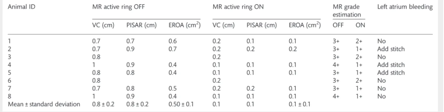
The postoperative intracardiac echocardiography showed MR in all animals, and the activation of the device allowed the re-duction of the MR in all cases (Fig.4). In all cases, the sliding element was brought at the P2 level in order to achieve the best correction of MR. Infive animals, the MR disappeared; in three animals, it was trivial. Vena contracta width decreased from 0.8 ± 0.2 to 0.1 cm; PISA radius decreased from 0.8 ± 0.2 to 0.1 cm and EROA decreased from 0.50 ± 0.1 to 0.1 ± 0.1 cm2. In two
animals, we were unable to calculate PISA and EOAR due to technical problems. Two animals had a reversal of systolic pulmonaryflow that normalized following the activation of the device. All corrections were reversible, causing the reappearance of MR. In three animals, additional sutures were placed to control bleeding from the left atrium. Table 1 reports the detailed results. Animals were sacrificed at the end of the procedure and devices retrieved.
DISCUSSION
‘Mitral repair is better than mitral replacement, whenever it’s pos-sible’ is one of the rare statements agreed on by both cardiolo-gists and cardiac surgeons. The advantages of mitral valve repair (MVR) include a low rate of thromboembolism, resistance to endocarditis and having no need of long-term anticoagulation [10]. However, repairing the mitral valve is a technically demand-ing procedure even for experienced surgeons and it is frustratdemand-ing when it culminates in residual regurgitation. These lead to a gen-erally low rate of repair. In a recent review, only about 44% of patients in the USA and 46% in Europe who required MV surgery for MR received MVR [10]. The persistence of trivial or mild (1+ to 2+) MR after the repair is generally tolerated, because an almost perfect repair is better than a second cardioplegia either to improve the correction or to change the valve. However, clinical studies have underlined the negative effect of residual MR on out-comes, particularly in ischaemic cardiopathy: revascularization alone did not eliminate the negative long-term effects of mild MR. Coronary artery bypass surgery patients with uncorrected mild or moderate MR are at increased risk of death (hazard ratio 1.34) and heart failure hospitalization (hazard ratio 1.34) [7,11].
Even when the repair is successful with excellent postoperative results, the progression of the degenerative disease and/or ischaemic cardiomyopathy are the major determinants of MR re-currence. In a recent study, Flamenget al. [12] reported that only 50% of the patients remain free from more than trivial mitral in-competence at 7 years after repair for degenerative disease.
Mitralflex ring seems to address the need for devices and/or techniques able to improve the clinical results of MVR. The device is implanted using the surgical technique initially described by Carpentier, with the exception of having a 3-mm thick cable piercing the left atrium. The Mitralflex ring provides the displacement of the posterior leaflet segments, keeping the anterior in place, because the anterior annulus of the mitral valve is tightly connected to the aortic valvular ring withfibrous tissue and is not easily dilated or deformed. Any deformation of the annulus at this level is difficult to achieve and could induce aortic regurgitation.
The animal model we proposed is based on historical data and is meant to reproduce the functional changes in MV movements observed in IMR due to restricted leaflet motion. The papillary muscles, normally parallel to the left ventricle long axis and per-pendicular to the leaflets efficiently balance the forces generated by ventricular pressure on the leaflet surface. Ischaemia or heart failure causes the myocardial segments underlying the PMs to
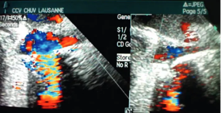
Figure 4:Intracadiac echocardiography showing severe MR (right) and its
cor-rection after device activation (left).
Figure 3: The Mitralflex ring is implanted using the interrupted suture
bulge posteriorly and outward, displacing the PMs, so that they pull the leaflets nonperpendicularly, away from their normal coaptation [13,14]. The distance between the papillary muscles tips and the annulus also increases, drawing the leaflets into the ventricle and restricting their motion towards closure [15,16].
Because the cable was a source of minor bleeding in three animals, we recommend placing additional sutures on the left atrium at the exit site in order to reduce bleeding complication.
Echocardiographic evaluation of the severity of MR is complex, and simple ‘eyeball’ grading of MR colour flow jets is prone to error. However, all quantitative measurement methods such as vena contracta width, regurgitate volume and EROA have inherent strengths and weakness. Vena contracta width is simple and good at identifying mild or severe MR, but not useful for multiple MR jets, and the small values could lead to a large per-centage of errors. PISA provides both lesion severity (EROA) and volume overload, but is cumbersome and not accurate in eccen-tric jets. Moreover, intracardiac ultrasound is not routinely per-formed and we personally lack experience with this technique. Therefore, we integrated the various echocardiographic mea-sures of MR severity, trying to minimize measure errors.
The reshaping of the mitral annulus induced by the activation of the device allowed the precise correction of 2+ MR in the postoperative phase. The activation of the ring was smooth, and under echocardiography control, we were able to reshape the posterior leaflet geometry to optimize leaflet coaptation. Because the correction was very precise and reversible, in all animals, we were able to adjust leaflet coaptation as many times as necessary to minimize MR, and this is a step forward with respect to other active mitral rings that provide only one direc-tion andfixed modification of valve geometry [17].
Although this ring has the same clinical indications as the clas-sical rigid mitral rings, it should provide particular benefit in IMR. Experimental and clinical studies have demonstrated that patients with IMR have decreased annular motion during the cardiac cycle [18] and both anterior and posterior portions of the annulus dilate proportionately to IMR severity, although the magnitude of the dilatation of the posterior annulus is greater [19]. Similarly, the intertrigonal distance is increased in IMR at end diastole, and these changes are the result of wall motion abnormalities due to myocardial infarction in the distribution of the dominant right coronary artery [20]. The altered geometry of the left ventricle
results in the loss of function of the valve secondary to excess tethering of the valve leaflets and resultant loss of zone coapta-tion. Surgical repair by annuloplasty effectively reduces the septal–lateral diameter of the mitral annulus [21] and, when acti-vated, the Mitralflex ring further reduces this distance, increasing the leaflets coaptation. It should represent an additional tool to improve immediate and mild-term correction results of MR.
Because the Mitralflex ring is thought to be activated even months or years after the implant, we could speculate that it could be helpful to correct recurrent MR and therefore to improve the clinical outcome of mitral repair, but further studies are necessary to confirm this statement.
Without adding complexity to the standard surgical procedure or additional risks for the patient, the Mitralflex ring expands the opportunity to perform effective, good mitral repair and to correct recurrent mitral regurgitation, probably improving clinical outcome.
Funding
This work was supported by the Swiss public grant (Centre Hospitalier Universitaire Vaudois) and a private grant from Kephalios SA, Paris.
Conflict of interest: Piergiorgio Tozzi and Didier Locca are company shareholders.
REFERENCES
[1] Carpentier A. Cardiac valve surgery: “The French correction”. J Thorac
Cardiovasc Surg 1983;86:323–37.
[2] Feldman T, Foster E, Glower DD, Kar S, Rinaldi MJ, Fail PS et al.
Percutaneous repair or surgery for mitral régurgitation. N Engl J Med 2011;15:1395–405.
[3] Bonow RO, Carabello BA, Chatterjee K, de Leon AC Jr, Faxon DP, Freed
MDet al. 2008 Focused update incorporated to the ACC/AHA 2006
guidelines for the management of patients with valvular heart disease : a report of the ACC/AHA task force on practice guidelines. J Am Coll Cardiol 2008;52:e1–142.
[4] Click RL, Abel MD, Schaff HV. Intraoperative transesophageal echocardi-ography: 5 years prospective review of impact on surgical management. Mayo Clin Proc 2000;75:241–7.
Table 1: Animal by animal quantification of mitral regurgitation (MR) induced by folding mitral chordae (active ring OFF) and after the activation of the Mitralflex ring (active ring ON)
Animal ID MR active ring OFF MR active ring ON MR grade
estimation
Left atrium bleeding
VC (cm) PISAR (cm) EROA (cm2) VC (cm) PISAR (cm) EROA (cm2) OFF ON
1 0.7 0.7 0.6 0.2 0.1 0.1 3+ 2+ No 2 0.7 0.9 0.7 0.2 0.2 0.2 3+ 1+ Add stitch 3 0.8 0.2 3+ 2+ No 4 1 0.9 0.4 0.1 0.1 0.1 4+ 1+ Add stitch 5 0.8 0.8 0.4 0.1 0.1 0.1 3+ 1+ Add stitch 6 0.8 0.2 3+ 2+ No 7 0.7 0.8 0.5 0.2 0.2 0.1 3+ 1+ No 8 1 0.9 0.4 0.1 0.1 0.1 4+ 1+ No
Mean ± standard deviation 0.8 ± 0.2 0.8 ± 0.2 0.50 ± 0.1 0.1 0.1 0.1 ± 0.1
MR, mitral regurgitation; PISA, proximal isovelocity surface area; EORA: effective regurgitant orifice area; VC: vena contracta.
BASIC
S
[5] Michel-Cherqui M, Ceddaha A, Liu N, Schlumberger S, Szekely B,
Brusset Aet al. Assessment of systematic use of intraoperative
transeso-phageal echocardiography during cardiac surgery in adults: a
prospect-ive study of 203 patients. J Cardiothorac Vasc Anesth 2000;14:45–50.
[6] McGee EC, Gillinov AM, Blackstone EH, Rajeswaran J, Cohen G, Najam F et al. Recurrent mitral regurgitation after annuloplasty for functional is-chemic mitral regurgitation. J Thorac Cardiovasc Surg 2004;128:96–24.
[7] Rizza A, Sulcaj L, Glauber M, Trianni G, Palmieri C, Mariani M et al.
Predictive value of less than moderate residual mitral régurgitation as assessed by TEE for the short-term outcomes of patients with mitral re-gurgitation treated with mitral valve repair. Cardiovasc Ultrasound 2007; 5:25.
[8] Carpentier AF, Lessana A, Relland JY, Belli E, Mihaileanu S, Berrebi AJ et al. The "physio-ring": an advanced concept in mitral valve annulo-plasty. Ann Thorac Surg 1995;60:1177–85.
[9] Lancellotti P, Moura L, Pierard LA, Agricola E, Popescu BA, Tribouilloy C et al. European Association of Echocardiography recommendations for the assessment of valvular régurgitation. Part 2: mitral and tricuspid re-gurgitation (native valve disease). Eur J Echocardiogr 2010;11:307–32. [10] Fedak PW, McCarthy PM, Bonow RO. Evolving concepts and
technolo-gies in mitral valve repair. Circulation 2008;117:963–74.
[11] Schroder JN, Williams ML, Hata JA. Impact of mitral valve regurgitation evaluated by intraoperative transesophageal echocardiography on long-term outcomes after coronary artery bypass grafting. Circulation 2005; 112:I293–8.
[12] Flameng W, Herijgers P, Bogaerts K. Recurrence of mitral valve regurgita-tion after mitral valve repair in degenerative valve disease. Circularegurgita-tion
2003;107:1609–13.
[13] Burch GE, DePasquale NP, Phillips JH. Clinical manifestations of papillary
muscle dysfunction. Arch Intern Med 1963;112:112–7.
[14] Burch GE, DePasquale NP, Phillips JH. The syndrome of papillary muscle
dysfunction. Am Heart J 1968;75:399–415.
[15] Silverman ME, Hurst JW. The mitral complex: interaction of the anatomy,
physiology, and pathology of the mitral annulus, mitral valve leaflets,
chordae tendineae, and papillary muscles. Am Heart J 1968;76:399–418. [16] Perloff JK, Roberts WC. The mitral apparatus: functional anatomy of
mitral regurgitation. Circulation 1972;46:27–39.
[17] Langer F, Borger MA, Czesla M, Shannon FL, Sakwa M, Doll Net al.
Dynamic annuloplasty for mitral regurgitation. J Thorac Cardiovasc Surg
2013;145:425–9.
[18] Hueb AC, Jatene FB, Moreira LF, Pomerantzeff PM, Kallás E, de Oliveira
SAet al. Ventricular remodeling and mitral valve modifications in dilated
cardiomyopathy: new insights from anatomic study. J Thorac Cardiovasc
Surg 2002;124:1216–24.
[19] Ahmad RM, Gillinov AM, McCarthy PM, Blackstone EH,
Apperson-Hansen C, Qin JXet al. Annular geometry and motion in human
ische-mic mitral regurgitation: novel assessment with three-dimensional
echocardiography and computer reconstruction. Ann Thorac Surg 2004;
78:2063–8.
[20] Gillinov AM, Wierup PN, Blackstone EH, Bishay ES, Cosgrove DM, White J et al. Is repair preferable to replacement for ischemic mitral regurgitation? J Thorac Cardiovasc Surg 2001;122:1125–41.
[21] Miller DC. Ischemic mitral regurgitation redux–to repair or to replace?
J Thorac Cardiovasc Surg 2001;122:1059–62.
APPENDIX. CONFERENCE DISCUSSION
Dr A. Ahlsson (Örebro, Sweden): I would like to take the position of an average mitral valve surgeon trying to understand the place of adjustable mitral rings in mitral valve repair surgery. The absolute majority of our patients undergo mitral valve surgery because of type II prolapse of one or more segments, and a failed mitral valve repair in this group of patients can rarely be explained by the choice of the annuloplasty ring. Maybe you think you have pinpointed this, but I would like you once more to state it. Can you
please define the group of MR patients in whom you think the use of
adjust-able mitral rings will improve long-term outcome?
Dr Tozzi: Initially, we were thinking about patients with functional mitral re-gurgitation, as for example, in Barlow’s disease. Then we thought that prob-ably this ring also has a place in the treatment of ischaemic mitral regurgitation. So basically this ring could be used as an alternative to the classic Carpentier ring.
Dr Ahlsson: For all patients? Dr Tozzi: Yes.
Dr Ahlsson: My second question deals with the practical problem of leaving a rotable tool in the left atrium piercing the atrial wall. I get a little worried about the risk of thrombosis, emboli or bleeding or if you are in heavy exercise. I would like your comment on this.
Dr Tozzi: Well, as you saw in the paper, we had two animals that had bleeding just because it was early in the experience. So we decided to improve the purse string suture where the cable pierced the atrium. And then concerning the thrombi, these were all acute animal studies, so we do not have long-term experience. But we are confident that if the ring is covered with biocompatible material as used in standard rings, like Dacron for example, such complications should be avoided.
Dr A. Diegeler (Bad Neustadt, Germany): In those patients with ischaemic mitral incompetence, usually the lack of coaptation is located more towards the postero-medial part. Has the ring any option to address this by a kind of asymmetric shrinking or does it always provide a symmetric shrinking?
Dr Tozzi: Yes, absolutely. This kind of ring can address the asymmetric placement of the posterior leaflet. So you can choose to reduce the AP dis-tance at the level of P3, for example, or P2 or P1 according to the need. And it is extremely selective, so it is not just a general shrinking of the annulus.