Publisher’s version / Version de l'éditeur:
Vous avez des questions? Nous pouvons vous aider. Pour communiquer directement avec un auteur, consultez la première page de la revue dans laquelle son article a été publié afin de trouver ses coordonnées. Si vous n’arrivez pas à les repérer, communiquez avec nous à [email protected].
Questions? Contact the NRC Publications Archive team at
[email protected]. If you wish to email the authors directly, please see the first page of the publication for their contact information.
https://publications-cnrc.canada.ca/fra/droits
L’accès à ce site Web et l’utilisation de son contenu sont assujettis aux conditions présentées dans le site LISEZ CES CONDITIONS ATTENTIVEMENT AVANT D’UTILISER CE SITE WEB.
Analytical Chemistry, 92, 1, pp. 1618-1627, 2019-12-06
READ THESE TERMS AND CONDITIONS CAREFULLY BEFORE USING THIS WEBSITE.
https://nrc-publications.canada.ca/eng/copyright
NRC Publications Archive Record / Notice des Archives des publications du CNRC :
https://nrc-publications.canada.ca/eng/view/object/?id=05cbb202-3514-4a60-90c6-530d92297448 https://publications-cnrc.canada.ca/fra/voir/objet/?id=05cbb202-3514-4a60-90c6-530d92297448
NRC Publications Archive
Archives des publications du CNRC
This publication could be one of several versions: author’s original, accepted manuscript or the publisher’s version. / La version de cette publication peut être l’une des suivantes : la version prépublication de l’auteur, la version acceptée du manuscrit ou la version de l’éditeur.
For the publisher’s version, please access the DOI link below./ Pour consulter la version de l’éditeur, utilisez le lien DOI ci-dessous.
https://doi.org/10.1021/acs.analchem.9b04937
Access and use of this website and the material on it are subject to the Terms and Conditions set forth at
Chemoenzymatic method for glycoproteomic N-glycan type
quantitation
Li, Henghui; Li, Leyuan; Cheng, Kai; Ning, Zhibin; Mayne, Janice; Zhang,
Xu; Walker, Krystal; Chen, Rui; Twine, Susan; Li, Jianjun; Figeys, Daniel
Chemoenzymatic Method for Glycoproteomic N‑Glycan Type
Quantitation
Henghui Li,
†Leyuan Li,
†Kai Cheng,
†Zhibin Ning,
†Janice Mayne,
†Xu Zhang,
†Krystal Walker,
†Rui Chen,
§Susan Twine,
§Jianjun Li,
§and Daniel Figeys
*
,†,‡†
SIMM-University of Ottawa Joint Research Center in Systems and Personalized Pharmacology and Ottawa Institute of Systems Biology and Department of Biochemistry, Microbiology and Immunology, Faculty of Medicine, University of Ottawa, 451 Smyth Road, Ottawa, Ontario K1H 8M5, Canada
‡
Molecular Architecture of Life Program, Canadian Institute for Advanced Research, Toronto M5G 1M1, Canada §
Human Health Therapeutics Research Centre, National Research Council Canada, Ottawa, Ontario K1A 0R6, Canada
*
S Supporting InformationABSTRACT: Glycosylation is one of the most important post-translational modifications in biological systems. Current glycoproteome methods mainly focus on qualitative identification of glycosylation sites or intact glycopeptides. However, the systematic quantitation of glycoproteins has remained largely unexplored. Here, we developed a chemoenzymatic method to quantitatively investigate glycoproteome based on the N-glycan types. Taking advantage of the specificity of different endoglycosidases and isotope dimethyl labeling, six N-glycan types of structures linked on each glycopeptide, including high-mannose/hybrid, biantennary, and triantennary with/without core fucose, were quantified. As a proof of principle, the glycoproteomic N-glycan type quantitative (glyco-TQ) method
was first used to determine the N-glycan type composition of the immunoglobulin G1 (IgG1) Fc fragment. Then we applied the method to analyze the glycan type profile of proteins from the breast cancer cell line MCF7, and we quantitatively revealed the N-glycan type microheterogeneity at the glycopeptide and glycoprotein level. The novel quantitative strategy to evaluate the relative intensity of the six states of N-glycan type glycosylation on each site provides a new avenue to investigate the function of glycoproteins in broad areas, such as cancer biomarker research, pharmaceuticals characterization, and antiglycan vaccine development.
G
lycosylation is one of the most common post-transla-tional modifications (PTMs).1 Glycans exhibit vast structural microheterogeneity which is mainly generated by variable glycan structures at each of their specific glycosylation sites. The N-linked glycans are generally attached to the Asn at Asn-X-Ser/Thr consensus sequence, where X is any amino acid other than Pro.2The biosynthesis of N-linked glycoproteins isunder a complex sequence of enzymatically catalyzed events, leading to a variety of diverse N-glycan structures. The diverse N-glycan structures are generally classified into three types: high mannose, hybrid, and complex type glycans, with all N-glycans sharing a common penta-saccharide (GlcNAc2Man3) core structure.3 Although the structure of glycan is variable,
evidence shows that the mammalian glycans are remarkably well conserved in certain organisms, expressing a distinct array of glycan profiles under defined conditions.4
Mass spectrometry (MS) is a powerful platform to comprehensively analyze protein glycosylation. However, due to the low abundance of glycosylated peptides and the heterogeneity of glycan structures, N-glycopeptide enrichment is required. Several enrichment methods have been reported,
including lectin5 and hydrazide chemistry-based methods,6,7
boronic acid enrichment,8 hydrophilic interaction liquid chromatography (HILIC),9 and metabolic labeling.10,11 In
general, these strategies for detecting N-linked glycosylated sites require an additional deglycosylation step by N-glycosidase F (PNGase F) before MS detection.5−7 Unfortu-nately, this results in the loss of the glycan structure information at the glycosylated sites as the glycans are removed from the peptides.
Recently, a site-specific glycoproteomic method was used to detect intact glycopeptide by MS with a variety of tandem MS techniques.10,12−14 This strategy allows the simultaneous detection of glycopeptide sequence, glycosylation site, and glycan structures in one MS/MS spectrum. Current state-of-the-art MS technology with multiple dissociation known as activated ion electron transfer dissociation methods (AI-ETD)
Received: October 29, 2019 Accepted: December 6, 2019 Published: December 6, 2019
Article
pubs.acs.org/ac Cite This:Anal. Chem. 2020, 92, 1618−1627
© 2019 American Chemical Society 1618 DOI:10.1021/acs.analchem.9b04937
Anal. Chem. 2020, 92, 1618−1627
allowed intact glycoproteomic identification of more than 1000 (∼1500) intact N-glycopeptides from a mouse brain tissue.13 The site-specific glycoproteomic strategies have been applied to quantitatively detect glycoprotein alteration using the isotope labeling,12 isobaric labeling strategies,15 and a label-ing-free method.16 However, due to missed detection of low abundant glycan structures, more than half of identified glycopeptides were linked with only one or two glycan structures using the intact glycopeptide method, hindering the comprehensive quantitation in complex biological samples.13
The diverse N-glycan structures play important roles in many key biological processes, including cell adhesion, receptor activation, molecular trafficking, signal transduction and disease progression, and immunotherapy.17,18 Some apparent changes associated with cancers are the over-expression of sialylation and core fucosylation, and complex branched N-glycans. For example, an increased core fucosylated type of N-glycan is an important signature of several cancers, such as hepatocellular carcinoma,19 lung cancer,20 and breast cancer.21 Therefore, quantitatively monitoring the N-glycan type changes in glycosylation is important for the diagnosis, prognosis, and understanding of molecular mechanisms involved in pathogenesis. Cao et al. introduced a MS-based method that used two glycosidases, PNGase F and endoglycosidase H (Endo-H), to assess the site occupancy and proportion of high-mannose and complex-type glycans of purified human immunodeficiency virus (HIV) envelope (Env) glycoprotein.22The NMR-based strategy was introduced to allow dissecting the glycan pattern of the IgE high-affinity receptor (FcεRIα), presenting of pauci-mannose, high-mannose, hybrid, and bi-, tri-, and tetra-antennary complex type N-glycans with different degrees of fucosylation and sialylation.23A purification step of glycoprotein is required in these methods; therefore, glycan type quantitation at the proteome level is urgently needed for complex biological samples.
To fulfill this analytical challenge, we developed a robust chemoenzymatic based method that quantitatively determined the proportion of N-glycan types at each glycopeptide. Briefly, three aliquots of trypsin proteolyzed sample are treated in parallel with three specific endoglycosidases and the aliquots are isotopically labeled using the three plex dimethyl labeling strategy and then combined. The cleaved N-glycopeptides are biotinylated and enriched by affinity chromatography, and the eluted N-glycopeptides are analyzed by MS. The glycoproteo-mic N-glycan type quantitative (Glyco-TQ) strategy was first applied on the standard glycoprotein IgG1 Fc fragment and further used to comprehensively investigate glycopeptides from the MCF7 breast cancer cell line. The data interpretation is convenient and compatible with the general proteomic platform, without the need of laboriously generating sample-specific spectral libraries, a complex data filtering process, or specialized commercial data analysis tools. The result showed that the novel strategy could provide quantitative information on important characteristics of glycoproteins, including the relative proportion of high-mannose and linkage-related complex type glycan and the proportion of nonfucosylated and core fucosylated type glycan at each glycosylated site. To our knowledge, this is the first report to quantify the proportion of N-glycan types on the glycopeptides using the data-dependent acquisition mode for a complex biological sample.
■
METHODSMaterials. Endoglycosidase S, F3, N-glycosidase F (PNGase F), β-N-acetylhexosaminidasef, and alkaline phos-phatase were from New England Biolabs. Mutant β1−4-galactosyltransferase (Gal-T1 Y289L)) and high capacity streptavidin agarose were obtained from Thermo Scientific. IdeS protease was purchased from Promega. UDP-GalNAz was from chemily Glycoscience. The immunoglobulin G1 (IgG1) from normal human plasma was obtained from Athens Research & Technology. 2-(4-((Bis((1-(tert-butyl)-1H-1,2,3-triazol-4-yl) methyl)amino)methyl)-1H-1,2,3-triazol-1-yl) ace-tic acid (BTTAA), photocleavable biotin alkyne (PC biotin alkyne) were purchased from Click Chemistry Tools. EDTA-free protease inhibitor cocktail was obtained from Roche Diagnostics. RapiGest SF surfactant and the Sep-Pak tC18 cartridge were obtained from Waters. All other chemical materials, if not specifically highlighted, were obtained from Millipore Sigma.
Preparing the Standard Glycoprotein.Immunoglobulin G1 (100 μg) was treated with 1000 units IdeS protease for 3 h in 1X phosphate buffered solution (PBS, pH 7.6). 200 μL of immobilized Protein A resin slurry (50% w/v) was added to the reaction buffer, and incubated with gentle mixing for 2 h at room temperature. Then the Protein A resin slurry was transferred into centrifuge columns and Protein A resin was washed with 1X PBS three times to remove unbound F(ab’)2 fragments (fragment antigen-binding). The Fc fragments (fragment crystallizable region) of immunoglobulin G1 (IgG 1) were eluted with 100 mmol/L glycine buffer, pH 2−3. The Fc fragments were immediately neutralized with 1 M Tris-HCl buffer, pH 8, and stored in −80 °C for further use.
Cell Culture, Protein Extraction, and Protein Diges-tion.The MCF-7 cell line was obtained from American Type Culture Collection (ATCC). MCF-7 cells were maintained in advanced MEM media (Gibco) with 10% (v/v) FBS, 1X GlutaMAX (Gibco), and 2.8 μg/mL Gentamicin (Gibco). The cells were cultured at 37 °C and 5% CO2. Once the cells reached 80% confluency, cells were harvested in the ice-cold RIPA buffer (50 mM HEPES pH 7.6, 150 mM NaCl, 1% NP-40, 1% sodium deoxycholate, 0.1% sodium dodecyl sulfate (SDS), and protease inhibitor cocktail (CompleteMini, Roche)) by scraping. Cell lysates were subjected to ultra-sonicate (10 s process with 10 s interval for 1 min) on ice using a Q125 Sonicator with 50% amplitude. The cell debris was removed through centrifugation at 16000g, 4 °C, 10 min. The protein in the supernatant was precipitated using 6-fold volume ice cold acetone overnight at −20 °C. Protein was pelleted by centrifugation at 16000g, 4 °C, 10 min and washed with the ice-cold acetone two times. For the in-solution trypsin digestion, the procedure was performed as in the previous report.24Briefly, the resulting protein was dissolved in 50 mM
ammonium bicarbonate and 8 M urea solution pH 7.8, reduced in 5 mM dithiothreitol (DTT) (56 °C, 30 min), and alkylated by with 10 mM iodoacetamide (25 °C, 40 min in the dark). Cell proteins were digested with the protein:trypsin (Worthington Biochemical Corp) at ratio, 50:1, in 50 mM ammonium bicarbonate, 1 M urea solution pH 7.8 at 37 °C for 20 h. After the digest, the solution was acidified (pH 2−3) by 0.5% formic acid and centrifuged to remove the debris, the supernatant was collected, and peptide was desalted by Sep-Pak tC18 cartridge (Waters). The peptide elution was dried by SpeedVac concentrator (Thermo Scientific).
Endoglycosidases Digestion and Dimethyl Labeling. To leave one acetylglucosamine for high mannose linked N-peptide, peptide from 1 mg protein was parallelly digested with 0.05 U Endo-H (sigma) in the 50 mM sodium acetate buffer, pH 6; 2000 units Endo-S (NEB) for biantennary N-glycan in 50 mM sodium acetate, 5 mM calcium chloride buffer pH 5.5; 100 units Endo-F3 (NEB) in 50 mM sodium acetate, pH 4.5, for 24 h, respectively. After the endoglycosidase was denatured at 95 °C, 5 min, β-N-acetylhexosaminidasef was added to remove O-GlcNAc for another 8 h. After desalted by Sep-Pak tC18 cartridge, the peptide was adjusted to pH 6 with HEPES buffer. For isotope dimethyl labeling, the peptides were treated with 10 μL 20% (v/v) CH2O, 15 μL 3 M NaBH3CN for the Endo-H treated sample; 10 μL 20% (v/v) CD2O, 10 μL 3 M NaBH3CN for the Endo-S treated sample; 15 μL 20% (v/v) CD2O, 15 μL 3 M NaBD3CN for the Endo-F3 treated sample, at 25 °C for 45 min with mixing. The reaction was quenched by adding 10 μL 20% (v/v) ammonia solution, combined, purified by Sep-Pak cartridge, and dried by Speedvac.
Glycopeptide Enrichment. All the peptide was resus-pended in the HEPES buffer (pH 7.9) containing 5 mM Zn2+, 2 μL phosphatase, 10 μL GalT1 T298L/1 mg peptide, and 25 μg UDP-GalNAz/1 mg peptide, incubated in 4 °C for 24 h. Excess UDP-GalNAz was remove by Sap-pak C18 cartridge. The peptide was dried in the speedVac and resuspended in the PBS buffer, pH 7.6. The copper(I)-catalyzed azide−alkyne cycloaddition (CuAAC) reaction reagents (25 nmol PC biotin alkyne, 300 μM CuSO4, 600 μM BTTAA, 1.50 mM sodium ascorbate) were mixed with the GalNAz labeled peptide, and the reaction was incubated for 3 h at 25 °C. 200 μL high capacity streptavidin agarose resin was add to the mixture and incubated overnight at 4 °C. The beads were extensively washed with 2 M urea ten times (pH 7.6), 1X PBS buffer (pH 7.6) ten times, and 20% (v/v) acetonitrile (ACN) ten times. The beads were then resuspended in 50% (v/v) ACN, transferred to clear thin-walled polymerase chain reaction
(PCR) tubes, and illuminated by 365 nm UV (VWR transilluminator, LM-20E) for 30 min at 4 °C with gentle mixing. The supernatant from each fraction was collected, lyophilized, and stored at −20 °C.
Mass Spectrometry Analysis. Glycopeptides were
analyzed by an Eksigent nanoLC liquid chromatograph that was connected in-line with an Q Exactive HF-X MS. The separation of peptides was performed on an analytical column (75 μm × 50 cm) packed with reverse phase beads (1.9 μm; 120-Å pore size; Dr. Maisch GmbH) with 2-hour gradient from 5 to 35% acetonitrile (v/v) at a flow rate of 200 nL/min. The full scan mass spectra were acquired over the range 300− 1800 (m/z) with the mass resolution setting 70000 at m/z 400. Maximum injection time 100 ms; AGC target value 1e6. The 12 most intense ions were selected for tandem mass spectrometry detection with the following parameters: collision energy, 30%; exclusion ions charge 1, 2, 7, 8, >8; resolution 17500, AGC target 1e5; maximum injection time 120 ms.
Data Analysis. The raw data were processed using the MaxQuant software and searched against with UniProt human database containing all proteins in the UniProt Human (Homo sapiens) database (20190802). The general parameters were performed during the search: 10 ppm precursor mass tolerances; digested with trypsin; two max missed cleavages; fixed modifications: carbamidomethylation of cysteine (+57.0214); variable modifications: oxidation of methionine (+15.9949). The common tag was also performed as variable modifications: modified amino acid, asparagine (N); compo-sition H30C19N6O10, GlcNAc-GalNAz- photocleavable tag (GalNAzPCt), 502.2023 Da; neutral losses, GlcNAc-GalNAzPCt, GalNAzPCt H17C11N5O5; diagnostic peaks, GalNAzPCt H17C11N5O5, GlcNAc H13C8NO5, GlcNAc-GalNAzPCt. If the N-glycopeptide was modified with core fucose, fucosylated linked was performed as variable modifications as following: modified amino acid, asparagine (N); composition H40C25N6O14, Fuc-GlcNAc-GalNAzPCt,
Figure 1.Enrichment and quantitative strategy for N-glycoproteomics. (a) The workflow for the N-glycopeptide enrichment and quantitation. (b) Six N-glycan types of glycan structures linked on the glycopeptide: nonfucosylated high mannose/hybrid type, nonfucosylated biantennary, nonfucosylated triantennary, core fucosylated high mannose/hybrid type, core fucosylated biantennary, core fucosylated bi/triantennary type.
Analytical Chemistry Article
DOI:10.1021/acs.analchem.9b04937
Anal. Chem. 2020, 92, 1618−1627 1620
648.2602 Da; neutral losses, Fuc H10C6O4, Fuc-GlcNAc-GalNAzPCt, Fuc-GlcNAc-GalNAzPCt, Fuc+GalNAzPCt H27C17N5O9; diagnostic peaks, GalNAzPCt, GlcNAc, Fuc, GlcNAc-Gal-NAzPCt, Fuc-GlcNAc-GalNAzPCt and Fuc-GlaNAzPCt H23C14NO9. Data are available via ProteomeXchange with identifier PXD015805.
■
RESULTSWe developed a method for the quantitative analysis of N-glycan type microheterogeneity at glycopeptide levels. The method includes the following steps (Figure 1a): (i) the trypsin proteolyzed peptides were divided into three aliquots, treated with one of three endoglycosidase (H, S, and F3), and incubated with β-N-acetylhexosaminidasefto remove O-linked N-acetylglucosamines (O-GlcNAc); Endoglycosidase H (Endo-H) releases high mannose and hybrid type N-glycans, including those that have a fucose residue attached to the core structure;25 endoglycosidase S (Endo-S) releases biantennary complex type glycans;26 and endoglycosidase F3 (Endo-F3)
releases core fucosylated biantennary complex type glycan and triantennary complex glycan from N-glycopeptides;27 (ii) the
three aliquots were dimethyl labeled with “light” isotope for Endo-H treated peptides, “intermediate” isotope for Endo-S treated peptides and “heavy” isotope for Endo-F3 treated peptides, respectively; (iii) the aliquots were combined and the retained GlcNAc on the N-glycopeptide was further trans-formed with N-azidoacetylgalactosamine (GalcNAz) through the catalysis of β-1,4-galactosyltransferase Y289L (GalT1 Y298L); (iv) the GalNAz labeled N-glycopeptides were covalently reacted with the photocleavable (PC) alkyne biotin through the copper(I)-catalyzed azide−alkyne cycloaddition
(CuAAC) reaction; (v) the biotin linked peptides were enriched by high capacity streptavidin agarose affinity chromatography. The nonglycopeptide and reagents were removed through extensively washing; (vi) the N-glycopep-tides were released by 365 nm ultraviolet irradiation and detected by MS. The detailed schematic for the PC alkyne biotin structure and reaction procedure is shown inFigure S1. We have tested different endoglycosidases for their specificity and selected three endoglycosidases for our method. To evaluate the specificity of the selected endoglycosidases, glycopeptides from MCF7 cells were incubated with Endo-H, Endo-S, and Endo-F3, respectively. The released glycans were collected, labeled with procainamide through reductive amination, and analyzed by nano LC-MS. The result showed that five high mannose and four hybrid N-glycans could be released by Endo-H (Figure S2a). The substrates for Endo-S were all biantennary complex glycans (Figure S2b). The Endo-F3 released the nonfucosylated triantennary and core fucosylated bi/triantennary glycans (Figure S2c). However, bisected biantennary structures with/without core fucose were not substrates for Endo-F3 (Figure S3). None of these three endoglycosidases showed any activity toward the more complex tetra-antennary structures. The detailed information on endoglycosidase specificity is listed inTable S1. Therefore, using the three endoglycosidases in our method, the quantitative assessment of six N-glycan types at glycopeptides was achieved and the six types of N-glycan were classified as follows: nonfucosylated high mannose/hybrid, nonfucosylated biantennary, nonfucosylated triantennary, core fucosylated high mannose/hybrid, core fucosylated biantennary, and core fucosylated bi/triantennary type glycans (Figure 1b).
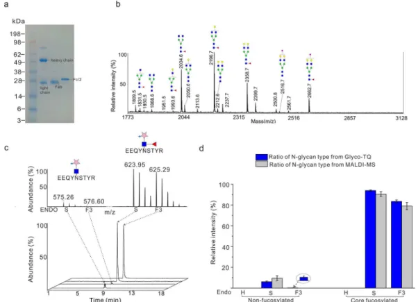
Figure 2.Quantitative analysis of the standard glycoprotein IgG1 Fc using the Glyco-TQ method. (a) Purification of Fc fragment from human serum IgG1. (b) MALDI-MS detection the glycan profile of Fc fragments. (c) Quantitative investigation of the proportion of Fc fragment glycopeptide, the MS profile of glycopeptide (up) and the corresponding extracted-ion chromatogram of glycopeptide. (d) Quantitative comparison of MALDI-MS method and Glyco-TQ method based on the glycan type.
Our method also includes a novel enrichment strategy. Previously, GalT1 Y298L was used to label the O-linked β-N-acetylglucosamine (GlcNAc) glycopeptide with GalNAz.28We discovered that the GalT1 Y298L could effectively label the N-GlcNAc (on average 95.3%) as shown inFigure S4. After the N-linked glycopeptides were modified with GalNAz, and covalently linked with photocleavable biotin through click reaction, the unlabeled peptides and other reagents were removed by extensive washing with urea and organic buffer. The PC biotin alkyne was selected for our method develop-ment because ultraviolet irradiation is a milder condition to release the labeled glycopeptides, when compared with chemical methods that use strong reductive hydrazine or oxidizing regents.29 Irradiation with 365 nm ultraviolet light
efficiently recovered glycopeptides from the agarose streptavi-din beads, with almost all glycopeptide released within 15 min (Figure S5).
Validating the Glyco-TQ Method on the Standard Glycoprotein IgG1 Fc.We first tested our approach using an immunoglobulin G1 (IgG1), containing one fragment crystallizable region (Fc fragments) and antigen-binding fragments (F(ab’)2fragment). It has one fixed N-glycosylated site at Asn297 of the Fc fragment while glycan occupancy on the F(ab’)2fragments is reported to be near 20%.30To obtain one standard N-glycoprotein with one fixed glycosylated site, IgG1 from human serum was treated with IdeS protease, to generate a homogeneous pool of F(ab’)2 and Fc/2 fragments and then the Fc fragments were enriched by protein A agarose chromatography (Figure 2a). As proof of principle for our quantitative method, the proportion of each glycan type was investigated by both matrix-assisted laser desorption/ioniza-tion-MS (MALDI-MS) and our novel Glyco-TQ method. The N-glycan spectrum of the Fc fragment was interrogated by MALDI-MS detection in Figure 2b. The proportion of different types of N-glycans was calculated through the peak intensity of each glycan structure based on the MALDI-MS spectrum. The endoglycosidase could release all the specified glycan structures from the intact glycopeptide (Figure S6) The detailed ratio information on each structure is shown inTable S2for the MALDI-MS detection. For the Glyco-TQ method, glycopeptides with sequence EEQYN#STYR were classified into six types based on their linked glycan structure: nonfucosylated high mannose/hybrid type, nonfucosylated biantennary type, nonfucosylated triantennary type, core fucosylated high mannose/hybrid type, core fucosylated biantennary type, and core fucosylated bi/triantennary type glycan. The MS spectra of enriched glycopeptides and their corresponding extracted-ion chromatograms are shown in
Figure 2c. The comparison of the proportion of glycan types based upon our Glyco-TQ method and MALDI-MS detection is shown inFigure 2d. The high mannose and hybrid glycan type was not detected using either method, showing the high specificity of Endo-H. As for the biantennary glycans released by Endo-S, the proportion of the core fucosylated biantennary glycan is 93.9% using our Glyco-TQ method and 90.5% using MALDI-MS detection; the proportion of the nonfucosylated biantennary is 6.1% using our Glyco-TQ method versus 9.5% using MALDI-MS detection. The proportion of the core fucosylated biantennary from our method is higher than using MALDI-MS detection (93.9% versus 90.5%), which may be due to their different ionization modes. The Endo-F3 did not release the core fucosylated bisecting biantennary glycans and nonfucosylated biantennary type glycans (showing only with
minor activity when compared with triantennary glycans) (Figure S4).31 Only 3.3% of nonfucosylated glycans were
released by the Endo-F3 in these experiments. In conclusion, our Glyco-TQ method quantitatively revealed glycan type and linkage of IgG1 Fc.
Identification of N-Glycopeptides from MCF7 Cells. Detection of Endogenous and Native N-Linked GlcNAc Glycopeptides.It has been reported that endo-β-N-acetylglu-cosaminidase (ENGase) acts as a deglycosylation enzyme for the misfolding proteins in the cytosol.32 Similar to the endoglycosidase, ENGase cleaves between the two core GlcNAc residues of the penta-saccharide core structure, leaving one N-acetylglucosamine residue attached to the asparagine. We first applied our enrichment method to enrich the endogenously and native existing N-linked GlcNAc glycopeptides from MCF7 total cell lysates in the absence of adding any endoglycosidase (Figure S7). Our enrichment strategy yielded 71 N-linked glycopeptides that mapped to consensus N-glycosylated sequences (Asn-X-Ser-Thr-Cys) and that were detected with modification of one N-linked GlcNAc residue (peptide-GlcNAc) (Table S3). At the same time, we also detected 63 O-linked GlcNAc modified peptides, which are located in the nucleus and cytoplasm by gene ontology (GO) analysis (Table S4). Caution should be taken when interpreting results since β-N-acetylhexosaminidasefcould not fully remove some abundant O-linked GlcNAc modifications. Detection of N-Glycopeptides from MCF7 Cells.To verify our enrichment strategy, we applied the three endoglycosidases (H, S, F3) to enrich all the high mannose, hybrid, and bi- and triantennary complex linked glycopeptides (Figure S8). To evaluate the reproducibility of our enrichment method, we performed three biological replicates with MCF-7 protein cell lysates and found 73% glycopeptides were identified in at least two replicates (Figure S9). We compared the glycopeptide and nonglycopeptide fractions in each parallel replicate, and showed that the specificity of our method is 55.4%. Our performance was better than the specificity of previously boronic acid and ZIC-HILIC enrichment method.8 After N-glycan was released by the endoglycosidase, the core fucose was retained on the core GlcNAc residue, which allowed us to simultaneously distinguish the nonfucosylated and core fucosylated peptides. In total, 1090 N-glycopeptides were detected, including 916 nonfucosylated and 174 core fucosylated glycopeptides, corresponding to 504 glycoproteins (Table S5). There were 116 glycopeptides with co-occurrence of the nonfucosylated and core fucosylated glycopeptides (Figure S10). As well, 58 glycopeptides were detected with only core fucosylated type glycans. Amino acid frequencies of sequences surrounding the N-glycosylated site are shown in
Figure S11 for both the canonical and atypical N-linked glycopeptides. The above was carried out using the stepped fragmentation strategy. The stepped fragmentation strategy could be used to fragment the same parent ion with different collision energies and combine all the fragment residues from all collisions into one spectrum. The advantage of this strategy is that it can combine the information on fragmentation for the fucosylated glycopeptide in the low collision energy and with the peptide sequence in the following high energy step. When the normalized collision energy (NCE) was set to 15, the fuc-GlcNAc linkage was prone to cleavage, which resulted in the parent ion and fucose neutral loss-ion as the highest peaks (Figure S12). When the fragmentation energy was set to NEC 30, the glycopeptide produced a series of b/y type ions and b/
Analytical Chemistry Article
DOI:10.1021/acs.analchem.9b04937
Anal. Chem. 2020, 92, 1618−1627 1622
y type ions attached with the glycan residue (Figure 3). For example, one MS/MS spectrum of atypical motif glycopeptide with sequence ISVN#NVLPVFDNLMQQK was identified with one core fucose as shown in Figure 3. In addition, the glycopeptides with common glycan tag (GlcNAc-GalNAzPCt) allowed identification of the glycopeptides with two glycosy-lated sites (Figure S13), overcoming significant challenges posed for their identification when using the intact glycopeptide method (5−7 kDa).33
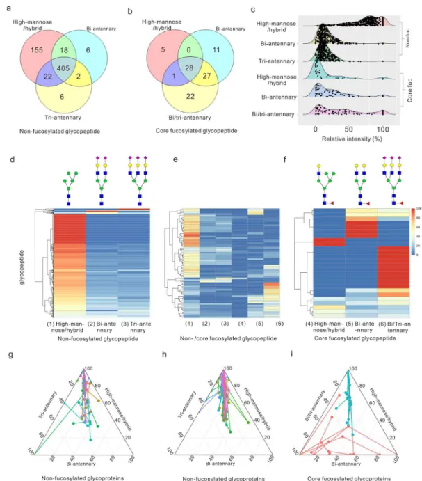
Applying the TQ Method to Analyze Glyco-peptides of the MCF7 Cell Line. Quantitative Micro-heterogeneity of Glycopeptides and Glycoproteins. The chemoenzymatic strategy allowed quantification of the N-linked glycan dynamics on a specific glycopeptide. After the peptides were treated with Endo-H, S, and F3, we combined and enriched the glycopeptides by affinity chromatography (Figure 1a). Relative quantitation of isotope peaks was calculated using the intensity of each peak area, which represented the proportion of each glycan type on the glycopeptide. First, we evaluated whether the different endoglycosidase treatments introduced biases during the glycan releasing steps. As show in Figure S14, high values from correlation coefficients (the value of Pearson’s correlation coefficient r > 0.96) were observed between the different endoglycosidase treatments after isotope labeling. Thus, we concluded that the endoglycosidase did not exhibit off-target protease activity and so did not introduce sample bias during glycan releasing steps. In total, we quantitatively detected 698 peptides that mapped to 378 proteins. All the glycopeptides and the relative ratio of each glycan type are shown inTable S6. Almost all the nonfucosylated glycopeptides (>97%) linked with the high-mannose or hybrid glycans (Figure 4a), while only 2 glycopeptides were linked with core fucosylated hybrid glycan and more than 97% core fucosylated peptides were linked with bi- and triantennary complex glycans (Figure 4b). The quantitative results exhibit distinct expression profiles for six glycan types on the glycopeptide as shown in theFigure 4c, highlighting the abundant expression of nonfucosylated
high-mannose and hybrid N-glycan. We use the heatmap to show the quantitative microheterogeneity of glycopeptide and relative distribution of glycan type present on the particular glycosylated site (Figure 4d−f). For the nonfucosylated glycopeptide, 143 glycopeptides were only linked with high-mannose and hybrid type glycan (Figure, 4d). Nonfucosylated glycopeptides (86.3%) were linked with high-mannose and hybrid type glycan, of which their proportion is larger than 50% (Table S6 and Figure, 4d). Consistent with previous reports, our results showed that N-glycans from the MCF7 cell line were predominant of nonfucosylated high-mannose/ hybrid type glycans.34 That is expected as all of the glycoproteins were first linked with high mannose type glycan during the biosynthesis.35 As for the quantitative micro-heterogeneity on the protein level, we studied internal connection of different glycopeptides from the same glycoprotein (Figure 4g−i). The distance between the connected dots represents the glycan type expressing divergence of glycopeptides from the same protein. Some proteins with more than one glycosylated site, such as hemicentin-1, membralin, and deoxyribonuclease-2-alpha, were detected only linked with the nonfucosylated high-mannose and hybrid type glycans, released by Endo-H. However, the majority of detected proteins with multiple glycosylated sites have differential N-glycan type profiles at each site for both the nonfucosylated (Figure 4g, h) and fucosylated glycoproteins (Figure, 4i). Due to the massive expression of nonfucosylated high-mannose and hybrid type, distribution of nonfucosylated proteins was constricted to a small region (Figure 4g, h), while the fucosylated glycoproteins were more widely distributed, based upon the quantitative information on six N-glycan types (Figure 4i). We also compared the location of nonfucosylated and fucosylated glycoproteins and found that more than 60% of fucosylated proteins were located in the extracellular exosome while it was 39.8% for nonfucosylated counterparts (Figure S15). That result indicates the N-linked core fucose may play significant roles in molecular trafficking.
Figure 3.MS/MS spectrum of atypical motif glycopeptide with sequence ISVN#NVLPVFDNLMQQK. * represents the b or y ions losing the glycan common tag. The neutral loss of fucose was shown between the parent ion at m/z 2608.2 and the fucose neutral loss-ion at m/z 2462.2. The oxonium ions from the common glycan tag were set as diagnostic ion peak, representing the fragment of GlcNAz at m/z 204.0, GalNAzCA at m/z 300.1, and GlcNAc-GalNAzPCt at m/z 503.2. The mass shift between b7 and b7* (* represents losing the common tag modification) is 618.3 Da, which is exactly the mass of the common glycan tag (Fuc-GlcNAc-GalNAzPCt).
Glycoproteins Related with Cancer. We quantitatively detected some glycoproteins, previously reported to be related with cancers. Mannose-6-phosphate receptor (M6PR), for example, can regulate cell growth and motility, and it functions as a breast cancer suppressor.36 We detected seven N-glycopeptides from that protein, which exhibited diverse structures on each site (Figure 5). The glycopeptides with sequences MN#FTGGDTCHK, TN#ITLVCKPGDLE-SAPVLR, and N#GSSIVDLSPLIHR had similar glycan type profiles, only expressing the nonfucosylated glycan on those
sites. In addition, the ratio of nonfucosylated high mannose/ hybrid type glycans is more than 77% in those three peptides. The glycopeptides with sequence MDGCTLTDEQL-LYSFN#LSSLSTSTFK only expressed the core fucosylated complex glycan, which could only be released Endo-F3, not Endo-S, indicating that the glycan structures were all triantennary core fucosylated N-glycans. The glycopeptide with sequence TGPVVEDSGSLLLEYVN#GSACTTSDGR had the most complex glycan profile, expressing both high mannose (14.9%) and fucosylated complex glycans (85.1%).
Figure 4.Quantitative detection MCF 7 cell derived glycopeptides by the Glyco-TQ method. (a) Detection of the nonfucosylated glycopeptide based on the glycan type. (b) Detection of the core fucosylated glycopeptide based on the glycan type. (c) The different N-glycan type ratio of glycopeptide. (d) Quantitative detection of nonfucosylated glycopeptide. (e) Quantitative detection of both nonfucosylated and core fucosylated glycopeptide. (f) Quantitative detection of core fucosylated glycopeptide. Each row indicates one specific glycopeptide, and each column indicates one type of glycan structure. The relative intensity of each glycan type on the glycopeptide was used for two-dimensional hierarchical clustering analysis. (g) The glycoprotein with two nonfucosylated glycosylation sites. (h) The glycoprotein with three or more nonfucosylated glycosylation sites. (i) The glycoprotein with two or more core fucosylated glycosylation sites, as the fucosylated glycopeptide includes the nonfucosylated section and the core fucosylated section: (blue circle) nonfucosylated section of core fucosylated glycopeptides; (red circle) fucosylated section of core fucosylated glycopeptides. The distribution of each glycopeptide onFigure 4g−i based on the relation ratio (%) of each N-glycan type. The glycopeptides from the same glycoprotein were linked together.
Analytical Chemistry Article
DOI:10.1021/acs.analchem.9b04937
Anal. Chem. 2020, 92, 1618−1627 1624
The M6PR showed heterogeneity of glycan structure and distinctive glycan profiles on each of its glycopeptides. Through investigating the proportion of glycan types at each site, we could get a better understanding of the expression of this glycoprotein and its glycoprotein variants, which will promote our understanding of glycoprotein function in cancer.
■
DISCUSSIONThe previously quantitative reports of glycoproteome generally focused on the difference of one specific structure of glycopeptides between samples. The distinctive characteristic of our research is that we can provide the relative proportion of N-glycan types on each glycopeptide. Our novel quantitative strategy provides broad information on each glycosylated site, such as the ratio of high-mannose and core fucosylated glycan and the construction of bi/triantennary about the complex glycans. The specific types of glycan contribute important properties of glycoproteins. For example, an antibody drug linked with high-mannose type glycan showed decreased complement activity and thermal stability.37,38 However, it is
challenging to detect the low abundance N-glycans, including the high-mannose and hybrid structures.35 We can directly
provide the proportion of high-mannose and hybrid structure with using glycol-TQ strategy, which has the potential to become a routine analytical strategy for pharmaceuticals. In addition, the increased expression of the core fucosylated type on specific glycoprotein is a potential biomarker in some cancers. For example, core fucosylation of α-fetoprotein (AFP) L3 showed a significant increase in samples from patients with hepatocellular carcinoma (HCC) than chronic hepatitis and liver cirrhosis, and so has been approved for the early detection of HCC.19Taking advantage of our method, we can provide
not only the expression level of fucosylated AFP L3 but also the relative ratio between nonfucosylated and core fucosylated AFP L3. Combining both levels of information may be a more sensitive and specific strategy to investigate the fucosylated
biomarker. Additionally, the proteomic method could simulta-neously detect multiple potential glycoproteins from single analysis. In the area of antiglycan drug development, the N-linked glycan of HIV-1 Env is the target for broadly neutralizing antibodies; therefore, routine analysis of glycan structure supports rational design and development of vaccine immunogens.22 Our Glyco-TQ method makes it possible to
quantitatively detect glycan types on each site of human immunodeficiency virus (HIV) envelope glycoprotein (Env) trimer without extensive purification. Therefore, our Glyco-TQ method has great potential in the area of biomarker research, antiglycan drug development, and fundamental biological research.
Our strategy used relatively mild-conditions and achieved high specificity through the biotin−avidin affinity chromatog-raphy. The high specificity was contributed: the strength of the biotin−avidin binding that allows us to extensively wash to remove nonspecific peptides. As well, the glycopeptides were observed with charge state of 3+ or more. Therefore, the 2+ nonspecific peptides would not be detected in the MS analysis, as we set the most abundant 3+ to 6+ peptides for the MS/MS analysis in the data-dependent acquisition mode. Unlike other modifications, such as phosphorylation and acetylation, the diverse glycan structures on glycoproteins make their analyses extraordinarily challenging by MS. In order to comprehensively investigate glycosylation sites, introducing a common tag could provide convenience for glycopeptide confirmation. PNGase F is the most common enzyme used to hydrolyze the glycosylamine linkage between N-glycans and asparagine, introducing a universal mass tag (0.98 Da shift) as the asparagine residue is converted to aspartic acid. However, spontaneous nonenzymatic deamidation of asparagine residues significantly affects the accuracy of the N-linked glycosylation site determination.39In our enrichment strategy, all processed glycopeptides contained one unique glycan residue (GlcNAc-GalNAzPCt, 502.2 Da or Fuc-GlcNAc-(GlcNAc-GalNAzPCt, 648.2 Da). That common tag not only reduced the false identification of glycopeptides but also distinguished the nonfucosylated and core fucosylated glycopeptide. For example, the peptide labeled with Fuc-GlcNAc-GalNAzPCt, 648.2 Da, was only mapped to the N-glycopeptide with a core fucose. Moreover, the common tags on the glycopeptides will help us to identify glycopeptide with atypical motifs and multiple glycosylated sites. One drawback in our research is that the HCD dissociation mode could not directly tell the glycosite of glycopeptides.40The glycan-peptide linkage is more labile than the amino acid linkages, which leads to the release of core GlcNAc residue from peptide rather than peptide dissociation under the high HCD energy. Although the N-glycopeptide canonical sequon (N-X-T/S) can be used to overcome most of the interference from O-linked GlcNAc modifications, that problem could be further resolved by using an EThcD dissociation strategy, which would be able to locate the modified site from the MS/MS spectrum.
Site-specific intact glycopeptide methods provide informa-tion on the exact N-glycan structure on the glycopeptide, while the intact glycopeptides generally have lower ionization efficiency, when compared with their peptide counterparts.41
Heterogeneity of the glycan structures from the intact glycopeptide produces a number of substoichiometric modifications, splitting MS signals of the same glycopeptide into a broad spectrum of ion species.42 Thus, the intact glycopeptide method qualitatively detects the most abundance
Figure 5. Quantitative analysis of glycosylation of mannose-6-phosphate receptor. # represents the glycosylated site.
structures for a glycopeptide, while the information on minor glycan structure on the same glycopeptide will be ignored during the MS detection. On the contrary, using our novel Glyco-TQ methods, six type structures based on the N-glycan linkage and terminal from the glycopeptide were quantified by the intensity of the MS signal. Therefore, site-specific intact glycopeptide detection and our Glyco-TQ method could become complementary strategies and come together to provide both qualitative and quantitative information, facilitating further understanding of the structure and function of glycoproteins. Finally, biologists could use our strategy to directly label their N-glycoproteins of interest. The GalNAz labeled glycoprotein could then be modified by PEG mass tag, resolved by SDS-PAGE, and visualized through immunoblot-ting with antibodies.43 That strategy would permit rapid quantitation of N-glycosylation levels of particular protein without the need for purification or expensive instruments, such as MS.
In conclusion, we provide a chemoenzymatic method to quantify the glycan type on the glycoproteins. All the procedures were performed under mild-conditions and the results showed high specificity of enrichment through affinity chromatography. We provide a new quantitative strategy based of the glycan type, which allows assessment of the micro-heterogeneity of glycopeptides and glycoproteins. The Glyco-TQ method has the potential to be broadly used including for biomarker research, pharmaceuticals, and fundamental bio-logical research.
■
ASSOCIATED CONTENT*
S Supporting InformationThe Supporting Information is available free of charge at
https://pubs.acs.org/doi/10.1021/acs.analchem.9b04937. (1) Procedure of Glyco-TQ method, (2) specificity of endoglycosidase, (3) glycan profile of IgG1 Fc, (4) glycopeptide labeled with GalNAz, (5) glycopeptide labeling efficiency, (6) intact glycopeptide, (7) enrich-ment of native GlcNAc glycopeptide, (8) enrichenrich-ment of all glycopeptides, (9) replication of enrichment glyco-peptide, (10) non/core-fucosylated glycoglyco-peptide, (11) glycosylated consensus sequence, (12) different collision energies, (13) peptide with two glycosylated sites, (14) influence of endoglycosidase, (15) cellular distribution of glycoprotein and substrates of endoglycosidase. N-glycan detected from (Table S1) MCF7 and (Table S2) IgG1 Fc. (PDF)
Table S3, Native N-Linked GlcNAc Glycopeptides; Table S4, O-linked GlcNAc glycopeptides; Table S5, MCF7 N-glycopeptides; Table S6 quantitative detection of MCF7 N-glycopeptide (XLSX)
■
AUTHOR INFORMATION Corresponding Author *E-mail:dfi[email protected]. ORCID Xu Zhang: 0000-0003-2406-9478 Rui Chen: 0000-0003-3149-9492 Daniel Figeys:0000-0002-5373-7546 Author ContributionsD.F., J.L., and H.L. designed the study. H.L., L.L., K.C., and K.W. performed the experiments and data analysis. R.C. and
S.T. were involved in discussion of the study design. D.F., J.L., H.L., X.Z., Z.N., and J.M. wrote the manuscript. All authors participated in the data interpretation, discussion, and edits of the manuscript.
Notes
The authors declare the following competing financial interest(s): D.F. co-founded Biotagenics and MedBiome, clinical microbiomics companies. The remaining authors declare no competing interests.
■
ACKNOWLEDGMENTSThis work was supported by the Government of Canada through Genome Canada and the Ontario Genomics Institute (OGI-114), CIHR grant (ECD-144627), the Natural Sciences and Engineering Research Council of Canada (NSERC, grant no. 210034), the Ontario Ministry of Economic Development and Innovation (REG1-4450), and The University of Ottawa. D.F. acknowledges a Distinguished Research Chair from the University of Ottawa.
■
REFERENCES(1) Ohtsubo, K.; Marth, J. D. Cell 2006, 126, 855−867.
(2) Stanley, P.; Taniguchi, N.; Aebi, M. In Essentials of Glycobiology; Varki, A., Cummings, R. D., Esko, J. D., Stanley, P., Hart, G. W., Aebi, M., Darvill, A. G., Kinoshita, T., Packer, N. H., Prestegard, J. H., Schnaar, R. L., Seeberger, P. H., Eds.: Cold Spring Harbor: New York, 2015; pp 99−111.
(3) Hebert, D. N.; Lamriben, L.; Powers, E. T.; Kelly, J. W. Nat. Chem. Biol. 2014, 10, 902−910.
(4) Gagneux, P.; Varki, A. Glycobiology 1999, 9, 747−755. (5) Zielinska, D. F.; Gnad, F.; Wisniewski, J. R.; Mann, M. Cell 2010, 141, 897−907.
(6) Wollscheid, B.; Bausch-Fluck, D.; Henderson, C.; O’Brien, R.; Bibel, M.; Schiess, R.; Aebersold, R.; Watts, J. D. Nat. Biotechnol. 2009, 27, 378−386.
(7) Zhang, H.; Li, X. J.; Martin, D. B.; Aebersold, R. Nat. Biotechnol. 2003, 21, 660−666.
(8) Xiao, H.; Chen, W.; Smeekens, J. M.; Wu, R. Nat. Commun. 2018, 9, 1692.
(9) Mysling, S.; Palmisano, G.; Hojrup, P.; Thaysen-Andersen, M. Anal. Chem. 2010, 82, 5598−5609.
(10) Woo, C. M.; Iavarone, A. T.; Spiciarich, D. R.; Palaniappan, K. K.; Bertozzi, C. R. Nat. Methods 2015, 12, 561−567.
(11) Xiao, H.; Wu, R. Chemical science 2017, 8, 268−277. (12) Sun, S.; Shah, P.; Eshghi, S. T.; Yang, W.; Trikannad, N.; Yang, S.; Chen, L.; Aiyetan, P.; Hoti, N.; Zhang, Z.; Chan, D. W.; Zhang, H. Nat. Biotechnol. 2016, 34, 84−88.
(13) Riley, N. M.; Hebert, A. S.; Westphall, M. S.; Coon, J. J. Nat. Commun. 2019, 10, 1311.
(14) Liu, M. Q.; Zeng, W. F.; Fang, P.; Cao, W. Q.; Liu, C.; Yan, G. Q.; Zhang, Y.; Peng, C.; Wu, J. Q.; Zhang, X. J.; Tu, H. J.; Chi, H.; Sun, R. X.; Cao, Y.; Dong, M. Q.; Jiang, B. Y.; Huang, J. M.; Shen, H. L.; Wong, C. C. L.; He, S. M.; et al. Nat. Commun. 2017, 8, 438.
(15) Stadlmann, J.; Taubenschmid, J.; Wenzel, D.; Gattinger, A.; Durnberger, G.; Dusberger, F.; Elling, U.; Mach, L.; Mechtler, K.; Penninger, J. M. Nature 2017, 549, 538−542.
(16) Mayampurath, A.; Song, E.; Mathur, A.; Yu, C. Y.; Hammoud, Z.; Mechref, Y.; Tang, H. J. Proteome Res. 2014, 13, 4821−4832.
(17) Pinho, S. S.; Reis, C. A. Nat. Rev. Cancer 2015, 15, 540−555. (18) Rodriguez, E.; Schetters, S. T. T.; van Kooyk, Y. Nat. Rev. Immunol. 2018, 18, 204−211.
(19) Sato, Y.; Nakata, K.; Kato, Y.; Shima, M.; Ishii, N.; Koji, T.; Taketa, K.; Endo, Y.; Nagataki, S. N. Engl. J. Med. 1993, 328, 1802− 1806.
(20) Liu, Y. C.; Yen, H. Y.; Chen, C. Y.; Chen, C. H.; Cheng, P. F.; Juan, Y. H.; Chen, C. H.; Khoo, K. H.; Yu, C. J.; Yang, P. C.; Hsu, T.
Analytical Chemistry Article
DOI:10.1021/acs.analchem.9b04937
Anal. Chem. 2020, 92, 1618−1627 1626
L.; Wong, C. H. Proc. Natl. Acad. Sci. U. S. A. 2011, 108, 11332− 11337.
(21) Potapenko, I. O.; Haakensen, V. D.; Luders, T.; Helland, A.; Bukholm, I.; Sorlie, T.; Kristensen, V. N.; Lingjaerde, O. C.; Borresen-Dale, A. L. Mol. Oncol. 2010, 4, 98−118.
(22) Cao, L.; Diedrich, J. K.; Kulp, D. W.; Pauthner, M.; He, L.; Park, S. R.; Sok, D.; Su, C. Y.; Delahunty, C. M.; Menis, S.; Andrabi, R.; Guenaga, J.; Georgeson, E.; Kubitz, M.; Adachi, Y.; Burton, D. R.; Schief, W. R.; Yates, J. R., III; Paulson, J. C. Nat. Commun. 2017, 8, 14954.
(23) Unione, L.; Lenza, M. P.; Arda, A.; Urquiza, P.; Lain, A.; Falcon-Perez, J. M.; Jimenez-Barbero, J.; Millet, O. ACS Cent. Sci. 2019, 5, 1554−1561.
(24) Li, L.; Abou-Samra, E.; Ning, Z.; Zhang, X.; Mayne, J.; Wang, J.; Cheng, K.; Walker, K.; Stintzi, A.; Figeys, D. Nat. Commun. 2019, 10, 4146.
(25) Maley, F.; Trimble, R. B.; Tarentino, A. L.; Plummer, T. H. Anal. Biochem. 1989, 180, 195−204.
(26) Collin, M.; Olsen, A. EMBO J. 2001, 20, 3046−3055. (27) Trimble, R. B.; Tarentino, A. L. J. Biol. Chem. 1991, 266, 1646− 1651.
(28) Clark, P. M.; Dweck, J. F.; Mason, D. E.; Hart, C. R.; Buck, S. B.; Peters, E. C.; Agnew, B. J.; Hsieh-Wilson, L. C. J. Am. Chem. Soc. 2008, 130, 11576−11577.
(29) Szychowski, J.; Mahdavi, A.; Hodas, J. J.; Bagert, J. D.; Ngo, J. T.; Landgraf, P.; Dieterich, D. C.; Schuman, E. M.; Tirrell, D. A. J. Am. Chem. Soc. 2010, 132, 18351−18360.
(30) Arnold, J. N.; Wormald, M. R.; Sim, R. B.; Rudd, P. M.; Dwek, R. A. Annu. Rev. Immunol. 2007, 25, 21−50.
(31) Tarentino, A. L.; Plummer, T. H., Jr. Methods Enzymol. 1994, 230, 44−57.
(32) Huang, C. C.; Harada, Y.; Hosomi, A.; Masahara-Negishi, Y.; Seino, J.; Fujihira, H.; Funakoshi, Y.; Suzuki, T.; Dohmae, N.; Suzuki, T. Proc. Natl. Acad. Sci. U. S. A. 2015, 112, 1398−1403.
(33) Khatri, K.; Pu, Y.; Klein, J. A.; Wei, J.; Costello, C. E.; Lin, C.; Zaia, J. J. Am. Soc. Mass Spectrom. 2018, 29, 1075−1085.
(34) Hamouda, H.; Kaup, M.; Ullah, M.; Berger, M.; Sandig, V.; Tauber, R.; Blanchard, V. J. Proteome Res. 2014, 13, 6144−6151.
(35) Moremen, K. W.; Tiemeyer, M.; Nairn, A. V. Nat. Rev. Mol. Cell Biol. 2012, 13, 448−462.
(36) Oates, A. J.; Schumaker, L. M.; Jenkins, S. B.; Pearce, A. A.; DaCosta, S. A.; Arun, B.; Ellis, M. J. Breast Cancer Res. Treat. 1998, 47, 269−281.
(37) Ponniah, G.; Nowak, C.; Gonzalez, N.; Miano, D.; Liu, H. Anal. Chem. 2015, 87, 2718−2726.
(38) Zheng, K.; Yarmarkovich, M.; Bantog, C.; Bayer, R.; Patapoff, T. W. mAbs 2014, 6, 649−658.
(39) Rivers, J.; McDonald, L.; Edwards, I. J.; Beynon, R. J. J. Proteome Res. 2008, 7, 921−927.
(40) Hu, H.; Khatri, K.; Klein, J.; Leymarie, N.; Zaia, J. Glycoconjugate J. 2016, 33, 285−296.
(41) Stavenhagen, K.; Hinneburg, H.; Thaysen-Andersen, M.; Hartmann, L.; Varon Silva, D.; Fuchser, J.; Kaspar, S.; Rapp, E.; Seeberger, P. H.; Kolarich, D. J. Mass Spectrom. 2013, 48, No. i.
(42) Marx, V. Nat. Methods 2017, 14, 667−670.
(43) Rexach, J. E.; Rogers, C. J.; Yu, S. H.; Tao, J.; Sun, Y. E.; Hsieh-Wilson, L. C. Nat. Chem. Biol. 2010, 6, 645−651.