HAL Id: inserm-01910162
https://www.hal.inserm.fr/inserm-01910162
Submitted on 12 Nov 2018HAL is a multi-disciplinary open access archive for the deposit and dissemination of sci-entific research documents, whether they are pub-lished or not. The documents may come from teaching and research institutions in France or abroad, or from public or private research centers.
L’archive ouverte pluridisciplinaire HAL, est destinée au dépôt et à la diffusion de documents scientifiques de niveau recherche, publiés ou non, émanant des établissements d’enseignement et de recherche français ou étrangers, des laboratoires publics ou privés.
Tight junction proteins in gastrointestinal and liver
disease
Mirjam Zeisel, Punita Dhawan, Thomas Baumert
To cite this version:
Mirjam Zeisel, Punita Dhawan, Thomas Baumert. Tight junction proteins in gastrointestinal and liver disease. Gut, BMJ Publishing Group, In press, Epub ahead of print. �10.1136/gutjnl-2018-316906�. �inserm-01910162�
Review 1
2
Tight junction proteins in gastrointestinal and liver disease 3
4
Mirjam B. Zeisel, PhD, PharmD1,2,3, Punita Dhawan, PhD4,5,6, Thomas F. Baumert, MD2,3,7
5 6
1Inserm U1052, CNRS UMR 5286, Cancer Research Center of Lyon (CRCL), Université de
7
Lyon (UCBL), Lyon, France; 2Inserm, U1110, Institut de Recherche sur les Maladies Virales
8
et Hépatiques, Strasbourg, France; 3Université de Strasbourg, Strasbourg, France;
9
4
Department of Biochemistry and Molecular Biology, 5Buffet Cancer Center, University of 10
Nebraska Medical Center, Omaha, NE; 6VA Nebraska-Western Iowa Health Care System, 11
Omaha, NE; 7Institut Hospitalo-Universitaire, Pôle hépato-digestif, Nouvel Hôpital Civil,
12
Strasbourg, France 13
14
Word count: abstract: 190 words; main text: 6831 words; 254 references; 4 figures; 3 tables 15
16
Key words: tight junction, claudin-1, hepatitis C virus, hepatocellular carcinoma, colorectal 17
cancer 18
19
Corresponding authors: Dr. Mirjam B. Zeisel, Inserm U1052 – CRCL, 151 cours Albert 20
Thomas, 69424 Lyon Cedex 03, France, Phone: +33472681970, Fax: +33472681971, E-21
mail: mirjam.zeisel@unistra.fr; Dr. Punita Dhawan, University of Nebraska for Medical 22
Sciences, 985870 Nebraska Medical Center | Omaha, NE 68198-5870, Phone: (402)-559-23
6587, Email : punita.dhawan@unmc.edu; and Prof. Thomas F. Baumert, Inserm U1110, 24
Institut de Recherche sur les Maladies Virales et Hépatiques, 3 rue Koeberlé, 67000 25
Strasbourg, France, Phone: +33368853703, Fax: +33368853724, Email: 26
thomas.baumert@unistra.fr 27
Abbreviations: APC: adenomatous polyposis coli; CLDN: claudin; CRC: colorectal cancer; 28
DAA: direct-acting antiviral; EA: esophageal carcinoma; ECL: extracellular loop; EGFR: 29
epidermal growth factor receptor; EMT: epithelial-to-mesenchymal transition; Fab: antigen-30
binding fragment; GI: gastrointestinal; HCC: hepatocellular carcinoma; HCV: hepatitis C 31
virus; IBD: inflammatory bowel disease; IgG: immunoglobulin G; IMAB: ideal monoclonal 32
antibody; JAM: junctional adhesion molecule; mAb: monoclonal antibody; miR/miRNA: 33
microRNA; MMP: matrix metalloprotease; TJ: tight junction; OCLN: occludin; SR-BI: 34
scavenger receptor BI; TAMP: tight junction-associated marvel proteins UC: ulcerative colitis; 35
ZO: zona occludens 36
Abstract 37
Over the past two decades a growing body of evidence has demonstrated an important role
38
of tight junction (TJ) proteins in the physiology and disease biology of gastrointestinal (GI)
39
and liver disease. On one side, TJ proteins exert their functional role as integral proteins of
40
TJs in forming barriers in the gut and the liver. Furthermore, TJ proteins can also be
41
expressed outside TJs where they play important functional roles in signaling, trafficking and
42
regulation of gene expression. A hallmark of TJ proteins in disease biology is their functional
43
role in epithelial-to-mesenchymal transition. A causative role of TJ proteins has been
44
established in the pathogenesis of colorectal cancer and gastric cancer. Among the best
45
characterized roles of TJ proteins in liver disease biology is their function as cell entry
46
receptors for hepatitis C virus – one of the most common causes of hepatocellular
47
carcinoma. At the same time TJ proteins are emerging as targets for novel therapeutic
48
approaches for GI and liver disease. Here we review our current knowledge of the role of TJ 49
proteins in the pathogenesis of GI and liver disease biology and discuss their potential as 50
therapeutic targets. 51
Tight junctions (TJs) are intercellular adhesion complexes that are essential to the barrier 53
function of epithelia and endothelia. They maintain cell polarity by limiting the movement of 54
proteins within the plasma membrane and by regulating paracellular solute and water flux 55
(for a recent review see[1]). Their functions are however not limited to these important 56
structural gate and fence functions as TJs also act as signaling hubs[1, 2]. TJs are highly 57
dynamic structures that constantly enable cells to adapt to their environment. Not 58
surprisingly, perturbation of TJ protein expression or function and/or disruption of TJ integrity 59
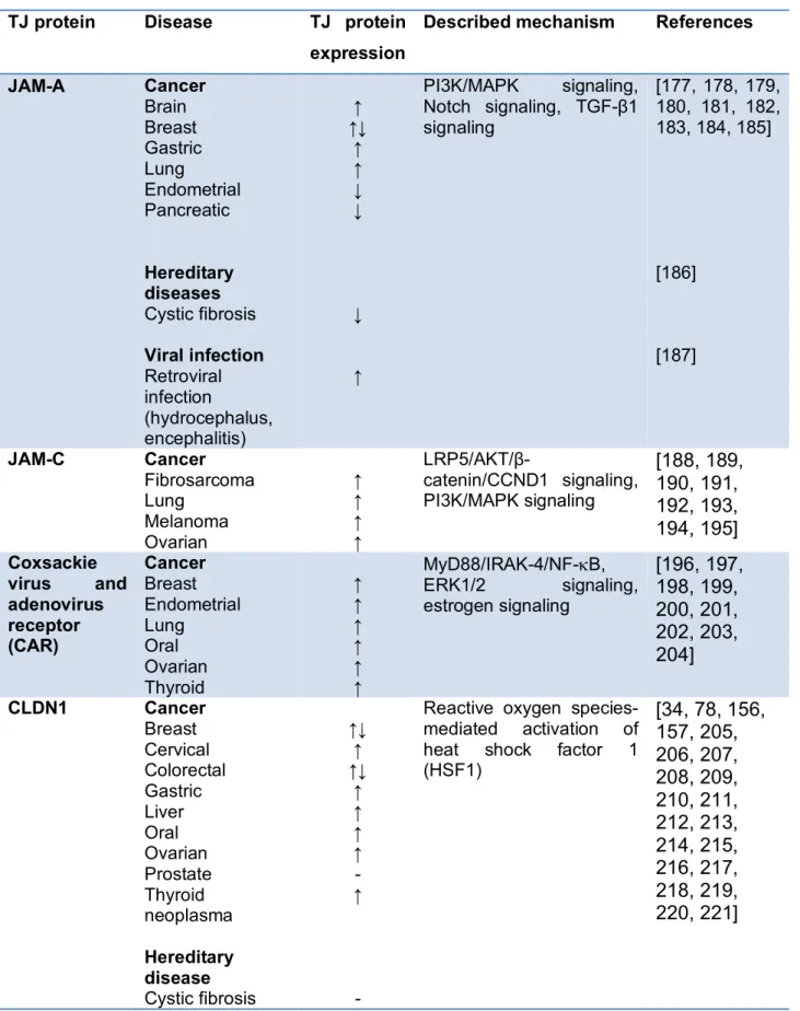
is associated with a variety of diseases, including skin, intestinal and lung diseases, and 60
cancers[1, 3] (Table 1). Furthermore, pathogens have evolved strategies to overcome 61
epithelial and endothelial barriers by using TJ components for their infection/invasion[1, 4]. 62
TJs are composed of transmembrane proteins, including different claudins (CLDNs), tight 63
junction-associated marvel proteins (TAMPs) such as occludin, junctional adhesion 64
molecules (JAMs) as well as cytosolic proteins, which form what has been termed the 65
junctional plaque and connect transmembrane components to the cytoskeleton[1] (Figure 1). 66
OCLN was the first identified integral membrane protein forming TJs. OCLN is a 65-67
kDa protein with 4 transmembrane domains. Posttranscriptional modification leads to several 68
splice variants. OCLN contains a small intracellular loop and two extracellular loops (ECLs), 69
ECL1 and ECL2, the latter being involved in homophilic interactions between OCLNs 70
expressed on adjacent cells. The N- and C-terminal parts are both located within the cell. 71
The C-terminal part is longer than the N-terminal part and its role in the modulation of TJ 72
assembly, structure and function via posttranslational modifications of OCLN as well as for 73
interaction with other TJ components and the cytoskeleton has been well studied[5] (Figure 74
1). OCLN contains a conserved four transmembrane marvel domain and is thus a member of 75
the tight junction-associated marvel proteins (TAMPs) that also include tricellulin and 76
marvelD3[6]. It has been shown that the three TAMPs have distinct but overlapping functions 77
at the TJ. Tricellulin localizes at tricellular junctions formed by the corners of three epithelial 78
cells while OCLN localizes at bicellular junctions. MarvelD3 has been reported to interact 79
The observation that TJs can form in the absence of OCLN[7] has led to the 81
identification of CLDNs as integral TJ components. CLDNs form a family of 25-27 kDa 82
proteins that in mammals comprises up to 27 members with high sequence homology. Like 83
OCLN, CLDNs can also be subject to posttranslational and postranscriptional regulation. 84
Their structure also resembles the one of OCLN, except for a shorter C-terminal part. The 85
ECLs contribute through homophilic or heterophilic interactions with CLDNs or other integral 86
membrane proteins located on adjacent cells to TJ formation[8]. The C-terminal part links the 87
protein to intracellular TJ components and the actin cytoskeleton (Figure 1). Interestingly, 88
CLDN expression patterns vary between different organs and cancers. CLDNs have thus 89
been suggested as diagnostic markers and targets for cancer therapy[9, 10, 11]. 90
Beside CLDNs and TAMPs that form TJ strands, additional transmembrane barrier 91
proteins, including JAMs and related proteins, have been described (reviewed in[12, 13]. The 92
best characterized JAM in the regulation of TJ barrier function is JAM-A, a member of the 93
immunoglobulin (Ig) superfamily. The dimerization of two JAM-A molecules expressed on the 94
same cell (cis-dimerization) contributes to the formation of a complex between 95
transmembrane TJ proteins and cytoplasmic scaffold proteins[12] (Figure 1). Furthermore, 96
JAM-A has been shown to act as a landmark for bicellular TJ formation[14] while lipolysis-97
stimulated lipoprotein receptor (LSR)/angulin-1, another member of the Ig superfamily of 98
proteins, defines cell corners for tricellular TJ formation[15]. 99
The best studied cytoplasmic proteins of TJs are zona occludens (ZO) proteins. ZO-1, 100
-2, and -3 can interact with each other as well as with several transmembrane proteins 101
(OCLN, CLDNs, JAM-A) and F-actin (Figure 1). Cytoplasmic proteins of TJs have thus been 102
suggested to act as scaffolds linking TJs to the actin cytoskeleton and microtubules[12]. 103
Importantly, TJ proteins have been reported to be also localized at sites outside TJs 104
(non-junctional expression). Indeed, CLDN, OCLN and ZO proteins can be expressed at the 105
basolateral membrane, in the cytoplasm and/or the nucleus where they have important 106
functions in addition to those observed in TJs. For TJ protein expressed at the basolateral 107
membrane these non-canonical functions include endosomal trafficking, signaling and 108
additional ion transport functions while TJ proteins in the nucleus have been shown to 109
regulate gene transcription (reviewed in[16]). Noteworthy, non-junctional TJ proteins do not 110
diffuse in a random manner throughout the membrane. Rather, by interacting with defined 111
molecules within the membrane and/or their phosphorylation, non-junctional CLDNs have 112
been shown to be stabilized in discrete domains within the basolateral membrane and 113
contribute to cell adhesion through interactions with the extracellular matrix[17, 18]. 114
Furthermore, CLDNs can regulate the expression/activity of matrix metalloproteases (MMPs) 115
that contribute to matrix remodelling[19, 20, 21, 22]. These functions can contribute to 116
epithelial-to-mesenchymal transition (EMT), a process by which polarized epithelial cells lose 117
their contacts to neighbouring cells and enable them to migrate. EMT has been shown to 118
play an important role in organogenesis (EMT type 1), homeostasis, inflammation and 119
fibrosis (e.g. wound healing, fibrogenesis; EMT type 2) but can also promote tumorigenesis, 120
invasion and metastasis (EMT type 3). 121
The role of TJ proteins in the physiology and disease biology of the GI system and 122
the liver deserves special emphasis. The GI tract epithelium has to maintain a delicate 123
however dynamic balance in allowing specific substances (food, ions and solutes) to pass 124
through the epithelium while not allowing many others (e. g. pathogens) in order to maintain 125
the delicate balance between immune tolerance and activation. These considerations are 126
further enriched by the recent findings that a feed-forward loop may exist between the gut 127
microbiota and mucosal barrier function in such regulatory schemes[23]. Moreover, studies 128
have now also revealed non-canonical roles of specific TJ integral proteins in regulating 129
cellular differentiation, proliferation and migration; cellular mechanisms implicated in normal 130
repair/regeneration as well as the oncogenic growth during tumorigenesis[24, 25]. 131
Accordingly, a causal role of TJ proteins in GI disease, including esophagitis, inflammatory 132
bowel disease (IBD) and cancers has been demonstrated. Similar to the GI tract, the liver 133
endothelial junctions are important for liver functions and TJ dysregulation has been 134
observed in chronic liver disease and hepatocellular carcinoma (HCC). Interestingly, TJ 135
virus (HCV)[26, 27]. Here we review our current knowledge of the role of TJ proteins in GI 137
and liver disease and discuss their potential as therapeutic targets focussing on GI cancer 138
and viral infection of the liver. 139
140
TJ proteins and the GI tract 141
The functional role of TJ proteins in the physiology of the GI system 142
The GI mucosal barrier plays an important role in the separation of the inside of the body 143
from the outside environment. TJs are present on the apical end of the lateral membrane 144
surface in epithelial cells and regulate paracellular transport and apicobasal cell polarity. The 145
expression of different TJs in the gut varies according to the gut’s functional properties as 146
well as localization in villus or crypt (small bowel vs colon). The proteins can be localized 147
strictly at the apical cell-cell adhesion or extend to the lateral or basolateral surfaces[28, 29, 148
30, 31]. Moreover, expression and cellular distribution of the TJ proteins – such as the CLDN 149
family of proteins – is associated with regulation of differentiation of the intestinal 150
epithelium[32, 33, 34]: CLDN1 is mainly expressed at the apex of the epithelial cells with a 151
reticular pattern in the colon. CLDN2 is expressed in both villus and crypt cells of the small 152
intestine but restricted to undifferentiated crypt cells in the colon. CLDN3, -4, -7 and -8 are 153
predominantly expressed in the distal parts (colon, sigmoid and rectum) of the GI tract while 154
CLDN10 and -12 show an ubiquitous expression pattern throughout the GI tract. 155
Loss- and gain-of-function studies in mice have revealed specific roles for a number 156
of CLDNs in the TJ barrier function, selective ion permeability, as well as their related 157
pathological phenotypes. For instance, knockout of CLDN7 in mice has severe intestinal 158
defects including mucosal ulcerations, epithelial cell sloughing and inflammation, which leads 159
to the death of the mice[35, 36]. Similarly, constitutive overexpression of CLDN1 in the 160
mouse gut epithelium (to mimic upregulated CLDN1 expression in colon cancer) 161
demonstrated a key role of CLDN1 in normal colonic epithelial homeostasis by regulating 162
Notch-signaling[21], while in combination with APC (adenomatous polyposis coli) mutation 163
(APCmin mice) CLDN1 overexpression induced colon tumorigenesis[37]. Similarly,
upregulated CLDN2 expression in the mouse gut epithelium demonstrated a critical and 165
complex role of CLDN2 in intestinal homeostasis by regulating epithelial permeability, 166
inflammation and proliferation[38, 39]. Moreover, CLDN8 contributed to regulation of 167
paracellular Na+ permeability, protecting the leakage of Na+ into the intestinal lumen[40].
168
CLDN16 is responsible for the defective absorption of Ca2+ in the intestine causing primary
169
hypercalciuria[41]. Furthermore, the interdependence between CLDN proteins in regulating 170
intestinal homeostasis is well demonstrated by in vivo loss-of-function studies for CLDN15. 171
CLDN15 knockout mice grow normally despite having a mega-intestine[42]. However, a 172
double knockout of CLDN15 with CLDN2 chronically reduces the paracellular flow of Na+
173
from the intestinal submucosa into the lumen resulting in shunting of the nutrient absorption, 174
malnourishment and death [43]. Of note, both CLDN2 and -15 are paracellular transporters 175
of Na+[44]. Overall, these findings show a critical functional role of these proteins in 176
regulating intestinal homeostasis. 177
Furthermore, OCLN and JAMs have been shown to contribute to regulate intestinal 178
homeostasis. For example, mice lacking JAM-A display an alteration of intestinal 179
homeostasis as shown by perturbed regulation of epithelial permeability, inflammation, and 180
proliferation, and significant alteration in CLDN protein expression[45]. Next generation gene 181
editing technology such as CRISPR and fluorescent gene-reporter tags will enable to better 182
understand the details of the function of TJ proteins in regulating GI physiology. 183
184
Functional role of TJ proteins during GI neoplastic transformations and growth 185
During neoplastic transformation in GI cancer, the expression and localization of TJ proteins 186
is perturbed by several mechanisms occuring at the transcriptional, translational and post-187
transcriptional level (Figure 2). Signaling mechanisms that are known to promote neoplastic 188
growth and cancer malignancy, including receptor tyrosine kinase signaling, inflammatory 189
signaling cascades and non-coding RNAs, have been shown to perturb TJ protein 190
expression and function. Perturbed TJ protein expression or function alters downstream 191
inflammation and cancer (Zeb-1/E-cadherin, Wnt signaling, MMP9/Notch signaling and 193
Src/PI3K/Akt signaling). Furthermore, disruption of TJs during infection or injury can result in 194
increased permeability with translocation of bacteria and luminal antigen, which in turn 195
increase IL-6/Stat3 signaling contributing to carcinogenesis (Figure 2). Delocalization of TJ 196
proteins from their normal membrane-tethered expression appears to be common among 197
inflammatory diseases and GI cancers. Aberrant cell signaling may contribute to this 198
process. Notably, in Ras-overexpressing MDCK cells, CLDN1, OCLN, and ZO-1 were absent 199
from the cell-cell contact sites however were present in the cell cytoplasm[46]. Inhibition of 200
MEK1 activity recruited all three proteins to the cell membrane leading to a restoration of the 201
TJ barrier function in these cells. In line with this, it has been shown that growth factor 202
receptors, including EGF, HGF and IGF receptors, as well as proinflammatory and tumor 203
promoting cytokines, including TNF-α, IFN-γ, IL-13, and IL-22, contribute to regulate CLDN 204
expression[47, 48, 49, 50, 51]. It has also been reported that nonsteroidal anti-inflammatory 205
drugs regulate CLDN expression in association with p38 MAPK activation in gastric epithelial 206
cancer cells[52]. Furthermore, protein modifications by phosphorylation, sumoylation, 207
palmitoylation[53, 54, 55] and endocytic recycling have emerged as potential mechanisms of 208
regulating TJ protein function and expression [56, 57, 58]. 209
Transcription factors known to be associated with cellular differentiation and EMT, 210
including Snail, Cdx-2, HNF-α, and GATA-4[34, 59, 60], can bind to the promoter regions of 211
specific CLDN genes to affect their expression in intestinal epithelial cells. In colon cancer 212
cells, caudal homeobox proteins (Cdx1 & Cdx2) and GATA-4 in cooperation with the Wnt 213
pathway are involved in CLDN1 promoter activation[34]. CLDN1 transcripts are regulated by 214
Smad-4 (a known tumor suppressor) and HDAC inhibitors supporting a complex multiprotein 215
regulatory scheme[61, 62]. Furthermore, transcription factor RUNX3, which is a gastric tumor 216
suppressor, upregulates CLDN1 expression by binding to the promoter region of CLDN1 in 217
gastric epithelial cells[63] and a similar regulatory scheme involving Cdx-proteins, GATA-4 218
and HNF-a has been demonstrated for CLDN2 and -4. Additionally, various epigenetic 219
regulatory mechanisms likely also contribute to the transcriptional regulation of CLDN 220
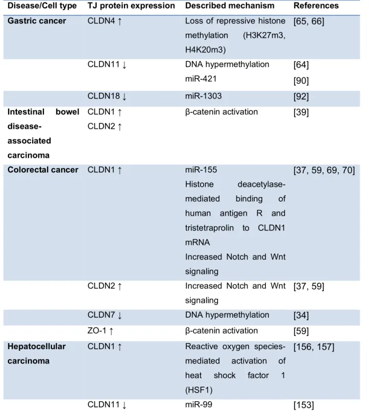
expression (Table 2). Indeed, it has been shown that DNA hypermethylation associated with 221
the downregulation of CLDN11 in gastric cancer cells[64] and CLDN7 in colon cancer 222
cells[34]. Furthermore, loss of repressive histone methylations, including H3K27me3 and 223
H4K20me3, is associated with the overexpression of CLDN4 in gastric cancer[65, 66]. 224
Finally, microRNAs (miRNAs) post-transcriptionally regulate TJ formation and barrier 225
function[67]. Indeed, different miRNAs have been shown to modulate CLDN1 expression 226
(Table 2): targeting of CLDN1 mRNA by miR-29 has been shown to regulate intestinal 227
permeability[68], while the regulation of CLDN1 mRNA by miR-155 plays an important role in 228
promoting colorectal cancer (CRC) cell migration and invasion[69]. Moreover, the histone 229
deacetylase has been shown to regulate CLDN1 mRNA stability in CRC cells through 230
modulating the binding of the human antigen R and tristetraprolin to the 3′ UTR of CLDN1 231
mRNA[70]. 232
233
TJ proteins and colorectal disease and cancer 234
Perturbation of the epithelial barrier function as well as TJ protein function and expression 235
are hallmarks of GI disease including CRC. The breakdown of polarized epithelial barrier 236
leads to the activation of specific signaling pathways as a response to injury. However, 237
chronic activation of these signaling pathways can also promote cancer formation in 238
premalignant epithelial tissues when TJs are chronically leaky. Furthermore, TJ proteins, 239
especially CLDNs, have now been demonstrated to play an essential role in cell proliferation 240
and neoplastic transformation during tumorigenic growth. 241
Understanding how these complex signaling networks are altered in cancer cells 242
represents a major challenge for the success of anti-cancer therapies. Upregulation or 243
aberrant tissue expression of CLDNs may contribute to neoplasia by altering TJ structure 244
and function or affecting cell signaling pathways. As stated above, the loss of cell polarity, 245
due to TJ deregulation can abrogate the normal check-points. Moreover, studies linking EMT 246
with the acquisition of stem cell characteristics have demonstrated important role of CLDNs 247
regulating the barrier properties, TJs also serve as hubs for a multitude of signaling proteins 249
including known tumor suppressor molecules like APC, PTEN (phosphatase and tensin 250
homolog) and polarity proteins like Par-3[74, 75, 76]. Silencing of the expression and/or 251
function of these proteins modulates CLDN expression and induces loss of polarity and EMT. 252
Interestingly, genetic modulation of CLDN proteins in mice or cancer cells can similarly affect 253
these signaling cascades, suggesting a feedback regulation. 254
Junctional proteins are known to play an important role or assist in cellular 255
transformation when mislocalized from their normal membrane localization and could serve 256
as oncogenic molecules. In this regard, the Wnt signaling pathway, essential for the 257
differentiation of epithelial cells and imbalanced during intestinal epithelial oncogenic 258
transformation is a major regulator of TJ protein expression[77]. For example, CLDN1 and 259
CLDN2 proteins are known target genes of the Wnt/β-catenin signaling pathway with binding 260
sites in the promoter of these genes[34, 59]. CLDN1 expression not only decreased 261
significantly in response to the reduction of intracellular β-catenin by adenovirus-mediated 262
transfer of wild-type APC into the APC-deficient colon cancer cells, but also two putative Tcf4 263
binding elements in the 5' flanking region of CLDN were confirmed to be responsible for 264
activating its transcription[34]. Importantly, in the intestine CLDN1 is weakly expressed at the 265
apical border of the lateral membrane of normal enterocytes but is strongly expressed at cell-266
cell boundaries as well as in the nucleus/cytoplasm of CRC cells. Many studies have 267
demonstrated that the expression of CLDN1 at the mRNA and protein levels is increased in 268
CRC tissue and correlates with tumor depth[78]. Additional studies using gene editing have 269
further shown that an intricate interdependence between the Notch and Wnt-signaling 270
upregulating CLDN1 expression to augment CRC progression[37] (Table 2). A role of the 271
nuclear effectors of the Wnt signaling pathway is to bind directly to the CLDN2 promoter 272
region and thereby enhance CLDN2 promoter activity. Also, a crosstalk between the Wnt 273
signaling and Cdx related transcriptional activation machinery has been implicated in 274
regulating CLDN2 promoter-activation[59]. Recent studies have also demonstrated that 275
levels of CLDN1 and CLDN2 are elevated in IBD-associated carcinoma[39] (Table 2). 276
Kinugasa et al.[79] demonstrated increased staining for CLDN1 in both high-grade dysplasia 277
and ulcerative colitis (UC)-associated CRC when compared with normal or UC samples. 278
CLDN1 overexpression modulates Notch-signaling in an MMP9-dependent manner to 279
modulate barrier properties and immune homeostasis to promote susceptibility to 280
inflammation-induced colitis and cancer[21]. Here it is worth noting that the outcome from a 281
series of studies have now provided ample evidence for a role of deregulated CLDN1 282
expression in promoting invasive and metastatic abilities of the colon cancer cells. Notably, 283
CLDN1 expression was sufficient to induce metastatic abilities in a colon cancer cell line that 284
normally does not metastasize well in vivo. In contrast, stable genetic inhibition of CLDN1 in 285
a poorly differentiated, highly metastatic and CLDN1 high colon cancer cell significantly 286
inhibited its metastatic abilities in a splenic model of CRC metastasis[78]. An increase in 287
CLDN2 expression also participates in promoting colorectal carcinogenesis potentially 288
dependent on the EGFR/ERK1/2 signaling[80, 81]. Here, overexpression of CLDN2 in 289
CLDN2 deficient CRC cells resulted in increased cell proliferation, anchorage-independent 290
growth and tumor growth[81]. A similar effect of the Wnt-/b-catenin signaling upon gene 291
expression of yet another component of the TJ complex, ZO-1, in human colonic cancer cell 292
lines with low endogenous β-catenin has been reported suggesting potential contribution to 293
the loss of epithelial polarization in neoplastic cells[59] (Table 2). Decreased ZO-1 294
expression was noted in the human digestive tract[82]. Using tissue biopsy samples, Mees et 295
al.[83] also found that CRC in human exhibits significantly elevated expression levels of 296
CLDN1 and -4 compared with normal mucosa. However, CLDN expression in colon cancer 297
tissues is differential and downregulation of CLDN7 and -8 has been reported in colorectal 298
adenoma samples compared with the normal intestinal tissues[84]. Collectively, these 299
studies suggest that these proteins may serve as potential biomarkers for CRC progression 300
and therapy resistance. 301
302 303
CLDNs and esophageal and gastric cancer 305
Similar to CRC, TJ proteins are also regulated aberrantly in the esophageal and gastric 306
cancers and this abnormal expression correlates with specific clinicopathologic parameters. 307
In the esophageal adenocarcinoma (EA), CLDN expression has been tested as potential 308
biomarker for the transition of the Barrett’s esophagus to the EA. Indeed, CLDN2, -3, −4 and 309
−7 are reported to have increased expression in EA compared to precancerous lesions and 310
normal esophageal squamous mucosa[85, 86]. JAMs, which comprise the integral parts of 311
TJs in the gastric epithelium, have been shown to promote proliferation, invasion, and inhibit 312
apoptosis. JAM-B was upregulated significantly in tumor samples compared with adjacent 313
normal tissues and was higher in high grade tumors than in the low grade and intermediate 314
grade tumors[87]. 315
An increased expression of CLDN2 is also associated with gastric cancer 316
progression[88]. Additionally, expression of CLDN11 and -23 is downregulated in gastric 317
cancer[89] and miR-421 was implicated in regulating CLDN11 expression to promote the 318
proliferation, invasion and metastasis of gastric cancer cells[90] (Table 2). In contrast, 319
CLDN23 positive expression was associated with poor prognostic outcomes of gastric cancer 320
patients and may therefore serve as an independent predictor of patient survival. Similarly, 321
upregulated expression of CLDN4 in gastric cancer was associated with cancer progression 322
and poor prognosis[91]. Furthermore, CLDN4-expressing gastric adenocarcinoma AGS cells 323
were found to have increased MMP2 and -9 expression, indicating that CLDN-mediated 324
increased invasion may be mediated through the activation of MMPs[91]. Overall, these 325
results suggest that CLDN4 overexpression may promote gastric cancer metastasis through 326
the increased invasion of gastric cancer cells. Yet another CLDN family protein, CLDN18 is 327
significantly downregulated in gastric cancer tissues and cell lines, which was associated 328
with tumor size, location invasion, histologic type and tumor-node-metastasis stage. miR-329
1303 was demonstrated to have putative binding sites in CLDN18 mRNA 3'-UTR and visibly 330
lower the expression of CLDN18[92] (Table 2). On the other hand, CLDN18.2 (isoform 2 of 331
claudin-18) was retained on malignant transformation and was expressed in a significant 332
proportion of primary gastric cancers and its metastases[9]. The expression of CLDN7 has 333
also been reported to have the potential of serving as an independent indicator of the poor 334
prognosis in gastric cancer[93]. 335
Taken together, TJ proteins are tissue-specific regulators of the epithelial and 336
endothelial barrier function and EMT, which cumulatively perturb the epithelial and immune 337
homeostasis leading to carcinogenesis in CRC as well as gastric and esophageal cancer. 338
339
TJ proteins and the liver
340
The liver plays an essential role in homeostasis by its metabolic and storage functions. It is 341
composed of different cell types, including two types of epithelial cells: hepatocytes and 342
cholangiocytes. Liver epithelial junctions are important for liver function. Hepatocytes are 343
liver parenchymal cells that exhibit a complex honeycomb morphology displaying at least two 344
basolateral membranes (facing the sinusoidal blood) and two intercellular apical membranes 345
(forming the bile canaliculi) separated by TJs. This peculiar architecture creates what has 346
been termed the blood-biliary barrier[94] and enables hepatocytes to perform distinct 347
secretory functions at the same time[95]. Hepatocytes produce and secrete bile into the bile 348
canaliculi from where it is transported via intrahepatic and extrahepatic bile ducts, formed by 349
cholangiocytes, to the gallbladder. Cholangiocytes contribute to modify the bile during its 350
transport to the latter. Hepatocyte polarity and bile duct TJs play a major role in the liver[96,
351
97] and thus defects in hepatocyte and/or cholangiocyte TJ integrity can result in 352
pathophysiological consequences. 353
Several lines of evidence indicate that disruption or loss-of-function of TJs contributes
354
to the pathogenesis of cholestatic diseases including primary biliary cholangitis and primary
355
sclerosing cholangitis[98, 99]. Interestingly, mutations in CLDN1 are associated with 356
neonatal ichthyosis and sclerosing cholangitis (NISCH) syndrome where deficient CLDN1 357
expression may contribute to paracellular bile leakage through deficient TJs[100]. Mutations 358
in ZO-2 have been described in familiar hypercholanemia[101]. The loss-of-function of TJ 359
proteins is not lethal and the clinical manifestation is variable including very mild 360
symptoms[102]. A comprehensive description of TJ protein alterations in biliary diseases is 361
reviewed in reference[97]. 362
Recent studies demonstrate that TJ protein expression is altered in HCC (primary 363
liver cancer) and cholangiocarcinoma (biliary tract cancer). For example, CLDN1 has been 364
shown to be up-regulated in advanced liver disease and HCC[103] and differential CLDN4 365
expression can help to distinguish these two forms of cancer at a molecular level[104]. Of 366
note, TJ alteration in epithelia outside the liver can also contribute to liver disease. Indeed, 367
dysfunction of the intestinal epithelial barrier - due to or unrelated to (aetiological factor(s) of) 368
the underlying liver disease - has been associated with the pathogenesis of chronic liver 369
disease and the development of complications in cirrhosis by favouring translocation of 370
bacteria and bacterial products from the intestinal lumen into the systemic circulation[105]. 371
372
CLDN1 and OCLN mediate hepatocyte entry of HCV 373
Ten years ago, expression cloning experiments uncovered CLDN1 to be required for HCV
374
infection[26]. The role of OCLN in HCV infection was uncovered two years later using
375
different approaches[27, 106, 107]. Subsequently several studies have characterized the
376
underlying molecular mechanisms and highlighted the essential function played by these
377
proteins in HCV entry and infection[4, 108, 109]. While over the past 20 years many host
378
factors have been reported to contribute to the early steps of HCV infection[108, 110],
379
CLDN1 and OCLN are regarded as two of the four major HCV host entry factors together
380
with CD81[111] and scavenger receptor BI (SR-BI)[112] (Figure 3).
381
First binding studies indicated that CLDN1 was unable to bind the HCV envelope
382
glycoprotein E2[26], suggesting that CLDN1 does not play the role of a primary receptor but
383
rather of a co-receptor, which contributes to (a) step(s) subsequent to viral binding[26]. This
384
was further confirmed in kinetic assays using anti-CLDN1 antibodies[113]. Several years
385
later it was shown that in contrast to soluble E2, HCV E1E2 complexes can interact with the
386
CLDN1 ECL1 and that this interaction is involved in viral fusion[114]. Based on the
Coxsackie B virus cell entry model, it was suggested that HCV may first interact with host
388
factors on the basolateral surface of hepatocytes and then move to the TJ co-entry factor(s),
389
e.g. through CD81-lateral membrane movements[26, 115]. Elegant fluorescence resonance
390
energy transfer studies showed that CLDN1 interacts with CD81 to promote viral
391
internalization[116, 117, 118]. Interestingly, no cellular function for CD81-CLDN1 interaction
392
has been reported so far and disruption of these complexes by defined anti-CLDN1
393
antibodies prevents HCV infection without affecting TJ integrity or any detectable adverse
394
effect[113, 119, 120]. Indeed, several lines of evidence support a model in which the
non-395
junctional form of CLDN1 rather than CLDN1 localized within TJ mediates HCV entry[117,
396
121]. The major pool of CLDN1 is expressed at TJs of hepatocytes and polarized hepatoma 397
cells but a minor fraction is also located at the basal membranes of these cells[117, 122]. Of 398
note, CD81-CLDN1 co-receptor association could only be detected at the basal membranes
399
but not in TJ-associated pools of CLDN1 and CD81[117]. Furthermore, the ECL1 appears to
400
be the critical part of the protein for HCV entry while the intracellular C-terminal part of
401
CLDN1 that plays an important role for its interaction with intracellular TJ components is not
402
required for this process[26, 121]. Interestingly, CLDN6 and CLDN9 - but not other members 403
of the CLDN family of proteins - have been shown to be able to promote HCV entry into 404
CLDN-deficient 293T-derived cell lines[123, 124]. This is most likely due to their ability to 405
form co-receptor associations with CD81 like CLDN1[118]. It is of interest to note that in 406
experimental model systems using a liver tumor cell lines some HCV genotypes have been 407
reported to be able to use either CLDN1 or CLDN6[125] through mutation in the HCV E1 408
envelope protein[126]. Whether CLDN6 or 9 can replace CLDN1 in the liver of HCV-infected 409
patients remains questionable since CLDN6 and CLDN9 expression is very low or absent in 410
human liver tissues[124, 125, 127]. Furthermore, treatment of HCV infection in human liver 411
chimeric mice with a monoclonal CLDN1-specific antibody did not reveal any detectable 412
escape (for a detailed review of the role of CLDN6 and CLDN9 in HCV entry please 413
see[128]). 414
Like CLDN1, OCLN does not appear to play the role of a primary HCV attachment
415
receptor but rather is required for late postbinding event(s) during the HCV entry
416
process[107, 129] (Figure 3). Nevertheless, HCV might interact with OCLN during viral entry
417
and/or in infected cells. Indeed, imaging studies evidenced a co-localization between OCLN
418
and HCV E2 in the endoplasmic reticulum of hepatoma cells[130]. Furthermore, it was shown
419
that an anti-E2 antibody could immunoprecipitate OCLN while GST-OCLN could pull down
420
E2[129, 130]. The OCLN ECL2 appears to be important for this interaction with HCV E2 as
421
well as for HCV entry[129]. However, the OCLN ECL2 was unable to pull down E2,
422
suggesting that either this interaction might not be direct or not be visualized in the utilized
423
experimental design[129]. Experiments using OCLN engineered to be recognized by
anti-424
FLAG antibodies are in favour of a HCV-OCLN interaction as different clones displaying the
425
FLAG epitope at different locations within ECL1 or ECL2 exhibited HCV isolate-dependent
426
host factor activity[131]. Using kinetic assays in polarized cells, this study also showed that
427
OCLN plays a role subsequent to SR-BI, CD81 and CLDN1[131]. These results were
428
recently confirmed in kinetic assays using anti-OCLN mAbs directed against either the ECL1
429
or ECL2 of OCLN[132]. How and where HCV interacts with OCLN as well as what pool(s) of
430
OCLN is(are) involved in this process remain to be further characterized. Although in liver
431
sections OCLN has been located at apical surfaces of hepatocytes[117], a minor pool of this
432
protein is expressed on the basolateral surface of hepatocytes. Indeed, OCLN is known to
433
traffic through the basolateral membrane towards TJs[133] and its subcellular localization
434
appears to be dependent on its phosphorylation status: phosphorylated forms of OCLN
435
mainly are found in TJs of epithelial cells, while less phosphorylated forms are localized at
436
the basolateral membrane and in the cytosol[134]. This is in line with a recent report showing
437
that tumor-associated calcium signal transducer 2 (TACSTD2) regulates HCV entry by
438
leading to the phosphorylation of CLDN1 and OCLN and regulates their subcellular
439
localization[135]. The importance of the subcellular localization of OCLN for HCV entry is
440
also underscored by the fact that only OCLN and its splice variant OCLN-ex7ext that both
441
localize to the plasma membrane are able to promote HCV entry in contrast to other OCLN
splice variants that exhibit an intracellular localization[136]. Of note, OCLN together with
443
CD81 define the HCV species specificity as HCV non-permissive mouse cells acquire
HCV-444
permissivity subsequent to human CD81 and OCLN expression both in vitro and in vivo[27,
445
137, 138, 139]. The species-specific determinants appear to be located within the second
446
extracellular loop of OCLN[27, 139].
447
Beside cell-free HCV entry, CLDN1 and OCLN have also been shown to be important
448
for HCV cell-to-cell transmission (Figure 3), a major mode of viral dissemination that enables
449
the virus to avoid the host's immune surveillance and to establish chronic infection[140, 141,
450
142, 143]. The exact localization at the plasma membrane of this process as well as the
451
form(s) of CLDN1 that contribute(s) to HCV cell-to-cell transmission remain unknown. The
452
importance of CLDN1 and OCLN for the pathogenesis of HCV infection in vivo has been
453
confirmed by observations of liver tissues from HCV-infected liver transplant patients. HCV
454
recurrence was associated with an increase in CLDN1 and OCLN expression levels in
455
hepatocytes over time after transplantation[144]. This is in line with findings indicating
456
increased CLDN1 and OCLN expression levels in HCV-infected livers[122, 145, 146]. In
457
contrast, in cell-based studies HCV infection was shown to downregulate CLDN1 and OCLN
458
expression to prevent superinfection[106]. Differences in TJ protein expression upon HCV
459
infection may thus exist depending on the analyzed samples.
460 461
CLDNs and HCC 462
The expression of several TJ proteins has been reported to be perturbed in liver tissue from 463
HCC patients. Many studies have shown different expression levels of the individual CLDNs 464
and OCLN and CLDN1 are being investigated as biomarkers for liver disease
465
progression[103, 122, 147, 148, 149, 150, 151]. From these studies, it appears that 466
expression of CLDNs is associated with more severe disease and/or bad prognosis in HCC 467
patients (Table 2): epigenetic silencing of CLDN14 was significantly associated with 468
advanced tumor state and tumor aggressiveness[152]; CLDN11 downregulation by miR-99 469
suggested to promote EMT via Wnt-b-catenin signaling[154]. However, more data are 471
needed to decipher the role of these TJ proteins in the pathogenesis of HCC. Several studies
472
have shown an increase in CLDN1 expression on basolateral and apical hepatocyte
473
membranes in cirrhotic livers and HCC compared to normal livers[103, 122, 149].
474
Interestingly, this increase was observed in tissues from both HCV-positive and -negative
475
patients, although it appeared to be stronger in tissues from HCV-infected patients, as well
476
as in HCC that developed on either cirrhotic or non-cirrhotic livers[103] and in paediatric
477
HCC[149]. In advanced HCC down-regulation of CLDN1 has been observed[147, 151] which
478
may correspond to the de-differentiation of cancer cells. Of note, a greater cytoplasmic
479
localization of CLDN1 was observed in some HCC in line with reports indicating that CLDN
480
localization has a causal role in cellular transformation[78, 155].
481
CLDN1 likely contributes to proliferation, motility and invasion by modulating cellular
482
signaling. Overexpression of CLDN1 increases the migration and invasiveness of human
483
hepatoma cells as well as normal liver cells through expression of MMP2 via the c-Abl-PKC
484
pathway[20] (Figure 4). Furthermore, increased CLDN1 expression has been associated with
485
mitochondrial dysfunction and invasiveness of hepatoma cells, and reactive oxygen
species-486
mediated activation of heat shock factor 1 (HSF1) was demonstrated to increase CLDN1
487
expression in these cells[156, 157] (Table 2). These data are in line with reports indicating
488
that CLDN1 enhances cell growth, migration and/or invasiveness of other cancer cell types
489
such as oral squamous cell carcinoma cells, CRC cells, ovarian cancer-initiating cells or
490
melanoma cells[69, 158, 159, 160, 161]. Taken together, these data suggest that by
491
promoting cell migration and increasing the invasive behaviour of cancer cells, CLDN1 can
492
contribute to cancer spread. Of note, CLDN1 has been shown to promote EMT in normal
493
liver cells and HCC cells that thereby acquire an invasive phenotype[162]. This process is
494
mediated by the c-Abl-Ras-Raf-1-ERK pathway and involves the transcription factors Slug
495
and Zeb1[162] (Figure 4). Confirming the functional role of these signaling pathways, an
496
CLDN1-specific antibody inhibits the HCV-induced increase in ERK1/2 phosphorylation in
human liver tissue [120]. Further studies are needed to understand the detailed role of
498
CLDNs in pathogenesis of liver disease and cancer.
499 500
Targeting TJ proteins for therapeutic approaches in the gut and the liver
501
CLDNs as targets for CRC 502
While the field related to the role and regulation of TJ proteins in GI pathologies and 503
oncogenic growth has taken a significant leap forward, therapeutic application of this 504
knowledge is now emerging. Significant progress has been made at several fronts including 505
the development of prognostic biomarkers, imaging and targeting. In this regard, CLDN 506
proteins are currently investigated as potential biomarkers for disease progression and 507
therapy resistance. A recent study has shown that serum levels of CLDN1 and CLDN7 may 508
be a useful tool in the differential diagnosis of CRC[163]. Furthermore, a progressive 509
increase in CLDN1 expression in colon cancer and the recent findings that infra-red imaging 510
using CLDN1-targeted conjugated peptide can enhance the ability of conventional 511
colonoscopy for detecting human colonic adenomas strongly supports the potential impact of 512
CLDN1 as a biomarker[164]. CRC has been found to arise from missed polypoid and flat 513
precancerous lesions which are more difficult to visualize by colonoscopy and the new 514
CLDN1 targeted fluorescent peptides may be used to improve screening of high-risk patients 515
with multiple polyps, inflammatory bowel disease, Lynch syndrome, or a family history of 516
CRC. 517
Aiming to develop targeted therapies, several antibodies against the extracellular 518
domain of CLDNs have been developed. Their therapeutic effects for cancer and metastasis 519
are summarized in Table 3. Ideal monoclonal antibodies (IMAB) specific to the proteins 520
expressed only on the tumor and hence avoiding potential off-target effects are actively being 521
developed. Currently, monoclonal antibodies (mAbs) have been generated against CLDN1,-522
2, -3, -4, -6, and -18.2. The antibody against CLDN18.2 (claudiximab) is in clinical 523
development for gastric cancer[9]. Interestingly, claudiximab significantly extends median 524
advanced gastric cancer[9]. Importantly, this target is not present in any healthy tissues 526
except the lining of the stomach, thereby minimizing treatment side effects. In addition, 527
recent studies have investigated the anti-tumor effect of anti-CLDN1 and anti-CLDN2 mAbs 528
using cancer cell models[11, 165] (Table 3). Importantly, anti-CLDN mAbs have been shown 529
to be safe and no relevant off-targets have been reported. Their marked therapeutic effects 530
combined with excellent safety profiles are highly encouraging for their development in 531
clinical applications. 532
533
CLDN1 and OCLN - targets for cure of HCV infection 534
CLDN1 was the first TJ protein to be explored as a therapeutic target for HCV infection using
535
anti-CLDN1 antibodies directed against its extracellular domain(s) (Table 3). Such antibodies
536
could be used to prevent liver graft infection in HCV-positive transplant recipients and as a
537
promising alternative for patients who fail current anti-HCV therapies[166]. The first
538
antibodies directed against human CLDN1 and blocking HCV infection were generated by
539
genetic immunization in rats[113, 119]. They recognize a conformational-dependent epitope
540
within the ECL1 and prevent CD81-CLDN1 co-receptor association at the basolateral
541
membrane[113, 119]. They are characterized by pan-genotypic inhibition of the infection by
542
all major HCV genotypes by blocking both cell-free virus entry and viral cell-to-cell
543
transmission[119, 143]. Of note, studies in human liver-chimeric mice demonstrated that the
544
lead antibody OM-7D3-B3 was not only able to prevent acute de novo infection with HCV
545
(i.e. the anticipated effect of an entry inhibitor) but also to cure already established chronic
546
HCV infection without detectable side or off-target effects[120]. These results highlighted the
547
importance of viral dissemination for maintenance of chronic HCV infection. It is of interest to
548
note that this antibody interfered with MAPK signalling suggesting an important role in this
549
pathway potentially also contributing to its antiviral effect[120]. The therapeutic potential of
550
this antibody is further underscored by the fact that it acts in synergy with HCV direct-acting
551
antivirals (DAAs), the current state-of-the-art antiviral therapy, to clear viral infection and is
552
also active on viral variants escaping DAAs[143, 167]. This antibody has recently been
successfully humanized (IgG4) for further clinical development[168]. Subsequently, other
554
CLDN1-specific antibodies inhibiting HCV infection have been reported: clones 3A2 and 7A5,
555
generated in mice and recognizing the human CLDN1 ECL2 can prevent HCV infection of
556
human liver-chimeric mice[169] while several antigen-binding fragments (Fab) and single
557
chain antibody fragments selected using phage display were demonstrated to inhibit HCV
558
infection in vitro when converted into human IgG1 or IgG4[170, 171]. Administration of
559
CLDN1-specific antibodies has been shown to be very safe in various animal and human-cell
560
based models without any adverse effects on the liver or other organs such as the gut or
561
skin[120, 168, 172]. This is most likely to the mechanism of action of CLDN1-specific
562
antibodies targeting the non-junctional expressed CLDN1 on the hepatocyte basolateral
563
membrane without affecting TJ barrier function as shown in several TJ model systems[120,
564
168, 172].
565
Antibodies directed against OCLN have been more difficult to generate than
anti-566
CLDN mAbs but very recently five mAbs with anti-HCV activity were described by two
567
different groups[132, 173]. The mouse mAb (67-2) - raised against a linear peptide within
568
OCLN ECL2 - was shown to recognize an epitope present in the ECL2 of both human and
569
mouse OCLN[173]. Interestingly, this mAb hardly inhibited the infection of human hepatoma
570
Huh7.5.1 cell monolayers with HCV when applied to the apical membrane of cells while it
571
was able to efficiently prevent HCV infection when applied to the basolateral membrane of
572
cells using a double-chamber culture system or a 3D culture model[173]. Of note, in line with
573
the hypothesis that mAb 67-2 interacts with OCLN monomers expressed on the basolateral
574
membrane, this mAb had no effect on TJ function in Eph4 cells[173]. Four rat mAbs -
575
generated by genetic immunization and directed against either the ECL1 or ECL2 of OCLN –
576
inhibited the entry of HCV into human hepatoma cells without affecting TJ barrier function of
577
polarized cells. Since the mAb directed against ECL2 appeared to be more potent in
578
inhibiting HCV infection than mAbs directed against ECL1 and in line with previous studies
579
using OCLN mutants/chimeras having demonstrated the essential function of ECL2 for HCV
block an essential function of OCLN in the HCV entry process while mAbs directed against
582
ECL1 may block HCV infection through steric hindrance. Two of those mAbs targeting either
583
ECL1 or ECL2 (1-3 and 37-5) were shown to inhibit HCV infection in human liver chimeric
584
mice without apparent side effects highlighting the possibility to target OCLN in vivo[132].
585
The positioning of CLDN1- and OCLN-specific antibodies in the widening arsenal of
586
anti-HCV therapies is most likely for patients with multi-resistance to DAAs or in organ
587
transplantation including HCV-positive donors where prevention of de novo infection may be
588
preferable to cure of an established HCV infection. They may offer also perspectives to
589
further shorten therapy regimens when combined with DAAs[175, 176].
590 591
Conclusions and future perspectives
592
Research in the last two decades has demonstrated an important role of TJ proteins in the
593
physiology and disease biology in GI and liver disease. TJ proteins exert their functional role
594
as integral proteins of TJs in forming barriers in the gut and the liver. Furthermore, TJ
595
proteins are expressed non-junctionally where they play important roles in signaling,
596
trafficking and regulation of gene expression outside the TJs. A hallmark of TJ proteins in
597
disease biology is their role in EMT, which is relevant for organogenesis and differentiation
598
(EMT type 1), inflammation and fibrosis (EMT type 2) and cancer metastasis/invasion (EMT
599
type 3). A causative role of TJ proteins has been established in the pathogenesis of CRC
600
and gastric cancer. Among the best characterized role of TJ proteins in liver disease biology
601
is their function as cell entry receptors for HCV – one of the most common causes of HCC.
602
At the same time TJ proteins are emerging as targets for novel therapeutic approaches for GI
603
and liver disease: these include treatment of CRC and gastric cancer as well as antiviral
604
therapy for chronic HCV infection complementing DAAs. Further studies are needed to study
605
their role in chronic inflammation, fibrosis and their role as drivers for carcinogenesis. The
606
understanding of these mechanisms offers new perspectives for novel therapeutic
607
approaches for key unmet medical needs in the gut including CRC and gastric cancer as well
608
as chronic liver disease and HCC.
Acknowledgements
610
The authors work is supported by ARC, Paris and Institut Hospitalo-Universitaire, Strasbourg
611
(TheraHCC IHUARC IHU201301187), the European Union (ERC-AdG-HEPCIR,
ERC-PoC-612
2016-PRELICAN, EU H2020-667273-HEPCAR, U Strasbourg Foundation HEPKIN), the
613
National Institutes of Health (NCI 1R21CA209940-01A1, NIAID R03AI131066, NIAID
614
5U19AI123862-02), the Institut Universitaire de France (IUF), the IdEx Program of the
615
University of Strasbourg, the Impulsion Program of the IDEXLYON, BX002086 (VA merit),
616
CA216746 (NIH/NCI) and a pilot project award from Fred and Pamela Buffet Cancer Center,
617
which is funded by a National Cancer Institute Cancer Center Support Grant under award
618
number P30 CA036727. This work has been published under the framework of the LABEX
619
ANR-10-LABX-0028_HEPSYS and benefits from funding from the state managed by the
620
French National Research Agency as part of the Investments for the future program.
621 622
Conflict of interests
623
TFB is a co-inventor of a patent/patent application of CLDN1-specific antibodies for
624
prevention and treatment of HCV infection. TFB and MBZ are co-inventors on patent
625
applications for anti-claudin 1 monoclonal antibodies for the prevention and treatment of liver
626
disease and HCC.
References
628
1 Zihni C, Mills C, Matter K, et al. Tight junctions: from simple barriers to multifunctional 629
molecular gates. Nat Rev Mol Cell Biol 2016;17:564-80. 630
2 Singh AB, Uppada SB, Dhawan P. Claudin proteins, outside-in signaling, and 631
carcinogenesis. Pflugers Arch 2017;469:69-75. 632
3 Brandner JM, Zorn-Kruppa M, Yoshida T, et al. Epidermal tight junctions in health and 633
disease. Tissue Barriers 2015;3:e974451. 634
4 Zeisel MB, Turek M, Baumert TF. Tight junctions and viral entry. Future Virology 635
2010;5:263-71. 636
5 Cummins PM. Occludin: one protein, many forms. Mol Cell Biol 2012;32:242-50. 637
6 Raleigh DR, Marchiando AM, Zhang Y, et al. Tight junction-associated MARVEL 638
proteins marveld3, tricellulin, and occludin have distinct but overlapping functions. Mol Biol 639
Cell 2010;21:1200-13. 640
7 Saitou M, Furuse M, Sasaki H, et al. Complex phenotype of mice lacking occludin, a 641
component of tight junction strands. Mol Biol Cell 2000;11:4131-42. 642
8 Van Itallie CM, Anderson JM. Claudin interactions in and out of the tight junction. 643
Tissue Barriers 2013;1:e25247. 644
9 Singh P, Toom S, Huang Y. Anti-claudin 18.2 antibody as new targeted therapy for 645
advanced gastric cancer. Journal of Hematology & Oncology 2017;10:105. 646
10 Osanai M, Takasawa A, Murata M, et al. Claudins in cancer: bench to bedside. 647
Pflugers Arch 2017;469:55-67. 648
11 Cherradi S, Ayrolles-Torro A, Vezzo-Vie N, et al. Antibody targeting of claudin-1 as a 649
potential colorectal cancer therapy. J Exp Clin Cancer Res 2017;36:89. 650
12 Van Itallie CM, Anderson JM. Architecture of tight junctions and principles of 651
molecular composition. Semin Cell Dev Biol 2014;36:157-65. 652
13 Luissint AC, Nusrat A, Parkos CA. JAM-related proteins in mucosal homeostasis and 653
inflammation. Semin Immunopathol 2014;36:211-26. 654
14 Severson EA, Parkos CA. Mechanisms of outside-in signaling at the tight junction by 655
junctional adhesion molecule A. Ann N Y Acad Sci 2009;1165:10-8. 656
15 Masuda S, Oda Y, Sasaki H, et al. LSR defines cell corners for tricellular tight junction 657
formation in epithelial cells. J Cell Sci 2011;124:548-55. 658
16 Hagen SJ. Non-canonical functions of claudin proteins: Beyond the regulation of cell-659
cell adhesions. Tissue Barriers 2017;5:e1327839. 660
17 Wu CJ, Mannan P, Lu M, et al. Epithelial cell adhesion molecule (EpCAM) regulates 661
claudin dynamics and tight junctions. J Biol Chem 2013;288:12253-68. 662
18 Van Itallie CM, Tietgens AJ, LoGrande K, et al. Phosphorylation of claudin-2 on 663
serine 208 promotes membrane retention and reduces trafficking to lysosomes. J Cell Sci 664
2012;125:4902-12. 665
19 Agarwal R, D'Souza T, Morin PJ. Claudin-3 and claudin-4 expression in ovarian 666
epithelial cells enhances invasion and is associated with increased matrix metalloproteinase-667
2 activity. Cancer Res 2005;65:7378-85. 668
20 Yoon CH, Kim MJ, Park MJ, et al. Claudin-1 acts through c-Abl-protein kinase Cdelta 669
(PKCdelta) signaling and has a causal role in the acquisition of invasive capacity in human 670
liver cells. J Biol Chem 2010;285:226-33. 671
21 Pope JL, Bhat AA, Sharma A, et al. Claudin-1 Regulates Intestinal Epithelial 672
Homeostasis through the Modulation of Notch Signaling. Gut 2014;63:622-34. 673
22 Miyamori H, Takino T, Kobayashi Y, et al. Claudin promotes activation of pro-matrix 674
metalloproteinase-2 mediated by membrane-type matrix metalloproteinases. J Biol Chem 675
2001;276:28204-11. 676
23 Grivennikov SI, Wang K, Mucida D, et al. Adenoma-linked barrier defects and 677
microbial products drive IL-23/IL-17-mediated tumour growth. Nature 2012;491:254-8. 678
24 Martin TA. The role of tight junctions in cancer metastasis. Semin Cell Dev Biol 679
2014;36:224-31. 680
25 Salvador E, Burek M, Forster CY. Tight Junctions and the Tumor Microenvironment. 681
26 Evans MJ, von Hahn T, Tscherne DM, et al. Claudin-1 is a hepatitis C virus co-683
receptor required for a late step in entry. Nature 2007;446:801-5. 684
27 Ploss A, Evans MJ, Gaysinskaya VA, et al. Human occludin is a hepatitis C virus 685
entry factor required for infection of mouse cells. Nature 2009;457:882-6. 686
28 Gunzel D, Yu AS. Claudins and the modulation of tight junction permeability. Physiol 687
Rev 2013;93:525-69. 688
29 Lu Z, Ding L, Lu Q, et al. Claudins in intestines: Distribution and functional 689
significance in health and diseases. Tissue Barriers 2013;1:e24978. 690
30 Amasheh S, Fromm M, Gunzel D. Claudins of intestine and nephron - a correlation of 691
molecular tight junction structure and barrier function. Acta Physiol (Oxf) 2011;201:133-40. 692
31 Rahner C, Mitic LL, Anderson JM. Heterogeneity in expression and subcellular 693
localization of claudins 2, 3, 4, and 5 in the rat liver, pancreas, and gut. Gastroenterology 694
2001;120:411-22. 695
32 Benoit YD, Pare F, Francoeur C, et al. Cooperation between HNF-1alpha, Cdx2, and 696
GATA-4 in initiating an enterocytic differentiation program in a normal human intestinal 697
epithelial progenitor cell line. Am J Physiol Gastrointest Liver Physiol 2010;298:G504-17. 698
33 Escaffit F, Boudreau F, Beaulieu JF. Differential expression of claudin-2 along the 699
human intestine: Implication of GATA-4 in the maintenance of claudin-2 in differentiating 700
cells. J Cell Physiol 2005;203:15-26. 701
34 Bhat AA, Sharma A, Pope J, et al. Caudal homeobox protein Cdx-2 cooperates with 702
Wnt pathway to regulate claudin-1 expression in colon cancer cells. PLoS One 703
2012;7:e37174. 704
35 Tanaka H, Takechi M, Kiyonari H, et al. Intestinal deletion of Claudin-7 enhances 705
paracellular organic solute flux and initiates colonic inflammation in mice. Gut 2015;64:1529-706
38. 707
36 Ding L, Lu Z, Foreman O, et al. Inflammation and disruption of the mucosal 708
architecture in claudin-7-deficient mice. Gastroenterology 2012;142:305-15. 709
37 Pope JL, Ahmad R, Bhat AA, et al. Claudin-1 overexpression in intestinal epithelial 710
cells enhances susceptibility to adenamatous polyposis coli-mediated colon tumorigenesis. 711
Molecular Cancer 2014;13:167-. 712
38 Ahmad R, Chaturvedi R, Olivares-Villagomez D, et al. Targeted colonic claudin-2 713
expression renders resistance to epithelial injury, induces immune suppression, and protects 714
from colitis. Mucosal Immunol 2014;7:1340-53. 715
39 Weber CR, Nalle SC, Tretiakova M, et al. Claudin-1 and claudin-2 expression is 716
elevated in inflammatory bowel disease and may contribute to early neoplastic 717
transformation. Lab Invest 2008;88:1110-20. 718
40 Yu ASL, Enck AH, Lencer WI, et al. Claudin-8 Expression in Madin-Darby Canine 719
Kidney Cells Augments the Paracellular Barrier to Cation Permeation. Journal of Biological 720
Chemistry 2003;278:17350-9. 721
41 Claverie-Martin F. Familial hypomagnesaemia with hypercalciuria and 722
nephrocalcinosis: clinical and molecular characteristics. Clinical Kidney Journal 2015;8:656-723
64. 724
42 Tamura A, Kitano Y, Hata M, et al. Megaintestine in Claudin-15–Deficient Mice. 725
Gastroenterology 2008;134:523-34.e3. 726
43 Wada M, Tamura A, Takahashi N, et al. Loss of Claudins 2 and 15 From Mice 727
Causes Defects in Paracellular Na+ Flow and Nutrient Transport in Gut and Leads to Death 728
from Malnutrition. Gastroenterology 2013;144:369-80. 729
44 Van Itallie CM, Holmes J, Bridges A, et al. The density of small tight junction pores 730
varies among cell types and is increased by expression of claudin-2. Journal of Cell Science 731
2008;121:298-305. 732
45 Laukoetter MG, Nava P, Lee WY, et al. JAM-A regulates permeability and 733
inflammation in the intestine in vivo. The Journal of Experimental Medicine 2007;204:3067-734
76. 735
46 Chen Y-h, Lu Q, Schneeberger EE, et al. Restoration of Tight Junction Structure and 736