Isoflavones and the prevention of breast and prostate cancer:
new perspectives opened by nutrigenomics
Claudia Steiner
1, Ste´phanie Arnould
2, Augustin Scalbert
1and Claudine Manach
1*
1INRA, UMR 1019, Unite´ Nutrition Humaine, Centre Clermont-Theix, F-63122 St Gene`s Champanelle, France
2Laboratoire d’Oncologie Mole´culaire, UMR 484 INSERM-UdA, Centre Jean Perrin, F-63011 Clermont-Ferrand, France
Epidemiological evidence together with preclinical data from animal and in vitro studies strongly support a correlation between soy isoflavone con-sumption and protection towards breast and prostate cancers. The biological processes modulated by isoflavones, and especially by genistein, have been extensively studied, yet without leading to a clear understanding of the cellular and molecular mechanisms of action involved. This review discusses the existing gaps in our knowledge and evaluates the potential of the new nutrigenomic approaches to improve the study of the molecular effects of isoflavones. Several issues need to be taken into account for the proper interpretation of the results already published for isoflavones. Too often knowledge on isoflavone bioavailability is not taken into account; supra-physiological doses are frequently used. Characterization of the indi-vidual variability as defined by the gut microflora composition and gene polymorphisms may also help to explain the discrepancies observed so far in the clinical studies. Finally, the complex inter-relations existing between tissues and cell types as well as cross-talks between metabolic and signalling pathways have been insufficiently considered. By appraising critically the abundant literature with these considerations in mind, the mechanisms of action that are the more likely to play a role in the preventive effects of isoflavones towards breast and prostate cancers are reviewed. Furthermore, the new perspectives opened by the use of genetic, transcriptomic, proteomic and metabolomic approaches are highlighted.
Isoflavones: prostate cancer: breast cancer: chemoprevention: nutrigenomics
Isoflavones are a class of bioactive phytochemicals that have been widely studied for their potential role in the prevention of various chronic diseases, such as cardiovascular diseases, neurodegenerative diseases, osteoporosis, cancers. . . Soy and its processed products (tofu, tempeh, miso, natto, soymilk, soy-based yoghurts and desserts) are the only sources provid-ing high quantities of isoflavones in the human diet. Isoflavone intake has been estimated to 25 – 50 mg/day in Asian countries,
with a maximum around 100 mg/d for elderly Japanese men(1).
Americans and Europeans, who have low soy content in their habitual diet, only consume few milligrams of isoflavones per day. However, advertising on the beneficial effects of isofla-vones, especially for relieving postmenopausal symptoms, has led to self-supplementation through isoflavone-rich foods or dietary supplements. Isoflavones in soybeans mainly include daidzein, genistein and glycitein, which are present in glycosylated or aglycone forms (Fig. 1). The structural similarity of aglycones with 17b-estradiol gives them the
capacity to bind estrogen receptors (ERs) and to induce hor-mone-like effects. Isoflavones and their protective role against various pathologies involving hormonal dysregulation have been extensively studied because of this particular property.
The current evidence provided by preclinical studies on the role of isoflavones in reducing prostate and breast cancer risk
has been recently reviewed(2 – 4). A summary of the main data
available will be made here, to highlight the present limi-tations in our understanding of their mechanisms of action, and to evaluate the potential of nutrigenomic approaches to further clarify these mechanisms.
Human studies
The lower risk of prostate and breast cancer in areas of high
soy and isoflavone intake, especially in Asia(5), as well as
the increased risk observed for Asian people who migrated
to Western countries or adopted a westernised lifestyle(6 – 8)
* Corresponding author: C. Manach, fax þ 33 473 62 46 38, email manach@clermont.inra.fr
Abbreviations:AF, activation functions; AP-1, activator protein 1; AR, androgen receptor; CDK, cyclin-dependent kinase; COMT, catechol-O-methyltransferase; CYP, cytochrome P450 monooxygenase; Da, daidzein; DHEA, dihydroepiandrosterone; DHT, dihydrotestosterone; EGF, epidermal growth factor; EGFR, epidermal growth factor receptor; Eq, equol; ER, estrogen receptor; ERE, estrogen responsive elements; ERK, extracellular signal-regulated kinase 1; FSH, follicel-stimulating hormone; Ge, genistein; GPx, glutathione peroxidase; GSR, glutathione reductase; GST, glutathione S-transferase; GTP-CH1, GTP cyclohydrolase 1; IGF-1, insulin-like growth factor 1; IGF-1R, insulin-like growth factor 1 receptor; IGFBP, insulin-like growth factor binding protein; JNK, c-jun N-terminal kinase; LDL, low density lipoprotein; LH, luteinising hormone; MAPK, mitogen-activated protein kinase; MMP, matrix metalloproteinase; MS, mass spectrometry; MT-1X, metallothionein 1X; mGST, microsomal glutathione S-transferase; NF-kB, nuclear factor-kappa B; NMR, nuclear magnetic resonance; PI3K, phosphatidylinositol-3 kinase; PIN, prostatic intra-epithelial neoplasia; PPAR, peroxisome proliferator-activated receptor; PR, progesterone receptor; PSA, prostate specific antigen; QR, quinone reductase; SD rats, Sprague – Dawley rats; SHBG, sex hormone binding globulin; SNPs, single nucleotide polymorphisms; SOD, superoxide dismutase; TEBs, terminal end buds; TGF-b, transforming growth factor b; TIMP, tissue inhibitor of metalloproteinase; TRAMP, transgenic adenocarcinoma of the mouse prostate; UDPGT, UDP-glucuronosyl-transferase; VEGFR, vascular endothelial growth factor receptor.
British Journal of Nutrition (2008), 99, E-Suppl. 1, ES78–ES108 doi:10.1017/S0007114508965788
qThe Authors 2008
British
Journal
of
Nutrition
https://www.cambridge.org/core . IP address: 118.70.52.165 , on 24 Jun 2021 at 12:13:37, subject to the Cambridge Core terms of use, available at
https://www.cambridge.org/core/terms
.
are well known. A recent meta-analysis compiling 2 cohort studies and 6 case – control studies in Western and Asian populations estimated that high soy food intake can be related to a 30 % reduction in prostate cancer risk (odds ratio 0·70,
95 % CI 0·59 – 0·83)(9). For breast cancer, a smaller risk
reduction (odds ratio 0·86, 95 % CI 0·75 – 0·99), stronger for premenopausal women, was found in a meta-analysis
compil-ing 6 cohort and 12 case – control studies(10). Asian women
whose soy intake was high during puberty experienced lower risk for breast cancer than women who did not consume
soy products or did only as adults(11,12).
Among hundreds of dietary components that have been pro-posed as potential cancer preventive agents, only a few have been used in clinical trials. The impact of isoflavone sup-plementation has been studied on some prostate cancer-related endpoints such as serum levels of Prostate-Specific Antigen (PSA), PSA velocity, plasma levels of testosterone, dihydro-testosterone (DHT), insulin-like growth factor 1 (IGF-1) and IGF binding protein 3 (IGFBP-3). Among the 11 trials
recently reviewed by Messina et al.(13), only 4 reported a
sig-nificant effect on PSA levels. However, reduction in prostate cancer risk may occur without any reduction in PSA levels. No beneficial effects were observed on levels of steroids, or on IGF-1/IGFBP-3 ratio. One study which compared the incidence of apoptosis in prostate tumours of patients sup-plemented or not with red clover isoflavones (160 mg/d for 7 – 54 days), reported significantly higher apoptosis in sup-plemented patients than in control subjects (1·48 % v. 0·25 %, P¼ 0·0007), specifically in regions of low to
mode-rate-grade cancer (Gleason grade 1 – 3)(14). It is worth noting
that no adverse effects were observed in any trial.
The whole available data have been considered sufficiently encouraging to justify the funding of additional Phase II
trials by the NIH(13).
Messina et al.(15) recently discussed the published and
ongoing clinical breast cancer studies. Three double-blind ran-domized controlled trials reported no effect of a 1 to 2-year isoflavone supplementation on mammographic density used
as a marker of breast cancer risk(16 – 18). A 2-week
adminis-tration of a soy supplement (45 mg/d isoflavones) increased epithelial cell proliferation and progesterone receptor (PR) expression in normal breast tissue, suggesting an estrogen
agonist effect(19). However, the potential link with
prolife-ration of breast cancer cells is difficult to assess. Conflicting results have been obtained regarding the impact of soy
isoflavones on hormone-related breast cancer risk factors such as plasma steroid hormone levels, Sex Hormone Binding Globulin (SHBG) plasma levels, urinary
2:16a-hydroxyes-trone ratio, and menstrual cycle length(20).
The inconsistencies in the results and the failure to observe clear clinical effects may be explained, at least in part, by dis-parities in experimental designs, in the form of isoflavone administration, the dose, or the duration of the study. Further-more, diverse subpopulations may respond differently to iso-flavone consumption due to age, sex, ethnic background, gene polymorphisms, history of cancer, known risk factors, nutritional status, hormonal status or colonic microbiota com-position. In this regard, the most critical parameters affecting the biological responses to isoflavone intake remain to be identified. The populations or subpopulations which may benefit, or possibly may experience some adverse effects while consuming isoflavones, also need to be identified.
The factors likely to affect isoflavone bioavailability may first modulate physiological responses to isoflavone intake. A compilation of 15 bioavailability studies in humans showed that plasma metabolite concentrations usually reach about 2 mmol/l after consumption of 50 mg isoflavones
(agly-cone equivalent)(21). Inter-individual variability in isoflavone
absorption and metabolism has never been assessed in large and ethnically non-homogeneous populations, however some bioavailability studies suggest that it may be high. Maximum plasma concentrations ranged between 4 and 27 mmol/l genis-tein among 20 American men with prostate cancer challenged
with a high pharmacological dose of genistein(22).
Further-more, a recent clinical study showed that genistein and daidzein bioavailability, evaluated by plasma pharmacoki-netics, was significantly lower in Caucasian than in Asian
men after single or 10-day isoflavone supplementation(23).
It would therefore be extremely useful to accurately measure the real individual exposure to isoflavones rather than only the isoflavone intake in clinical studies.
The intestinal microflora also plays a crucial role in the metabolism of isoflavones. Various bacterial metabolites are known to be produced, among which some may exert
biologi-cal activities(24 – 26). Equol is the most studied metabolite
since it has been shown to be more estrogenic than its precur-sor daidzein in many in vitro studies and in animal
models(27). There is great inter-individual variability in the
capacity to produce equol, and only 30 – 50 % of the Western
population exhibits the ‘equol-producer’ phenotype(28). Song
et al. recently reported that the equol-producer phenotype was more frequent in Korean American than in Caucasian American women (51 % v. 36 %) living in the same area of
the US(29). Several studies suggested that equol-producers
may gain more benefits from soy consumption than non-equol-producers. In a case – control study in Japanese and Korean men, the proportion of equol-producers was 1·5- to 2-fold higher in the control group than in the group of
men with prostate cancer(30). More favourable hormonal
profiles (lower plasma concentrations of estrone, estrone-sul-fate, testosterone, androstenedione, dehydroepiandrosterone (DHEA), DHEA-sulfate, and midluteal progesterone) and higher concentrations of SHBG were also found in equol-pro-ducing premenopausal women compared to non-equol-produ-cers, which was consistent with a lower risk for breast
cancer(31). Frankenfeld et al. did not find such hormonal
O O HO OH OH Genistein O O HO OH MeO Glycitein O O HO OH Daidzein HO OH 17β-Estradiol
Fig. 1. Structures of the soy isoflavones genistein, daidzein and glycitein in comparison to 17b-estradiol.
Isoflavones and the prevention of breast and prostate cancer ES79
British
Journal
of
Nutrition
https://www.cambridge.org/core . IP address: 118.70.52.165 , on 24 Jun 2021 at 12:13:37, subject to the Cambridge Core terms of use, available at
https://www.cambridge.org/core/terms
.
differences in postmenopausal women, but equol-producing women had higher 2:16a-hydroxyestrone ratio, which has
also been related to a lower breast cancer risk(32). Similar
results were obtained in breast cancer survivors(33).
Mammo-graphic density was 39 % lower in equol-producing postme-nopausal women compared with non-producers, which is again consistent with a lower breast cancer risk in
equol-producers(34). Niculescu et al. studied the effects of a
high-dose isoflavone supplementation (900 mg/d for 84 days) on gene expression changes in lymphocytes of 30 postmenopau-sal women. Using microarray analysis they showed that iso-flavone treatment in equol-producer subjects differentially affects gene expression as compared with non-producers, with a stronger effect on some estrogen-responsive genes in
equol-producers(35). It is not yet clear whether equol itself
is the active metabolite responsible for these beneficial effects, or whether it is only a biomarker for a particular microbial community composition able to elicit favourable metabolisms in addition to equol production. Some finger-printing methods, such as terminal restriction fragment length polymorphism (TRFLP) analysis, are now available to compare the overall patterns of gut microbial composition
between individuals(36). These high-throughput methods
should enlighten the role of specific bacteria and help to ade-quately stratify individuals in future human intervention and observational population-based studies.
Metabolomics is another high-throughput method that may be useful to deal with inter-individual variability. Nuclear magnetic resonance (NMR) and mass spectrometry (MS) tech-niques allow the simultaneous detection of hundreds or thou-sands of compounds present in human plasma, urine or
tissues(37). Multivariate statistical analyses of the datasets
obtained for groups of individuals that differ for one controlled criterion, such as isoflavone intake, microflora composition or type of cancer, provide a list of variables dis-criminating the groups. The identification of these discriminat-ing variables may lead to the discovery of novel biomarkers for the particular criterion studied. Metabolic profiling using these biomarkers could then be used in clinical or cohort studies to better assess individual exposure to isoflavones, as well as to stratify individuals on the basis of their metabolic capacities or other phenotypes.
Another new possibility, offered by nutrigenetics, is the access to valuable information on individual genotypes through sequencing of targeted single nucleotide
polymorph-isms (SNPs)(38). As some polymorphisms in genes coding for
membrane transporters, protein carriers or metabolic
enzymes such as glucuronosyl-transferases could affect the nature and concentrations of isoflavone metabolites present in target tissues, it may be useful to search for correlations between individual genotypes, isoflavone bioavailability and isoflavone effects. However, the relevant SNPs that would be related to the responsiveness to isoflavone consumption are not identified to date. Beyond the gene polymorphisms that may affect bioavailability, two recent studies demon-strated the major interest of considering the inter-individual variability in the genetic background of a population to evaluate the isoflavone biological effects. Hedelin et al. stu-died the interactions between dietary phytoestrogen intake, ERb polymorphisms, and the risk of prostate cancer in a
case – control study performed in 2096 Swedish men(39).
For one particular SNP of the ERb gene, a higher phytoes-trogen and isoflavone consumption was associated to a sig-nificantly reduced prostate cancer risk (respectively 57 % and 27 %) in subjects who were homozygous or heterozy-gous for the variant allele, whereas no association could be found in subjects who were homozygous for the wild-type allele (58 % of the population). In another cohort of 1988 menopausal women from UK, isoflavone exposure was positively associated with SHBG levels only in the frac-tion of the populafrac-tion that carried the Asn variant of the SHBG D356N polymorphism, which is associated with a
reduced SHBG clearance(40). As higher SHBG levels have
been associated with a lower breast cancer risk, isoflavone
consumption may be particularly beneficial to these
women. It is worth noticing that the authors would have con-cluded that isoflavone exposure was not associated to SHBG levels if they had considered the population as a whole, without stratification according to the genotype. Gene-isofla-vone interactions may partly explain some of the discrepan-cies observed in the first clinical studies conducted so far, especially if the allele frequencies differed between the populations considered. Nevertheless, high statistical pow-ered studies will be required in order to take the relevant gene polymorphisms into account in the investigation of iso-flavone effects.
Another element that may explain the inconsistencies observed in clinical studies is the relevance of the endpoint biomarkers used. Biomarkers for staging and monitoring the evolution of prostate and breast cancer development are criti-cally needed to evaluate the impact of any therapeutic inter-vention. Considering that cancers are multi-factorial and variable diseases, it appears today unrealistic to rely on a single biomarker (such as PSA for prostate cancer) to assess diagnosis, prognosis, and prediction of response to therapy. Complex combinations of markers coming from transcrip-tomic, proteomic as well as metabolomic approaches are cer-tainly more appropriate and promising perspectives can already be seen. Rhodes et al. have analysed gene expression profiles in 40 published cancer microarray data sets, compris-ing 38 million gene expression measurements from . 3700
cancer samples(41). This study generated a set of 69 genes
that were commonly dysregulated in 12 types of cancers including prostate and breast cancers. Proteomic data have also been used on a blinded cohort to identify patients with
prostate cancer with a high specificity(41 – 43). There is now a
great hope that these high-throughput methods will lead to the discovery of early biomarkers for preclinical metabolic dysregulations associated with the first stages of carcinogen-esis. Many novel important genes involved in prostate carcino-genesis, such as Hepsin or Alpha-methylacyl-CoA racemase
(AMACR), have been identified by using microarray
analysis(44). NMR-based metabolomic analyses to target
biomarkers for prostate cancer are under development(45).
Novel early biomarkers will improve our understanding of the initial stages of the diseases and will be essential to study the preventive effects of dietary intervention such as isoflavone supplementation.
Therefore, nutrigenomic approaches should allow to better assess individual metabolism and to better evaluate the effects of isoflavones at the individual level and at early stages of the diseases. C. Steiner et al. ES80
British
Journal
of
Nutrition
https://www.cambridge.org/core . IP address: 118.70.52.165 , on 24 Jun 2021 at 12:13:37, subject to the Cambridge Core terms of use, available at
https://www.cambridge.org/core/terms
.
Animal studies
As demonstrated above, human studies are still hampered with major weaknesses, and more controllable animal and cellular models are very useful to investigate isoflavone effects. Different animal models of prostate cancer such as Lobund-Wistar rats (spontaneous tumours), rats treated with various type of carcinogens [N-methylnitrosourea (NMU),
7,12-dimethylbenz(a)anthracene (DMBA),
2-amino-1-methyl-6-phenylimidazopyridine (PhIP)] or implants of testosterone propionate, transgenic mice (TRAMP), xenograft models (e.g. immunodeficient mice implanted with human tumour cell lines) have been used to explore the effects of isoflavones on prostate cancers. In more than 40 such studies, isoflavones were shown to reduce both the incidence and the size of
pros-tate tumours(2,3,46). Isoflavones may act at different stages of
carcinogenesis, by inhibiting the appearance of prostatic intra-epithelial neoplasia (PIN) or their progression into carci-nomas, but the determination of their timing of action requires further experiments.
Much more conflicting results have been obtained in animal models of breast cancer, as isoflavones have been shown to inhibit as well as to promote the growth of mammary tumours, depending on the studies. Many parameters may explain such discrepancies including the various natures of the animal models used, the test compounds, the dose and the mode of administration, or the timing of isoflavone exposure. Overall, isoflavones were mainly shown to prevent or to delay chemi-cally-induced mammary carcinogenesis in prepubertal or pre-menopausal adult female rats. The protective effects of isoflavones at the prepubertal period have been linked to a promotion of early mammary gland differentiation, leading to a decreased number of immature terminal end buds (TEBs), sites for malignant transformation, and an increased
number of more differentiated lobules(47 – 51). Complex
cross-talks between steroids, growth factors and other pathways are involved in the differentiation process. Hilakivi-Clarke et al. reported that the pro-differentiating effect of isoflavones was associated with a repression of ERa and PR expression
but an increase of ERb expression in mammary glands(52).
Lamartiniere et al. suggested that isoflavone exposure in the prepubertal period mainly promotes mammary epithelium differentiation through the transient activation of epidermal
growth factor (EGF) signalling pathway(50).
The most frequent types of breast cancer are the estrogen-dependent breast tumours in postmenopausal women. Widely used animal models for these types of tumours are immunode-ficient mice implanted with estrogen-dependent human breast cancer MCF-7 cells, combined with ovariectomy and in some cases with estradiol implants releasing high doses of estradiol that stimulate the growth of MCF-7 implanted cells. The group of Helferich published several consistent studies show-ing that genistein at nutritionally relevant doses (0·025 – 0·1 % in the diet) stimulates tumour growth or negates the inhibitory
effect of tamoxifen in such xenograft models(53 – 56). In such
models the circulating levels of endogenous estrogens are either much lower (ovariectomy) or much higher (estradiol implants) than the physiological levels observed in postmeno-pausal women. Hence the relevance of the xenograft models to evaluate the effects of weak estrogenic compounds, the activity of which may depend on the levels of endogenous
estrogens, has been criticised. Recently, Ju et al. developed a mouse model with slow-growing estrogen-dependent tumours in ovariectomised athymic mice, using silastic implants to deli-ver estradiol at levels similar to those observed in
postmeno-pausal women(57). Again, genistein administered in the diet at
500 ppm was shown to act in an additive manner with the low levels of circulating estradiol and stimulated the growth of the tumorigenic MCF-7 cells in this model.
Several authors reported that in utero exposure to high-dose isoflavones (s.c. injections) increased the susceptibility of rats to chemically-induced mammary cancers later in life, in the same way as in utero exposure to high levels of endogenous
estrogens(49,58). This effect has been related to an increased
number of TEBs, and a reduction in epithelial differentiation in the mammary gland. However, other authors reported that a more nutritional exposure to soy protein isolate (but not to genistein alone) in utero through maternal diet protected young adult rats from NMU-induced mammary
tumourige-nesis(59). Early exposure to isoflavones or at least to genistein
before puberty may cause transient and persistent effects on mammary gland development, as well as on hormone receptor
levels(60). These imprinting-like effects may thus differ
depending on dose and timing of exposure and lead to adverse or beneficial effects in later life.
Dolinoy et al. provided the first evidence that early in utero exposure to genistein can produce permanent epigenetic
changes(61). They showed that maternal dietary genistein
sup-plementation (250 ppm) of mice during gestation shifted the
coat colour of heterozygous viable yellow agouti (Avy/a)
off-spring towards pseudoagouti and decreased the incidence of obesity during adulthood. These marked phenotypic changes were mediated by an increased methylation of six CpG sites located in a retrotransposon upstream of the Agouti gene which determines coat colour. An exciting hypothesis of research has been put forward with this study. DNA methyl-ation is known to control cellular differentimethyl-ation in the mam-mary epithelium. Thus, epigenetic events associated with in utero or prepubertal exposure to genistein may also contribute to the effects observed on animal mammary gland
differen-tiation(62). Such imprinting phenomena are extremely difficult
to study in humans. Extrapolation of data from animal models must take into account some differences in isoflavone bioavailability in rodents and in humans. For example, all laboratory animals have been shown to be constitutive equol-producers, and equol is the major circulating metabolite in animals while it is only present in a low proportion in
humans(27).
Animal studies also showed that isoflavones exert various effects depending on their structures and application forms. The large majority of studies have been conducted with genis-tein. When used alone, daidzein was shown to be equivalent to genistein at the same dose (250 ppm in the diet) in delaying
mammary tumour development in MMTV-neu mice(63). In
contrast, prepubertal exposure of Sprague – Dawley female rats to 250 ppm daidzein in the diet neither enhanced mammary gland differentiation nor suppressed DMBA-induced mammary tumour development as previously demonstrated for
genis-tein(64). Furthermore, dietary daidzein only had a slight
stimu-latory effect on MCF-7 tumour growth in athymic mice,
whereas (^ )-equol did not stimulate tumour growth(65). Thus,
the effects observed with genistein should not be extrapolated
Isoflavones and the prevention of breast and prostate cancer ES81
British
Journal
of
Nutrition
https://www.cambridge.org/core . IP address: 118.70.52.165 , on 24 Jun 2021 at 12:13:37, subject to the Cambridge Core terms of use, available at
https://www.cambridge.org/core/terms
.
to other isoflavones, and the various isoflavones may affect different metabolic or signalling pathways. Moreover, con-sumption of genistein in pure or highly enriched forms (as in soy protein isolates) has a greater stimulatory effect on MCF-7 cells implanted in immunodeficient mice than the same
con-tent of dietary genistein in less purified soy flour(66). Other
authors reported a higher beneficial effect of soymilk or soy protein isolate toward chemically-induced cancer as compared
to pure genistein(59,67). Soybeans contain a wide variety of
other phytochemicals such as saponins, protease inhibitors like the Bowman-Birk inhibitor, phenolic acids, phytic acid, and lignans that may exert additional or synergistic effects with isoflavones. Taking supplements may thus have different effects than increasing soy food consumption.
Cell models
Isoflavones have been shown to inhibit proliferation and/or induce apoptosis in various dependent or androgen-independent prostate cancer cell lines. The concentrations used were generally high, allowing cytotoxic effects, but anti-proliferative effects have also been observed at lower
concen-trations (5 mM) of genistein(2,3). Again, the impact of genistein
is more complex in breast cancer cell lines than in prostate cell lines, since genistein stimulates cell growth of
estrogen-depen-dent cells when used at concentrations lower than 10 mM but
inhibits their growth at higher concentrations(3,53,68,69). High
concentrations of genistein also inhibit the growth of ER-nega-tive cell lines such as MDA-MB-231, indicating that this isoflavone can exert cellular effects independently from ER.
One major limit of such in vitro studies is that they use isoflavone concentrations far exceeding the concentrations commonly achieved in human plasma or tissues after soy con-sumption. The available data indicate that concentrations as high as 1 – 2·5 nmol/g of each isoflavone may be achieved in human prostate or prostatic fluid after short-term supplemen-tation with high nutritional doses of isoflavones and that in Asian men basal levels of total isoflavones are about
0·4 nmol/g in prostate(70 – 74). Isoflavone concentrations in
breast tissue are still unknown. Maubach et al. suggested in an isoflavone supplementation study in 9 women, that they
may be markedly lower than serum concentrations(75).
The question of the dose is critical, since increasing the dose of exposure does not necessarily produce more intense effects but may also lead to completely different effects. Dang
et al.(76)clearly showed that genistein elicits different
biologi-cal effects in osteoprogenitor KS483 cells depending on the
concentration of exposure. At low concentrations (, 1 mM),
genistein acts as an estrogen, stimulating osteogenesis and
inhi-biting adipogenesis, whereas at high concentrations (. 1 mM), it
acts as a ligand for PPARg, leading to the upregulation of adi-pogenesis and downregulation of osteogenesis.
Daidzein and genistein glycosides and aglycones, when ingested with soy foods, are extensively metabolised in the human intestine and liver and systematically recovered in
plasma as 7-O- and 40-O-glucuronides, along with low
amounts of aglycones and sulfate esters(77 – 79).
Mono-glucuro-nides of genistein and daidzein were recently shown to also be
the main metabolites present in the human prostate(74).
How-ever, the effects of these conjugated forms of isoflavones on breast or prostate cell proliferation have not been investigated
so far. The nature of the physiological metabolites should yet be taken into consideration, because the biological properties of conjugated metabolites have already been shown to differ from those of the corresponding aglycones. The affinity of iso-flavone glucuronides for ERs has been reported to be 10 – 40
times lower than that of the aglycones(80). Furthermore,
sulfa-tion of isoflavones was shown to decrease their antioxidant activity and their effect on platelet aggregation, inflammation,
cell adhesion and chemotaxis(81,82). Tumour cell lines may
have lost some membrane carriers, which would limit the uptake of anionic metabolites such as glucuronides or sulfates by the cells. For example, MCF-7 and T47D cells do not express the OATP-B carrier which is normally expressed in the mammary gland and transports a wide range of sulfated and glucuronidated conjugates of endogenous and exogenous
compounds(83). Thus, exposure to the same isoflavone
tration could result in quite different intracellular concen-trations of isoflavone metabolites depending on the cell type used. To facilitate comparisons between in vitro studies, as well as extrapolation to the in vivo setting, information on intracellular concentrations of isoflavone metabolites should be provided in future studies on cultured cells. Another limit of in vitro studies is that they rarely used non-tumourigenic cells. Even if tumourigenic cell lines are of great interest to study specific signalling or metabolic pathways or to assess the potential therapeutic effects of isoflavones, these highly modified cells are not the best models to investigate the pre-ventive effects of isoflavones.
Mechanisms of action
Although many studies have been conducted to understand the effects of soy isoflavones on breast and prostate cancer, avail-able data are inconsistent and the mechanisms of action are still not entirely elucidated. Studies on animal and cellular models have firmly established that isoflavones are active com-pounds, showing quite variable effects depending on the dose, the form of administration, or the timing and duration of exposure. Serious concerns were raised about a possible estro-gen-like detrimental effect of genistein through growth stimu-lation of pre-existing estrogen-sensitive mammary tumours. In the present state of our knowledge, the increasing isoflavone self-supplementation should be considered with caution until the mechanisms of action of isoflavones are better understood.
As questioned in the titles of S. Barnes, “Isoflavones ¼
phytoestrogens and what else?”(84), or M. McCarty “Isoflavones
made simple – Genistein’s agonist activity for the beta-type
estrogen receptor mediates their health benefits”(85), a largely
debated issue is to establish whether isoflavones only act through their phytoestrogenic properties or whether ER-independent mechanisms of action may also play a role. Isoflavones have been reported to modulate steroid biosynthesis, transport and metabolism, as well as carcinogen activation and detoxification, to inhibit cell proliferation induced by growth factors, to induce cell cycle arrest or apoptosis, to favour cell differentiation, to reduce oxidative stress or to inhibit angiogenesis, cell
invasive-ness and metastasis(3,86). They may act through modulation of
cell signalling (direct binding to nuclear receptors, modification of the phosphorylation state of some signal transduction pro-teins), regulation of gene expression and/or specific inhibition of some key enzyme activities.
C. Steiner et al. ES82
British
Journal
of
Nutrition
https://www.cambridge.org/core . IP address: 118.70.52.165 , on 24 Jun 2021 at 12:13:37, subject to the Cambridge Core terms of use, available at
https://www.cambridge.org/core/terms
.
The mechanisms of action of isoflavones are reviewed here in the context of breast and prostate cancer. Table 1 summari-zes the most relevant studies that have been published for each putative mechanism of action and notifies whether in vivo data exist or whether the mechanism has only been observed in vitro. In vitro studies using isoflavone concentrations up
to 10 mM have been distinguished from studies using higher
pharmacological concentrations.
The major focus in isoflavone research so far lies on the modulation of steroid hormone receptor signalling. Estrogens regulate many physiological processes in hormone-dependent tissues, including cell growth and differentiation, apoptosis and tissue-specific gene regulation, but also influence the patho-logical processes of hormone-dependent diseases, such as breast and prostate cancers. The biological actions of estrogens are mediated by the binding to one of the two specific nuclear
receptors, ERa and ERb(87), which can induce gene
transcrip-tion of estrogen-responsive target genes. A general hypothesis is that estrogens acting via ERa exert strong proliferation stimulatory effects while those interacting with ERb tend to reduce this stimulation. Furthermore, ERb has been shown to
repress ERa-controlled transcription(88,89). Cell response
would therefore depend on the balance between ERa and ERb expression levels within a given cell type. This balance
is altered in favour of ERa during tumour progression(90),
emphasizing the protective role of ERb signalling and the use-fulness of ERb-selective agonists in cancer prevention.
Eight in vitro and in vivo studies consistently reported a downregulation of ERa and an upregulation of ERb mRNA
and protein levels by genistein at 1 – 10 mM in breast cancer
cells(91 – 93). A decrease in ERa protein expression was also
observed in mammary tumours of Sprague – Dawley rats
after consumption of a soy extract(94). In the dorsolateral
pros-tate of rats and mice, exposure to dietary genistein ($ 25 ppm for rats; 500 mg/g bw for mice) resulted in a downregulation of
ERa and ERb mRNA and protein expression(95 – 97).
Activation of ERs is an established effect of isoflavones that has been documented in the low nanomolar range, concen-trations commonly achieved at nutritional levels of soy isofla-vone intake. It has been shown in vitro that genistein has an agonist activity for both ER subtypes, but its affinity for ERb is considerably greater than for ERa, with binding affinities
of 8·4 nMand 145 nM, respectively(98,99). For daidzein, the
cor-responding values are 100 nM and 420 nM, indicating a much
lower affinity for these receptors(98,99). The transactivating
functions of ERa and ERb are mediated by two transcription activation functions (AF) located in the ligand-binding domain, AF-1 (N-terminal) and AF-2 (C-terminal). AF-1 is very active in ERa on a variety of estrogen-responsive promo-ters, but minimal in ERb, whereas AF-2-mediated transcrip-tional activities of ERs are dependent on recruitment and interactions with cofactor proteins (coactivators and
corepres-sors) to estrogen-responsive promoters(100). The
conformation-al change of the AF-2 transactivation helix induced by the formation of the ERb-genistein complex is closer to that induced by ER antagonists than by ER agonists and could
there-fore account for the partial agonistic behaviour of genistein(101).
The higher affinity of isoflavones for ERb is paralleled by their ability to activate transcription with ERb at lower
concen-trations than with ERa(102). Genistein, daidzein and equol
were reported to increase the binding rate of ERs to estrogen
responsive elements (ERE), with a more prominent effect on ERb than ERa. The concentrations of genistein, daidzein and equol which would increase this binding rate by 50 % were
determined to be 30 nM, 350 nM and 400 nM for ERb and
15 mM, . 300 mMand 3·5 mMfor ERa, respectively(103).
The activation of ERs by isoflavones subsequently lead to a modulation of the expression of their target genes, and thus, to a modulation of cellular processes such as proliferation and apop-tosis. Well-known target genes for the ERs are PR, pS2, bcl-2
and cyclin D1(104,105). PR mRNA and protein levels were
shown to be upregulated by isoflavones in the majority of the in vitro and in vivo studies. In MCF-7 cells, an upregulation
was observed with 1 – 10 mMgenistein(106), and in
premenopau-sal cynomolgus macaques and ovariectomised athymic nude mice implanted with MCF-7 cells, the doses ranged between
240 and 750 ppm in the diet(66,107). In contrast, a natural soy
extract (0·7 % in the diet), containing 12 % isoflavones and 35 % saponins, decreased PR protein expression in mammary
tumours of female Sprague – Dawley rats(94). Furthermore,
soy isoflavones stimulated the expression of the ER target gene pS2 in breast cells, as shown in twelve in vivo and in vitro studies. As for the modulation of PR, low isoflavone
concentrations (between 0·001 and 10 mM) were effective
in vitro in MCF-7 cancer cells(106,108,109), whereas in vivo
doses above 500 ppm in the diet in mouse xenograft models
or 45 mg daily in premenopausal women were used(55,66,110).
Only one in vivo study reported a converse decrease in mRNA expression of pS2 in premenopausal cynomolgus maca-ques after administration of 240 ppm isoflavones in
combi-nation with 0·09 mg/g estradiol(107). Other ER target genes
such as bcl-2 and cyclin D1 were also significantly increased
in breast cells after genistein treatment. At 1 mM, it increased
bcl-2 mRNA expression in MCF-7 cells(111,112). Moreover,
750 ppm genistein in the diet increased cyclin D1 mRNA
expression in a mouse xenograft model(66). Increased
transcrip-tional and translatranscrip-tional levels of the aforementioned estrogen-responsive target genes rather suggest estrogen-agonistic activities of isoflavones in breast cancer cells.
Since the androgen receptor (AR) pathway plays a pivotal role in prostate cell growth, differentiation and function, agents that minimise or eliminate AR transactivation are con-sidered useful to prevent and treat prostate cancer. Although
genistein does not seem to act as a ligand for AR(113,114), it
has been shown to exert anti-androgenic effects in prostate cells, and to downregulate the expression and secretion of
the typical androgen-responsive gene PSA(113,115). The
mech-anisms underlying isoflavone anti-androgenic effects are still not entirely elucidated. In more than seven in vitro and in vivo studies, a decrease in AR expression at mRNA and protein levels was detected in prostate cells exposed to low
isoflavone concentrations (0·1 – 1 mM in vitro; 250 ppm in
diet in vivo)(96,113,116). Long-term soy protein isolate
consump-tion also lowered AR expression in prostate of men with high
prostate cancer risk(117). Isoflavones may also decrease
andro-gen levels(118,119). Moreover, a direct binding of DHT by
equol has been reported in vitro, as well as a stimulation of testosterone inactivation through intracellular glucuronidation
by biochanin A(120,121). Interestingly, Bektic et al.
demon-strated with the use of a pure anti-estrogen that the down-regulation of the AR was mediated by ERb in LNCaP cells. A cross-talk between ER and AR was also described by
Isoflavones and the prevention of breast and prostate cancer ES83
British
Journal
of
Nutrition
https://www.cambridge.org/core . IP address: 118.70.52.165 , on 24 Jun 2021 at 12:13:37, subject to the Cambridge Core terms of use, available at
https://www.cambridge.org/core/terms
.
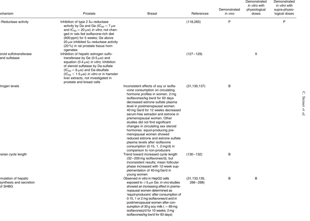
Table 1. Mechanisms of action of isoflavones in prostate and breast tissues from cell, animal and human studies
Mechanism Prostate Breast References
Demonstrated in vivo Demonstrated in vitro with physiological doses Demonstrated in vitro with supra-physio-logical doses ER and AR pathway
ER mRNA and protein expression
Downregulation of ERa mRNA in pros-tate of rats fed Ge (250 ppm from conception to day 70, or 250 – 1000 ppm from day 56 to day 70) and in prostate of mice fed 500 mg Ge/g bw
Downregulation of ERa mRNA levels in MCF-7 breast cell lines exposed to 1 – 10 mMGe
Decrease of ERa protein expression in mammary tumours of SD rats fed a soy extract (0·7 % in the diet)
(91,92,95 – 97, 106,184) (94) P B B
Downregulation of ERb mRNA in pros-tate of rats fed Ge (250 ppm from conception to day 70, or 250 – 1000 ppm from day 56 to day 70)
Upregulation of ERb mRNA levels in breast cell lines exposed to 1 – 10 mMGe
(92,93,95,96) P B
AR mRNA and protein expression
Soy protein isolate consumption (107 mg/d isoflavones) for 6 mo lowered AR but not ERb protein expression levels in prostate of men at high prostate cancer risk
(117) P
Downregulation of AR mRNA in pros-tate of rats fed Ge (250 ppm from conception to day 70, or 250 – 1000 ppm from day 56 to day 70) or a phytoestrogen-rich diet (600 ppm); no effect of Ge (250 ppm in diet) on AR protein level in TRAMP mice prostate
(96,180,263) P
Downregulation of AR at the mRNA and protein level in LNCaP cells exposed to Ge (0·1 – 1 mM); this effect was abolished by the pure antiestrogen ICI164 384, suggesting an action via ERb. Da (up to 50 mM) had no effect (113,116) P ER target gene expression (PR, pS2, cyclin D1, bcl-2) Downregulation of PR protein expression in the prostate of C57BL/6 mice fed with 500 mg Ge/g bw
Upregulation of PR mRNA and protein expression by Ge (1 – 10 mMin MCF-7 cells; 240 – 750 ppm in diet in premenopausal cynomolgus maca-ques and ovariectomized athymic nude mice implanted with MCF-7 cells)
(66,97,106, 107)
B, P B
Upregulation of pS2 mRNA and pro-tein expression by Ge (0·001 – 10 mM in MCF-7 cells; 500 ppm in diet in ovariectomized athymic nude mice implanted with MCF-7 cells)
(55,66,106, 108 – 110)
B B
Upregulation of pS2 protein level in nipple aspirate fluid of premenopau-sal women supplemented with iso-flavones (45 mg/d for 14 d) (110) B C. Steiner et al. ES84
Table 1. Continued
Mechanism Prostate Breast References
Demonstrated in vivo Demonstrated in vitro with physiological doses Demonstrated in vitro with supra-physio-logical doses Induction of cyclin D1 mRNA
expression in a xenograft mice model by Ge (750 ppm)
(66) B
Induction of bcl-2 mRNA expression in MCF-7 cells exposed to Ge (1 – 10 mM)
(111,112) B
Androgen-stimulated transcription (androgen responsive genes promote prostate cancer development and progression)
Ge does not function as a ligand agonist for AR in either PC3 or LNCaP cells; no effect of Ge (1 mM) was detected on the activation of AR-dependent transcription in reporter gene assays
(113,114) P
Ge (10 mM) decreased AR binding to ARE, thereby inhibited transcription of androgen-responsive genes such as PSA in LNCaP cells
(116) P
Downregulation of PSA mRNA level in LNCaP cells exposed to Ge (1 – 5 mM); Ge (10 nM) dose-depen-dently decreased PSA secretion in both androgen-treated and untreated LNCaP cells (unknown mechanism); biochanin A (1 – 5 mM) significantly decreased testoster-one-stimulated release of PSA by LNCaP cells through upregulation of UDPGT and intracellular glucuroni-dation of testosterone
(113,115,121) P
Ge (10 mM) enhanced AR-driven gene expression (agonist R1881) through activation of the Raf-MEK-ERK signalling pathway in PC3 cells, as shown by the use of the specific inhibitor PD098059; Ge (0·5 – 5 mM) enhanced b-estradiol-induced PSA expression in LNCaP cells
(114,116) P
Synthesis and metabolism of steroids
17b-Hydroxysteroid dehydrogenase Inhibition of enzyme activity by 1 – 10 mMGe and Da in several in vitro models, not investigated in prostate cells
Inhibition of enzyme activity by 1 mM Ge in T47D breast cancer cells
(69,122, 123,125)
X, B
3b-Hydroxysteroid dehydrogenase
Inhibition of enzyme activity by 1 – 10 mMGe and Da in several in vitro models, not investigated in prostate and breast cells
(69,123,264) X
Aromatase Inhibition of human aromatase in vitro
by Ge at high doses (Ki¼ 123 mM), not investigated in prostate and breast cells (124) X Isoflavones and the prevention of breast and prostate cancer ES85
Table 1. Continued
Mechanism Prostate Breast References
Demonstrated in vivo Demonstrated in vitro with physiological doses Demonstrated in vitro with supra-physio-logical doses
5a-Reductase activity Inhibition of type 2 5a-reductase
activity by Da and Ge (IC50¼ 7 mM and IC50¼ 20 mM) in vitro; not chan-ged in rats fed isoflavone-rich diet (600 ppm) for 5 weeks; Ge above 20 mMinhibited 5a-reductase activity (20 %) in rat prostate tissue hom-ogenates
(118,265) P P
Steroid sulfotransferase and sulfatase
Inhibition of hepatic estrogen sulfo-transferase by Ge (0·5 mM) and equation (0·4 mM) in vitro; inhibition of steroid sulfatase by Da-sulfate (IC50¼ 6 mM) and Da-disulfate (IC50,1·5 mM) in vitro or in hamster liver extracts, not investigated in prostate and breast cells
(127 – 129) X
Estrogen levels Inconsistent effects of soy or
isofla-vone consumption on circulating hormone profiles in women. 2 mg isoflavones/kg bw/d for 93 days decreased estrone sulfate plasma level in postmenopausal women. 40 mg Ge/d for 12 weeks decreased serum-free estradiol and estrone in premenopausal women. Other studies did not find significant changes in circulating sex steroid hormones. equol-producing pre-menopausal women showed reduced estrone and estrone sulfate plasma levels after isoflavone consumption (0·15, 1, 2 mg/d) in comparison to non-producers
(31,130,137) B
Ovarian cycle length Trend toward increased cycle length
(32 – 200 mg isoflavones/d), but inconsistent results; mean follicular phase increased with 12-week sup-plementation of 40 mg Ge/d in young women
(130 – 132) B
Stimulation of hepatic synthesis and secretion of SHBG
Observed in vitro in HepG2 cells exposed to $5 mMGe; in vivo studies showed an increasing effect in preme-nopausal women determined as ‘equol-producers’ after consumption of 0·15, 1 or 2 mg isoflavones/d and in postmenopausal women after con-sumption of 30 g soy milk ( ¼ 69 mg isoflavones)/d for 10 weeks; 2 mg isoflavones/kg bw/d for 93 days)
(31,133,135, 266 – 268) B B C. Steiner et al. ES86
Table 1. Continued
Mechanism Prostate Breast References
Demonstrated in vivo Demonstrated in vitro with physiological doses Demonstrated in vitro with supra-physio-logical doses
Androgen levels Plasma testosterone and
androstene-dione levels significantly lowered in rats fed a phytoestrogen-rich diet (600 ppm) for 5 weeks; genistein (20 ppm in diet) reduced testoster-one and DHT levels in male mice orthopically implanted with LNCaP cells; inconsistent effects of soy or isoflavone consumption on circulat-ing hormone profiles in men. Most studies did not find significant changes in circulating sex steroid hormones
(117 – 119) P
Eq specifically binds circulating and intracellular DHT with an apparent Kdof 1·32 nMand prevent DHT’s biological actions
(120) P
Hypothalamic-pituitary-testicular (HPT) axis
No significant differences in plasma LH in rats fed a phytoestrogen-rich diet (600 ppm) for 5 weeks. Lack of significant effects of soy protein iso-late supplementation on serum LH in men; 2 week-supplementation with clover phytoestrogens (240 mg/d) increased serum LH and modestly decreased serum testos-terone concentrations
in men with prostate cancer and induced testicular resistance to LH
(118,269,270) P
Cell cycle control and apoptosis
Cell cycle arrest G1 or G2/M arrest only observed in
vitro at high Ge concentrations (20 – 60 mM); not observed at 10 mM Ge in LNCaP cells
G2/M arrest observed with 5 – 10 mM Ge in breast cancer cell lines and $15 mMin non-neoplastic breast cells
(147 – 152) B P
Downregulation of cyclin B expression; inhibition
of cdc2 activity
Observed in LNCaP cells exposed to high concentration of Ge (20 – 60 mM)
Observed in MCF-10F, MCF-7 and MDA-MB-231 cells exposed to high concentration of Ge (45 – 100 mM) (148,149,152,155, 157,164,271) P, B Upregulation of cyclin-dependent inhibitors (p21WAF1; p27KIP1)
Observed in LNCaP cells exposed to high concentration of Ge (20 – 60 mM)
Observed in mammary gland of rats fed Ge or soy protein isolate (0·5 mg/kg bw) and in MCF-7, MDA-MB-231 and AU565 cells exposed to Ge ($ 1 mM)
(148,149,155, 160 – 164)
B B P
Induction of apoptosis Observed in PC3 and LNCaP cells
exposed to high concentration of Ge (. 20 – 50 mM)
Observed in mammary gland of rats fed Ge or soy protein isolate (250 ppm Ge or 394 ppm isofla-vones in diet); observed in MCF-7 and MDA-MB-231 cells exposed to high Ge concentration ($ 25 mM) (3,86,112,160, 163,271) B P, B Isoflavones and the prevention of breast and prostate cancer ES87
Table 1. Continued
Mechanism Prostate Breast References
Demonstrated in vivo Demonstrated in vitro with physiological doses Demonstrated in vitro with supra-physio-logical doses Downregulation of antiapoptotic Bcl-2,
Bcl-x and upregulation of proapopto-tic Bax, Bak, Bok expression
Not modified in prostate cancer cells exposed to Ge concentration lower than 40 mM
Observed in mammary gland of rats fed Ge or soy protein isolate (250 ppm Ge or 394 ppm isofla-vones in diet) and in MDA-MB-231 and 184-B5/HER cells exposed to Ge ($ 5 mM)
(3,112,148,160, 163,272)
B P
Inhibition of proteasome activity (which plays essential role in promoting tumour cell proliferation and protec-ting tumour cells from apoptosis)
Inhibition of chymotrypsin-like activity (2 30 %) of purified proteasome from LNCaP cell extracts by 1 mM Ge, but much higher Ge concen-tration needed to inhibit proteasome in intact LNCaP cells (IC50¼ 70 mM)
Inhibition of chymotrypsin-like activity (2 30 %) of purified proteasome from MCF-7 cell extracts by 1 mM Ge, but much higher Ge concen-tration needed to inhibit the protea-some in intact MCF-7 cells (IC50¼ 65 mM)
(165) P, B
Angiogenesis
Microvessel density in tumours Reduced vessel density in SCID mice inoculated with LNCaP cells and fed diets supplemented with three levels of soy phytochemical concentrate (415 ppm, 756 ppm, 2120 ppm iso-flavones)
Reduced vessel density and decreased protein levels of VEGF and TGFß in tumours and serum from nude mice implanted with MDA-MB-231 and MCF-7 cells and treated by s.c. injections of Ge (0·1 – 0·5 mg/kg bw)
(147,154) P, B
No effect on VEGF and VEGFR expression in prostate of TRAMP mice fed 250 ppm Ge
(180)
Invasion and metastasis
Cell adhesion and migration Ge ($ 10 mM) and Da (100 mM)
suppressed cell migration and cell adhesion of highly invasive MDA-MB-231 cells
(169) B B
Matrigel invasion Reduction of the invasive potential
and upregulation of KAI1 mRNA and protein levels in TRAMP-C2 prostate cancer cells exposed to 5 mMGe; induction of PC-3 and DU-145 cell invasion through Matrigel by plasminogen is counteracted by Ge (dose unknown)
Induction of MDA-MB-231 cells invasion through Matrigel by arachidonic acid or DHA is reversed by Ge (10 mM)
(140,170,171) P, B P
Matrix metalloproteinases (MMP)
Inhibition of MMP-2 expression in PC3 and LNCaP cells by Ge (. 18 mM); Ge (. 10 nM) inhibited TGF-b-mediated induction of MMP-2 in six cell lines including normal, early cancer and established cancer prostate cell lines, possibly through inhibition of p38 MAPK
Downregulation of MMP-9 and upregulation of TIMP-1 mRNA expression in tumours from nude mice implanted with MDA-MB-231 or MCF-7 cells and treated by s.c. injections of Ge (0·1 – 0·5 mg/kg bw)
(154,172 – 174) B P
Osteopontin Ge (250 or 500 mg Ge/kg bw) reduced
OPN mRNA levels in TRAMP mice prostates displaying advanced pros-tate cancer (175) P C. Steiner et al. ES88
Table 1. Continued
Mechanism Prostate Breast References
Demonstrated in vivo Demonstrated in vitro with physiological doses Demonstrated in vitro with supra-physio-logical doses Regulation of cell signalling pathways
Growth factors
EGF signalling Downregulation of EGFR expression
in rat and TRAMP mice prostate with 250 – 1000 ppm Ge in the diet; Not seen in LNCaP cells
Initial upregulation of EGFR protein in mammary gland of young rats, pre-pubertally treated with Ge (250 ppm in diet or 500 mg/g bw) and sub-sequent downregulation of EGFR in mammary gland (stimulation of early differentiation)
(179 – 183) P, B
Inhibition of EGF-stimulated growth of LNCaP and DU145 cells by Ge and Da (15 – 60 mM), without inhibition of EGFR autophosphorylation
Inhibition of DNA synthesis by 0·1 mM Ge in presence of 1 ng/ml EGF; induction of DNA synthesis by 1 – 10 mMGe in presence of 1 ng/ml EGF in breast cancer cells
(68,176) B P
IGF-1 signalling Downregulation of IGF-1R but not of
IGF-1 in TRAMP mice fed 250 ppm genistein
Upregulation of IGF-1R, IRS-1 mRNA and protein expression in MCF-7 cells by 1 mMGe
(180,184) P B
Inhibition of IGF-1 stimulated prolifer-ation of rat prostatic cancer cells by Ge (10 mM)
Induction of IGF-1-induced DNA syn-thesis by 0·1 – 10 mMGe; inhibition of IGF-I-stimulated proliferation of MCF-7 cells by Ge (. 25 mM)
(68,177) P, B
Positive association between soy pro-tein intake (food frequency question-naire) and serum IGF-1 levels in Asian men
40 g soy protein for 3 months increased IGF-1 serum levels stron-ger than milk protein; 83 mg/d soy isoflavone for 12 mo did not modu-late serum IGF in old men; 3 – 6 mo-supplementation with 100 mg/d of soy isoflavone did not
significantly change serum levels of IGF-1 or IGFBP-3 levels in men with prostate cancer
No significant association between soy product and soy isoflavone intake and serum IGF-1 in Asian premenopausal and European pre-and postmenopausal women Increased IGF-I plasma levels after
40 g soy protein intake for 3 months in postmenopausal women; 80 mg total phytoestrogens daily increased IGF-I and IGFBP-3 plasma levels after 1 week in healthy women; low isoflavone-enriched diet (1 mg/kg bw þ d) for 3 months increased IGF-1 and IGFBP-3 serum levels in pre-menopausal women, high
(186 – 188,192) (189 – 191, 193 – 195) P, B P, B isoflavone-enriched diet (2 mg/kg bw þ d) for 3 months decreased IGF-1 and IGFBP-3 serum levels in post-menopausal women
Decreased serum IGF-1 levels in Han:SPRD-cy rats fed a soy pro-tein-based diet for 6 weeks and in mice after supplementation with soy protein isolate (20 % in diet) and soy phytochemical concentrate (1 % in diet) (147,185) P Isoflavones and the prevention of breast and prostate cancer ES89
Table 1. Continued
Mechanism Prostate Breast References
Demonstrated in vivo Demonstrated in vitro with physiological doses Demonstrated in vitro with supra-physio-logical doses Akt and NFkB pathways and their cross-talk
Akt pathway (activation promotes cell survival, by inactivation of pro-apoptotic factors; also regulates NFkB pathway)
Inhibition of Akt phosphorylation by 10 mMGe in DU145 cells Inhibition of Akt kinase activity by
50 mMGe in non-stimulated PC3 cells, and abrogation of EGF-stimu-lated Akt activation
Inhibition of Akt phosphorylation in MCF-7 cells exposed to Ge (. 1 mM) Decrease in phosphorylated Akt1 and
total Akt1 in non-stimulated MDA-MB-231 cells exposed to Ge (30 mM); abrogation of EGF-stimu-lated Akt activation by Ge (50 mM) in MDA-MB-231 cells
(161,196)
(199,273)
P, B
P, B
Incorporation of Ge in the diet (250 and 1000 ppm) of transgenic male TRAMP/FVB mice resulted in a drastic reduction of Akt activation, subsequent activation of GSK-3b, culminating in a dose-dependent post-transcriptional reduction in cyclin D1 levels, and maintenance of the cadherin-1 complex via downregulation of snail-1
(197) P
Induction of PTEN gene expression in PC-3 and LNCaP cells by Ge (20 mM). Decreased expression of VEGF, ERa and ERb genes may be via repression of Akt pathway by PTEN
Induction of PTEN protein expression in SD rats fed Ge or soy protein iso-late (250 ppm Ge or 394 ppm isofla-vones in diet) and in MCF-7 cells exposed to Ge (. 0·1 nM)
(161,163,168) B B P
NFkB (control of cell growth, differentiation, apoptosis, inflammation, stress response)
Inhibition of IkB phosphorylation by Ge (50 mM) in LNCaP and PC3 cells
Induction of IkB protein expression in MCF-7 cells by 100 mMGe (86,165,201, 273,274) P, B Inhibition of NFkB DNA-binding activity by Ge (. 30 mM) in LNCaP and PC3 cells Inhibition of NFkB DNA-binding activity in MDA-MB-231 breast can-cer cells exposed to Ge (. 30 mM)
(86,199,201, 273,274)
P, B
Ge (. 30 mM) abrogated NFkB acti-vation by DNA damaging agents (H2O2and TNF-a) in LNCaP and PC3 cells
Ge (50 mM) abrogated EGF- and AKT-induced NFkB activation in MDA-MB-231 breast cancer cells
(86,199,201, 273,274)
P, B
Inhibition of arachidonic acid-stimu-lated NFkB mRNA expression by Ge (2·5 mM) in MDA-MB-231 cells
(140) B
0·5 mMGe activated NFkB and elev-ated expression of Mn-SOD through activation of ERK1/2 in MCF-7 cells
(203) B
Ge ($ 10 mM) and Da (100 mM) inhib-ited constitutively active transcrip-tion factors NFkB and AP-1 in highly invasive MDA-MB-231 cells
(169) B C. Steiner et al. ES90
Table 1. Continued
Mechanism Prostate Breast References
Demonstrated in vivo Demonstrated in vitro with physiological doses Demonstrated in vitro with supra-physio-logical doses MAPK signalling
JNK Induction of activation of JNK protein
by Ge (0·1 – 25 mM) in MCF-7 cells
(111) B
p38 Increase of p38 phosphorylation by
Ge (0·5 – 45 mM) in MCF-10F cells
(158) B
ERK1/2 Decrease of phosphorylated ERK1/2
protein in prostate of TRAMP mice fed Ge (250 ppm in diet)
Decrease of phosphorylated ERK1/2 protein in MCF-10F cells exposed to Ge (45 mM)
(158,180) P B
Ge (1 – 10 mM) increased ERK1/2 activity in non-tumourigenic prostate epithelial cells RWPE-1 cells via an estrogenic-dependent mechanism and in tumourigenic LNCaP and PC3 cells, while supra-physiological concentrations (. 25 mM) decreased ERK1/2 signalling; glycitein (0·01 – 100 mM) induces sustained ERK1/2 activation in RWPE-1 cells. This activation was independent of estrogen and androgen receptor sig-nalling but dependent on VEGFR signalling
(114,177, 204,205)
P
c-jun, c-fos (member of the AP-1 protein)
Decrease of the c-jun:c-fos mRNA ratio in nude mice implanted with MDA-MB-231 or MCF-7 cells and treated by s.c. injections of Ge (0·1 – 0·5 mg/kg bw)
(154) B
PPARg pathway
Ge binds a human full-length PPARg receptor expressed in bacteria (Ki¼ 5·7 mM)
(139) X
Ge (1 – 10 mM) stimulated PPAR g-directed gene expression in various cell types (T47D, MDA-MD-231, KS483, RAW 264.7, HUVEC cells)
(76,138) B
Induction of PPARg mRNA expression in MDA-MB-231 cells exposed to Ge (2·5 mM) in combination with PUFAs (140) B Isoflavones and the prevention of breast and prostate cancer ES91
Table 1. Continued
Mechanism Prostate Breast References
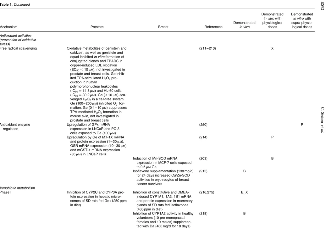
Demonstrated in vivo Demonstrated in vitro with physiological doses Demonstrated in vitro with supra-physio-logical doses Antioxidant activities (prevention of oxidative stress)
Free radical scavenging Oxidative metabolites of genistein and
daidzein, as well as genistein and equol inhibited in vitro formation of conjugated dienes and TBARS in copper-induced LDL oxidation (EC50,10 mM), not investigated in prostate and breast cells. Ge inhib-ited TPA-stimulated H2O2 pro-duction in human
polymorphonuclear leukocytes (IC50¼ 14·8 mM) and HL-60 cells (IC50¼ 30·2 mM). Ge (. 10 mM) sca-venged H2O2in a cell-free system. Ge (100 – 200 mM) inhibited O22
for-mation. Ge (0·1 – 10 mM) suppresses TPA-mediated H2O2formation in mouse skin, not investigated in prostate and breast cells
(211 – 213) X
Antioxidant enzyme regulation
Upregulation of GPx mRNA expression in LNCaP and PC-3 cells exposed to Ge (100 mM)
(250) P
Upregulation by Ge of MT-1X mRNA and protein expression (1 – 30 mM), GSR mRNA expression (10 – 30 mM) and mGST-1 mRNA expression (30 mM) in LNCaP cells
(214) P
Induction of Mn-SOD mRNA expression in MCF-7 cells exposed to 0·5 mMGe
(203) B
Isoflavone supplementation (138 mg/d) for 24 days increased Cu/Zn-SOD activities in erythrocytes of breast cancer survivors
(215) B
Xenobiotic metabolism
Phase I Inhibition of CYP2C and CYP3A
pro-tein expression in hepatic micro-somes of SD rats fed Ge (1250 ppm in diet)
Inhibition of constitutive and DMBA-induced CYP1A1, 1A2, 1B1 mRNA and protein expression in mammary glands of SD rats fed isoflavones (430 ppm in diet)
(216,275) B, X
Inhibition of CYP1A2 activity in healthy volunteers (10 pre-menopausal females and 10 males) supplemen-ted with Da (400 mg/d for 10 days)
(218) B C. Steiner et al. ES92
Table 1. Continued
Mechanism Prostate Breast References
Demonstrated in vivo Demonstrated in vitro with physiological doses Demonstrated in vitro with supra-physio-logical doses Inhibition of CYP1A1 and 1B1
activi-ties in MCF-7 cells exposed to Ge (Ki¼ 15·4 mMand Ki ¼ 0·7 mM, respectively)
(217) B
Phase II Induction of GSTP1 mRNA, protein
expression and enzyme activity in non-tumourigenic MCF-10A cells exposed to Ge (1 – 30 mM)
(219) B
Induction of quinone reductase (QR) mRNA and protein expression in MDA-MB-231 and MCF-7 cells exposed to 10 mMGe
(220) B
Inhibition of COMT activity in cytosolic fractions of human mammary tis-sues exposed to 30 mMGe
(276) B
Induction of liver GPx, GR, kidney GST, colon QR and colon UDPGT activity in female SD rats fed isofla-vones (0·81 mg/g diet)
(277) X
Induction of UDPGT activity in LNCaP cells exposed to 5 mMGe
(121) P
Inhibition of SULT1A1 activity in human liver tissues by Ge (IC50¼ 2 mM)
(278) X
DNA repair
Time- and concentration-dependent upregulation of BRCA1 and BRCA2 protein levels in LNCaP and DU145 cells exposed to Ge (0·5 – 7·5 mM)
Upregulation of BRCA1 and BRCA2 protein levels in MCF-7 and T47D cells exposed to Ge (0·5 – 1 mM)
(209) B, P
Upregulation of BRCA2 mRNA levels in ovariectomised rats fed isoflav-one-formulated Soylife (8 mg/g bw); upregulation of BRCA1 protein leve-ls in rats fed Ge (1·25 – 3·3 mg/g bw)
(51,210) B
Vitamin D metabolism
Synthesis of the antiproliferative and prodifferentiative metabolite 1,25-dihydroxy-vitamin D3
Downregulation of CYP24 and CYP27B1 expression in DU-145 cells exposed to Ge (. 25 mM)
Increase of CYP27B1 mRNA, protein expression and enzyme activity in MCF-7 cells exposed to 50 mMGe
(221,222) B, P
Inhibition of topoisomerase II (maintains DNA structure)
Inhibition of topoisomerase II by Ge (. 20 mM) in vitro, not investigated in prostate and breast cells
(223) X
Telomerases
Repression of hTERT transcriptional activity in PC3 and DU145 cells exposed to Ge (50 mM)
Significant reduction of telomere length in MCF-7 cells exposed to Ge (100 mM)
(196,224) B, P
P, prostate; B, breast; X, other cells/tissues.
Isoflavones and the prevention of breast and prostate cancer ES93