HAL Id: hal-02795220
https://hal.inrae.fr/hal-02795220
Submitted on 5 Jun 2020
HAL is a multi-disciplinary open access
archive for the deposit and dissemination of
sci-entific research documents, whether they are
pub-lished or not. The documents may come from
teaching and research institutions in France or
abroad, or from public or private research centers.
L’archive ouverte pluridisciplinaire HAL, est
destinée au dépôt et à la diffusion de documents
scientifiques de niveau recherche, publiés ou non,
émanant des établissements d’enseignement et de
recherche français ou étrangers, des laboratoires
publics ou privés.
Temperature impact on yeast fluidity
Marine Froissard
To cite this version:
Standard Project
Experimental Report
Proposal title: Temperature impact on yeast fluidity
Proposal number:
99140076
Beamline: DISCO Date(s) of experiment:
from: 31/05/2014 to: 01/06/2014 Date of report: 12/09/2014 Shifts: 6 shifts Local contact(s): Frédéric JAMME Matthieu REFREGIERS Date of submission: 12/09/2014 Objective & expected results (less than 10 lines):
The main objective of the experiment is to design a procedure for measurement of membrane fluidity, i.e. fluorescence anisotropy, on living yeasts at 4°C and 22°C. For this purpose, we will performed preliminary tests to evaluate the technical environment necessary with the TELEMOS microscope such as convenient objectives, controlled temperature stage using Peltier effect, polarizer rotation, software image acquisition procedure, etc. Various staining conditions with TMA-DPH and Laurdan dyes have also to be tested. During this beamtime, we will expect a set of good quality parallel and perpendicular images convenient for fluorescence anisotropy calculation.
Results and the conclusions of the study (main part):
Biological material
Saccharomyces cerevisiae and Debaryomyces arctica cells were grown 48h at 28°C and 15 days at 4°C in complete medium
(YPG) in INRA laboratory before arrival at SOLEIL synchrotron. One millilitre of each culture was incubated with 10 µM final concentration of 1-(4-trimethylammoniumphenyl)-6-phenyl-1,3,5-hexatriene (TMA-DPH) or 6-Dodecanoyl-2-Dimethylaminonaphthalene (Laurdan). Incubations were performed on ice for cells grown at 4°c or at room temperature for cells grown at 28°C. Cells were washed once with PBS before observation. Cell suspensions (6 µL) were placed between two quartz slides.
Image acquisition
Experiments were done on TELEMOS microscope equipped with polarization filters and controlled temperature stage using Peltier effect. Unfortunately, we encountered microscope and software problems and lost a lot of experimental time. So, during the remaining time, we decided to work on cells stained with TMA-DPH to obtain a coherent set of experimental data. Parallel and perpendicular image acquisitions were performed for S. cerevissiae and D. arctica grown at 4°C and 28°C.
Results
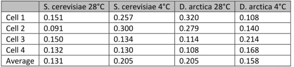
Fluorescence anisotropy intensity was calculated using ImageJ software as described in Figure 1 and Table 1 on a subset of data.
Table 1: anisotropy intensity for individual yeast cells
S. cerevisiae 28°C S. cerevisiae 4°C D. arctica 28°C D. arctica 4°C
Cell 1 0.151 0.257 0.320 0.108 Cell 2 0.091 0.300 0.279 0.140 Cell 3 0.150 0.134 0.114 0.214 Cell 4 0.132 0.130 0.108 0.168 Average 0.131 0.205 0.205 0.158 Conclusions
At this time, it is not reasonable to draw robust conclusions but we are convinced that it will be possible with technical improvements such as those listed below.
We observed that cell grown 15 days at 4°C are not in a good physiological state for microscopy and decided to optimize our growth conditions for further experiments. In particular, we observed a lot of cell death in the samples. It will be necessary to preserve the quality of biological samples by reducing the acquisition time. For this purpose, we
concluded that motorization of polarizers and automatized acquisition have to be developed
Acquisition of images out of focus has a negative impact on the calculation of fluorescence anisotropy. So, acquisition of Z-stacks will resolve this problem
We observed that the cells present a large heterogeneity in size (figure 2) in a same culture, between species and between culture conditions (28°C and 4°C), so it will be necessary to normalize anisotropy signal according to cell surface to make valuable comparisons
Figure 2: D. arctica size heterogeneity
Justification and comments about the use of beam time (5 lines max.):
The beamtime allowed for this experiment was 6 shifts and was efficiently used to explore some technical requirements and for the development of convenient protocols for a further in-depth study of fluorescence anisotropy on yeast cells with TMA-DPH but also with other lipid dyes such as DPH or Laurdan. At this time, DISCO staff is hardly working to upgrade the beamline and answer our technical requirements. Now, the motorization of the polarization filters is available on the beamline.
Publication(s): -