HAL Id: inserm-00188488
https://www.hal.inserm.fr/inserm-00188488
Submitted on 20 Nov 2007
HAL is a multi-disciplinary open access
archive for the deposit and dissemination of
sci-entific research documents, whether they are
pub-lished or not. The documents may come from
teaching and research institutions in France or
abroad, or from public or private research centers.
L’archive ouverte pluridisciplinaire HAL, est
destinée au dépôt et à la diffusion de documents
scientifiques de niveau recherche, publiés ou non,
émanant des établissements d’enseignement et de
recherche français ou étrangers, des laboratoires
publics ou privés.
3D Multi-Object Segmentation of Cardiac MSCT
Imaging by using a Multi-Agent Approach.
Julien Fleureau, Mireille Garreau, Dominique Boulmier, Alfredo Hernandez
To cite this version:
Julien Fleureau, Mireille Garreau, Dominique Boulmier, Alfredo Hernandez. 3D Multi-Object
Seg-mentation of Cardiac MSCT Imaging by using a Multi-Agent Approach.. Conference proceedings : ..
Annual International Conference of the IEEE Engineering in Medicine and Biology Society. IEEE
En-gineering in Medicine and Biology Society. Annual Conference, Institute of Electrical and Electronics
Engineers (IEEE), 2007, 1, pp.6003-6006. �10.1109/IEMBS.2007.4353716�. �inserm-00188488�
3D Multi-Object Segmentation of Cardiac MSCT Imaging by using a
Multi-Agent Approach
Julien Fleureau, Mireille Garreau, Dominique Boulmier, Alfredo Hern´andez
Abstract— We propose a new technique for general pur-pose, semi-interactive and multi-object segmentation in N-dimensional images, applied to the extraction of cardiac struc-tures in MultiSlice Computed Tomography (MSCT) imaging. The proposed approach makes use of a multi-agent scheme combined with a supervised classification methodology allowing the introduction of a priori information and presenting fast computing times. The multi-agent system is organised around a communicating agent which manages a population of situated agents which segment the image through cooperative and competitive interactions. The proposed technique has been tested on several patient data sets. Some typical results are finally presented and discussed.
I. INTRODUCTION
Cardiac Computed Tomography is a fast, non invasive, sub-millimeter resolution, volumetric imaging modality. The development of multi-slice CT has made it an important tool for cardiac imaging, giving access in the same time to differ-ent parts of the heart anatomy (right and left vdiffer-entricles, atria, myocardium, vessels) but also to its associated movements during one cardiac cycle. The acquisition of these dynamic 3D images leads to very large data sets to analyse from which the cardiac structures have to be extracted under strong constraints to face clinical requirements. 3D visualization tools are used in clinical practice but they need a very important part of human interaction and a long time to remove structures which do not belong to the heart. Nu-merous techniques have also been described for segmenting the left ventricle of the heart in images from different types of medical scanners but rarely for extracting simultaneously all parts of the heart. This paper is focused on the detection of the heart components from each entire 3D volume of the cardiac MSCT dynamic sequence. This step of segmentation is actually very complex due to the nature of the acquired image. The similarity of intensities and the connectedness of two different structures present within the image, the fuzziness and complexity of the frontiers, the artifacts of reconstruction, or even the heterogeneous dispersion of con-trast product, represent some of the problems associated with segmentation, and justify the development of robust algorithms. This paper presents a method, robust enough to answer our problem of cardiac structures extraction in 3D
This work was supported by the European ALFA IPECA project. J. Fleureau, M. Garreau, D. Boulmier and A. Hern´andez are with INSERM, U642, Rennes, F-35000, France; and with Universit´e de Rennes 1, LTSI, Rennes, F-35000, France julien.fleureau, mireille.garreau, [email protected]
D. Boulmier is also with Centre Cardio-Pneumologique, CHU Pontchail-lou, Rennes, [email protected]
imaging and, in a more general perspective, to propose a new way to segment any kind of 3D anatomical structure while keeping strong constraints in computing time and ergonomy. Several approaches have been proposed to solve segmenta-tion problems in 3D imaging as well as many works exist in segmenting the left ventricle in 2D or 3D data sets from various imaging modalities. A first category of methods, identified as boundary-based methods, like snakes or Live-Wire [5] try, with more or less interactivity, to minimize an energetic criteria in order to find the more accurate border of one only object. Such approaches, focused on frontiers detection, may pose problems when the object to segment presents holes or discontinuities in its border (such as tra-becular structures, which are present at the borders of the left ventricle). Region-based approaches have consequently been privileged. In this perspective, Fuzzy Connectedness, Graphcut and Level Set methodologies seem to provide the best results in medical contexts. Fuzzy Connectedness methods [7], [4] are fuzzy approaches which associate seed points (selected interactively) with each object to segment and then build a connectedness map which symbolizes the strengthness of the link (connectedness) between each spel (generalization of the pixel or voxel notion) of the map and the seed point. Then, the user thresholds this map to keep the area matching with the object of interest. In Graphcuts methods [1], the user has to select one region of the back-ground and one region of the object for the initialization of the process and the minimisation of an energy by graph-cut algorithm is applied. In Level Set methods [10], negative and positive region matching with region of interest and background evolve via an iterative energetic criteria. Much of the work done on 3D cardiac segmentation has been model-based (see [6] and [9] for reviews or [2], [8], [12] for more recent works.
Most of these approaches are often strictly mono-object, don’t offer the possibility to bring a priori information with few paramaters, can be sensitive to initialization or can take a long computing time. A way to answer these questions can be to consider hybrid methods, but it will necessary lead to loss in simplicity and even higher computational loads. Our objectives are : i) to ease the integration of prior information (essentially spatial prior information); ii) to integrate the notion of texture; iii) to face with real problem of multi-object segmentation; iv) to avoid step of preprocessing; v) to minimize computing costs and be user-friendly.
Our proposed solution is relevant from region-based ap-proaches and distributed low level vision systems based on a multi-agent architecture (with distributed time
implemen-HAL author manuscript inserm-00188488, version 1
HAL author manuscript
tation) and integrating a supervised classification approach. Several image segmentation techniques based on multi-agent methodologies have been presented in the litterature [3], [11] and showed the interest of using distributed approaches to split the work of segmentation or to combine techniques (contour and region-based). The proposal multi-agent system (MAS) is meant to gain in decision for the segmentation process by merging cooperative and competitive behaviours associated to specific agents related to different objects of the scene. The methodology that we propose thus differs from region growing approach by the MAS implemented archi-tecture (which allows interactions between subprocesses or even tasks distribution) and is not relevant from deformable models or level sets since no explicit cost functions are to be minimized here. Moreover the method allows multi-object detection without preprocessing stage. The outline of this paper is as follows : section II describes the adopted method-ology for the segmentation process. Section III presents some typical results obtained on real data and section IV gives a conclusion with some perspectives.
II. A MULTI-AGENTSYSTEM
The proposed Multi-Agent System (MAS) performs object segmentation in a 3D image, based on a first step in which the user selects interactively one or more seed points for each object of interest. In this application, an object is considered as an entity satisfying the following conditions (Figure 1):
• homogeneity in terms of intensity and texture (the
intensity reflects the density of an object, and one object is characterized by a quite homogeneous density).
• connexity in 3D space, so that an organ is composed of connected and compact subparts.
• availability of a priori information: actually, the left ventricle and the left atrium shown in Figure 1 have the same intensity, the same texture and are also connected structures. The only way to differentiate them lies in the introduction of prior anatomical knowledge.
Fig. 1. An original cardiac MSCT slice
The MAS is composed of a set of purely situated agents, called Workers, that cooperate and compete so as to max-imize their territory (satisfying a criterion of texture and intensity) and one purely communicating agent that coor-dinates the behaviors of all worker agents (Figure 2). This architecture corresponds to a micro-social organization, since
the society is only composed of a few agents. The environ-ment is defined as the volume of voxels (a 3D image) to be segmented and the different agent behaviors are completely deterministic. The following subsections will describe each agent type and their interactions.
Fig. 2. MAS Architecture
A. Situated Agents : Workers
Each situated agent is initially located in its environment and associated with a specific label (according to the object to be segmented), by the user. Several agents with the same label can obviously coexist and their goal is to associate their label to one region of the image. The behavior of these agents can be decomposed into three steps: As intentional agents, they firstly learn local information of intensity and texture concerning the object which they are associated with. More precisely, local intensity transitions inside the image that we called “transition vectors sets” are analyzed and generalized in the intensity plane through parametrical shapes (quadric for example) and allow the workers to acquire information on its object in terms of intensities and local texture. In a second step, each agent will browse the environment from its initial point, trying to reach its first order connected neigh-bourhood and defining its browsing direction in function of the neighbor spels which satisfy its texture and intensity criteria in the image. At each displacement, they ask for the communicating agent to acquire it, i.e. to associate their label to this new part of the image. Once this request has been proposed, the agent becomes reactive and simply waits for the approbation, or the reject of the controller. Specifical worker called inhibitor agents have been also introduced which role is just to prevent other agents to reach wrong territory by initiating an “artificial” situation of competition. B. Communicating Agent : Controller
Situated agents depend on one other particular agent, the communicating one that we label ”Controller”. This agent is clearly a cognitive agent and especially an intentional one. Its main role consists in deciding if a region of the image can be acquired by one of the workers and to inform it of its decision. To decide, it updates and uses two specific maps: a
“Segmentation Map” which is a labeled copy of the original image and a “Travel Map” which records, for each spel, the length of the path of the agent which acquired it. Using these two maps, the controller may have to face with three kinds of request:
• Simple Acquisition: A worker makes a request for the acquisition of a spel which is not yet owned by any other agent. In this case, the controller first accepts the acquisition and updates the segmentation map. It then evaluates the length of the path required by the current worker to reach the current spel and updates the travel map. It finally informs the worker that the acquisition was accepted and that it can keep on its progression through this spel.
• Cooperation: A worker would like to acquire a spel
that has been already acquired by an agent of its group (same label). In this case, the controller compares the length of the paths needed to arrive to this spel by the two cooperating agents. The agent with the shortest path is retained, the Travel Map is updated accordingly and only this agent is allowed to keep on its progression through the current spel.
• Competition: A worker asks for the acquisition of a spel which is already attributed to another group of workers (another label). This situation implies that the texture and intensity of the two corresponding objects are similar, or even perfectly identical. To solve this ambiguity, the controller attributes the critical spel to the agent with the shortest path.
III. APPLICATION TO CARDIACMSCT IMAGES We present the new segmentation framework applied to cardiac MSCT images. Our goal is to extract different anatomical parts of the heart from the whole 4D database, in particular, the left ventricle (LV) and atrium (LA), the right ventricle (RV) and atrium (RA) (we will then consider the grouping of the right ventricle and right atrium for conve-nience) and the myocardium (MY) which surrounds the left ventricle cavity. The method can simultaneously segment and label different structures, according to the objects requested by the user. We will also highlight the improvement of the results due to the addition of inhibitor agents associated to “competitor objects” (CO). Finally, we will present how this approach can be evaluated by the extraction of clinical quantitative parameters.
A. 3D Segmentation
The proposed algorithm has been tested on several databases (around twenty) for the detection of cardiac struc-tures cited before. We present here some typical results. For each example, we precise the type of cardiac structures requested by the user, the number and initial configuration of the workers (i.e. the seed points and the number of slices in which they have been positionned in 3D) and the computing time. All the presented results have been obtained on a 3 Ghz with 1GB RAM computer using a dedicated interface
(libraries developped under FLTK and VTK). They corre-spond to results obtained by the 3D segmentation process which are illustrated both in 2D slices for comparison with original slices and in 3D visualization.
1) Competition situations without inhibitor agents : Differentiating left ventricle and left atrium is very difficult in MSCT images because of their connexity and their great similarity in terms of intensity and texture. Here specifying three objects (LA, LV and RV), selecting seed points in three slices of the volume (see Table I), lead to a competitive situation which enhances the final 3D objects (Figures 3 and 4).
Acquisition Device MSCT GE Lightspeed VCT 64 Rings Data Dimensions 512 × 512 × 350 Segmentation Time 4 mn Objects RV LA LV Seeds Number Slice 1 3 3 3 Slice 2 2 3 4 Slice 3 0 2 2 TABLE I
CONFIGURATION FOR THE3DSEGMENTATION(RV, LA, LV)
Fig. 3. Illustration of competition for left atrium segmentation : on the left, one original slice extracted from the whole volume to segment (with the seed points) and on the right the associated segmentation results for RV (green color), LV (red color), LA (blue color)
Fig. 4. Competition for left atrium segmentation, 3D vizualisation of extracted structures (with part of their associated vascular trees) : RV (green color), LV (red color), LA (blue color). On the left, one inferior view of the heart and on the right, one anterior view.
2) Inhibitor agents and competition : In the second situation, the segmentation at the same time of the left ventricle (grouped with the left atrium), the right ventricle (grouped with the right atrium) and the myocardium around the left ventricle cavity is researched. The major problem comes here from the myocardium structure. In fact this anatomical part appears, on MSCT images, very similar and
connected to one part of the background so that, by the only association of agents to this part, the agents spread in the whole region associated to the myocardium but also to one part of the background (see Figure 5). To face with that problem, inhibitor agents (associated with CO) have been introduced (see Table II and Figure 5) and show clearly their good influence on the segmentation.
Acquisition device MSCT GE Lightspeed VCT 64 Rings Image Dimensions 512 × 512 × 350 Segmentation Time 4 mn
Objects VG VD MY CO Seeds Number Slice 1Slice 2 53 53 76 149
TABLE II
CONFIGURATION FOR THE3DSEGMENTATION(VG, VD, MY, CO)
Fig. 5. Myocardium segmentation without (up) and with (down) inhibitor agents : on the left, one original slice extracted from the whole volume to segment (with the seed points) and on the right the associated segmentation results for MY (green color), LV (blue color), RV (red color), CO (yellow color)
B. Extraction of Quantitative Parameters
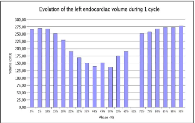
A first way to evaluate the performance of the proposed methodology lies in the extraction of clinical parameters which can be qualitatively analysed and compared to refer-ence values. We can see in Figure 6, for the same database, the different volumes of the left ventricle (without the left atrium) which have been obtained by the application of the method on each 3D volume acquired during the cardiac cycle (for different phases or instants expressed in percentage of the whole cycle). The resulting volume variation curve phys-iologically validates our methodology thanks to its relative continuity, its time consistency with diastolic and systolic phases of the cardiac cycle and the rightness of its values. The ejection fraction has also been computed and compared with CT and echocardiographic measures obtained in clinical practice, giving encouraging results.
IV. CONCLUSION AND PERSPECTIVES
In this paper, we have presented a new 3D multi-object segmentation method, based on a mutli-agent scheme com-bined with a supervised classification methodology and a
Fig. 6. Left ventricle volume during one cardiac cycle
region growing process. Its originality lies in its low user in-teraction, in its capacity to handle multi-object segmentation in N-dimensional spaces, and also in its performance in terms of noise sensitivity (no preprocessing required) and rapidity (the different computing times encountered during our tests remained acceptable for a clinical application). More than a quantitative improvement, this multi-object approach allows a multi-agent and a competitive implementation which leads as we showed to qualitative enhancement, especially in critical situations.
A more complete validation of this method is now in progress and it will be also quite interesting to compare it with well-known methods of the litterature.
REFERENCES
[1] Y. Boykov and V. Kolmogorov. An experimental comparison of min-cut / max-flow algorithms for energy minimization in vision. IEEE Trans. Pattern Anal. Mach. Intell., 26(9):1124–1137, 2004. [2] T. Chen, D. N. Metaxas, and L. Axel. 3D cardiac anatomy
recon-struction using high resolution CT data. In MICCAI, volume 1, pages 411–418, 2004.
[3] E. Duchesnay, J.J. Montois, and Y. Jacquelet. Cooperative agents soci-ety organized as an irregular pyramid: A mammography segmentation application. Pattern Recognition Letters, 24(14):2435–2445, 2003. [4] J.K. Udupa et al. Relative fuzzy connectedness and object definition:
Theory, algorithms, and applications in image segmentation. IEEE Trans. Pattern Anal. Mach. Intell., 24(11):1485–1500, 2002. [5] A. Falcao, J. Udupa, S. Samarasekera, and S. Sharma. User-steered
image segmentation paradigms: Livewire and livelane. Graphical Models and Image Processing, 60:233–260, 1998.
[6] A.F. Frangi, W.J. Niessen, and M.A. Viergever. Three-dimensional modeling for functional analysis of cardiac images: A review. IEEE Trans on Medical Imaging, 20(1):2–25, 2001.
[7] G.T. Herman and B.M. Carvalho. Multiseeded segmentation using fuzzy connectedness. IEEE Trans. Pattern Anal. Mach. Intell., 23(5):460–474, 2001.
[8] M. Lorenzo-Valdes, G.I. Sanchez-Ortiz, A.G. Elkinton, R.H. Mohi-addin, and D. Rueckert. Segmentation of 4D cardiac MR images using a probabilistic atlas and the EM algorithm. Medical Image Analysis, 8(3):255–265, 2004.
[9] T. McInerney and D. Terzopoulos. Deformable models in medical image analysis : A survey. Medical Image Analysis, 1(2), 1996. [10] S. Osher and J.A. Sethian. Fronts propagating with curvature
depen-dent speed : Algorithm based on hamilton-jacobi formulations. Journal of Computational Physics, 79:12–49, 1988.
[11] N. Richard, M. Dojat, and C. Garbay. Automated segmentation of human brain MR images using a multi-agent approach. Artificial Intelligence in Medicine, 30(2):153–176, 2004.
[12] M. Sermesant, C. Forest, X. Pennec, H. Delingette, and N. Ayache. Deformable biomechanical models: application to 4D cardiac image analysis. Medical Image Analysis, 7(4):475–488, 2003.