HAL Id: hal-02905823
https://hal.archives-ouvertes.fr/hal-02905823
Submitted on 2 Oct 2020HAL is a multi-disciplinary open access archive for the deposit and dissemination of sci-entific research documents, whether they are pub-lished or not. The documents may come from teaching and research institutions in France or abroad, or from public or private research centers.
L’archive ouverte pluridisciplinaire HAL, est destinée au dépôt et à la diffusion de documents scientifiques de niveau recherche, publiés ou non, émanant des établissements d’enseignement et de recherche français ou étrangers, des laboratoires publics ou privés.
marker of polyps with malignant potential
Florence Renaud, Christophe Mariette, Audrey Vincent, Agnès Wacrenier,
Vincent Maunoury, Julie Leclerc, Lucie Coppin, Michel Crépin, Isabelle van
Seuningen, Emmanuelle Leteurtre, et al.
To cite this version:
Florence Renaud, Christophe Mariette, Audrey Vincent, Agnès Wacrenier, Vincent Maunoury, et al.. The serrated neoplasia pathway of colorectal tumors: Identification of MUC5AC hypomethylation as an early marker of polyps with malignant potential. International Journal of Cancer, Wiley, 2016, 138, pp.1472-1481. �10.1002/ijc.29891�. �hal-02905823�
For Peer Review
The serrated neoplasia pathway of colorectal tumours: identification of MUC5AC hypomethylation as an early
marker of high-risk polyps
Journal: International Journal of Cancer Manuscript ID: IJC-15-1083
Wiley - Manuscript type: Early Detection and Diagnosis Date Submitted by the Author: 22-May-2015
Complete List of Authors: Renaud, Florence; Institut de Pathology,
Mariette, Christophe; Lille University Hospital, Digestive Surgery Vincent, Audrey; Inserm UMRS 1172, Team 'Mucins, Epithelial Differentiation and carcinogenesis'
Wacrenier, Agnes; Lille University Hospital, Pathology Institute Maunoury, Vincent; Lille University Hospital, Gastroenterology
Leclerc, Julie; Lille University Hospital, Molecular Oncology and Genetics, Biochemistry and Molecular Biology Institute
Coppin, Lucie; Lille University Hospital, crépin, michel; Lille university hospital,
Van Seuningen, Isabelle; Inserm UMRS 1172, Team 'Mucins, Epithelial Differentiation and carcinogenesis'
Leteurtre, Emmanuelle; Lille University Hospital, Pathology Institute Buisine, Marie-Pierre; Lille University Hospital, Molecular Oncology and Genetics, Biochemistry and Molecular Biology Institute
Key Words: hyperplastic polyp, sessile serrated adenoma, colonic cancer, mucin,
methylation
For Peer Review
The serrated neoplasia pathway of colorectal tumours: identification of MUC5AC hypomethylation as an early marker of high-risk polyps
Short title: MUC5AC hypomethylation in high-risk colorectal serrated polyps
F Renaud1,2,3, C Mariette1,3,4, A Vincent1,3,5, A Wacrenier2, V Maunoury6, J Leclerc1,3,7, L Coppin1,3,7, M Crépin7, I Van Seuningen1,3,5, E Leteurtre1,2,3, MP Buisine1,3,7
1 Inserm, UMR-S1172, Team ‘Mucins, Epithelial Differentiation and Carcinogenesis’,
Jean-Pierre Aubert Research Center, Lille, France
2
Pathology Institute, Biology Pathology Center, Lille University Hospital, Lille, France
3
North of France Lille 2 University, Lille, France
4
Department of Digestive Surgery, Claude Huriez Hospital, Lille University Hospital, Lille, France
5
Lille University Hospital, Lille, France
6
Department of Gastroenterology, Claude Huriez Hospital, Lille University Hospital, Lille, France
7
Department of Molecular Oncology and Genetics, Biochemistry and Molecular Biology Institute, Biology Pathology Center, Lille University Hospital, Lille, France
Key words: hyperplastic polyp, sessile serrated adenoma, colonic cancer, mucin, methylation.
Abbreviations: CA, conventional adenoma; CIMP, CpG island methylation phenotype; CIMP-H, CIMP-high; CIMP-L, CIMP-low; CRC, colorectal carcinoma; GCHP, goblet cell-rich hyperplastic polyp; MSI, microsatellite instability; MSS, microsatellite stable; MVHP, 2 3 4 5 6 7 8 9 10 11 12 13 14 15 16 17 18 19 20 21 22 23 24 25 26 27 28 29 30 31 32 33 34 35 36 37 38 39 40 41 42 43 44 45 46 47 48 49 50 51 52 53 54 55 56 57
For Peer Review
microvesicular hyperplastic polyp; SSA, sessile serrated adenoma; SSA/D, sessile serrated adenoma with dysplasia; TSA, traditional serrated adenoma.
Grant sponsors: SIRIC ONCOLille; Lille University Hospital; French Society of Pathology.
Correspondence to: Marie-Pierre Buisine, Inserm UMR-S1172, Team ‘Mucins, Epithelial Differentiation and Carcinogenesis’, Jean-Pierre Aubert Research Center, 59045 Lille cedex, France. Tel.: 33-3-20-29-88-50, Fax: 33-3-20-53-85-62, E-mail: marie-pierre.buisine@inserm.fr
Appropriate article category: Early detection and diagnosis
2 3 4 5 6 7 8 9 10 11 12 13 14 15 16 17 18 19 20 21 22 23 24 25 26 27 28 29 30 31 32 33 34 35 36 37 38 39 40 41 42 43 44 45 46 47 48 49 50 51 52 53 54 55 56
For Peer Review
ABSTRACT
The serrated neoplasia pathway accounts for 20 to 30% of colorectal cancers (CRC), which are characterised by extensive methylation (CpG Island Methylation Phenotype, CIMP), frequent BRAF mutation and high microsatellite instability (MSI). We recently identified MUC5AC mucin gene hypomethylation as a specific marker of MSI CRC. The early identification of preneoplastic lesions among serrated polyps is currently challenging. Here we performed a detailed pathological and molecular analysis of a large series of colorectal serrated polyps and evaluated the usefulness of mucin genes MUC2 and MUC5AC to differentiate serrated polyps and to identify high-risk lesions. A series of 330 colorectal polyps including 218 serrated polyps (42 goblet cell-rich hyperplastic polyps (GCHP), 68 microvesicular hyperplastic polyps (MVHP), 100 sessile serrated adenoma (SSA), 8 traditional serrated adenoma (TSA)) and 112 non-serrated polyps was analysed for BRAF/KRAS mutations, MSI, CIMP, MLH1 and MGMT methylation, and MUC2 and MUC5AC expression and methylation. We show that MUC5AC hypomethylation is an early event in the serrated neoplasia pathway, and specifically detects MVHP and SSA, arguing for a filiation between MVHP, SSA, and CIMP-H/MSI CRC, whereas GCHP and TSA arise from a distinct pathway. Moreover, MUC5AC hypomethylation specifically identified serrated lesions with BRAF mutation, CIMP-H or MSI, suggesting that it may be useful to identify serrated neoplasia pathway-related precursor lesions. Our data suggest that MVHP and SSA should benefit from the same therapeutic and surveillance strategies.
2 3 4 5 6 7 8 9 10 11 12 13 14 15 16 17 18 19 20 21 22 23 24 25 26 27 28 29 30 31 32 33 34 35 36 37 38 39 40 41 42 43 44 45 46 47 48 49 50 51 52 53 54 55 56 57
For Peer Review
What’s new?
The identification of high-risk colorectal serrated polyps is challenging since benign hyperplastic polyps and sessile serrated adenoma may share the same morphology and since carcinogenesis in this pathway is faster than in the conventional adenoma-carcinoma pathway. We identified MUC5AC hypomethylation as a new early marker of serrated neoplasia pathway, specifically detecting CIMP-H/MSI CRC precursors MVHP and SSA, suggesting they should benefit from the same surveillance for accurate management of patients.
2 3 4 5 6 7 8 9 10 11 12 13 14 15 16 17 18 19 20 21 22 23 24 25 26 27 28 29 30 31 32 33 34 35 36 37 38 39 40 41 42 43 44 45 46 47 48 49 50 51 52 53 54 55 56
For Peer Review
INTRODUCTION
Most colorectal cancers (CRC) develop through a conventional adenoma (CA)-carcinoma sequence. However, 20 to 30% of CRC develop through the ‘serrated neoplasia pathway’, named as for the serrated appearance of crypt in the precursor polyps.1-3
Serrated lesions represent a heterogeneous group of polyps, divided into hyperplastic polyps (HP), sessile serrated adenomas (SSA) and traditional serrated adenomas (TSA) by the latest World Health Organisation (WHO) classification of tumors of the digestive system (fourth edition 2010).4 However a diagnostic ‘grey-zone’ exists between HP and SSA, with a wide inter-observer variability due to the lack of consensus diagnostic criteria.5-9 In addition, contradictory guidelines were given for the diagnosis of serrated polyps by the WHO and then by an expert panel,10 resulting in some confusion among pathologists.9 Yet, it is important to distinguish SSA from HP, since the former are associated with a malignant potential whereas the later are commonly considered to be benign. Furthermore, neoplastic progression through this pathway has been reported to be faster than within the conventional adenoma-carcinoma pathway. Underdiagnosis of serrated neoplasia pathway precursors in pathologic practice may result in inadequate surveillance and thus contribute to interval carcinomas.9
SSA were underdiagnosed for many years due to their resemblance to HP and it currently remains unanswered whether SSA arise de novo or derive from pre-existing HP.11,12
Molecular characterization has improved our understanding of serrated polyps and associated-risk of malignant progression. SSA are mainly observed in the proximal colon and are associated with frequent BRAF mutation and high CpG island methylation phenotype (CIMP-H), suggesting that SSA are precursors of sporadic CIMP-H CRC, most of which display high microsatellite instability (MSI) through epigenetic inactivation of MLH1.2,11,13 TSA seem to be less strongly associated with the serrated neoplasia pathway, but they are 2 3 4 5 6 7 8 9 10 11 12 13 14 15 16 17 18 19 20 21 22 23 24 25 26 27 28 29 30 31 32 33 34 35 36 37 38 39 40 41 42 43 44 45 46 47 48 49 50 51 52 53 54 55 56 57
For Peer Review
associated with frequent KRAS or BRAF mutations, low CIMP (CIMP-L) and are supposed to be precursors of CIMP-L microsatellite stable (MSS) CRC.
Moreover, serrated pathway-related polyps and CIMP-H/MSI CRC have similar phenotype, including frequent mucinous pattern with increased expression of the intestinal mucin MUC2 and aberrant expression of the gastric mucin MUC5AC.14,15 We recently showed that abnormal expression of MUC2 and MUC5AC in CIMP-H/MSI CRC was closely related to altered methylation of their promoter.16 Interestingly, MUC5AC demethylation was specific to MSI CRC, suggesting that it may serve as a specific marker of the serrated neoplasia pathway.
In this study, 330 polyps including 218 serrated polyps were selected in order to (1) further characterise molecular features of serrated polyps and (2) evaluate the potential interest of mucin genes MUC2 and MUC5AC to discriminate serrated polyps and to identify high-risk lesions. We demonstrate that MUC5AC hypomethylation is an early and specific marker of preneoplastic lesions in the serrated neoplasia pathway of the colorectum.
2 3 4 5 6 7 8 9 10 11 12 13 14 15 16 17 18 19 20 21 22 23 24 25 26 27 28 29 30 31 32 33 34 35 36 37 38 39 40 41 42 43 44 45 46 47 48 49 50 51 52 53 54 55 56
For Peer Review
MATERIAL AND METHODS
Patients and tissues
Specimens of colorectal serrated lesions and conventional adenoma were retrieved from the archives of the Department of Pathology of the Lille University Hospital. All cases were collected from patients who underwent endoscopic mucosal resection, polypectomy or colectomy at Lille University Hospital from 2009 to 2013. Polyps were selected on the basis that they fulfilled the diagnostic criteria, were well oriented, and had sufficient available tissue for both immunohistochemical and molecular analyses. Clinical and endoscopic data were available for all patients. Clinical information retrieved included sex and age, colonic localisation (classified as proximal or distal relative to the splenic flexure) and size of the polyp. Informed consent was obtained from all patients. Approval of this study was obtained from the Institutional Review Board of Lille University Hospital.
Histological analysis
All specimens were reviewed by three experienced gastrointestinal pathologists (EL, AW, FR) blinded to the clinical and molecular information. Serrated lesions (HP, SSA and TSA) were classified on the basis of the criteria from the latest WHO classification.4 Representative serrated polyps included in this study are shown in Figure 1. Mixed polyps or cases with hyperplastic polyposis were excluded from this study. HP were defined as serrated polyps with simple tubular architecture composed of serrated crypts in the upper half of the lesion, narrow and straight crypts at the base. HP were subdivided into two groups: microvesicular HP (MVHP) and goblet cell-rich HP (GCHP) defined as per previously published criteria.4 MVHP were characterized by elongated and relatively straight crypts, with serration that are visible mainly near the luminal end of the crypts, epithelial cells with microvesicular (small 2 3 4 5 6 7 8 9 10 11 12 13 14 15 16 17 18 19 20 21 22 23 24 25 26 27 28 29 30 31 32 33 34 35 36 37 38 39 40 41 42 43 44 45 46 47 48 49 50 51 52 53 54 55 56 57
For Peer Review
droplet) mucin and absence of cytologic dysplasia (Figure 1A). GCHP were characterized by elongated and slightly serpiginous crypts mostly composed of mucin-rich goblet cells (Figure 1B). SSA were defined as serrated polyps with basal crypt dilatation, crypt branching, horizontal extension of crypts at the base, prominent serrations, mitoses in the upper half of crypts and dystrophic goblet cells (Figure 1C). To classify a polyp as SSA, there should be at least two or three contiguous SSA-type crypts. Dysplasia was defined by the presence of narrow elongated hyperchromatic nuclei, nuclei stratification and basophilic cytoplasm (Figure 1D). TSA were defined as serrated polyps composed of a uniform population of cells showing abundant eosinophilic cytoplasm, nuclear elongation, ectopic crypt formations and nuclear hyperchromasia with a tubular or villous architecture and mild crypt or surface slit-like serration (Figures 1E and 1F).
In total, 330 polyps from 330 patients were included in the study. They contained 218 serrated polyps with 110 HP (68 MVHP, 42 GCHP), 100 SSA (41 SSA with dysplasia), 8 TSA (3 with dysplasia). Non-serrated adenomas (112 CA including 17 tubular, 18 villous, 77 tubulovillous) diagnosed during the same period were also included as controls.
Immunohistochemistry
Immunohistochemical studies were carried out on 4-µm-thick formalin-fixed, paraffin-embedded whole tissue sections using antibodies against mucins MUC2 and MUC5AC as previously described.17 The average percentage of stained tumour cells and intensity of staining were calculated using a semiquantitative histological scoring method (Hscore).18 Hscore above or equal the threshold of 20 was considered as positive expression.
DNA methylation analysis
2 3 4 5 6 7 8 9 10 11 12 13 14 15 16 17 18 19 20 21 22 23 24 25 26 27 28 29 30 31 32 33 34 35 36 37 38 39 40 41 42 43 44 45 46 47 48 49 50 51 52 53 54 55 56
For Peer Review
The promoter methylation status of 6 genes commonlyused to define CIMP status (CDKN2A (p16), RUNX3, CACNA1G, SOCS1, NEUROG1, and IGF2), DNA repair genes MLH1 and MGMT, and mucin genes MUC2 and MUC5AC was determined by quantitative bisulfite pyrosequencing assays as previously described.16 Amplification of MGMT and MLH1 was performed using the PyroMark kits (Qiagen). The percentage of methylation for a given gene was calculated as the mean methylation levels of all CpG sites analysed. Tumours with a methylation level above or equal to the threshold of 10% were considered as methylation-positive. Tumours were defined as CIMP-H when ≥ 3/6 of the CIMP-specific gene markers were methylated. Tumours with 1 or 2 methylated markers and tumours with no methylated marker were considered as CIMP-L and CIMP-negative, respectively. Hypomethylation was defined by a methylation level below the thresholds of 70% for MUC5AC and 60% for MUC2, as previously described.16
MSI and mutation analysis
MSI was determined using a six-marker panel as previously described.19 Mutations in codons 12 and 13 of KRAS and in codon 600 of BRAF were identified by pyrosequencing as previously described.16
Statistical analysis
SPSS version 19.0 software (SPSS, Chicago, IL) and GraphPad Prism version 6.02 were used for statistical analyses. Discrete variables were compared using the Chi square test or the Fischer exact test as appropriate. Continuous variables were compared using the Mann– Whitney U test. A 2-tailed probability value (P) was calculated. A P value < 0.05 was considered statistically significant. CIMP-L and CIMP-negative tumours were grouped vs CIMP-H tumours for statistical analyses.
2 3 4 5 6 7 8 9 10 11 12 13 14 15 16 17 18 19 20 21 22 23 24 25 26 27 28 29 30 31 32 33 34 35 36 37 38 39 40 41 42 43 44 45 46 47 48 49 50 51 52 53 54 55 56 57
For Peer Review
RESULTS
Clinicopathological characteristics of the cohort
Table 1 summarises the clinical and endoscopic findings of the patients and their polyps. Among the 330 polyps, 153 were from women and 177 from men, with overall median age of 65 years (range, 32 to 88). Patients with TSA were significantly older than patients with other polyps (P = 0.04). There were no significant different sex ratio between the subtypes of polyps or between patients with serrated polyps (HP, SSA, TSA) and those with CA.
Overall, the median size of polyps was 10 mm (range, 3 to 35 mm) (Table 1). Among serrated polyps, HP were significantly smaller (median, 4.9 mm) than SSA (median, 9 mm), SSA/D (median, 10) and TSA (median, 20 mm) (P < 0.0001, P < 0.0001 and P < 0.0001, respectively).
In terms of tumour site, 44% were proximal and 56% were distal (Table 1). Compared to CA, serrated polyps were more frequently found in the proximal colon (P < 0.001). Among serrated polyps, 53% of MVHP, 69% of SSA, and 71% of SSA/D were proximal, whereas only 5% of GCHP and 25% of TSA were proximal.
Mutational and methylation characteristics of polyp subtypes
Activating mutations in BRAF or KRAS, MSI and abnormal methylation of a number of genes are common events in preneoplastic colorectal lesions. All cases were successfully analysed for these molecular events. Molecular alterations within the different polyp types are shown in Figure 2.
Compared to CA, serrated polyps displayed higher frequency of BRAF V600E mutation (21.6% vs 1.8%), MSI (17.9% vs 0%) and CIMP-H (24.8% vs 5.4%) and lower frequency of 2 3 4 5 6 7 8 9 10 11 12 13 14 15 16 17 18 19 20 21 22 23 24 25 26 27 28 29 30 31 32 33 34 35 36 37 38 39 40 41 42 43 44 45 46 47 48 49 50 51 52 53 54 55 56
For Peer Review
KRAS mutations (11.5% vs 33.9%). BRAF and KRAS mutations were mutually exclusive in all types of polyps.
Among GCHP, 0% displayed BRAF mutation, whereas 47.6% displayed KRAS mutation, and 0% was MSI. Only 2.4% were CIMP-H, but 11.9% were CIMP-L and 9.5% displayed abnormal methylation of MGMT. Conversely, among MVHP, 33.8% displayed BRAF mutation, 0% KRAS mutation, 8.8% were MSI, and 13.2% were CIMP-H. BRAF mutations, MSI and CIMP were more common in proximal MVHP compared to distal MVHP (47.2% vs 18.7%, P = 0.02, 16.7% vs 0%, P = 0.03, and 25.0% vs 0%, P =0.0025, respectively).
In SSA and SSA/D, BRAF mutations were significantly more frequent than KRAS mutations (22.0% vs 2.0%), 33.0% were MSI with methylation of MLH1. 41.0% of SSA were CIMP-H and 8.0% displayed methylation of MGMT. BRAF mutations, MSI and CIMP were more common in proximal SSA compared to distal SSA (28.0% vs 9.7%, P = 0.06, 43.0% vs 9.0%, P = 0.001, and 59.5% vs 4.0%, P < 0.0001, respectively).
In TSA, KRAS mutations were detected in 37.5% of cases, BRAF in 25.0% and all were MSS. TSA were essentially CIMP-negative (50.0%) or CIMP-L (37.5%), and were particularly prone to MGMT methylation (37.5%). Notably, CIMP-L, detected in 7.3% of all polyps, was significantly associated with MGMT methylation (P < 0.0001).
Taken together, our findings argue for a relationship between MVHP and SSA, GCHP and TSA being apart. Moreover, comparison of methylation patterns between the different sub-groups showed an increase in the number of methylation-positive CIMP-target genes and in the methylation levels from MVHP to SSA to SSA/D (Figure 2 and Figure 3A).
Mucin expression in subtypes of polyps
Immunohistochemistry for the intestinal mucin MUC2 and the gastric mucin MUC5AC was performed and analysed in all polyps. Frequencies and representative images of expression 2 3 4 5 6 7 8 9 10 11 12 13 14 15 16 17 18 19 20 21 22 23 24 25 26 27 28 29 30 31 32 33 34 35 36 37 38 39 40 41 42 43 44 45 46 47 48 49 50 51 52 53 54 55 56 57
For Peer Review
within the different types of polyps are shown in Figure 4 and Additional supporting information.
MUC2 and MUC5AC expressions were more frequent in serrated polyps than in CA (95% vs 63%, P < 0.0001 and 96% vs 59%, P < 0.0001, respectively). Moreover, in HP (GCHP and MVHP), SSA, SSA/D and TSA, MUC2 and MUC5AC staining intensity was generally strong to moderate and homogeneously distributed along the epithelium, whereas it was more heterogeneous in CA.
Mucin gene methylation in subtypes of polyps and association with clinicopathologic features
Methylation of MUC2 and MUC5AC mucin genes was analysed in all polyps. Results are shown in Figure 2 and Figure 3B.
MUC2 mean methylation level was 59.2% in serrated polyps and 63.8% in CA. MUC2 hypomethylation was more frequent in serrated polyps than in CA (36.7% vs 18.8%, P < 0.0001). Among serrated polyps, MUC2 hypomethylation was detected in 50.0% of GCHP, 20.6% of MVHP, 44.1% of SSA, 43.9% of SSA/D and 12.5% of TSA. In GCHP, MUC2 hypomethylation was significantly associated with KRAS mutation (P = 0.006). In MVHP, MUC2 hypomethylation was associated with proximal location (P = 0.001) and CIMP (P = 0.002). In SSA, MUC2 hypomethylation was associated with proximal location (P < 0.0001), BRAF mutation (P < 0.0001), MSI (P < 0.0001), and CIMP (P < 0.0001).
MUC5AC mean methylation level was 82.5% in serrated polyps and 92.3% in CA. MUC5AC hypomethylation was detected in 18.8% of serrated polyps and none of the CA, indicating that MUC5AC hypomethylation is specific of the serrated pathway (P < 0.0001). Furthermore, in serrated polyps, MUC5AC hypomethylation was detected in 8.8% MVHP, 25.4% SSA, and 43.9% SSA/D, but in none of GCHP or TSA. In MVHP, MUC5AC 2 3 4 5 6 7 8 9 10 11 12 13 14 15 16 17 18 19 20 21 22 23 24 25 26 27 28 29 30 31 32 33 34 35 36 37 38 39 40 41 42 43 44 45 46 47 48 49 50 51 52 53 54 55 56
For Peer Review
hypomethylation was associated with proximal location (P = 0.011), BRAF mutation (P = 0.017), MSI (P < 0.0001), MLH1 methylation (P < 0.0001), and CIMP-H (P < 0.0001). In SSA (with or without dysplasia), MUC5AC hypomethylation was associated with proximal location (P < 0.0001), BRAF mutation (P < 0.0001), MSI (P < 0.0001), MLH1 methylation (P < 0.0001), and CIMP (P < 0.0001).
MUC5AC hypomethylation is predictive of high-risk serrated lesions
We further evaluated the performances of MUC5AC hypomethylation for the identification of lesions harbouring molecular alterations, which should thus be considered as at high risk of progression to carcinoma.
MUC5AC hypomethylation was detected in 52.7% (39/74) of serrated polyps harbouring BRAF mutation, CIMP-H, or MSI and in none (0/144) of the other serrated polyps, which corresponds to a positive predictive value of 100% and a negative predictive value of 81.4% for the identification of high-risk serrated lesions.
By comparison, MUC2 hypomethylation was detected in 52.7% (39/74) of serrated polyps harbouring BRAF mutation, CIMP-H or MSI, but also in 27.8% (40/144) of the others, which corresponds to a positive predictive value of 49.4% and a negative predictive value of 75.9% for the identification of high-risk serrated lesions.
2 3 4 5 6 7 8 9 10 11 12 13 14 15 16 17 18 19 20 21 22 23 24 25 26 27 28 29 30 31 32 33 34 35 36 37 38 39 40 41 42 43 44 45 46 47 48 49 50 51 52 53 54 55 56 57
For Peer Review
DISCUSSION
Despite the improved recognition of the clinicopathological significance of serrated lesions of the colorectum, some questions remain to be resolved, such as the differential diagnosis of serrated polyp subtypes and especially the identification of those which require close surveillance. In this study, we performed a detailed pathological and molecular analysis of secreted mucin genes MUC2 and MUC5AC. We show that MUC5AC hypomethylation is specific of high-risk serrated polyps, suggesting that MUC5AC hypomethylation may serve as a marker to identify serrated pathway neoplasia-related precursors.
Clinicopathological and molecular features were characterized in our series of polyps. Clinicopathological data were in agreement with previous reports, 20-23 confirming that most SSA and MVHP are proximal, whereas most TSA and GCHP are distal. In line with recent reports,11,13,24,25 we found that SSA, especially proximal SSA/D, harboured frequent BRAF mutation, were often CIMP-H and MSI due to MLH1 methylation. Moreover, although the number of TSA in our series was small, we confirmed that they may display KRAS or BRAF mutations,26-28 were MSS, and often CIMP-L with methylation of MGMT. Regarding HP, MVHP differed from GCHP in that they harboured frequent BRAF mutation, some of them being CIMP-H and MSI. Conversely, GCHP showed exclusively KRAS mutations, were MSS, CIMP-negative or CIMP-L, but prone to MGMT methylation.
A common feature of serrated pathway-related polyps and CIMP-H/MSI CRC is frequent mucin-rich/mucinous pattern with increased expression of secreted mucins. We analysed the expression of the secreted mucins MUC2 and MUC5AC in our series of polyps. According to prior studies,29,30 increased expression of the intestinal mucin MUC2 and aberrant expression 2 3 4 5 6 7 8 9 10 11 12 13 14 15 16 17 18 19 20 21 22 23 24 25 26 27 28 29 30 31 32 33 34 35 36 37 38 39 40 41 42 43 44 45 46 47 48 49 50 51 52 53 54 55 56
For Peer Review
and SSA. Accumulating evidence indicates that serrated pathway-associated colorectal tumours may display aberrant gastric-type differentiation. However, MUC5AC expression was also observed in CA, although it was less frequent and weaker than in serrated polyps. Our findings, in line with recent ones,30 demonstrate that gastric-type expression is not exclusive to serrated pathway-associated lesions.
Based on our recent data showing that abnormal expression of MUC2 and MUC5AC in CRC was closely related to demethylation of their promoters,16 we determined the methylation status of MUC2 and MUC5AC in our series of polyps. Hypomethylation of MUC2 and MUC5AC was detected in a proportion of serrated lesions including HP, indicating that the demethylation process is an early event in polyp development. This mechanism may contribute to increased production of mucus and to acquisition of their particular mucin-rich phenotype.
It has been suggested that SSA may originate from certain HP. The frequent finding of MVHP-like areas within large SSA and identification of excessive methylation and BRAF mutations in both MVHP and SSA are evidence supporting that SSA may represent ‘advanced’ forms of MVHP, but the relationship between these two entities has not been clearly established.31-33
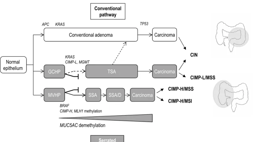
A key finding of this study was that MUC5AC hypomethylation was restricted to MVHP and SSA, suggesting it may be useful to discriminate GCHP from MVHP and SSA. Moreover, there was a gradual increase in the frequency of MUC5AC hypomethylation from MVHP to SSA and SSA/D suggesting that SSA arise from MVHP. Interestingly, our recent data in CRC showed that MUC5AC hypomethylation was specific to MSI/CIMP-H CRC.16 Altogether, our data argue for a progression of MVHP to SSA, SSA/D, and then to MSI/CIMP-H CRC (Figure 5). Some authors have questioned the utility of distinguishing 2 3 4 5 6 7 8 9 10 11 12 13 14 15 16 17 18 19 20 21 22 23 24 25 26 27 28 29 30 31 32 33 34 35 36 37 38 39 40 41 42 43 44 45 46 47 48 49 50 51 52 53 54 55 56 57
For Peer Review
MVHP and GCHP.10,34 Our data reinforce the concept that MVHP and GCHP are distinct morphological but also biological entities, highlighting a true relevance to diagnose MVHP and to precise the HP subtype in pathological reports.
We further evaluated the performances of MUC5AC hypomethylation in identifying high-risk lesions. MUC5AC hypomethylation accurately identified only serrated lesions with BRAF mutation, CIMP-H or MSI, with a positive predictive value of 100% and a negative predictive value of 81.4%, validating the utility of MUC5AC hypomethylation to assess the malignant risk of precursor lesions.
Furthermore, our data suggest that MUC5AC demethylation may play a role in the serrated pathway to colorectal cancer. Understanding the mechanisms leading to MUC5AC demethylation and subsequent aberrant expression of the gastric-type MUC5AC mucin could help to elucidate whether it is directly involved in the progression of tumours through this pathway.
Until recently, information regarding TSA was very scarce, probably due to their rarity, accounting for less than 1% of all colorectal polyps.4 Recent studies on large series of TSA have enriched our knowledge regarding their clinicopathological and molecular features.27,28 Whereas TSA are morphologically fairly similar, they are characterized by molecular heterogeneity, showing BRAF or KRAS mutation and variable CIMP, suggesting at least two pathways of neoplastic progression. Frequent identification of SSA-type lesions in proximal and BRAF mutant TSA and frequent association with MVHP led to the hypothesis that some TSA develop from SSA.28 However, no MUC5AC hypomethylation was detected in TSA in our series, especially in the two proximal TSA with BRAF V600E mutation. Although the number of TSA was small in our series, our data suggest a pathway distinct from the MVHP-SSA-CIMP-H CRC pathway, as previously suggested by Tsai et al.27 Our hypothesis is 2 3 4 5 6 7 8 9 10 11 12 13 14 15 16 17 18 19 20 21 22 23 24 25 26 27 28 29 30 31 32 33 34 35 36 37 38 39 40 41 42 43 44 45 46 47 48 49 50 51 52 53 54 55 56
For Peer Review
supported by recent findings obtained in vivo in a BRAF-mutant mouse model, which showed that the resulting mice developed serrated polyps which progressed to TSA, but not to SSA.35 Conversely, we observed similarities between TSA with KRAS mutation and some GCHP that were CIMP-L with MGMT methylation, suggesting that some TSA may develop from GCHP and progress to conventional adenoma and CIMP-negative or CIMP-L MSS CRC (Figure 5).27
Our data have major implication at the clinical level. The identification of high-risk serrated polyp is currently challenging, but is essential to guide adequate cancer screening and prevention care, especially the neoplastic progression within this pathway has been reported to be faster than within the classical adenoma-carcinoma pathway.36,37
In current clinical practice, whereas the diagnosis of SSA is followed by the same colonoscopic surveillance as for patient with CA,34 the diagnosis of HP including MVHP does not lead to a follow-up. Identification of MVHP as precursor lesions of SSA could impact medical practice.
In conclusion, this study further confirms the importance to accurately diagnose serrated lesions. We have shown that MUC5AC hypomethylation may be useful to distinguish serrated polyps and to identify high-risk lesions. Moreover, our findings suggest that some MVHP may progress to SSA, SSA/D, and CIMP-H/MSI CRC. In this respect, MVHP and SSA should benefit from the same therapeutic and surveillance strategies.
Acknowledgements:
The authors thank B. Duchêne, RM Siminsky and MH Gevaert for excellent technical assistance. 2 3 4 5 6 7 8 9 10 11 12 13 14 15 16 17 18 19 20 21 22 23 24 25 26 27 28 29 30 31 32 33 34 35 36 37 38 39 40 41 42 43 44 45 46 47 48 49 50 51 52 53 54 55 56 57
For Peer Review
References
1. Snover DC, Jass JR, Fenoglio-Preiser C, Batts KP. Serrated polyps of the large intestine: a morphologic and molecular review of an evolving concept. Am J Clin Pathol 2005;124:380-91.
2. Jass JR. Classification of colorectal cancer based on correlation of clinical, morphological and molecular features. Histopathology 2007;50:112-130.
3. Bettington M, Walker N, Clouston A, Brown I, Leggett B, Whitehall V. The serrated pathway to colorectal carcinoma: current concepts and challenges. Histopathology 2013;62:367-86.
4. Snover D, Ahnen DJ, Burt RW, et al. Serrated polyps of the colon and rectum and serrated polyposis. In: Carneiro F, Flejou JF, eds. WHO classification of Tumours of the Digestive System. 4th ed. Lyon: IARC Press;2010.
5. Farris AB, Misdraji J, Srivastava A, Muzikansky A, Deshpande V, Lauwers GY, Mino-Kenudson M. Sessile serrated adenoma: challenging discrimination from other serrated colonic polyps. Am J Surg Pathol 2008;32:30-5.
6. Torlakovic EE, Gomez JD, Driman DK, Parfitt JR, Wang C, Benerjee T, Snover DC. Sessile serrated adenoma (SSA) vs. traditional serrated adenoma (TSA). Am J Surg Pathol 2008;32:21-9. Erratum in: Am J Surg Pathol 2008;32:491.
2 3 4 5 6 7 8 9 10 11 12 13 14 15 16 17 18 19 20 21 22 23 24 25 26 27 28 29 30 31 32 33 34 35 36 37 38 39 40 41 42 43 44 45 46 47 48 49 50 51 52 53 54 55 56
For Peer Review
7. Hetzel JT, Huang CS, Coukos JA, Omstead K, Cerda SR, Yang S, O'Brien MJ, Farraye FA. Variation in the detection of serrated polyps in an average risk colorectal cancer screening cohort. Am J Gastroenterol 2010;105:2656-64.
8. Ensari A1, Bilezikçi B, Carneiro F, Doğusoy GB, Driessen A, Dursun A, Flejou JF, Geboes K, de Hertogh G, Jouret-Mourin A, Langner C, Nagtegaal ID, et al. Serrated polyps of the colon: how reproducible is their classification? Virchows Arch 2012;461:495-504.
9. Bettington M, Walker N, Rosty C, Brown I, Clouston A, Wockner L, Whitehall V, Leggett B. Critical appraisal of the diagnosis of the sessile serrated adenoma. Am J Surg Pathol 2014;38:158-66.
10. Rex DK, Ahnen DJ, Baron JA, Batts KP, Burke CA, Burt RW, Goldblum JR, Guillem JG, Kahi CJ, Kalady MF, O'Brien MJ, Odze RD, et al. Serrated lesions of the colorectum: review and recommendations from an expert panel. Am J Gastroenterol 2012;107:1315-29.
11. Spring KJ, Zhao ZZ, Karamatic R, Walsh MD, Whitehall VL, Pike T, Simms LA, Young J, James M, Montgomery GW, Appleyard M, Hewett D, et al. High prevalence of sessile serrated adenomas with BRAF mutations: a prospective study of patients undergoing colonoscopy. Gastroenterology 2006;131:1400-7.
12. Batts KP. The pathology of serrated colorectal neoplasia: practical answers for common questions. Modern Path 2015;28:S80-S87.
2 3 4 5 6 7 8 9 10 11 12 13 14 15 16 17 18 19 20 21 22 23 24 25 26 27 28 29 30 31 32 33 34 35 36 37 38 39 40 41 42 43 44 45 46 47 48 49 50 51 52 53 54 55 56 57
For Peer Review
13. Kambara T, Simms LA, Whitehall VL, Spring KJ, Wynter CV, Walsh MD, Barker MA, Arnold S, McGivern A, Matsubara N, Tanaka N, Higuchi T, et al. BRAF mutation is associated with DNA methylation in serrated polyps and cancers of the colorectum. Gut 2004;53:1137-44.
14. Bara J, Forgue-Lafitte E and Buisine MP. Expression of gastric MUC5AC mucin during colon carcinogenesis. In: Immunohistochemistry and in situ hybridization of human carcinomas. M.A Hayat Eds 2005: 167-182.
15. Gibson JA, Hahn HP, Shahsafaei A, Odze RD. MUC expression in hyperplastic and serrated colonic polyps: lack of specificity of MUC6. Am J Surg Pathol 2011;35:742-9.
16. Renaud F, Vincent A, Mariette C, Crépin M, Stechly L, Truant S, Copin MC, Porchet N, Leteurtre E, Van Seuningen I, Buisine MP. MUC5AC hypomethylation is a predictor of microsatellite instability independently of clinical factors associated with colorectal cancer. Int J Cancer 2015;136:2811-21.
17. Buisine MP, Desreumaux P, Leteurtre E, Copin MC, Colombel JF, Porchet N, Aubert JP. Mucin gene expression in intestinal epithelial cells in Crohn's disease. Gut 2001;49:544-51.
18. Cappia S, Righi L, Mirabelli D, Ceppi P, Bacillo E, Ardissone F, Molinaro L, Scagliotti GV, Papotti M. Prognostic role of osteopontin expression in malignant pleural mesothelioma. Am J Clin Pathol 2008;130:58-64. 2 3 4 5 6 7 8 9 10 11 12 13 14 15 16 17 18 19 20 21 22 23 24 25 26 27 28 29 30 31 32 33 34 35 36 37 38 39 40 41 42 43 44 45 46 47 48 49 50 51 52 53 54 55 56
For Peer Review
19. Pagin A, Zerimech F, Leclerc J, Wacrenier A, Lejeune S, Descarpentries C, Escande F, Porchet N, Buisine MP. Evaluation of a new panel of six mononucleotide repeat markers for the detection of DNA mismatch repair-deficient tumours. Br J Cancer 2013;108:2079-87.
20. Fujita K, Yamamoto H, Matsumoto T, Hirahashi M, Gushima M, Kishimoto J, Nishiyama K, Taguchi T, Yao T, Oda Y. Sessile serrated adenoma with early neoplastic progression: a clinicopathologic and molecular study. Am J Surg Pathol 2011;35:295-304.
21. Patil DT, Shadrach BL, Rybicki LA, Leach BH, Pai RK. Proximal colon cancers and the serrated pathway: a systematic analysis of precursor histology and BRAF mutation status. Mod Pathol 2012;25:1423-31.
22. Salaria SN, Streppel MM, Lee LA, Iacobuzio-Donahue CA, Montgomery EA. Sessile serrated adenomas: high-risk lesions? Hum Pathol 2012;43:1808-14.
23. Bettington M, Walker N, Clouston A, Brown I, Leggett B, Whitehall V. The serrated pathway to colorectal carcinoma: current concepts and challenges. Histopathology 2013;62:367-86.
24. Jass JR, Baker K, Zlobec I, Higuchi T, Barker M, Buchanan D, Young J. Advanced colorectal polyps with the molecular and morphological features of serrated polyps and adenomas: concept of a 'fusion' pathway to colorectal cancer. Histopathology 2006;49:121-31. 2 3 4 5 6 7 8 9 10 11 12 13 14 15 16 17 18 19 20 21 22 23 24 25 26 27 28 29 30 31 32 33 34 35 36 37 38 39 40 41 42 43 44 45 46 47 48 49 50 51 52 53 54 55 56 57
For Peer Review
25. Kim YH, Kakar S, Cun L, Deng G, Kim YS. Distinct CpG island methylation profiles and BRAF mutation status in serrated and adenomatous colorectal polyps. Int J Cancer 2008;123:2587-93.
26. Fu B, Yachida S, Morgan R, Zhong Y, Montgomery EA, Iacobuzio-Donahue CA. Clinicopathologic and genetic characterization of traditional serrated adenomas of the colon. Am J Clin Pathol 2012;138:356-66.
27 Tsai JH, Liau JY, Lin YL, Lin LI, Cheng YC, Cheng ML, Jeng YM. Traditional serrated adenoma has two pathways of neoplastic progression that are distinct from the sessile serrated pathway of colorectal carcinogenesis. Mod Pathol 2014;27:1375-85.
28. Bettington ML, Walker NI, Rosty C, Brown IS, Clouston AD, McKeone DM, Pearson SA, Klein K, Leggett BA, Whitehall VL. A clinicopathological and molecular analysis of 200 traditional serrated adenomas. Mod Pathol 2015;28:414-27.
29. Fujita K, Hirahashi M, Yamamoto H, Matsumoto T, Gushima M, Oda Y, Kishimoto J, Yao T, Iida M, Tsuneyoshi M. Mucin core protein expression in serrated polyps of the large intestine. Virchows Arch 2010;457:443-9.
30. Kim JH, Kim KJ, Rhee YY, Bae JM, Cho NY, Lee HS, Kang GH. Gastric-type expression signature in serrated pathway-associated colorectal tumours. Hum Pathol 2015;46:643-56. 2 3 4 5 6 7 8 9 10 11 12 13 14 15 16 17 18 19 20 21 22 23 24 25 26 27 28 29 30 31 32 33 34 35 36 37 38 39 40 41 42 43 44 45 46 47 48 49 50 51 52 53 54 55 56
For Peer Review
31. Rosty C, Buchanan DD, Walsh MD, Pearson SA, Pavluk E, Walters RJ, Clendenning M, Spring KJ, Jenkins MA, Win AK, Hopper JL, Sweet K, et al. Phenotype and polyp landscape in serrated polyposis syndrome: a series of 100 patients from genetics clinics. Am J Surg Pathol 2012;36:876-82.
32. Rosty C, Parry S, Young JP. Serrated polyposis: an enigmatic model of colorectal cancer predisposition. Patholog Res Int 2011:157073. doi: 10.4061/2011/157073.
33. Kwon HJ, Cho NY, Chang MS, Kim YS, Kang GH. Intermediate serrated polyp as an intermediate lesion of hyperplastic polyp and sessile serrated polyp/adenoma in terms of morphological and molecular features. Hum Pathol. 2014;45:1759-65.
34. Lieberman DA, Rex DK, Winawer SJ, Giardiello FM, Johnson DA, Levin TR; United States Multi-Society Task Force on Colorectal Cancer. Guidelines for colonoscopy surveillance after screening and polypectomy: a consensus update by the US Multi-Society Task Force on Colorectal Cancer. Gastroenterology 2012;143:844-57.
35. Rad R, Cadinanos J, Rad L, Varela I, Strong A, Kriegl L, Constantino-Casas F, Eser S, Hieber M, Seidler B, Price S, Fraga MF, et al. A genetic progression model of BRAFV600E-induced intestinal tumourigenesis reveals targets for therapeutic intervention. Cancer Cell 2013;24:15-29.
36. Lazarus R, Junttila OE, Karttunen TJ, Mäkinen MJ. The risk of metachronous neoplasia in patients with serrated adenoma. Am J Clin Pathol 2005;123:349-59.
2 3 4 5 6 7 8 9 10 11 12 13 14 15 16 17 18 19 20 21 22 23 24 25 26 27 28 29 30 31 32 33 34 35 36 37 38 39 40 41 42 43 44 45 46 47 48 49 50 51 52 53 54 55 56 57
For Peer Review
37. Goldstein NS. Serrated pathway and APC (conventional)-type colorectal polyps: molecular-morphologic correlations, genetic pathways, and implications for classification. Am J Clin Pathol 2006;125:146-53. 2 3 4 5 6 7 8 9 10 11 12 13 14 15 16 17 18 19 20 21 22 23 24 25 26 27 28 29 30 31 32 33 34 35 36 37 38 39 40 41 42 43 44 45 46 47 48 49 50 51 52 53 54 55 56
For Peer Review
Table 1. Clinical and endoscopic findings
Polyp type No. of cases HP 110 SSA 59 SSA/D 41 TSA 8 CA 112 Male:Female ratio 60:50 35:24 18:23 3:5 61:51 Median age (y) (range) 65.5 (34-88) 61 (32-83) 63 (43-82) 71 (60-85) 65 (45-82) Median size (mm) (range) 4.9 (3-8) 9 (5-20) 10 (5-30) 20 (15-30) 15 (8-35) Proximal:Distal colon 38:72 41:18 29:12 2:6 34:78
HP, hyperplastic polyp; SSA, sessile serrated adenoma; SSA/D, sessile serrated adenoma with dysplasia; TSA, traditional serrated adenoma; CA, conventional adenoma 2 3 4 5 6 7 8 9 10 11 12 13 14 15 16 17 18 19 20 21 22 23 24 25 26 27 28 29 30 31 32 33 34 35 36 37 38 39 40 41 42 43 44 45 46 47 48 49 50 51 52 53 54 55 56 57
For Peer Review
Figure Legends.
Figure 1. Representative serrated colorectal polyps included in the study.
A. Microvesicular hyperplastic polyp (MVHP) located in the proximal colon, 20 x magnification. Relatively straight crypts are mostly composed of microvesicular epithelial cells.
B. Goblet cell-rich hyperplastic polyp (GCHP) located in the distal colon, with slightly serpiginous crypts and numerous mucin-rich goblet cells, 40 x magnification.
C. Sessile serrated adenoma (SSA) located in the proximal colon, 20 x magnification. Note the serrations that extend to the crypt base, basal crypt dilatation, horizontal growth and absence of cytologic dysplasia.
D. SSA with low-grade dysplasia (SSA/D), defined by enlarged hyperchromatic nuclei, nuclear stratification and loss of polarity, 20 x magnification.
E. Traditional serrated adenoma (TSA), with protuberant villous architecture, 5 x magnification.
F. TSA with low-grade dysplasia, 10 x magnification. Note slit-like serrations (arrows), epithelial cells with abundant brightly eosinophilic cytoplasm with central nuclei, ectopic crypts formations.
Figure 2. Schematic representation of anatomic, mutational and methylation characteristics of polyps.
Horizontal rows represent individual polyp. Each column shows the tumour site and the presence or absence of mutation (KRAS, BRAF), microsatellite instability (MSI), abnormal promoter methylation (MLH1, MGMT, CIMP-related genes, MUC2, MUC5AC), CpG island methylator phenotype (CIMP). Dark grey squares indicate proximal site, presence of 2 3 4 5 6 7 8 9 10 11 12 13 14 15 16 17 18 19 20 21 22 23 24 25 26 27 28 29 30 31 32 33 34 35 36 37 38 39 40 41 42 43 44 45 46 47 48 49 50 51 52 53 54 55 56
For Peer Review
mutation, MSI, hypermethylation of MLH1, MGMT, CIMP markers and high CIMP; light grey squares indicate low CIMP; white squares indicate distal site, absence of mutation, MSI, methylation for MLH1, MGMT, CIMP markers and hypomethylation of MUC2 or MUC5AC (see materials and methods for details). GCHP, goblet cell-rich hyperplastic polyps; MVHP, microvesicular hyperplastic polyp; SSA, sessile serrated adenoma; SSA/D, sessile serrated adenoma with dysplasia; TSA, traditional serrated adenoma; CA, conventional adenoma.
Figure 3. Methylation profiles in colorectal polyps.
These graphs show individual methylation levels for CIMP markers (A) and mucin genes (B) in the different polyp subtypes: goblet cell-rich hyperplastic polyp (GCHP), microvesicular hyperplastic polyp (MVHP), sessile serrated adenoma (SSA), sessile serrated adenoma with dysplasia (SSA/D), traditional serrated adenoma (TSA) and conventional adenoma (CA). Each circle represents an individual specimen; the horizontal bars represent means values. Indicated comparisons correspond to GCHP vs MVHP, and to each serrated polyp subtype vs CA (Fisher exact test). Only significant P-values are shown. Note the restriction of MUC5AC hypomethylation to MVHP, SSA and SSA/D and the increase in methylation levels from MVHP to SSA and to SSA/D for CIMP markers and mucin genes.
Figure 4. Mucin immunohistochemical expression in colorectal polyps.
Representative photomicrographs of immunohistochemistry for MUC2 and MUC5AC in colorectal polyps. Note the positivity of MUC2 and MUC5AC in hyperplastic polyps (HP), sessile serrated adenomas (SSA) and in traditional serrated adenomas (TSA). The staining was homogeneous along the crypt, with a moderate or high intensity. The staining was more heterogeneous in conventional adenoma (CA) in terms of frequency, intensity and distribution. 2 3 4 5 6 7 8 9 10 11 12 13 14 15 16 17 18 19 20 21 22 23 24 25 26 27 28 29 30 31 32 33 34 35 36 37 38 39 40 41 42 43 44 45 46 47 48 49 50 51 52 53 54 55 56 57
For Peer Review
Figure 5. Schematic representation of putative pathways to colorectal cancer.
GCHP, goblet cell-rich hyperplastic polyp; MVHP, microvesicular hyperplastic polyp; SSA, sessile serrated adenoma; SSA/D, sessile serrated adenoma with dysplasia; TSA, traditional serrated adenoma; CA, conventional adenoma; CIN, chromosomal instability; CIMP, CpG island methylator phenotype; CIMP-H, CIMP-high; CIMP-L, CIMP-low; MSI, microsatellite instability; MSS, microsatellite stable.
2 3 4 5 6 7 8 9 10 11 12 13 14 15 16 17 18 19 20 21 22 23 24 25 26 27 28 29 30 31 32 33 34 35 36 37 38 39 40 41 42 43 44 45 46 47 48 49 50 51 52 53 54 55 56
For Peer Review
Figure 1. A B C D E F 2 3 4 5 6 7 8 9 10 11 12 13 14 15 16 17 18 19 20 21 22 23 24 25 26 27 28 29 30 31 32 33 34 35 36 37 38 39 40 41 42 43 44 45 46 47 48 49 50 51 52 53 54 55 56 57 58 59 60For Peer Review
F ig ur e 2 . GCHP MVHP SSA SSA/D CA TSAJohn Wiley & Sons, Inc.
2 3 4 5 6 7 8 9 10 11 12 13 14 15 16 17 18 19 20 21 22 23 24 25 26 27 28 29 30 31 32 33 34 35 36 37 38 39 40 41 42 43 44 45 46 47 48 49 50 51 52 53 54 55
For Peer Review
P < 0.0001 P < 0.0001 P < 0.0001 P < 0.0001 P = 0.0068 P = 0.0126 RUNX3 CDKN2ANEUROG1 SOCS1 IGF2
A. Figure 3. CACNA1G P = 0.0241 P = 0.0159 P < 0.0001 P = 0.0190 P < 0.0001 P < 0.0001 P = 0.0034 P = 0.0124 P = 0.0106 P < 0.0001 P < 0.0001 P < 0.0001 P < 0.0001 P < 0.0001 P < 0.0001
John Wiley & Sons, Inc.
2 3 4 5 6 7 8 9 10 11 12 13 14 15 16 17 18 19 20 21 22 23 24 25 26 27 28 29 30 31 32 33 34 35 36 37 38 39 40 41 42 43 44 45 46 47 48 49 50 51 52 53 54 55
For Peer Review
P = 0.0002 MUC2 MUC5AC Figure 3. (continued) B. P = 0.0006 P = 0.0029 P = 0.0017 P < 0.0001 P < 0.0001 P = 0.0025John Wiley & Sons, Inc.
2 3 4 5 6 7 8 9 10 11 12 13 14 15 16 17 18 19 20 21 22 23 24 25 26 27 28 29 30 31 32 33 34 35 36 37 38 39 40 41 42 43 44 45 46 47 48 49 50 51 52 53 54 55
For Peer Review
Figure 4. 2 3 4 5 6 7 8 9 10 11 12 13 14 15 16 17 18 19 20 21 22 23 24 25 26 27 28 29 30 31 32 33 34 35 36 37 38 39 40 41 42 43 44 45 46 47 48 49 50 51 52 53 54 55 56 57 58 59 60For Peer Review
MUC5AC demethylation Carcinoma Conventional adenoma Carcinoma SSA KRAS APC TP53 BRAF CIN CIMP-H/MSS CIMP-H/MSI CIMP-H, MLH1 methylation Carcinoma TSA MVHP Normal epithelium GCHPFigure 5. Schematic representation of putative pathways to colorectal cancer
KRAS CIMP-L, MGMT Serrated pathways Conventional pathway CIMP-L/MSS SSA/D
John Wiley & Sons, Inc.
2 3 4 5 6 7 8 9 10 11 12 13 14 15 16 17 18 19 20 21 22 23 24 25 26 27 28 29 30 31 32 33 34 35 36 37 38 39 40 41 42 43 44 45 46 47 48 49 50 51 52 53 54 55