Université de Montréal
Mitochondrial dysfunction in survivors of acute
lymphoblastic leukemia
par Jade Leahy
Département de Nutrition Faculté de Médicine
Mémoire présenté à la Faculté des études supérieures en vue de l’obtention du grade de Maîtrise ès sciences (M.Sc.)
en Nutrition
Août 2017
Résumé
La leucémie aigüe lymphoblastique (LAL) est le cancer le plus commun chez l’enfant. Fort heureusement, les avancées thérapeutiques ont permis qu’environ 90% de patients diagnostiqués LAL survivent au moins 5 ans post traitements. Toutefois, deux tiers des survivants présentent au moins une complication secondaire à long terme suite au caractère de la maladie et surtout aux traitements agressifs. Le syndrome métabolique et ses composantes sont reconnus comme facteurs de risque de maladies cardiovasculaires et sont présents chez les survivants de LAL. De façon surprenante, très peu d’informations sont disponibles sur le statut mitochondrial chez les survivants de LAL. Pourtant, ces organelles cellulaires jouent un rôle central dans les voies métaboliques, et leurs dysfonctions sont associées à des complications cardiométaboliques. L’objectif de notre étude est donc de déterminer si l’émergence des complications cardiométaboliques chez les survivants pédiatriques LAL est associée à une altération de la composition des protéines clés de la mitochondrie menant à leur dysfonction. Méthode. Nous avons évalué le profile cardiométabolique de 52 survivants de LAL que nous avons repartis en deux groupes; l’un métaboliquement sain et l’autre muni de composantes du syndrome métabolique. Les mitochondries des globules blancs des survivants ont été isolées par ultracentrifugation, caractérisées dans certaines de leurs fonctions, et évaluées par l’approche protéomique. La peroxydation lipidique a été déterminée et l’expression de protéines fonctionnelles a été examinée par immunobuvardage et ELISA. Résultats. Les résultats ont montré des différences dans l’expression des protéines entre les contrôles et le groupe de survivants. Les survivants de la LAL expriment moins de protéines antioxydantes comme la superoxide dismutase et plus de stress oxydatif comme l’indique le marqueur malondialdehyde. L’augmentation de l’expression protéique du cytochrome c indique une élévation de l’apoptose alors que la diminution du facteur de transcription PGC1-α souligne un amoindrissement de la biogenèse mitochondriale. L’analyse protéomique a permis d’identifier 957 protéines mitochondriales chez nos survivants LAL et contrôles. Des altérations entre les groupes ont été observés dans la régulation des protéines liées aux voies de signalisation, l’inflammation, d’apoptose, de la production énergétique, de la β-oxydation, de l’oxydoréduction et transport et métabolisme de protéines. Conclusions. L’analyse protéomique et fonctionnelle chez les survivants LAL révèlent des différences dans la présence, concentration et fonction des protéines
mitochondriales, ce qui suggère des dysfonctions mitochondriales associées aux troubles cardiométaboliques. Des études plus approfondies seraient nécessaires pour confirmer si ces dysfonctions mitochondriales sont responsables du développement des complications cardiométaboliques dans cette population.
Mots-clés: leucémie aigüe lymphoblastique pédiatrique, mitochondrie, syndrome métabolique,
désordres cardiométaboliques, survivant de cancer, obésité, résistance à l’insuline, hypertension et dyslipidémie.
Abstract
Background. Childhood acute lymphoblastic leukemia (cALL) is the most prevalent form of
paediatric cancer. Fortunately, advances in treatment have allowed for approximately 90% of those diagnosed with cALL to survive at least 5 years post treatments. However, almost two thirds of cALL survivors present at least one considerable complication due to long-term secondary effects following aggressive treatments. The metabolic syndrome (metS) and its components are present among this group of paediatric cancer survivors and are risk factors of cardiovascular diseases. Surprisingly, little information is available on mitochondrial status in paediatric cancer survivors even if these organelles display central roles in metabolic processes and their dysfunction has been connected to the development of cardiometabolic complications. The objective of this study is to determine if the appearance of cardiometabolic complications in cALL survivors is associated with alterations in the composition and functions of key mitochondrial proteins leading to dysfunctional mitochondria. Methods. We evaluated the cardiometabolic profile of a group of 52 cALL survivors classing them as either metabolically healthy or metabolically unhealthy. Mitochondria from white blood cells were isolated, assessed in several of their characteristic functions, and analyzed by the proteomic approach. Lipid peroxidation was measured and the expression of functional proteins was examined by western blot analysis and commercial ELISA kits. Results. Our findings demonstrated differences in protein expression among controls and the cALL survivor group. cALL survivors expressed lower antioxidant superoxide dismutase along with increased oxidative stress as reflected by raised malondialdehyde levels. Increased cytochrome c indicated elevated apoptosis whereas decreased expression of PGC1-α transcription factor revealed mitochondrial biogenesis reduction. Proteomic analysis identified 957 proteins within our groups. Furthermore we could evidence alterations among groups in the regulation of mitochondrial proteins including those related to signalling pathways, inflammation, apoptosis, energy production, β-oxidation, oxidoreduction, and protein transport and metabolism. Conclusion. Our proteomic and functional analysis among cALL survivors reveal differences in the presence, concentration and function of mitochondrial proteins compared to controls. These results may indicate an association between dysfunctional mitochondria and cardiometabolic complications among cALL survivors. Further studies are necessary to confirm
whether mitochondrial dysfunctions are responsible for the development of cardiometabolic complications in this population.
Keywords: acute lymphoblastic leukemia, cancer survivors, paediatric, mitochondria,
TABLE OF CONTENTS
Résumé ... i
Abstract ... iii
TABLE OF CONTENTS ...v
LIST OF TABLES ... vii
LIST OF FIGURES ... viii
LIST OF ABBREVIATIONS ... ix CHAPTER 1- INTRODUCTION ...1 1. Cancer...2 1.1 Definition ...2 1.2 Causes of cancer ...2 1.3 Treatments of cancer ...3
1.4 Complications after cancer and its treatments...3
1.5 Childhood Acute Lymphoblastic Leukemia ...4
1.5.1 Definition and description of cALL ...4
1.5.2 Treatment of cALL...5
1.5.3 Prevalence and outcome statistics of cALL ...6
1.5.4 Long-term adverse health effects in cALL survivors...6
2. The metabolic syndrome ...8
2.1 Definition of the metS ...8
2.1.1 Abdominal obesity ...9
2.1.2 Insulin resistance ...10
2.1.3 Dyslipidemia ...10
2.1.4 Hypertension ...12
3. The mitochondrion ...12
3.1 Mitochondrial structure and dynamics ...13
3.1.1 Fission and fusion dynamics ...14
3.1.3 Mitochondrial biogenesis ...15
3.2 Mitochondrial functions ...17
3.2.1 Metabolic functions of the mitochondrion ...17
3.2.2 Mitochondrial signalling pathways ...21
3.3 Mitochondrial dysfunctions and cardiometabolic complications ...27
3.3.1 Oxidative stress and metabolic complications ...28
4. cALL and metabolic complications ...36
4.1 Obesity in cALL survivors ...37
4.2 Insulin resistance in cALL survivors ...37
4.3 Dyslipidemia in cALL survivors ...37
4.4 Hypertension in cALL survivors ...38
4.5 Cardiometabolic complications in connection to genetics, cancer and cancer treatments ...38
5. Mitochondrial dysfunction in cancer...40
5.1 Mitochondrial biogenesis and turnover in cancer ...41
5.2 Mitochondria fission and fusion dynamics in cancer ...43
5.3 Control of cell death in cancer ...44
5.4 Oxidative stress in cancer ...44
5.5 Metabolic processes in cancer ...45
5.6 mtDNA mutations in cancer ...46
6. Rational behind this study ...46
7. Aim of this study ...47
CHAPTER 2- ARTICLE ...48
CHAPTER 3- DISCUSSION ...126
CONCLUSION ...132
BIBLIOGRAPHY ... i
LIST OF TABLES
LIST OF FIGURES
Figure 1: The various causes of cancer………..3
Figure 2: Process of development of cALL in blood cells………..………..5
Figure 3: Health and quality-of-life related outcomes in survivors of childhood and adolescent cancers………...……….7
Figure 4: Risk factors and consequences of the metS…….………..……….……...8
Figure 5: Development and consequences of dyslipidemia……….……....11
Figure 6: Shape, structure and components of the mitochondrion organelle………...13
Figure 7: Regulation of expression of PPARα, ERRα, NRF1 and NRF2 by transcription factor PGC1-α driving biogenesis and oxidative metabolism……….….16-17 Figure 8: ATP production by the FA β-oxidation pathway………..…...19
Figure 9: Enzymes and complexes involved in the OXPHOS pathway………..…20
Figure 10: Hypothesized mechanisms of the effect of calcium on the mitochondrion………....…22
Figure 11: Mechanisms of the intrinsic and extrinsic pathways of apoptosis………….….…..23-24 Figure 12: Normal functions of ROS and the factors involved in these processes………….…….25
Figure 13: Endogenous and exogenous sources of ROS production………..….…28
Figure 14: Involvement of oxidative stress pathways with components of the MetS…….….…...30
Figure 15: ROS production in adipocytes and its contribution and pathogenesis in the metS……31
Figure 16: Obesity mediated inflammation and ROS production in the cell involving ER and mitochondrion organelles………...………33
Figure 17: Functions of mitochondria biology contributing to tumourigenesis………...……..41
Figure 18: Mitochondrial biogenesis proteins c-Myc and mTOR in their oncogenic forms supporting rapid cell growth………...42-43 Figure 19: The sensitivity of normal cells compared to cancer cells in response to ROS.………..45
LIST OF ABBREVIATIONS
8-OHdG: 8-hydroxy-2’-deoxyguanosine AA: Amino acids
ACO2: Aconitase 2
ADP: Adenosine diphosphate ALL: Acute lymphoblastic leukemia ALEs: Advanced lipoxidation end-products AML: Acute myeloid leukemia
AMP: Adenosine monophosphate
AMPK: 5’ adenosine monophosphate-activated protein kinase AngIII: Angiotensin III
AOPPs: Advanced oxidized plasma protein AP-1: Activating protein-1
APAF-1: Apoptotic protease activating factor 1 Apo-B: Apolipoprotein B
ATP: Adenosine triphosphate
ATP5B: ATP synthase, H+ transporting, mitochondrial F1 complex, beta subunit precursor. BAX: Bcl-2-associated X protein
BAK: Bcl-2 homologous antagonist/killer Bcl2: B-cell lymphoma 2
Bclx: B-cell lymphoma X
BH3: Interacting-domain death agonist
BID: BCL-2-homology domain 3 (BH3) interacting-domain death agonist BMI: Body mass index
BP: Blood pressure
CAD: Coronary artery disease
cALL: Childhood acute lymphoblastic leukemia
cAMP: Cyclic adenosine 3,5’-monophosphate-activated protein kinase CAT: carnitine translocase
CCSS: Canadian cancer survivor study CE: Cholesteryl ester
CETP: Cholesterol ester transfer protein CGL: Chronic granulocytic leukaemia CHO: Carbohydrate
CHU: University hospital center CNS: Central nervous system CoA: Coenzyme A
COX-2: Cyclooxygenase-2
CPT1B: Carnitine palmitoyltransferase 1b CPT1: Carnitine palmitoyltransferase I CPT2: Carnitine palmitoyltransferase II
CREB: C-AMP Response Element-binding protein CRP: C-creative protein
CRT: Cranial radiotherapy CV: Cardiovascular
CYCS: Cytochrome c, somatic
DISC: Death-inducing signalling complex eNOS: Endothelial nitric oxide synthase ER: Endoplasmic reticulum
ERRα: Estrogen-related receptor-alpha ETC: Electron transport chain
FA: Fatty acids
FACS: Fatty acyl-CoA synthase
FADD : Fas-associated death domain-containing protein FasL: Fas ligand.
FFA: Free fatty acid
GPX: Glutathione peroxidase GSH: Glutathione
GSH-Px: Glutathione peroxidase HDL: High density lipoprotein
HDL-c: High density lipoprotein cholesterol HFD: High fat diet
HIFs: Hypoxia inducible factors HNE: 4-hydroxynonenal
HOMA-IR: Homeostasis model assessment HT: Hypertension
ICAM: Intracellular adhesion molecule
IDH3A: Isocitrate dehydrogenase 3, alpha subunit IGT: Impaired glucose tolerance
IFG: Impaired fasting glucose IKK: I- B kinase
IL-1: Interleukin 1 IL-6: Interleukin 6
IMM: Inner mitochondrial membrane iNOS: Inducible nitric oxide synthase IR: Insulin resistance
IRS: Insulin receptor substrates IRS1: Insulin receptor substrate 1 JNK: c-jun N-terminal kinase
JNK1: jun-N-terminal kinase 1
LDL: Low density lipoprotein
LDL-c: Low density lipoprotein cholesterol LPL: Lipoprotein lipase
MAPK: Mitogen activated protein kinases MCAD: Medium-chain acyl-coA dehydrogenase MCP-1: Monocyte chemoattractant protein 1 MDA: Malonyldialdehyde
metS: Metabolic syndrome MFN2: Mitofusin-2
MMP: Mitochondrial membrane permeabilization MnSOD: Manganese superoxide dismutase mRNA: Messenger RNA
mtDNA: Mitochondrial DNA
mTOR: Mammalian target of rapamycin NAD: Nicotinamide adenine dinucleotide
NADH: Nicotinamide adenine dinucleotide + hydrogen NADPH: Nicotinamide adenine dinucleotide phosphate nDNA: Nuclear DNA
NF-kB: Nuclear factor-kappa B NO: Nitric oxide
NRF-1: Nuclear respiratory factor 1 NRF2: Nuclear respiratory factor 2 O2-: Superoxide anion
OMM: Outer mitochondrial membrane ONE: 4-oxy-2-nonenal
ONOO-: Peroxynitrite OxS: Oxidative stress
OXPHOS: Oxidative phosphorylation PAI-1: Plasminogen activator inhibitor-1 PBMC: Peripheral blood mononuclear cells
PETALE: Prévenir les effets tardifs des traitements de la leucémie aigüe lymphoblastique chez l’enfant
PGC1-α: Proliferator-activated receptor gamma coactivator 1-alpha
PGC1- : Peroxisome proliferator-activated receptor gamma coactivator 1-beta Pi: Inorganic phosphate
PINK1: PTEN-induced kinase 1
PPAR: Peroxisome proliferator-activated receptors
PPARα: Peroxisome proliferator-activated receptors-alpha PRDX3: peroxiredoxin 3
RNS: Reactive nitrogen species ROS: Reactive oxygen species RT: Radiotherapy
Sd-LDL: Small dense LDL SOD: Superoxide dismutase SOD2: Superoxide dismutase 2 s-TG: Serum triglycerides T2D: Type 2 diabetes
TAS: Total antioxidant status TCA: Tricarboxylic acid
Tfam: Mitochondrial transcription factor A TG: Triglycerides
TGF- 1: Transforming growth factor beta-1
TIMM13: Translocase of inner mitochondrial membrane 13 TNF: Tumour necrosis factor
TNF-α: Tumour necrosis factor alpha TNFR-1: Tumour necrosis factor receptor 1 TNFR-2: Tumour necrosis factor receptor 2 TNFRs: Tumour necrosis factor receptors TRAF: TNF receptor-associated factor TRAF2: TNF receptor-associated factor 2 UPR: Unfolded protein response
VDAC: Voltage-dependent anion channel VLDL: Very low density lipoprotein WAT: White adipose tissue
I dedicate this master’s thesis to my parents
ACKNOWLEDGMENTS
I would first like to thank my research supervisor Dr. Emile Levy who included me in his team and allowed me to work on this research project. His encouragement to pursue my studies and his passion for science was inspirational.
Furthermore I would like to thank Schohraya Spahis (Zola) for her guidance in every step of my graduate studies. Her support was key in helping me complete this phase of my studies. Carole Garofalo was also fundamental in helping me complete my master’s project. Her knowledge and help with lab work and love for science is admirable. Thank you to Alain Sainé for all the tips you gave me regarding lab experiments and helping me throughout all my western blotting.
Thank you to all the students who I’ve met, worked with and developed strong friendships during this process. You have all made this experience exceptional. Sophia Morel, who I worked with while doing my first stage increased my knowledge in the field of nutrition and her motivation and work ethic was something to look up to. Pantea, Maryse, Veronique whom I spent much of my time with and who helped me every step of the way.
Finally, I would like to thank my family for supporting me through this journey and encouraging me never to give up.
CHAPTER 1- INTRODUCTION
The survival rate for childhood cancer has increased due to the advances of cancer therapy [1]. Acute lymphoblastic leukemia (ALL) is the most common childhood cancer but luckily 90% of patients aged between 0-19 years at diagnosis will survive 5 years post treatment [2]. Although the prognosis for childhood ALL (cALL) is promising, the toxic treatments, the cancer itself or genetics can lead to late onset effects in some survivors [3]. For example, childhood cancer survivors can develop long term cardiovascular (CV) complications and/or diabetes due to the presence of components of the metabolic syndrome (metS) (i.e. obesity, insulin resistance, hypertension and dyslipidemia) [4]. Furthermore, they are significantly more likely than siblings to develop adverse cardiac outcomes [5]. Many studies have confirmed the presence of later complications among cancer survivors, however, the mechanisms of action in cALL survivors remains unclear. Recent evidence suggests that certain cancer therapies may lead to later onset cardiometabolic derangements by damaging mitochondrial functions [6]. Intriguingly, to our knowledge, this has never been studied in cALL survivors. Mitochondria are important organelles that play various cellular roles, namely in metabolic functions [7]. It is now understood that a wide range of metabolic diseases are associated with mitochondrial dysfunctions [8]. Furthermore, chemical medications are now known to be a major cause of mitochondrial damages [9]. In view of these literature observations, we have hypothesized that cALL survivors may present dysfunctional mitochondria. Therefore, the aim of this research project is to study important mitochondrial functions of cALL survivors and compare to a healthy control group. In addition, proteomic mitochondrial analyses were performed to more fully characterize the molecular changes and provide valuable insight into the underlying causes of metabolic disturbances in cALL. This may lead to biomarkers that could help us identify the risk of developing metabolic abnormalities among the cALL population and to eventually plan preventative interventions or closer follow up for those at risk.
1. Cancer
1.1 Definition
The Cancer Research Society defines cancer as a disease arising from a single normal cell that transforms into a tumour in multiple stages beginning as a pre-cancerous lesion. Cancer can be the result of genetic factors and/or external agents. The abnormal cells carrying different mutations evade apoptosis and divide uncontrollably leading to many mutated cells with the ability to invade other tissues [10].
Since 2005, cancer has been the leading cause of death in Canada and is expected to continue to increase [10, 11]. Fortunately the survival rates are gradually rising due to advancement of treatments. The 5-year survival rate of cancer is about 50% for adults and 80% for the younger population (i.e. children, adolescents and young adults) [12]. Survival rates depend on the type of cancer, the patient’s characteristics, and medical care [13] [14].
1.2 Causes of cancer
The chance of developing cancer depends on the persons demographics, occurrence of risk factors and their life expectancy [14]. As displayed in figure 1 (page 3) development of cancer can be due to different cancer causing agents such as genetic and external factors that include physical, chemical and biological carcinogens [10]. Additionally, thirty percent of cancer mortality is due to avoidable behavioral and dietary risk factors (i.e. tobacco use, overweightness, lack of fruit and vegetable intake, sedentary behavior, alcohol consumption) [10, 15-17].
Figure 1. The various causes of cancer. Cancer causing agents can affect the many different
steps involved in tumour development. Adapted from [10].
1.3 Treatments of cancer
There are two categories of available treatments (e.g. systemic and targeted) that vary depending on the type of cancer. Systemic treatments (e.g. conventional cytotoxic chemotherapy, hormonal therapy, immunotherapy) act in eliminating all actively dividing cells, which include cancer cells but also normal cells leading to side effects [18]. Targeted therapies (e.g. monoclonal antibodies, small-molecule drugs, surgery) are given in lower doses, have fewer side effects and are directed towards the cancer cells to eliminate the primary tumour and to avoid killing the normal cells [19, 20]. The most common treatments of cancer include surgery, chemotherapy and radiation therapy [20].
1.4 Complications after cancer and its treatments
Although cancer treatments save lives, we now recognize that many survivors present both physical and psychological long-term and late effects [12]. Late effects that have been studied in relation to cancer have included metabolic derangements, cardiac perturbations, neuropsychological system abnormalities, bone problems and quality of life outcomes.
The second most common cause of death in the paediatric population in developed countries is cancer [21]. Survivors of childhood cancer represent 1% of all new cancers diagnosed in the US [22], and despite the good prognosis, they are unfortunately at risk of developing long-term late effects. Survivors of cALL, the most common form of cancer in
Cancer causing agents
GeneticsExternal factors (physical, chemical and biological carcinogens)
children, are also at risk of developing complications after treatment.
1.5 Childhood Acute Lymphoblastic Leukemia
1.5.1 Definition and description of cALL
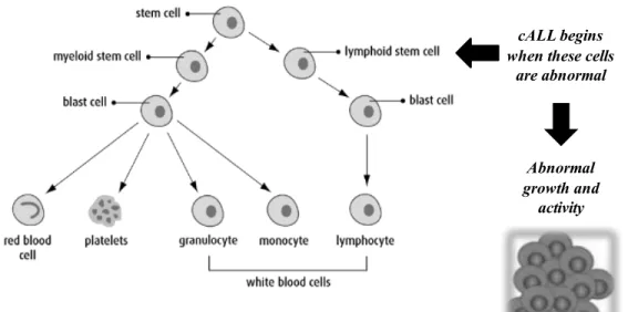
cALL is a malignant disease that begins primarily in the bone marrow and spreads to other parts of the body. However, it may arise from extramedullary sites (i.e. thymus or intestine) that invade the bone marrow [23]. It can originate from abnormal lymphoid stem cells that avoid apoptosis and undergo abnormal growth and activity, as shown in figure 2 (page 5). In other cases cALL is initiated from different genetic mutations in a single B or T-lymphoid progenitor that communicate the ability for unlimited self-renewal or that bring about the exact stage-specific halt in development [23]. Immunoglobin or T-cell receptor genes display clonal rearrangements in mutated cALL [23]. Their antigen-receptor molecules and differentiation-linked cell-surface glycoproteins are similar to the ones found on immature lymphoid progenitor cells at the early normal T and B lymphocyte development stages [23]. Furthermore the mutated cells are spread through white blood cells or immune cells that are crucial in fighting infections [24]. The outcome includes increased blast cell proliferation, maturation and survival, and eventually the accumulation of leukemic cells can be lethal, illustrated in figure 2 (page 5) [23]. Bone marrow can acquire too many immature white blood cells, making it harder for the body to fight infections, leaving the child with frequent infections, fever, bruises, weakness, etc. [24, 25]. Unfortunately the exact cause of cALL development is still unclear [23]. Only a small number of cases of cALL have been associated with inherited predisposing genetic syndromes (e.g. Down’s syndrome, Bloom’s syndrome, ataxia-telangiectasia, Nijmegan breakage syndrome) and ionizing radiation or exposure to chemotherapeutic drugs [23]. Moreover, it is believed that cALL aetiology can be provoked by prenatal influences [26], and many studies have associated the increased risk of development of cALL with high birth weight [26-28] as well as accelerated foetal growth [29]. The proposed rational behind this is that pregnancies generating higher birth weight babies have higher levels of circulating growth factors that may carry oncogenic properties to the developing immune system, which brings about an increased risk of cALL progression [26]. Other causes that have been studied but present conflicting data include parental occupation, maternal reproductive history, parental
tobacco and alcohol use, maternal diet, exposure to residual power-frequency magnetic fields, and several infection based assumptions [23].
Figure 2. Process of development of cALL in blood cells. Adapted from Canadian Cancer
Society [30]. Cancer initiates when abnormal lymphoid cells divide and develop into a malignant tumour. Cancer cells can escape the primary tumour and travel through the bloodstream leading the spread of cancer, otherwise known as metastasis.
1.5.2 Treatment of cALL
Treatment of cALL generally includes 4-6 weeks of induction chemotherapy initially administered in the hospital, followed by months of consolidation chemotherapy and finally 2-3 years of maintenance chemotherapy [18, 31]. Radiation therapy is a highly toxic treatment and is now only used on a small portion of patients with a high risk of CNS relapse [32]. Allogeneic bone marrow transplantation is recommended for some children with high-risk leukemia at diagnosis as well as those who develop a recurrence after their remission or when there is no remission after many courses of induction chemotherapy [31].
cALL begins when these cells
are abnormal
Abnormal growth and
activity
Development of cALL Cancer Cells Development of Blood Cells
1.5.3 Prevalence and outcome statistics of cALL
Cancer in children is rare, nonetheless cALL is the common childhood cancer, accounting for 25% of all cases and has peak incidence between ages 2-5 years [31, 32]. Even though the overall incidence of cALL has increased from 1975-2010, the mortality rate has been in a steep decline [31]. The National Cancer institute found that 90% of children ages 0-19 years who are diagnosed with cALL will survive at least 5 years post treatment [31]. This number has increased from the 57% survival rate in the 1970s [31].
ALL is rare in adults and survival rates of cALL far exceed those of adult ALL [33]. Partial explanation for the difference in prognosis includes the more intensive regimens, decreased side effects, and higher compliance to the aggressive treatments in children. In adults, the toxic effects represent an important limit to the administration of the proper drug doses and timing of chemotherapy [33].
It must be noted that the elevated 5-year survival rate does not come without consequence. cALL survivors are at increased risk of developing other cancers, chronic diseases and/or functional impairments over time [31].
1.5.4 Long-term adverse health effects in cALL survivors
cALL survivors display long-term adverse health anomalies that include fertility problems, metabolic dysfunction, psychological issues, neurocognitive deficits [34], growth deficiencies and are at increased risk of a second cancer [31]. Many studies have focused on the development of the metS, or its components (i.e. obesity, insulin resistance, dyslipidemia, hypertension), as well as CV conditions (i.e. congestive heart failure, coronary artery disease, myocardial infarction, cardiac arrest and cerebrovascular accidents) and diabetes, which may arise as a consequence of metabolic anomalies in cALL survivors early into their remission period [1, 4-6, 35-38]. More specifically, studies have confirmed that there is a high prevalence of cALL survivors affected by the metS, its components and related complications [4, 12, 37-41].
The presence of adverse outcomes in this population of childhood cancer survivors may be due to a combination of factors related to the primary malignancy, demographics, underlying genetic predisposition and pre-morbid conditions, and health related behaviours may adjust the
risks associated with cancer treatment [40]. However, investigators suggest that the adverse late effects are determined by treatment-specific factors [40], especially when treatment involves total body irradiation (TBI) or abdominal radiotherapy (RT) [36].
Figure 3. Health and quality-of-life related outcomes in survivors of childhood and adolescent cancers. Survivors of childhood and adolescent cancers may suffer from many
adverse late effects including psychosocial problems, fertility and reproductive problems, organ dysfunction, altered growth and development and secondary carcinogenesis. Reproduced with permission [40].
Evidently, metabolic complications bear significant health consequences for cALL survivors even after they are treated and cured from their primary cancer. Late effects are expected to continue to increase as the person ages, signifying the importance of research to understand the aetiology of metabolic disease in cALL survivors, plan for earlier detection and establish prevention efforts to improve the long-term health of cALL survivors [36].
2. The metabolic syndrome
As mentioned previously, the metS and its components can emerge as adverse late effects in cALL survivors. Our research focused on metabolic complications in cALL survivors, therefore this section will describe the metS and its components in detail.
2.1 Definition of the metS
The metS is described as a cluster of metabolic abnormalities, including obesity, insulin resistance (IR), dyslipidemia and hypertension (HT) [42]. The interrelated risk factors have been independently associated with an increased risk for atherosclerotic CV disease and mortality [42, 43], type 2 diabetes (T2D), and their resulting complications [44]. The metS components share underlying mediators, mechanisms and pathways [43] and predominant risk factors include obesity and IR [44], which usually occur collectively. We must note that people with isolated components of the metS are still at risk for CV complications, however their risk remains lower than those diagnosed with the complete metS.
Figure 4. Risk factors and consequences of the metS. Altered lipoprotein levels, HT, IR and
visceral obesity are the components involved in making up the metS. MetS development due to these risk factors may bring about further complications including CV complications and T2D. Abbreviations: HDL-c: high-density lipoprotein cholesterol, TG: triglycerides, HT: hypertension, IR: insulin resistance, MetS: metabolic syndrome, CV: cardiovascular.
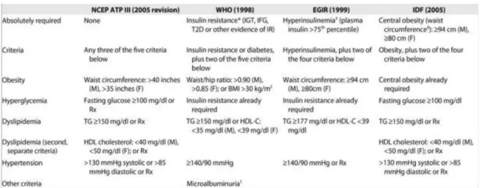
Screening for diagnosis of the metS includes identifying at least 3 of 5 of the following: waist circumference (i.e. visceral obesity), high s-TG levels, low HDL-c levels, increased fasting glycaemia or IR, and increased blood pressure (BP) [44, 45]. Table I displayed below describes the criteria for these risk factors, however cut-off points may vary depending on the population of study.
Table I: The different metS screening tools displaying diagnosis criteria.
This table describes the criteria of different screening tools used to diagnose the metS [43]. NCEP ATP III: National Cholesterol Education Program Adult Treatment Panel III, WHO: World Health Organization, EGIR: European Group for the Study of Insulin Resistance, IDF: International Diabetes Federation, IGT: impaired glucose tolerance, IFG: impaired fasting glucose, T2D: type 2 diabetes, IR: insulin resistance, BMI: body mass index, Rx: medical prescription, TG: triglycerides, HDL: high-density lipoprotein.
2.1.1 Abdominal obesity
Continued overweightness can eventually progress into obesity if not controlled or properly treated. The World Health Organization (WHO) describes obesity as a growing health and economic problem in Canadian society and its incidence is elevated and increasing in adults and unfortunately even in children. Statistics Canada found that approximately 20% of Canadian adults were classified as being obese [14]. Obesity is caused by an imbalance in intake of calories and energy expenditure, leading to an excess accumulation of body fat. The degree of adiposity is closely correlated with physiological parameters, including BP, systemic
insulin sensitivity, and circulating lipid levels [46]. In fact, obesity is directly related to the prognosis of the metS [46, 47]. Moreover, it can be an important precursor of the metS [41] thereby influencing CV risks and is actually an independent risk factor for cardiometabolic disorders (e.g. myocardial infarction, stroke, T2D) [46].
Weight reduction appears to improve certain features of the metS [43], hence its significance in this health disorder. Visceral adiposity rather than BMI is an especially important measure to consider because of its association with a number of pathologies (e.g. those associated with atherogenic and diabetogenic abnormalities) [45, 47].
2.1.2 Insulin resistance
Insulin is produced by the pancreas, an organ with endocrine and exocrine functionality. It is released when blood glucose levels are high stimulating the uptake of glucose by tissues, including the liver, skeletal muscle and adipose tissue. Insulin reduces circulating glucose by turning it into fat and glycogen, the two storage molecules. Under the manifestation of IR, the tissues are not able to respond to insulin and therefore cannot incorporate glucose leading to elevated blood glucose levels that can be pathological [43]. Noteworthy, IR is at the heart of the metS and can lead to metabolic complications. Additionally, the mechanism for which IR affects the body may advance into abnormalities in the vascular system (e.g. hyperglycaemia, advanced glycation products, toxicity from free fatty acids (FFAs), dyslipidemia and inflammatory agents) which consequently may incline T2D and atherosclerosis [43].
2.1.3 Dyslipidemia
Lipids, though physiologically essential, can contribute to disease if consumed in considerable amounts or if their metabolism is altered. Lipids include triglycerides (TG), phospholipids, FFAs, cholesterol and fat-soluble vitamins. TG are the main dietary lipid (95%), and along with cholesterol they are the primary contributors to disease. Lipoproteins are synthesized in the liver and transport endogenous TG and cholesterol through the blood until TG are taken up by peripheral tissues or until lipoproteins are cleared by the liver. TG and two types of lipoproteins are measured when determining the manifestation of dyslipidemia. These include LDL-c and HDL-c levels, or the bad and the good cholesterol, respectively. They are made up of different concentrations of TG, cholesteryl ester (CE) and phospholipids. LDL-c is
the most cholesterol rich lipoprotein and is associated with an increased risk of coronary artery disease [39] due to lipid accumulation on artery walls causing narrowing of the vessels. Furthermore, small dense LDL-c is atherogenic and linked to visceral adiposity, hypertriglyceridemia and IR [39]. HDL-c is synthesized by the liver and enterocytes and is important in collecting cholesterol from other lipoproteins and peripheral tissues to meet cell needs or for cholesterol clearance, a process known as the reverse cholesterol system. Due to its role in clearing cholesterol, HDL-c is an anti-atherogenic lipoprotein. It is not surprising then that TG, HDL-c and LDL-c are frequent components of the metS [48].
Figure 5. Development and consequences of dyslipidemia. Adapted from [49]. Dyslipidemia
can include decreased concentrations of HDL-c and/or increased TG and LDL-c, which can consequently lead to progression of T2D and atherosclerosis, both risk factors for CAD. IR: insulin resistance, FFA: free fatty acids, TG: triglycerides, Apo-B: apolipoprotein B, LDL-R: low density lipoprotein receptor, apo-CIII: apolipoprotein CIII, CE: cholesteryl ester, LPL: lipoprotein lipase, VLDL: very low density lipoprotein, CETP: cholesterol ester transfer protein, LDL: low density lipoprotein, HDL2: high density lipoprotein 2, HDL3: high density lipoprotein 3, sd-LDL: small dense low density lipoprotein, T2D: type 2 diabetes mellitus, CAD: coronary artery disease.
2.1.4 Hypertension
Hypertension (HT) is defined as a state of abnormally high BP mostly due to modifiable lifestyle and dietary factors (e.g. tobacco, alcohol, physical inactivity, high sodium diet, low potassium and/or calcium diet, aging, obesity, IR) [50], consequently generating great physiological stress. Unfortunately, about 7.5 million people in Canada live with HT, which is a critical problem since it is the pivotal pathophysiological cause of CV morbidity and mortality [51]. HT is especially triggered by the interaction between genetics (e.g. epigenetic modifications in vessel wall smooth muscle cells) [52] and environmental factors [53] affecting smooth muscle cells of blood vessels, further playing a role in their dysfunction and leading to disease [54]. The association with vascular endothelial dysfunction is the final shared pathway concerning CV risk factors and is the central pathogenesis in atherosclerosis development [43, 55]. In the long run, HT can lead to a number of complications (e.g. CAD, CVD, heart failure, stroke, end-stage renal disease and death from the mentioned causes) [50] by the narrowing of blood vessels and is a key component in the diagnosis of the metS.
Mitochondria are organelles at the center of metabolic functions and their dysfunction has been associated with diseases related to components of the metS.
3. The mitochondrion
Discovered in the 1890s [56], the mitochondrion is a double membrane organelle otherwise known as the powerhouse of the cell due to its role in the production of energy in the form of adenosine triphosphate (ATP). This organelle is extremely dynamic and controls its morphology through the balance of fission and fusion, and has a unique structure that is crucial in the way mitochondria function. Furthermore, mitochondria exercise importance in maintaining normal structure, function and survival of tissues [57]. Aside from the nucleus, the mitochondrion is the only other subcellular organelle containing DNA, of which is maternally inherited [9].
Mitochondria are present in every human cell except mature erythrocytes and play major roles in metabolic processes (e.g. cellular respiration, FA β-oxidation, calcium metabolism, biosynthetic metabolism) due to their large quantity in the most metabolically active cells (e.g.
skeletal muscle, cardiac muscle, liver, brain) [9]. Other key functions include their role in apoptosis- and ROS-signalling pathways, and inflammatory processes. These functions make them important cellular stressors and supportive in the cell’s adaptation to the environment [56].
3.1 Mitochondrial structure and dynamics
The Mitochondrion is made up of an outer mitochondrial membrane (OMM) surrounding its inner mitochondrial membrane (IMM). The IMM has a large surface area due to its ability to fold into large highly flexible tubular junctions called cristae [58]. In the case where the cell responds to a metabolic state and/or matrix volume, these junctions can endure morphological changes [58]. Furthermore, the shape and morphology of the mitochondrion’s network in a cell depends on mitochondrial dynamics and its metabolic status [58] and the oxidative capacity of the cell depends on the number and size of mitochondria [57]. This underlines the importance of mitochondria structure in the cell.
Figure 6. Shape, structure and components of the mitochondrion organelle [59]. The
mitochondrion is a double membrane organelle consisting of an inner membrane, which has a large surface area and folds into cristae, and an outer membrane. The mitochondrial matrix is located within the inner membrane and holds mtDNA, ribosomes, and ATP synthase particles.
3.1.1 Fission and fusion dynamics
Mitochondria have a generally tubular external appearance. However, there is great variability in their shape depending on the organism and tissue, which may represent their different physiological and cellular states [60]. Mitochondria continually go through processes of fission and fusion, evidently changing their morphology, which is essential for the health and functioning of mitochondria and the cells [61]. Some mitochondrial shapes found inside the cell include small vesicles, short rods and reticular networks, and the main proteins responsible for this dynamic mitochondrial process are fission and fusion proteins such as the dynamin family of large GTPases [60].
Fusion and fission dynamics of mitochondria have been studied in relation to many pathological conditions. Some disorders related to metabolic and CV diseases have revealed dysfunctional mitochondria with alterations in their structure [60]. Therefore, dynamic processes have contributed to demonstrating the importance of mitochondria in human health.
3.1.2 Mitochondrial DNA
The IMM encloses a protein rich matrix where mtDNA is contained. The endosymbiotic theory explains that the mitochondrion began as a free living prokaryotic organism, thereby explaining the presence of mtDNA [62]. The human mitochondrion contains 16.6kb of circular maternally inherited mtDNA comprising 37 genes [63]. These genes code for 13 of the 90 proteins essential to the respiratory chain and for the 24 RNA (22tRNAs and 2 rRNAs) genes necessary for the synthesis of intra-mitochondrial proteins [64]. Proteins encoded by mtDNA are all subunits of the ETC complexes I, III, IV, and V, and are essential for ATP production by oxidative phosphorylation (OXPHOS). Thus, mtDNA is essential in cellular energy metabolism [65]. Moreover, the majority of mitochondrial proteins are encoded by nuclear DNA (nDNA) and synthesized on free ribosomes in the cytosol [66]. Additionally, they contain cytosolic precursors of mitochondrial proteins that hold information directing them towards the mitochondrion where they are imported by specialized systems [62, 66, 67]. The mitochondrion has receptors on its surface that recognize nuclear encoded proteins [66]. The transport process of nuclear encoded proteins into the mitochondrion is energy (i.e. ATP and membrane potential)
dependent and the import and intra-mitochondrial sorting of proteins is mediated by translocases on the OMM (TOM complex) and IMM (TIM23 complex) [66].
Mitochondria are able to maintain their genome and they have well-developed machinery for replication, transcription and translation [67]. However, unlike nDNA, mtDNA is not protected by histones and is therefore at risk of damage [9]. There is evidence that a number of inherited diseases are a result of mtDNA mutations and loss of mtDNA, giving rise to respiratory chain defects, modifying the synthesis of ATP and leading to organ dysfunction [64].
3.1.3 Mitochondrial biogenesis
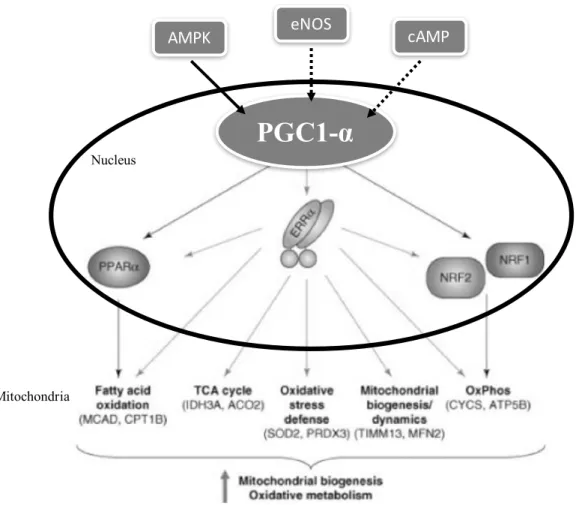
Growth and division of existing mitochondria are defined as processes of mitochondrial biogenesis [63]. Proper mitochondrial biogenesis is dependent on the coordinated synthesis and import of proteins encoded by the nuclear genome in addition to the proper coordination of mitochondrial dynamics (i.e. fission and fusion) and mtDNA replication [63]. Biogenesis can be influenced by environmental stressors, including caloric restriction, exercise, low temperature, oxidative stress (OxS), cell renewal, cell differentiation and cell division [63]. Moreover, mitochondrial biogenesis is driven by its master regulator, the transcription factor peroxisome proliferator-activated receptor gamma coactivator 1-alpha (PGC1-α) [68] in the nucleus. Expression of PGC1-α can be controlled by signalling cascades including endothelial nitric oxide synthase (eNOS), cyclic adenosine 3,5’-monophosphate (cAMP) and 5’ adenosine monophosphate-activated protein kinase (AMPK) signalling [57, 69] among others, illustrated in figure 7 (page 16). Mechanisms of PGC1-α regulation by eNOS are not well established, however high levels of NO results in cGMP-dependent signalling inducing PGC1-α, mitochondrial transcription factor A (Tfam) and nuclear respiratory factor 1 (NRF-1), and this has been correlated with increased mitochondrial biogenesis in some cell lines [69]. Under moments of energy deprivation (e.g. during exercise), AMPK induces PGC1-α transcription and enhances PGC1-α co-transcriptional activity by phosphorylating threonine-177 and serine-538, all leading to increased mitochondrial biogenesis [70]. Additionally under cold or fasting, the thermogenic signalling cascade increases cyclase activity and increases cAMP production who’s elevated levels phosphorylate CREB, activating it and further inducing PGC1-α [69].
Furthermore, PGC1-α implements its functions by binding to and regulating the activity of many transcription factors including peroxisome proliferator-activated receptors (PPAR)γ and α, nuclear respiratory factor (NRF) 1 and 2 and estrogen-related receptor-alpha (ERRα), provoking the expression of their target genes shown in figure 7 (page 16) [57, 70]. The regulation of these proteins by PGC1-α modifies genes involved in metabolic pathways (e.g. FA oxidation, OXPHOS, gluconeogenesis, etc.) [70]. For example, NRF-1 regulates the expression of mitochondrial genes (e.g. OXPHOS genes & Tfam) needed for mitochondrial gene expression and mitochondrial genome replication [57].
Figure 7. Regulation of expression of PPARα, ERRα, NRF1 and NRF2 by transcription factor PGC1-α driving biogenesis and oxidative metabolism. Adapted with permission from
[71]. Abbreviations: eNOS: endothelial nitric oxide synthase, cAMP: cyclic adenosine 3,5’-monophosphate, AMPK: 5’ adenosine monophosphate-activated protein kinase, PGC1-α:
Nucleus
Mitochondria
PGC1-α
eNOSperoxisome proliferator-activated receptor gamma coactivator 1-alpha, PGC1-β: peroxisome activated receptor gamma coactivator 1-beta, PPARα: peroxisome proliferator-activated receptor-alpha, ERRα: estrogen-related receptor-alpha, NRF1: nuclear respiratory factor 1, NRF2: nuclear respiratory factor 2, TCA: tricarboxylic acid, MCAD: medium-chain acyl-CoA dehydrogenase, CPT1B: carnitine palmitoyltransferase 1b, IDH3A: isocitrate dehydrogenase 3 alpha subunit, ACO2: aconitase 2, SOD2: superoxide dismutase 2, PRDX3: peroxiredoxin 3, TIMM13: translocase of inner mitochondrial membrane 13, MFN2: mitofusin-2, CYCS: cytochrome c somatic, ATP5B: ATP synthase, H+ transporting, mitochondrial F1 complex, beta subunit precursor.
Biogenesis of mitochondria is significant in the way they function and a decreased number of mitochondria may cause decreased mitochondrial function [57]. Consequently a decline in the mitochondrion’s electron transport activity is partly due to decreased mitochondrial content or number, which may induce an interruption in normal cell metabolic functions.
3.2 Mitochondrial functions
3.2.1 Metabolic functions of the mitochondrion
The mitochondrion’s abundant metabolic pathways are important in the metabolism of carbohydrates (CHO), lipids and amino acids (AA). The production of metabolic energy in eukaryotic cells is a critical role implemented by the mitochondrion and is achieved by its oxidative respiration and metabolism of nutrients [57], especially from the breakdown of CHO and fatty acids (FA) [72]. Cellular respiration is a catabolic process in which glucose molecules from food fuels, are broken down releasing energy that can be captured to form ATP [56]. Mitochondria, with the help of oxygen, completes the breakdown of food by producing CO2, water and large amounts of ATP for cellular use [56].
Production of ATP involves anaerobic and aerobic processes. Firstly, glycolysis comprises the breakdown of glucose to pyruvate coupled with the anaerobic synthesis of ATP in the cytosol [73]. However, most of the ATP is harvested through the aerobic processes that initiate when pyruvate enters the mitochondrion producing acetyl-CoA and on setting the
tricarboxylic acid (TCA) cycle [73]. The TCA cycle produces intermediates that donate electrons to the OXPHOS pathway generating energy in the form of ATP. Most of the energy produced (~95%) [74] in the cells comes from the OXPHOS pathway and the remainder arises through glycolysis [57]. The three mitochondrial pathways of energy production, i.e. TCA cycle, OXPHOS and FA β-oxidation, will be further discussed.
A) TCA cycle
The TCA cycle is the central metabolic pathway of the cell, taking place inside the mitochondrial matrix. It involves a series of oxidation-reduction reactions resulting in two molecules of carbon dioxide from the oxidation of an acetyl group. In addition, this is the final common pathway where fuel molecules, including AA, FA and CHO, are oxidized. Any molecule that can be transformed into an acetyl group or dicarboxylic acid can enter into aerobic metabolism. Carbon fuels, that mostly enter the cycle as acetyl CoA, are oxidized and act as a precursor source for biosynthetic processes, as well as for storage forms of fuels, and for building blocks of AA, cholesterol, porphyrin and nucleotide bases [73]. However, the primary function of this cycle is to use carbon fuels to yield high-energy electrons. The TCA cycle is important in the production of energy. However, this process alone doesn’t produce a significant amount of ATP but it forms NADH and FADH2 that carry electrons to the OXPHOS pathway (i.e. ETC and chemiosmosis) where most of the energy is produced.
B) Fatty acid β-oxidation
In moments of fasting or stress, the human body uses metabolic fuels to function. FA are essential metabolic fuels [75] that are activated in the cytosol, however oxidation of FA occurs largely in the mitochondrion. This process includes a series of enzymatic steps generating acetyl-CoA. First, fatty acyl-coA must cross the impermeable IMM and achieves this with the help of carnitine palmitoyltransferase I (CPT1), carnitine translocase (CAT) and carnitine palmitoyltransferase II (CPT2) enzymes, shown in figure 8 (page 19). Once inside the matrix the process of β-oxidation oxidizes short chained fatty acyl-CoA ester until it becomes acetyl-CoA [75]. As discussed previously, acetyl-coA initiates the TCA cycle leading to energy production. Thus FA oxidation fuels the TCA cycle and yields important amounts of NADH and FADH2, which are needed in the OXPHOS pathway [67].
Figure 8. ATP production by the FA β-oxidation pathway [76]. This image shows the
different enzymes and transporters involved in carrying FA from outside the cell into the mitochondrial matrix for ATP production. Abbreviations: FACS: fatty acyl-CoA synthase, CPT1: carnitine palmitoyl transferase 1, CPT2: carnitine palmitoyl transferase 2, CAT: carnitine translocase, TCA: tricarboxylic acid, NADH: nicotinamide adenine dinucleotide + hydrogen, FADH2: flavin adenine dinucleotide, ATP: adenosine triphosphate.
C) Oxidative phosphorylation
High-energy intermediates in the mitochondrial matrix, including NADH, NADPH and FADH2 produced by the mitochondrion from the TCA cycle and FA β-oxidation metabolic pathways, are electron donors involved in the production of energy [67, 77, 78]. They are used to fuel the ETC complexes and ATP synthase in the IMM in a process called OXPHOS or cellular respiration [56, 57, 78]. This process yields the highest amount of energy in the form of ATP [78].
OXPHOS consists of a system of five multisubunit complexes- NADH: ubiquinone oxidoreductase (complex I), succinate: ubiquinone oxidoreductase (complex II), ubiquinol: cytochrome c oxidoreductase (complex III), cytochrome c oxidase (complex IV), and F0 F1-ATP-synthase (complex V), depicted in figure 9 (page 20) [67]. This pathway can be separated into two different functional parts: complexes I-IV make up the ETC whereas complex V is
responsible for the synthesis of ATP through a process known as chemiosmosis [67]. The ETC consists of passing electrons from one molecule to another, using enzymes to oxidize nutrients releasing ATP. When electrons are transported along the ETC, protons are released. Some of the energy collected during this process is then used to pump protons out of the matrix resulting in a pH gradient and charge gradient across the IMM [78]. The proton motive force produced by disruption in gradients pumps protons into the mitochondrial matrix where it couples with ATP synthase and energy is produced [78].
OXPHOS has been demonstrated as having critical control over metabolic pathways [79]. In addition, the number and size of mitochondria is related to the mitochondrial oxidative capacity. For example, decreased mitochondrial oxidative capacity is joined by decreased expression of mitochondrial proteins [e.g. encoded by mtDNA (e.g. cyt c oxidase 1) and nDNA (e.g. succinate dehydrogenase & pyruvate dehydrogenase)] [57].
Figure 9. Enzymes and complexes involved in the OXPHOS pathway [80]. The OXPHOS
pathway can be separated into 2 parts, the ETC and chemiosmosis, utilizing and producing many enzymes leading to the production of ATP in the final step (complex V). Abbreviations: NAD: nicotinamide adenine dinucleotide, NADH: nicotinamide adenine dinucleotide + hydrogen, ADP: adenosine diphosphate, ATP: adenosine triphosphate, Pi: inorganic phosphate.
3.2.2 Mitochondrial signalling pathways
Aside from metabolic functions, it is understood that mitochondria possess signalling pathways allowing them to communicate their bioenergetic and biosynthetic states with other parts of the cell [7, 81]. Mitochondria are established in the cytosol and evidently communicate with this part of the cell through the release of proteins, ROS, or metabolites, by interacting with other organelles, and by acting as a framework in the arrangement of signalling molecules [7]. Signalling processes ensure that the mitochondrial fitness is adequate in order to carry out its many functions and ensures that mitochondria have the capacity to meet their functional demands [7].
A) Calcium signalling
The transport of calcium into the mitochondrion is a highly regulated system that is vital for the cell. Calcium from the cytosol crosses the outer membrane into the intermembrane space through the voltage-dependent anion channel (VDAC) situated on the OMM [82]. The main role of calcium in the mitochondrion is the stimulation of OXPHOS occurring at many levels, more specifically by activating the enzymes involved in the OXPHOS machinery [82]. When mitochondrial calcium is increased, the entire OXPHOS machinery is upregulated causing rapid respiratory chain activity and increased output of energy [82]. So, depending on cellular demand, ATP output can be controlled by calcium.
Under normal circumstances, calcium positively influences mitochondrial function, though any disturbance in mitochondrial or cytosolic calcium homeostasis can have consequences on cell function, as displayed in figure 10 (page 22). In addition, pathologies where calcium concentration is high can alter mitochondrial function.
Figure 10. Hypothesized mechanisms of the effect of calcium on the mitochondrion [82].
Calcium positively affects mitochondrial function under physiological conditions, however under pathological stimulus, increased calcium can harm mitochondrial function. Abbreviations: ATP: adenosine triphosphate, ROS: reactive oxygen species.
B) Apoptosis signalling
Another essential physiological process of the mitochondrion used by multicellular organisms in development and morphogenesis is controlling the regulation of cell death. This process is known as apoptosis and regulates the number of cells in tissues [67]. Apoptosis is characterized by distinct morphological characteristics and its biochemical reactions are energy-dependent [83]. In addition, when cells are damaged, apoptosis acts as a defense mechanism to eliminate damaged cells [83]. For example, during a stress response, it is important that mitochondria are under a normal physiological state for cellular homeostasis and cell death [84].
Cell death by apoptosis depends on mitochondrial membrane permeabilization (MMP), this may include the inner or outer MMP [84]. Apoptosis involves mitochondrial signalling by releasing cytochrome c from the mitochondrion into the cytosol. Cytochrome c release is an all or nothing process of apoptosis [85] where the cell is ultimately broken down by proteins called caspases.
Caspases can be activated via 2 different pathways; the intrinsic (i.e. mitochondrial pathway) and extrinsic (i.e. death receptor pathways) pathways displayed in figure 11 (page 23). During the intrinsic pathway, caspases are activated from signals within the cell, for example from DNA damage beyond repair or severe stress. This pathway depends on the balance of 2 sets of mitochondrial membrane proteins, anti-apoptotic (Bcl2, Bclx) and pro-apoptotic (BAX, BAK) proteins [84]. In a healthy cell, the anti-apoptotic proteins bind to the pro-apoptotic
proteins blocking their actions. If a cell is damaged or stops receiving survival signals, these stimuli will alter the inner mitochondrial membrane resulting in the opening of the mitochondrial permeability pore and loss of mitochondrial transmembrane potential [83]. By locking Bcl2 and Bclx, Bax and Bak are free to pierce a series of punctures in the mitochondrion membrane allowing cytochrome c to leak out into the cytoplasm. Cytochrome c binds to the APAF-1 and procaspase-9 protein creating a compound (i.e. apoptosome) that activates the caspase cascade, leading to cell death [83]. The extrinsic apoptotic pathway is triggered by external signals from outside the cell. For example, T-lymphocytes have a surface molecule (i.e. Fas-ligand) that recognizes receptors on the surface of target cells (i.e. Fas receptor), leading to a chain of events resulting in apoptosis. This is mediated by the Fas associated domain protein (FADD) triggering caspases, which then activate each other in a self-amplifying process called the caspase cascade. The activation of caspase initiates the phase of apoptosis breaking down the cellular material, resulting in cell death. Once the cells have undergone apoptosis, their debris are cleared by macrophages.
Altered apoptosis homeostasis has been implicated in many human conditions involving excessive or deficient apoptosis activity [86].
Figure 11. Mechanisms of the intrinsic and extrinsic pathways of apoptosis. Apoptosis can
occur through 2 different cellular pathways which lead to cell death and clearance by macrophages. Abbreviations: FADD: Fas-associated death domain-containing protein, DISC:
death-inducing signalling complex, BID: BCL-2-homology domain 3 (BH3) interacting-domain death agonist, APAF-1: apoptotic protease activating factor 1, FasL: fas ligand. Reproduced with permission [87].
C) ROS signalling
ROS are ubiquitous, highly reactive, short lived oxygen metabolites with functions such as the striping of electrons from other molecules (i.e. oxidizing), donating electrons to other molecules (i.e. reducing), or reacting with parts of molecules (i.e. oxidative modification) [88]. ROS, when isolated, are very unstable molecules with powerful oxidative effects on the cells proteins, lipids and DNA, consequently impairing cellular functions [89]. Under normal conditions mitochondria are responsible for 90% of free radical production. Furthermore, the ETC is an essential cellular process, however when it is inhibited it creates the superoxide anion radical, a toxic reactive by-product [55]. ROS accumulation can damage cell structure and function [90] while causing inflammation and cell death [91]. Furthermore, mitochondria and mtDNA are vulnerable to the ROS they produce. There are several reasons for this vulnerability, including: 1. mtDNA is located near the inner mitochondrial membrane where the ETC forms free radicals; 2. mtDNA is not protected by histones; and 3. mitochondria have limited DNA repair ability. Since ETC components are encoded by mtDNA, mutations induced by ROS can damage OXPHOS, leading to more mitochondria and cellular damage because of the additional ROS production [9, 55, 57]. Once mitochondria are damaged, cellular energy requirements are increased in order to repair it, and direct damage to mitochondria reduces their affinity for coenzymes or substrates [9].
ROS have had a negative image due to the previous thought that they were the respiratory processes damaging by-products responsible for aging and oxidative damage [82]. However, the ROS/RNS systems of the mitochondrion have crucial signalling roles [86], that under physiological conditions, are able to communicate with the rest of the cell [7, 81]. These systems are important in normal cell functions, including growth, migration, apoptosis, and remodelling [88], making them fundamental for life [92]. Under hypoxic conditions (i.e. low oxygen) mitochondria release ROS stabilizing hypoxia inducible factors (HIFs) and inducing genes responsible for the metabolic adaption to low oxygen conditions, summarized in figure 12
(page 25) [7]. Moreover, low levels of mitochondrial ROS (mROS) are essential in maintaining homeostatic biological processes, and slightly higher levels of mROS can stimulate adaption to stress pathways [7].
Figure 12. Normal functions of ROS and the factors involved in these processes.
Abbreviations: ROS: reactive oxygen species, HIFs: hypoxia inducible factors, PI3K: phosphoinositide 3-kinase, NF-κB: nuclear factor kappa-light-chain-enhancer of activated B cells, MAPK: mitogen activated protein kinases. Adapted with permission [93].
Superoxide (O2), the primary source of ROS produced by the mitochondrion, is converted to H2O2 by spontaneous dismutation or by the superoxide dismutase (SOD) enzyme [82], an important antioxidant. Antioxidant enzymes (e.g. Cu/Zn-superoxide dismutase (SOD), MnSOD, endothelial cell SOD, glutathione peroxidase (GPX), and catalase) as well as other substances with antioxidant activity (e.g. vitamin C, vitamin E, glutathione (GSH)) protect against ROS [89]. OxS depends on the balance of ROS and antioxidant. When the antioxidant system is overwhelmed due to too much ROS, this results in OxS [89] and may lead to pathological conditions.
D) Inflammation
Inflammation is a normal biological process supporting the repair or prevention of damage to tissues or cells in response to pathogens [94]. Furthermore, inflammation is involved in the aging process by changing OxS-induced inflammatory responses and redox status [94, 95]. Mitochondria can be released from the cell to promote inflammation by stimulating pro-inflammatory signalling processes [96]. Hence, they have an important role in pro-pro-inflammatory
processes yet their function may also be affected by pro-inflammatory mediators [94]. Additionally, altered mitochondrial function has been associated with diseases involving acute and chronic inflammation [94].
There is indication that mitochondrial ROS are fundamental signalling activators of the innate immune response [91]. Under physiological conditions antioxidant systems can repress OxS in the mitochondrion but under pathological conditions excess O2- ions are produced and redox-sensitive transcription factors are activated (e.g. κB, cytokines, iNOS, etc.) [94]. NF-κB, is a transcription factor involved in the activation of genes implicated in inflammation [97], OxS and endothelial dysfunction. It is activated by the proinflammatory cytokine tumour necrosis factor alpha (TNF-α) [98], IL-1, bacterial products and physical stress forms [97]. Cellular I-κB proteins interact with NF-κB in an uninduced state masking its nuclear location sequence so it remains in the cytoplasm [97]. The TNF-α mediated inflammatory event begins with the binding of its heterotrimeric ligand to either TNFR-1 or TNFR-2 surface receptors [99], where TNFR-1 seems to be the main mediator [97]. TNFRs can activate gene expression by recruitment TNF receptor-associated factor (TRAF) family members [97]. This binding then allows TNF to activate the I-κB kinase (IKK) complex leading to the proteolytic degradation of I-κB by phosphorylation and ubiquitination [98]. This liberates NF-κB where it can be further phosphorylated and translocated into the nucleus [97] thereby inducing the transcription of NF-κB responsive genes leading to inflammation [100]. To note, the IL-6 proinflammatory cytokine uses a similar transduction mechanism to activate IKK and NF-κB [101].
Additionally, research throughout the last decade has proposed that TNF-α is connected to mitochondrial damage, by functionally damaging certain components of the mitochondrial ETC and causing structural changes to its morphology [102]. Some cell types actually have protection against TNF, for example, overexpression of the mitochondrial enzyme manganese SOD shows increased resistance against TNF implying that the overexpressed superoxide radicals or other ROS may play a role in TNF-mediated cytotoxic pathway [102]. In addition, TNF cytotoxic activity can either be increased or blocked by certain electron transport inhibitors [102]. Furthermore, TNF alters the mitochondrial ultrastructure by a degenerative clumping of mitochondrial cristae, inhibits the electron transport chain by early functional damage to electron flow thereby decreasing the mitochondrion’s ability to oxidize succinate and NADH-linked substrates therefore decreasing synthesis of ATP, and by mediating mitochondrial


![Figure 5. Development and consequences of dyslipidemia. Adapted from [49]. Dyslipidemia](https://thumb-eu.123doks.com/thumbv2/123doknet/2043620.4907/28.918.242.734.381.723/figure-development-consequences-dyslipidemia-adapted-dyslipidemia.webp)
![Figure 6. Shape, structure and components of the mitochondrion organelle [59]](https://thumb-eu.123doks.com/thumbv2/123doknet/2043620.4907/30.918.240.661.548.828/figure-shape-structure-components-mitochondrion-organelle.webp)
![Figure 8. ATP production by the FA β-oxidation pathway [76]. This image shows the](https://thumb-eu.123doks.com/thumbv2/123doknet/2043620.4907/36.918.268.695.115.447/figure-atp-production-fa-oxidation-pathway-image-shows.webp)
![Figure 9. Enzymes and complexes involved in the OXPHOS pathway [80]. The OXPHOS](https://thumb-eu.123doks.com/thumbv2/123doknet/2043620.4907/37.918.170.739.549.845/figure-enzymes-complexes-involved-oxphos-pathway-oxphos.webp)
