Synthesis and Structural Study of Triphenylbismuth
Bis (Salicylate)
Kheira Feham1, Abdelkarim Benkadari2, Abdelkader Chouaih2*, Abdellah Miloudi1,3,
Gérard Boyer4, Douniazed El Abed1
1Laboratoire de Chimie Fine (LCF), Université d’Oran, Es-Senia, Algérie 2Laboratoire SEA2M, Université de Mostaganem, Mostaganem, Algérie
3Département de Physique-Chimie, ENSET d’Oran, Oran, Algérie 4Laboratoire HIT, Université Paul Cezanne, Marseille, France
Email: *achouaih@gmail.com
Received October 29, 2012; revised December 5, 2012; accepted December 18, 2012
ABSTRACT
The crystal of triphenylbismuth bis (Salicylate) pentavalent was synthesized from the reaction of triphenylbismuth di-chloride with salicylic acid dissolved in methylene di-chloride at room temperature. The molecular and crystal structures of triphenylbismuth bis (Salicylate) were determined by X-ray diffraction analysis. This compound crystallizes in the triclinic space group P1 with crystallographic parameters: a = 11.2937 (3) Å, b = 14.6516 (3) Å, c = 17.8253 (4) Å, α = 78.2958 (7)˚, β = 76.232 (6)˚, γ = 85.351 (6)˚,6.332 mm, V = 2803.59 (11) Å, Z 2, 3
1.693g cm
Dc ,
. The final residual factor is 0.0602 for 5806 reflexions with
000
1392,F T 293 2 K
I 2
I . The bismuthatom of the compound has a distorted trigonal-bipyramidal configuration.
Keywords: Organobismuth; Structure; X-Ray Diffraction; Salicylate; Synthesis
1. Introduction
Bismuth is nontoxic and relatively cheap, and bismuth compounds have been widely used in catalysis and or-ganic synthesis [1]. Inoror-ganic bismuth compounds such as bismuth halides have been used as Lewis acid cata- lysts in a number of organic reactions [1-5]. However, the utilization of organobismuth compounds trivalent and pentavalent for organic synthesis is rarely reported partly due to the unstable nature of the Bi-C bonds [6,7]. Recent developments show that the incorporation of a bulky substituent in pentavalent organobismuth com- plex can result of organobismuth compounds that have stable Bi-C bonds [8-10]. In our research group, we have been working on the synthesis of stable pentava- lent organobismuth compounds. In previous works we have synthesized organobismuth compounds such as pentavalent triphenylbismuth dichloride, triphenylbis- muth diacetate and triphenylbismuth bis (thiophene car- boxylate) [7,11]. In this work, we report synthesis and structure determination of the organobismuth compound triphenylbismuth bis (salicylate), I, where I is the pen- tavalent complex, bulky and more stable at room tem- perature.
2. Materials and Methods
2.1. SynthesisThe compound triphenylbismuth bis (salicylate) I was prepared from the triphenyl dichloride and salicylic acid in solution of methylene chloride. The mixture is heated to react for one hour of stirring. At the end of the reaction, the mixture is removed and then recrystallized from di- chloromethane/pentane (1.1) [12] (Scheme 1).
The obtained compound has the empirical formula C32H25BiO6 containing an OH and carboxylate groups that were chosen for our investigation for the following considerations: 1) the hydroxyl group can act as both proton acceptor to promote the formation of intermo- lecular hydrogen bond; 2) the oxygen atoms of the car-
Bi Cl Cl OH OH O CH2Cl2 Bi O O O O HO HO + RT / 1h I
Scheme 1. The synthetic route of the triphenylbismuth bis *Corresponding author.
bismuth (V) centre, hence it could act as a proton
2.2. Single Crystal X-Ray Analysis
by single
crys-aining part exhibits es in di
acbonyl of carboxylate ligand is weakly associated with theceptor and participate in the formation of hydrogen bonds; 3) The oxygen atom of the carboxylate links to the central atom of bismuth, forming a stable coordina- tion bond. The resulting compound is studied using X-ray diffraction to obtain molecular crystal structure.
The crystal structure of I was determined
tal X-ray diffraction method. X-ray data were collected at room temperature (293 K) on a Kappa CCD diffracto-meter (Bruker Nonius, 1998) [13] using MoK radiation
(k = 0.71073 Å), at a voltage of 50 kV and current of 20 mA. Cell parameters were obtained from refinement of 25 reflections collected from a random searching. Data reduction was performed with Denzo and Scalepack software [14]. Data obtained were processed with the WinGX integrated system software package for single crystal X-ray diffraction data solution, refinement and analysis [15]. The crystal structure was solved by direct methods and was refined by full-matrix least-squares refinement on F2 using the software package SHELX-97 [16]. Molecular graphics were done with ORTEP-3 [17]. In the absence of significant anomalous scattering effects Friedel pairs have been merged. The crystal data and structure refinement details are listed in Table 1.
3. Results and Discussion
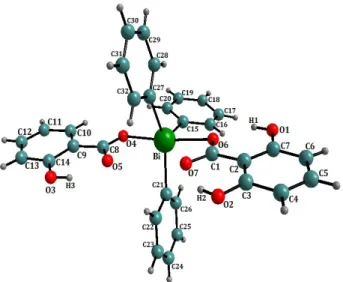
3.1. Bond Length and Angles In Figure 1, the central bismuth-cont
a distorted pseudo-trigonal-bipyramidal structure. The average values of bond distances and angl fferent aryl rings are in agreement with the literature
Figure 1. Perspective view of the molecule of
triphenyl-for
Chemical formula C32H25BiO6
bismuth bis (salicylate), I showing the labelling scheme.
Table 1. Crystal data and structure refinement details C32H25BiO6 compound. CCDC deposit 737,905 Colour colourless Crystal di m) 1429 T mensions (m 0.15 0.10 0.10 Formula weight
Crystal system riclinic
Space group P 1 Unit cell di 1.29 7 (3) Cell volu (Å3) 2803. Temper re (K) 293 (2) Density mensions a (Å) 1 3 b (Å) 14.6516 (3) c (Å) 17.8253 (4) α(˚) 78.2958 (7) β (˚) 76.232 (6) γ(˚) 85.351 (6) me 59 (11) Z 2 atu
g cm 3
Absorp icient 1.693 tion coeff
mm1
6.332 Diffractometer Kappa Radiation, Mo Reflections collected/unique 3274 Range of h, k, lAbsorption correction Sortav (Blessing, 1997)
Data/r s 5806/ Final CCD λ (Å) K , 0.71073 Theta min-max (˚) 1.68 - 27.89 8/12722 13 h 14, 18 19, 0 23 k l estraints/parameter 0/712 Goodness of fit on F2 0.857 R indices R F 22
F2 0. all data) 0602 R indices ( 0.1564alues [18]. The bond distances and valence angles of ata of compound I an
v
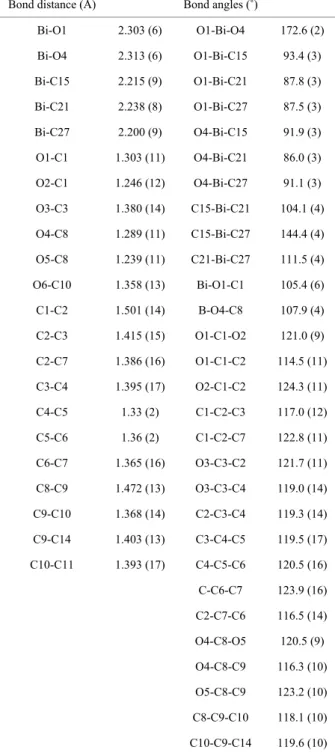
molecule I are shown in Table 2. According to X-ray diffraction d
d as shown in Figure 2, Bi atom has distorted trigo-nal-bipyramidal coordination with oxygen atoms in api-cal positions. The O(1) and O(4) atoms are located at the apical positions and the C(21), C(27) and C(15) atoms are at the equatorial positions. The sum of angles in
Table 2. Bond distances and bond angles for non-hydrogen atoms (e.s.d.’s are given in parenthesis).
Bond distance (Å) Bond angles (˚)
Bi-O1 2.30 3(6) O1-Bi-O4 6 172. (2) Bi-O4 2.313 (6) O1-Bi-C15 93.4 (3) Bi-C15 2.215 (9) O1-Bi-C21 87.8 (3) Bi-C21 2.238 (8) O1-Bi-C27 87.5 (3) Bi-C27 2.200 (9) O4-Bi-C15 91.9 (3) O1-C1 1.303 (11) O4-Bi-C21 86.0 (3) O2-C1 1.246 (12) O4-Bi-C27 91.1 (3) O3-C3 1.380 (14) C15-Bi-C21 104.1 (4) O4-C8 1.289 (11) C15-Bi-C27 144.4 (4) O5-C8 1.239 (11) C21-Bi-C27 111.5 (4) O6-C10 1.358 (13) Bi-O1-C1 105.4 (6) C1-C2 1.501 (14) B-O4-C8 107.9 (4) C2-C3 1.415 (15) O1-C1-O2 121.0 (9) C2-C7 1.386 (16) O1-C1-C2 114.5 (11) C3-C4 1.395 (17) O2-C1-C2 124.3 (11) C4-C5 1.33 (2) C1-C2-C3 117.0 (12) C5-C6 1.36 (2) C1-C2-C7 122.8 (11) C6-C7
The Bi atom does not almost extend from the equato-rial plan as we can view in Figure 2.
9) Å) respectively. Th
Å (F
ngles from 120˚ in the equatorial pl
o aromatic rings A and B are perpendicular to
1.365 (16) O3-C3-C2 121.7 (11) C8-C9 1.472 (13) O3-C3-C4 119.0 (14) C9-C10 1.368 (14) C2-C3-C4 119.3 (14) C9-C14 1.403 (13) C3-C4-C5 119.5 (17) C10-C11 1.393 (17) C4-C5-C6 120.5 (16) C-C6-C7 123.9 (16) C2-C7-C6 116.5 (14) O4-C8-O5 120.5 (9) O4-C8-C9 116.3 (10) O5-C8-C9 123.2 (10) C8-C9-C10 118.1 (10) C10-C9-C14 119.6 (10)
quatorial plan and the axial angle O-Bi-O for the title e
compound are 360˚ (144.4˚, 104.1˚ and 111.5˚) and 172.6˚, respectively.
2 2 1 w Fo2 0.0437P 2.0512P where
2 2 2
3 P Fo Fc .However, the (Bi-C(15), Bi-C(21), Bi-C(27)) distances are (2.215 (9), 2.238 (8) and 2.200 (
e Bi-O(1) and Bi-O(4) distances are (2.303 (6) Å) and (2.313 (6) Å) respectively. We note that, from Figure 3 that the ligands of carboxylate groups are in cis-position compared to the phenyl group. In the other hand, inter- molecular interaction between the bismuth atom and the two carbonyl groups forms the Van-Der-Waals bond.
The distances between valence-non-bonded Bi-O(2) and Bi-O(5) are respectively 2.814 (4) and 2.861 (3)
igure 3) which indicate that oxygen atoms are weakly coordinated with the bismuth atom and form a cis con- formation together.
This conformation apparently causes significant devia-tion of the C-Bi-C a
an.
However, Figure 4 shows, in the equatorial plane, that the tw
Figure 2. Three dimensional structure of compound I showing different angles in equatorial plan.
Figure 3. Bond lengths of atoms surrounding the bismuth atom.
Figure 4. A, B and C rings compared to Bi-O bond apical position. A and B rings are in the sam
is parallel to
n Table 3. We can see that,
n hydrogen and carbon, hy-
cial roles in the crystal pack-e plan.
Bi-O bond while the third aromatic ring C
Bi-O. The axial positions are occupied by electronegative atoms and the equatorial ones by carbon atoms of the aromatic groups. The distortion of the coordination ge- ometry at bismuth is mainly due to the restrictions im- posed by the chelate rings.
The bond distances and valence angles of carbon (C15, C21 and C27) and oxygen (O2 and O4) atoms surround- ing the bismuth central atom are shown in Figure 5. 3.2. Dihedral Angles
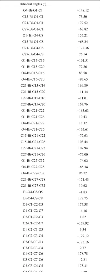
Torsion angles are reported i
the carboxyl groups are coplanar (Bi-O(1)-C(1)-O(2) and Bi-O(4)-C(8)-O(5)). These two dihedral angles are about −8.01˚ and −1.83˚ respectively (Table 3).
3.3. Hydrogen Bonds The bond distance betwee
drogen and oxygen noted X-H (Å) and the possible in- tramolecular interactions by hydrogen bonds noted X-H (Å) of molecule I is shown in Table 4.
These different hydrogen bonds showed in Figure 6 are responsible of the molecular packing in the unit cell. 3.4. Crystal Packing
Hydrogen bonds played cru
ing. Figure 7 shows the presence of two molecules in the unit cell
Z 2
which corresponds to the centrosym- metric triclinic space group P1 with two general posi- tions
x y z, ,
and
x y z, ,
.4. Conclusions
The new pentavalent triphenylbismuth bis (salicylate)
bo
was synthesized with a good yield and its crystal struc-ture was determined by X-ray diffraction analysis at room temperature. This work shows that coordination by the salicylate group is more stable, when compared to pentaphenylbismuth.
This is caused by rigidity of the ligand between the bismuth and oxygen atoms, and the presence of hydrogen nding and the Van-Der-Waals bonding. The three-di-
Table 3. Dihedral angles in degrees with e.s.d.’s given in parenthesis. Dihedral angles (˚) O4-Bi-O1-C1 −148.12 C15-Bi-O1-C1 75 .50 C21-Bi-O1-C1 179.52 C27-Bi-O1-C1 −68.82 O1- Bi-O4-C8 155.21 C15-Bi-O4-C8 −68.34 C21-Bi-O4-C8 −172.36 C27-Bi-O4-C8 76.14 O1-Bi-C15-C16 −101.51 O1-Bi-C15-C20 77.26 O4-Bi-C15-C16 83.58 O4-Bi-C15-C20 −97.65 C21-Bi-C15-C16 169.89 C21-Bi-C15-C20 11.34 C27-Bi-C15-C16 11.01 C27-Bi-C15-C20 167.76 O1-Bi-C21-C22 165.63 O1-Bi-C21-C26 10.43 O4-Bi-C21-C22 18.32 O4-Bi-C21-C26 165.61 C15-Bi-C21-C22 72.63 C15-Bi-C21-C26 103.44 C27-Bi-C21-C22 107.94 C27-Bi-C21-C26 76.00 O1-Bi-C27-C32 76.02 O4-Bi-C27-C28 85.34 O4-Bi-C27-C32 96.72 C21-Bi-C27-C28 171.43 C21-Bi-C27-C32 10.62 Bi-O4-C8-O5 1.83 Bi-O4-C8-C9 178.75 O1-C1-C2-C3 177.38 O1-C1-C2-C7 4.16 O2-C1-C2-C3 1.62 O2-C1-C2-C7 179.92 C1-C2-C3-O3 3.34 C1-C2-C3-C4 179.12 C7-C2-C3-O3 175.16 C7-C2-C3-C4 2.37 C1-C2-C7-C6 178.78 C3-C2-C7-C6 2.81 O3-C3-C4-C5 175.31 C2-C3-C4-C5 2.29
Continued C3-C4-C5-C6 2.76 C4-C5-C6-C7 3.45 1 1 C5-C6-C7-C2 3.39 O4-C8-C9-C10 177.60 O4-C8-C9-C14 2.98 O5-C8-C9-C10 3.00 C8-C9-C10-O6 2.49 C8-C9-C10-C11 179.81 C14-C9-C10-O6 178.08 C14-C9-C10-C11 0.37 C8-C9-C14-C13 179.33 C10-C9-C14-C13 1.26 O6-C10-C11-C12 177.42 C9-C10-C11-C12 0.40 C10-C11-C12-C13 0.29 C11-C12-C13-C14 0.60 C12-C13-C14-C9 1.36 Bi-C15-C16-C17 179.16 C20-C15-C16-C17 2.17 Bi-C15-C20-C19 178.95 C16-C15-C20-C19 2.24 C15-C16-C17-C18 0.17 C16-C17-C18-C19 1.73 C17-C18-C19-C20 1.64 C18-C19-C20-C15 0.29 Bi-C21-C22-C23 75.91 C26-C21-C22-C23 0.20 Bi-C21-C26-C25 176.08 C22-C21-C26-C25 0.05 C21-C22-C23-C24 0.53 C22-C23-C24-C25 0.62 C23-C24-C25-C26 0.37 C24-C25-C26-C21 0.04 Bi-C27-C28-C29 78.00 Bi-C27-C32-C31 178.10
Figure 6. Hydrogen bonds representation ensuring the mo- lecular packing in the unit cell of compound I.
Figure 7. Molecular packing in the unit cell showing two molecules in symmetric positions (x, y, z) and (−x, −y, −z). Hydrogen atoms are shown as small spheres for clarity. Table 4. Hydrogen bonds X-H and X…H of molecule I.
Distance of virtual bond
X…H (Å)
Distance of actual bond
X-H (Å) H3O2 1.822 O3-H3 0.820 H6O5 1.798 O6-H6 0.820 H7O1 2.533 C7-H7 0.929 H14O4 2.513 C14-H14 0.930 H16O5 2.556 C16-H16 0.931 H22O4 2.369 C22-H22 0.930 H26O1 2.453 C26-H26 0.929 H28O2 2.777 C28-H28 0.930 H28O5 2.636
bond the V r-Waals g. The -di- mensional representation of the m shows xial position groups and the equatorial posit e thre yls. This shows that the crystal struc iphenylbismuth bis te) pe lent has a shape distorted trigonal-bipyramid.
ing and an-De bondin three olecule the a of the two salicylate
ion of th e phen
ture of tr (Salicyla ntava Figure 5. Bond distances and valence angles around the
REFEREN
[1] zuki a atano, anobis
he-rdam, 2
[2] mamot . Ishiha Acid Ca
rganic sis, Wil einhe 08. [3] rd-Ilou e and C. Le Roux, “Bismuth(III) Triflate in Organic Synthesis,” European Journal of
Or-CES
In: H. Su nd Y. M Eds., Org muth C mistry, Elsevier, Amste 001.
In: H. Ya
Modern O o and H Synthe ey-VCH, Wra, Eds., im, 20talysis in H. Gaspa ghman
ganic Chemistry, Vol. 2004, No. 12, 2004, pp. 2517-2532.
doi:10.1002/ejoc.200300754
[4] R. M. Hua, “Recent Advances in Bismuth-Catalyzed Or- ganic Synthesis,” Current Organic Synthesis, Vol. 5, No. 1, 2008, pp. 1-27. doi:10.2174/157017908783497518 [5] R. M. A. Pinto, J. A. R. Salvador, C. L
Paixão, “Bismuth(III) Triflate-Catalyzed
e Roux and J. A. Direct Conver- sion of Corticosteroids into Highly Functionalized 17-Ke-tosteroids by Cleavage of the C17-Dihydroxyacetone Side Chain,” Journal of Organic Chemistry, Vol. 74, No. 21, 2009, pp. 8488-8491. doi:10.1021/jo9018478
[6] M. Bao, E. Hayashi and S. Shimada, “Cationic Organo-
nd Reactivity of Triphenyl-bismuth Complex with 5,6,7,12-Tetra Hydrodibenz [c,f][1,5] Azabismocine Framework and Its Coordination Com- plexes with Neutral Molecules,” Organometallics, Vol. 26, No. 7, 2007, pp. 1816-1822.
[7] A. Miloudi, D. El-Abad, G. Boyer, J.-P., Galy and J.-P., Finet, “Synthesis, Structure a
bismuth Bis(2-Thiophenecarboxylate),” Main Group Metal
Chemistry, Vol. 24, No. 11, 2001, pp. 767-774.
doi:10.1515/MGMC.2001.24.11.767
[8] M. Chovancová, R. Jambor, A. Růžička, R. Jirásko, I. Císařová and L. Dostál, “Synthesis, Structure, and Reac- tivity of Intramolecularly Coordinated Organoantimony and Organobismuth Sulfides,” Organometallics, Vol. 28, No. 6, 2009, pp. 1934-1941. doi:10.1021/om801194h [9] P. Simon, F. de Proft, R. Jambor, A. Ruzicka and L. Dos-
tál, “Monomeric Organoantimony(I) and Organobismuth(I) Compounds Stabilized by an NCN Chelating Ligand: Syn- theses and Structures,” Angewandte Chemie International
Edition, Vol. 49, No. 32, 2010, pp. 5468-5471.
doi:10.1002/anie.201002209
[10] A. Soran, H. J. Breunig, V. Lippolis, M. Arca and C. Sil- vestru, “Syntheses, Solid-State Structures, Solution Be-
0.01.004 havior of Hypervalent Organobismuth(III) Compounds [2-(Et2NCH2)C6H4]nBiX3−n and DFT Characterization of [2-(Me2NCH2)C6H4]nBiX3−n [X = Cl, Br, I; n = 1–3],”
Journal of Organometallic Chemistry, Vol. 695, No. 6,
2010, pp. 850-862. doi:10.1016/j.jorganchem.201
yer, J.-P., Galy, J.-P., Finet [11] A. Miloudi, D. ElAbad, G. Bo
and S. Didier, “Reactivity of 2-Aminothiazole and 2- or 6-Aminobenzothiazole Derivatives towards the Triphenyl- bismuth Diacetate/Catalytic Copper Diacetate Phenyla- tion System,” European Journal of Organic Chemistry, Vol. 2004, No. 7, 2004, pp. 1509-1516.
doi:10.1002/ejoc.200300656
[12] T. Arnauld, D. H. R. Barton and E. Doris, “The Che- mistry of Pentavalent Organobismuth Reagents. Part 14. Recent Advances in the Copper-Catalyzed Phenylation of Amines,” Tetrahedron, Vol. 53, No. 12, 1997, pp. 4137- 4144.
[13] E. Nonius, “CAD-4 Express Software,” Delft, 1996. [14] Z. Otwinowski and W. Minor, “Processing of X-Ray Dif-
fraction Data Collected in Oscillation Mode,” Methods in
Enzymology, Vol. 276, 1997, pp. 307-326.
doi:10.1016/S0076-6879(97)76066-X
[15] L. J. Farrugia, “WinGX Suite for Small-Molecule Single- Crystal Crystallography,” Journal of Applied Crystallog-
raphy, Vol. 32, 1999, pp. 837-838.
doi:10.1107/S0021889899006020
[16] G. M. Sheldrick, “A Short History of SHELX,” Acta
Crystallographica, Vol. 64, No. 1, 2008, pp. 112-122.
[17] L. J. Farrugia, “ORTEP-3 for Windows—A Version of ORTEP-III with a Graphical User Interface (GUI),” Jour-
nal of Applied Crystallography, Vol. 30, 1997, pp. 565-
567. doi:10.1107/S0021889897003117
[18] E. Prince and A. J. C. Wilson, “International Tables for X-ray Crystallography,” 2nd Edition, Kluwer Academic Press, Boston, 1992.