POLYMORPHISM IN HOLOTHURIA (PLATYPERONA) SANCTORI FROM THE ALGERIAN COASTAL
AREA
Karim Mezali *
Universite abdelhamid iben badis F.S.N.V Departement des sciences de la mer et de l'aquaculture - [email protected]
Abstract
The morphological, endoskeleton and genetic criteria were used to compare between individuals of two morphotypes of H.
(Platyperona) sanctori. In this study, we concluded that both morphotypes (A & B) constitute the same species.
Keywords: Echinodermata, Genetics, Systematics, Algerian Basin
1 1
Introduction
The systematic study of holothurians “sea cucumbers” is quite complex. The morphology, size and distribution of ossicles in the body wall tissues are key characters in the determination of these species [1]. Holothuria (P.) sanctori is a southern species, which is distributed through the Mediterranean [2, 3] and is widely dominant in many parts of the Algerian Infralittoral [4].
Material and methods
The animals were collected in 3 stations [Sidi Fredj (Algiers), Figuier plage (Boumerdes) and Stidia (Mostaganem)] (Fig. 1B) and then anesthetized with Mgcl .6H 0 to describe their morphology [1]. The ossicles isolated from a tissue taken in the bleach (12 °) were measured using light microscope (X 20). The fraction of the 16S mtDNA gene and the primers AR CGCCTGTTTATCAAAAACAT-3') and BR (5'-GCCGGTCTGAACTCAGATCACGT-3') [5] were used. PCR amplification was performed on 49 μL with ddH O (30.8 μL), buffer 10 X (5 μL), dNTPs (5
μL), AR (2μL), BR (2 μL), Taq polymerase (0.2 μL), MgCl (4 μL) and 1 μL of
DNA template. The phylogenetic tree is constructed from the obtained sequences and mtDNA sequence of Holothuria (Platyperona) forskali is used as an out group.
Results
1. Morphology and endoskeleton analysis
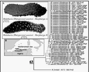
The morphotype A is dark brown color and the morphotype B has a brownish color and is easily recognized underwater by its white spots clearly visible in dark middle and lightly visible in light middle (Fig. 1A). Ossicle measurements resulting from the comparative study are indicated in table 1.
Fig. 1. The studied morphotypes (A); studied sites (B). Left: The 16S neighbour-joining tree using Kimura-2- parameter distance model. The (%) bootstrap1000 replicate is represented.
2 2
2
2
Tab. 1. The ossicles measurement. N = Number of ossicles.
2. Molecular analyses
The phylogenetic tree clearly shows that both H. (P.) sanctori morphotypes are closely grouped to a single clade with few alternative sites (Fig. 1 left). They are in fact monophyletic.
Discussion
The two morphotypes of H. (P.) sanctori are genetically identical and thus represent the same species rather than different species. These two polymorphic and ecotypic forms are characterized by the reduction of their ossicles in size and in number.
References
1 - Samyn Y., Vandenspiegel D., and Massin C., 2006. Taxonomie des holothuries des Comores. ABC Taxa., (1): 1-130.
2 - Rowe F.W.E., 1969. A review of the family Holothuroidae (Holothurioidea: Aspidochirotida). Bull. Br. Mus. Nat. Hist., Zool., 18(4): 119-170.
3 - Gustato G., and Villari A., 1977. Sulla systematic e frequenza di specie del genere Holothuria in una zona del Golfo di Napoli. Boll. Soc. Natur., Napoli, 86: 283-314.
4 - Mezali K. 2004. Micro-répartition des holothuries aspidochirotes au sein de l’herbier de Posidonies de la presqu’île de Sidi-Fredj – Algérie. Rapports P.V. Commission International pour l’Exploration Scientifique de la Mer Méditerranée, Monaco, Vol. 37, 534p.
5 - Palumbi S.R., Martin, A., Romano, S., McMillan, W.O., Stice, L., and Grabowski, G. (1991).- A simple fools guide to PCR, vers. 2.0. Special publication of the University of Hawaii. Department of Zoology and Kewalo Marine Laboratory. University of Hawaii, Honolulu.