HAL Id: hal-02445553
https://hal.archives-ouvertes.fr/hal-02445553
Submitted on 14 Dec 2020HAL is a multi-disciplinary open access archive for the deposit and dissemination of sci-entific research documents, whether they are pub-lished or not. The documents may come from teaching and research institutions in France or abroad, or from public or private research centers.
L’archive ouverte pluridisciplinaire HAL, est destinée au dépôt et à la diffusion de documents scientifiques de niveau recherche, publiés ou non, émanant des établissements d’enseignement et de recherche français ou étrangers, des laboratoires publics ou privés.
Handing off iron to the next generation: how does it get
into seeds and what for?
Stéphane Mari, Christophe Bailly, Sébastien Thomine
To cite this version:
Stéphane Mari, Christophe Bailly, Sébastien Thomine. Handing off iron to the next generation: how does it get into seeds and what for?. Biochemical Journal, Portland Press, 2020, 477 (1), pp.259–274. �10.1042/BCJ20190188�. �hal-02445553�
1
title
1
Handing off iron to the next generation: how does it get into seeds and
2
what for?
3 4
Stéphane Mari1, Christophe Bailly2, Sébastien Thomine3* 5
6
1
BPMP, INRA, CNRS, Univ Montpellier, Montpellier SupAgro, Montpellier, France 7
2 Sorbonne Université, CNRS, Laboratoire de Biologie du Développement, F-75005 Paris,
8
France 9
3
Institute for Integrative Biology of the Cell (I2BC), CEA, CNRS, Univ. Paris‐Sud, 10
Université Paris‐Saclay, 91198, Gif‐sur‐Yvette cedex, France 11
*to whom correspondence should be addressed 12 Sebastien.thomine@i2bc.paris-saclay.fr 13 +33 1 69 82 46 32 14 15 Abstract 16
To ensure the success of the new generation in annual species, the mother plant transfers a 17
large proportion of the nutrients it has accumulated during its vegetative life to the next 18
generation through its seeds. Iron (Fe) is required in large amounts to provide the energy and 19
redox power to sustain seedling growth. However, free Fe is highly toxic as it leads to the 20
generation of reactive oxygen species. Fe must therefore be tightly bound to chelating 21
molecules to allow seed survival for long periods of time without oxidative damage. 22
Nevertheless, when conditions are favorable, the seed Fe stores have to be readily remobilized 23
to achieve the transition towards active photosynthesis before the seedling becomes able to 24
take up Fe from the environment. This is likely critical for the vigor of the young plant. Seeds 25
constitute an important dietary source of Fe, which is essential for human health. 26
Understanding the mechanisms of Fe storage in seeds is key to improve their Fe content and 27
availability in order to fight the Fe deficiency. Seed longevity, germination efficiency and 28
seedling vigor are also important traits that may be affected by the chemical form under which 29
Fe is stored. In this review, we summarize the current knowledge on seed Fe loading during 30
development, long-term storage and remobilization upon germination. We highlight how this 31
knowledge may help seed Fe biofortification and discuss how Fe storage may affect seed 32
quality and germination efficiency. 33
2 34
Introduction
35
Seed is a very special stage in the life of a plant. It is the basis for the next generation and the 36
survival of the species. To ensure the success of the new generation, the mother plant transfers 37
a large proportion of the nutrients it has accumulated during its vegetative life to its seeds. 38
Most work on seed nutrient storage focuses on carbohydrate, lipids and proteins, which 39
provide the bulk energy, carbon and nitrogen required to initiate development. Depending on 40
species, 30 to 90% of plant nitrogen is allocated to seeds at the end of the plant life cycle1. 41
However, mineral stores are also very important to sustain seedling growth. Especially, iron 42
(Fe) is required in large amount in mitochondria, which provide the energy and redox power 43
during the initial stage of seedling development. For example, in the case of Arabidopsis 44
(Arabidopsis thaliana), 50 to 55% of the iron (Fe) is allocated to seeds at the end of the life 45
cycle 2, 3. Moreover, the nutrients need to be stored in a compact and stable form. Seed storage 46
proteins form pseudo crystalline aggregates in protein bodies within seed embryo cells. Free 47
Fe is highly toxic as it leads to the generation of reactive oxygen species through the Fenton 48
reaction. In cells, Fe must therefore be tightly bound to chelating molecules to prevent the 49
formation of ROS. This is probably even more important in seeds than in other organs, as 50
seeds represent resistance forms designed to survive for long periods of time, often several 51
years, and through stresses. Nevertheless, when conditions are favorable, the nutrients stored 52
in seeds have to be readily remobilized to sustain the early development of the seedling before 53
it acquires photosynthetic capacity and becomes able to take up nutrients from the 54
environment. Iron is a major component of photosystem I and its availability to the young 55
seedling is necessary to achieve the transition towards active photosynthesis. This is likely 56
critical for the vigor of the young plant. 57
From the point of view of animals and especially human beings, seeds are a major food 58
resource. They provide carbohydrates, proteins and fat but also micronutrients such as Fe and 59
zinc, which are essential for human health. About 2 billion human beings suffer from Fe 60
deficiency, which leads to asthenia, anemia and in extreme cases to death. The prevalence of 61
Fe deficiency is highest in populations that rely mostly on a plant based diet because plants 62
and seeds in particular do not provide sufficient Fe 4. Understanding the mechanisms of Fe 63
storage in seeds is key to improve their Fe content and availability in order to fight Fe 64
deficiency using biofortification. From the agricultural perspective, seed longevity, 65
germination efficiency and seedling vigor are also very important traits that breeders and 66
farmers seek to improve. Here also, seed nutrient content and especially the content and 67
3
chemical form of Fe may be critical. In this review, we will summarize the current knowledge 68
on seed Fe loading during development, long-term storage and remobilization upon 69
germination, updating the last review on the topic 5. We will highlight how this knowledge 70
may help seed Fe biofortification, longevity, germination efficiency and stand establishment. 71
72
Iron uptake and translocation to the shoots
73
Plants take up iron using distinct strategies according to species 6, 7. Most plant species use 74
strategy I, which is based on the reduction of Fe in the rhizosphere followed by its uptake by a 75
transporter for Fe(II). In contrast, graminaceous species, which include the major grain crops 76
such as rice, wheat and barley, have evolved strategy II. Strategy II is based on the release of 77
small Fe(III) chelating molecules called phytosiderophores in the rhizosphere and subsequent 78
uptake of siderophore-Fe(III) complexes. Under Fe deficiency, strategy I plants activate the 79
expression of the genes involved in Fe solubilization and uptake. The molecular players in 80
this response have been identified in Arabidopsis but are essentially conserved in other 81
species. The proton pump Arabidopsis plasma membrane H+-ATPase 2 (AHA2) drives 82
rhizosphere acidification which is important to solubilize Fe 8. The coumarin transporter 83
Pleiotropic Drug Resistance 9 (PDR9) allows the secretion of coumarin with iron binding and 84
reducing properties in the rhizosphere. Enzymes of the coumarin biosynthetic pathway are 85
induced along with PDR9 9, 10. The membrane bound Ferric chelate Reductase FRO2 reduces 86
chelate-bound Fe(III) to Fe(II), which is taken up into root cells by the divalent metal Iron 87
Regulated Transporter 1 (IRT1) 11, 12. IRT1 is not specific for Fe(II) and drives the uptake of a 88
range of other divalent metal cations including Zn, Mn, Co, Cd, Ni 13-15. A sophisticated 89
mechanism enables the removal of IRT1 from the plasma membrane when intracellular 90
concentrations of non-Fe metals raise 16. In addition, several genes involved in the 91
sequestration of these metal cations are activated under Fe deficiency 17. In graminaceous 92
species such as rice, maize and barley, the components that are upregulated for Fe acquisition 93
under Fe deficiency include the phytosiderophore efflux and Fe-sirerophore influx 94
transporters as well as the biosynthetic pathway of phytosiderophores of the mugineic acid 95
family 7. The S-adenosyl methionine cycle is activated to produce methionine, which is used 96
by the nicotianamine synthase (NAS) to produce nicotianamine (NA) the precursor of 97
mugineic acids. Whereas NA is also synthetized in strategy I species, mugineic acids are 98
unique to graminaceous species. NA is converted to mugineic acid by the nicotianamine 99
aminotransferase (NAAT) and to deoxymugineic acid by the deoxymugineic acid synthase 100
(DMAS). MA and DMA are secreted to the rhizosphere by an efflux transporter, Transporter 101
4
Of Mugineic acid 1 (TOM1) in rice (Oryza sativa), Yellow Stripe 3 (YS3) in maize (Zea 102
mais) 18, 19. Fe-siderophore complexes are taken up by specific influx transporters, Yellow
103
Stripe 1 (YS1) in maize and YS1-Like 15 (YSL15) in rice 20-22. Despite the difference in their 104
uptake strategies, the control of Fe deficiency responses involves a similar network of 105
negatively and positively regulating bHLH (basic Helix Loop Helix) in graminaceous plants 106
and the other species 23, 24. In addition, Hemerythrin Domain containing RING proteins that 107
act as negative regulators of Fe deficiency responses by mediating the Fe dependent 108
degradation of specific bHLH, are also conserved among strategy I and strategy II plants 25-27. 109
To be translocated to the shoots, Fe has to go through the endodermis cell layer and to be 110
released into the xylem sap together with ligands that maintain it in soluble form. Recently, 111
suberin deposition around the endodermis has been shown to provide a control point for Fe 112
translocation. Under Fe deficiency, the suberin layer is degraded allowing the transfer of Fe 113
from the root cortex to the central cylinder where it can be loaded into the xylem for 114
translocation to the aerial parts 28, 29. Based on its expression pattern and plasma membrane 115
localization, the Fe efflux transporter Ferroportin (FPN1)/Iron REGulated 1 (IREG1) has been 116
proposed to mediate Fe loading in the xylem in Arabidopsis 30. The MATE family citrate 117
efflux transporters, Ferric Reductase Deficient 3 (FRD3) in Arabidopsis and FRD3- Like 118
1(FRDL1) in rice, play an important role in the translocation of Fe and other metals, by 119
loading citrate which chelates Fe and prevent its precipitation in the xylem sap 31-35. 120
121
Remobilization from other organs to the seeds
122
At the vegetative stage, most of the iron translocated to aerial parts is used for photosynthesis 123
in chloroplasts in mesophyll cells. Photosystem I and Ferredoxin, which are very abundant in 124
chloroplasts, contain numerous FeS clusters. This assumption based on biochemical needs is 125
supported by cell fractionation experiments and Fe imaging in leaves 36-38. Upon transition to 126
the reproductive stage, a large part of the Fe present in vegetative tissues is transferred to the 127
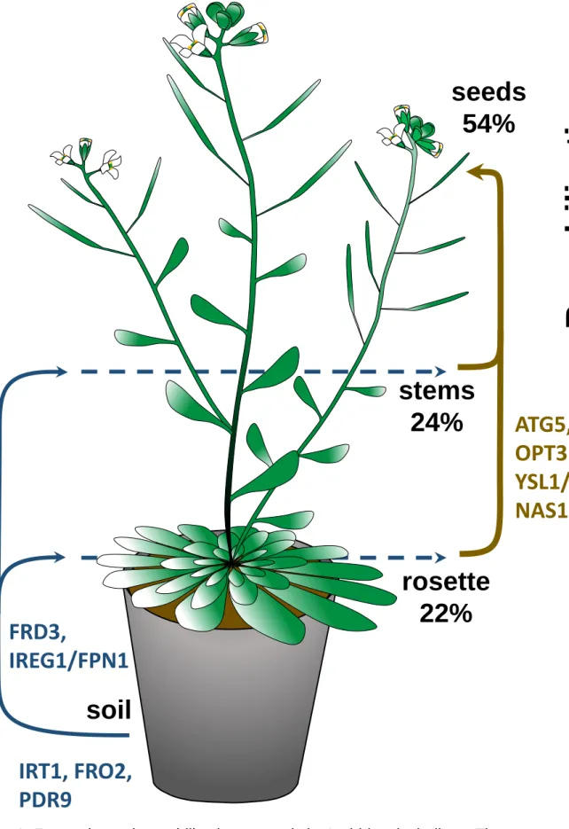
seeds. In Arabidopsis, 50 to 55% of total plant Fe ends up in the seeds at the end of the life 128
cycle (the Iron Harvest Index, Figure 1)2, 3. A recent study based on Fe isotope labeling 129
indicated that most of the Fe stored in seeds originates from vegetative tissue, implying a 130
major remobilization of Fe 2. 131
Prior to its movement from the vegetative tissues to the seeds, Fe has to be made available in 132
the senescing tissues. Autophagy was proposed to play a key role in nutrient remobilization 133
from vegetative tissues to seeds 39. This was based on the finding that autophagy is involved 134
in nitrogen remobilization to seeds 40. Recently, the finding that a mutation in Autophagy 5 135
5
(ATG5), which strongly impairs autophagy, decreases the proportion of Fe allocated to seeds 136
from 50-55% to 15-20% highlighted the importance of autophagy for Fe remobilization 2. 137
Similar results were obtained for other micronutrients such as Zn and Mn. The role of 138
autophagy is thus clearly not limited to Fe. Recently, several pathways have been implicated 139
in the degradation of chloroplasts, which contain most of the cellular Fe, during leaf 140
senescence 41. They include (i) the senescence-associated vacuoles (SAV) that are important 141
for the degradation of stromal proteins such as RuBisCo and glutamine synthase, (ii) 142
chlorophagy in which whole chloroplast are encapsulated in autophagosomes 42 (iii) a 143
vesicular pathway depending on the ATG8 Interacting protein (ATI) which degrades envelope 144
and thylakoid membranes in addition to stromal proteins 43 and (iv) CV-Containing vesicles 145
(CCVs) that contain the CV protein (Chloroplast Vesiculation) responsible for chloroplast 146
destabilization 44. In contrast to the others, the CCV pathway does not require autophagy. It 147
will be interesting to determine the respective importance of these pathways for Fe 148
remobilization during vegetative organ senescence, as they could allow to target some 149
nutrients more specifically than autophagy. 150
In addition to autophagy, the timing of senescence is also an important factor controlling 151
remobilization. If senescence occurs too fast, the time window for remobilization is reduced 152
and less nutrients are allocated to seeds. Hence, NAC transcription factors, which control the 153
onset of senescence, also play a major role in the control of nutrient remobilization from 154
leaves to seeds 45-47. More work will be required to understand how the interplay between 155
autophagy and senescence controls Fe allocation to seeds in different species. 156
After it has been made available in vegetative tissue, Fe has to be transported to developing 157
seeds through the phloem. Many lines of evidence have pointed to a key role of NA in this 158
process. Complete loss of NA synthesis leads to sterility 48-50. This has initially prevented a 159
direct analysis of the role of NA in Fe transport to seeds, as the mutants did not set seeds. The 160
first evidence that NA is important for Fe transport to the seeds came from the study of 161
mutants impaired in YS1-like genes (YSL). The strong expression of OsYSL2 in phloem tissues 162
of rice aerial parts and developing seeds and the ability of this transporter to take up NA-Fe 163
and NA-Mn complexes when expressed in Xenopus oocytes provided the first indication that 164
NA is important for Fe transport through the phloem to the seeds 51. Analysis of rice lines in 165
which OsYSL2 expression was silenced further supported this hypothesis 52. In these lines, Fe 166
transport to the shoots and the seeds was strongly decreased. Conversely, increased OsYSL2 167
expression under a phloem specific promoter led to strong increase in seed endosperm Fe 168
concentration 52. More recently, another YSL transporter from rice, OsYSL9, was implicated 169
6
in Fe delivery to developing rice grain. OsYSL9 expression is upregulated by Fe deficiency in 170
roots but down regulated in leaves under the same conditions 53. The role of YSL in metal 171
transport to seeds is not restricted to rice. Similar conclusions were obtained from the analysis 172
of Arabidopsis knockout mutants targeting AtYSL1 gene or double mutants affecting AtYSL1 173
and AtYSL3 54-56. Like OsYSL2, AtYSL1 is expressed in phloem tissues of leaves as well as in 174
developing siliques. Similar to OsYSL9, it is down regulated under Fe deficiency. Loss of 175
function mutants in AtYSL1 accumulated higher levels of NA in shoots but strongly decreased 176
NA and Fe concentrations in mature seeds 55. Combining a mutation in AtYSL1 with a 177
mutation in another Arabidopsis YSL strongly expressed in phloem, AtYSL3, leads to more 178
severe phenotypes: the ysl1ysl3 mutants display symptoms of strong Fe deficiency with 179
interveinal chlorosis as well as decreased fertility. Like ysl1, they also accumulate lower 180
concentrations of Fe in their seeds when provided with a high Fe concentration in the 181
medium, which alleviates the defect in fertility and up-regulate YSL1 expression 55, 56. Further 182
inflorescence grafting experiments showed that expression of AtYSL1 and AtYSL3 in the 183
rosette was sufficient to restore fertility but not seed Fe content 54. Furthermore, the 184
identification of a quadruple mutant combining knockout in 3 out of the 4 genes encoding 185
NAS in Arabidopsis and a knockdown in the fourth one allowed to obtain fertile plants with 186
drastically decreased levels of NA 48. In this quadruple nas mutant, Fe and NA levels are 187
lower in seeds, similar to the phenotype observed in ysl1. Interestingly, in NA biosynthesis 188
mutants, Fe accumulates in phloem cells indicating that NA is required to retrieve Fe from the 189
phloem and provide it to sink tissues, such as seeds 57. This role may be shared between NA 190
and mugineic acids in graminaceous plants. On the other hand, all YSL transporters 191
characterized so far using expression in yeast or in Xenopus oocytes mediate the influx of 192
NA-Fe or MA-Fe complexes into cells. The finding that NA and YSL are important for 193
phloem transport of Fe does not imply that Fe is transported as NA-Fe complex in the phloem. 194
Another important player in Fe transport from vegetative organs to the seeds is the 195
oligopeptide transporter OligoPeptide Transporter 3 (OPT3). Full loss of function of OPT3 196
leads to embryo lethality suggesting that the substrate of OPT3 is an essential cellular 197
metabolite 58. Even though the closest homologue of OPT3, BjGT1, is a glutathione 198
transporter 59, transport assays in yeast failed to show OPT3 ability to transport GSH 60. 199
Instead, expression of OPT3 could complement the yeast fet3fet4 mutant impaired in Fe 200
uptake, suggesting that it transports Fe complexes. The exact substrate of OPT3 remains to be 201
identified. The identification of a weak allele of opt3, opt3-2 allowed investigating its 202
function at the reproductive stage 61. In opt3-2 mutants, Fe concentration is increased in 203
7
vegetative organs but decreased in seeds, indicating a role in Fe transfer from vegetative 204
organs to seeds. More recently, OPT3 was shown to be targeted to the plasma membrane and 205
mainly expressed in phloem cells 60, 62. In opt3-2 mutants, Fe concentration was strongly 206
increased in the xylem sap and decreased in the phloem sap, pointing to role of OPT3 in Fe 207
transfer from the xylem to the phloem 60. Combined with its expression in phloem cells, this 208
suggests that OPT3 is involved in Fe loading into the phloem. Hence, both OPT3 and NA 209
play critical roles in long distance transport of Fe in the phloem to sink organs and especially 210
seeds: OPT3 would be necessary for loading Fe into the phloem, while NA would be required 211
for Fe retrieval from the phloem 57, 60. Interestingly, OPT3 is also important for Fe deficiency 212
signaling and opt3-2 up-regulated Fe-deficiency responsive gene expression even under Fe 213
sufficient conditions 60, 62. In rice, OsOPT7, a homologue of OPT3, was characterized 63. 214
OsOPT7 could not transport GSH or Fe when expressed in Xenopus oocytes and yeast. Like 215
AtOPT3, OsOPT7 is induced under Fe deficiency but its expression is more widespread than
216
that of OPT3 61, 63. Moreover, the phenotype of opt7 mutant does not resemble that of opt3-2. 217
Whether graminaceous plant genomes carry orthologues of OPT3 remains to be determined. 218
219
Iron loading into seed tissues
220
There is no continuity between the vasculature of the mother plant and the pro-vasculature of 221
the embryo. Therefore, Fe, as other nutrients, has to be released from the phloem and re 222
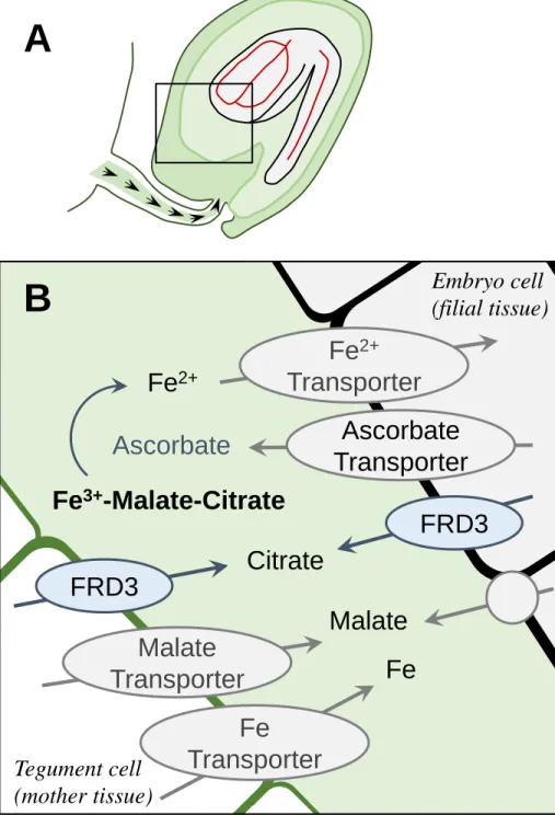
absorbed by the embryo. In Arabidopsis and other dicotyledonous plants, nutrients are 223
released at the level of the chalaze and the nucellus into the embryo sac fluid from which the 224
embryo takes up nutrients (Figure 2). Accordingly, AtYSL1 expression is detected in the 225
funiculus and the chalazal endosperm 55. Analyses of Fe speciation in the embryo sac fluid of 226
developing pea seeds revealed that Fe is present as ferric Fe bound to citrate and malate in this 227
extracellular compartment 64. In agreement with this finding, the citrate efflux transporter 228
FRD3 is expressed in the peripheral cell layer of the embryo and the cell layer of the tegument
229
facing the embryo sac during seed development (Figure 2)34. This suggest that FRD3 secretes 230
citrate in the embryo sac to maintain Fe solubility and availability for uptake by the embryo. 231
In dicotyledonous plants, Fe is taken up as ferrous Fe. This implies that Fe must be reduced 232
prior to uptake by the embryo. However, genetic analyses in Arabidopsis failed to identify a 233
membrane bound ferric chelate reductase that is important for Fe acquisition by the embryo 234
among FRO2 homologues. Instead, further analysis of the embryo sac fluid in pea showed the 235
presence of a high concentration of ascorbate, sufficient to reduce Fe(III) to Fe(II) prior to its 236
uptake by the embryo 64. Consistently, in Arabidopsis vtc (vitamin c) mutants deficient in 237
8
ascorbate biosynthesis, the seed Fe content is decreased. The speciation of Fe in the embryo 238
sac is thus similar to that encountered in other extracellular compartments of the plant, such as 239
the xylem sap. In contrast, it is striking that ascorbate is used for Fe reduction, whereas 240
membrane bound ferric chelate reductases are used for uptake in roots and leaves as well as in 241
intracellular organelles such as mitochondria and plastids 11, 65, 66. The transporters responsible 242
for secreting ascorbate and Fe in the embryo sac remain to be identified. In the case of Zn, the 243
Heavy Metal pumping P-type ATPases HMA2 and HMA4 release Zn from the mother tissues 244
for subsequent uptake by the embryo 67. In the case of Fe, the plasma membrane Fe efflux 245
transporter IREG1/FPN1 may be involved in this process but so far no defect in Fe supply to 246
the embryo has been reported for ireg1/fpn1 mutant 30. 247
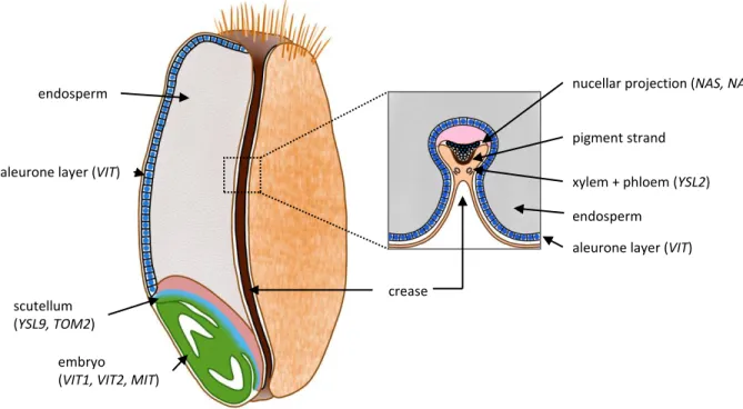
In wheat and barley, the nutrients, including Fe, are provided to the grain by a single vascular 248
strand along the ventral crease 68. Fe is mostly provided by the phloem and moves through 249
several specialized cell layers: the crease vascular parenchyma, the pigment strand and the 250
nucellar projection. Fe ends up in the transfer cells that are facing the embryo. From there, Fe 251
has to be released in the extracellular space separating the mother plant from the embryo 252
(Figure 3). The transfer cells, as the modified aleurone cells facing them on the side of the 253
embryo, have highly invaginated plasma membranes favoring nutrient release and 254
reabsorption 68. Fe accumulation and speciation in these structures show marked contrasts in 255
the mature wheat grain. Fe is highly concentrated in the nucellar projection and co-localizes 256
with sulfur. Fe accumulates to a lesser extend in the modified aleurone, from where it is 257
probably distributed to the other aleurone cells, the embryo and the endosperm 69. X-ray 258
absorption spectra indicate that in the nucellar projection, Fe is mostly associated with NA, 259
whereas in the modified aleurone it is associated with phytate 69. In agreement with the 260
speciation, barley homologues of AtVIT1, which drives Fe influx into the vacuole where 261
phytate is localized, are strongly expressed in the aleurone 68, 70-72. In contrast, the genes 262
encoding NAAT, NAS and YSL, that favor Fe mobility, are expressed at high levels in 263
transfer cells 68, 73(Figure 3). In the future, it will be important to determine the specific 264
expression pattern and the role of each of the genes involved in Fe transport in this complex 265
structure. This should allow the identification of the key transporters that release Fe to the 266
extracellular space from the transfer cells as well as those responsible for taking it up into the 267
embryo in the modified aleurone cells. 268
269
Iron storage in seeds
9
The localization of Fe in seeds differs according to species and seed developmental stage. The 271
localization and subcellular localization are strongly associated with Fe speciation, i.e. the 272
nature of the ligand that binds Fe and determines it bioavailability 74. For example, Fe phytate 273
complexes that are stored in vacuoles are notoriously poorly bioavailable 75. In contrast, 274
ferritin Fe stored in plastids constitutes a highly bioavailable source of Fe 76. 275
Iron and other metals are not distributed evenly in seed tissues. In contrast, Fe distribution 276
follows striking patterns. For example, in Arabidopsis mature embryo, Fe is highly 277
concentrated around the vascular tissues 71, 77-81. Interestingly, the pattern of Fe distribution is 278
distinct from that of other metals: Mn is concentrated in the sub-epidermal cell layers of 279
Arabidopsis cotyledons in mature seeds while Zn is evenly distributed in Arabidopsis embryo 280
71, 77, 78, 81
. The patterns of metal localization were initially discovered using synchrotron X-ray 281
Fluorescence imaging of intact seeds and were then confirmed and refined using additional 282
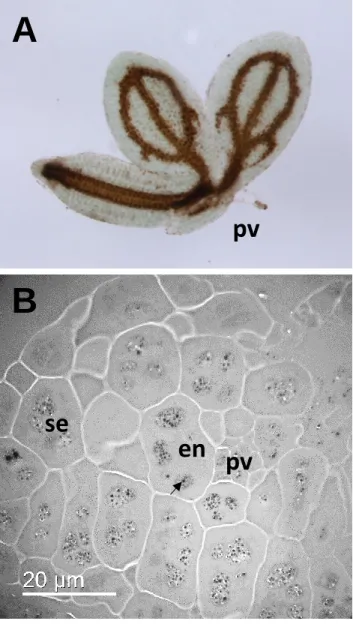
approaches. The use of Perls staining intensified by Diaminobenzidine (Perls DAB) allowed 283
identification of the precise cell type accumulating Fe as the proto endodermis of the embryo 284
80
. The use of micro Particle Induced X-ray Emission (PIXE) allowed quantification of the 285
pattern: even though it is concentrated in proto-endodermal cells, Fe is present in other cells, 286
albeit at much lower concentrations, and the Fe concentrated around pro-vascular tissues 287
accounts for 50% of total seed Fe 81. Finally, the use of Electron Dispersive X-ray imaging 288
coupled to Transmission Electron Microscopy (TEM EDX) provided higher spatial resolution 289
allowing the determination of the subcellular localization of Fe in the globoids of vacuoles of 290
proto-endodermal cells (Figure 4)79, 82. This subcellular localization is in agreement with the 291
finding that mutations in AtVIT1 (Vacuolar Iron Transporter 1) disrupt the pattern of Fe 292
distribution 71, 79. In vit1 mutants, Fe is not concentrated around vascular tissues but instead 293
co-localizes with Mn in sub-epidermal cells 71, 79. Nevertheless, Fe is still localized in 294
vacuoles in vit1 mutants 79. Recently, the vacuolar Mn transporter AtMTP8 (Metal Tolerance 295
Protein 8) was shown to be responsible for concentrating Mn in sub-epidermal cells in wild 296
type seeds and Fe localization in these cells in the vit1 mutant background 77, 78. In mtp8 297
knockout mutant seeds, Mn is concentrated around the vascular tissues together with Fe. In a 298
double mutant combining vit1 and mtp8 mutations, Mn and Fe are evenly distributed in 299
Arabidopsis embryo 78. Therefore, by creating strong sinks for Fe or Mn, vacuolar metal 300
transporters determine the pattern of metal distribution in mature Arabidopsis embryo. The 301
phenotype of vit1 mutant, which survives poorly when germinated under Fe-deficient 302
conditions, indicates that adequate tissue localization in the embryo is crucial 71, 79. Little 303
information is available on how the Fe distribution pattern is generated during embryo 304
10
development. Consistent with a strong expression of VIT1 during seed development, Fe 305
patterning is already apparent at the torpedo stage at the center of the cotyledons where the 306
provascular tissue will differentiate 71, 80. However, in Brassica napus, high resolution Perls 307
DAB imaging detects Fe in the nuclei of all embryo cells at the torpedo stage 83. Further 308
during development, at the bent cotyledon stage, Fe is detected in cytoplasmic vesicles and 309
finally ends up in the vacuoles of the cells surrounding the vasculature in mature embryo 83. 310
The observation of Fe in the nucleus at the torpedo stage in B. napus is consistent with the 311
detection of high Fe concentrations in the nucleus of embryo cells in developing pea (pisum 312
sativum) seeds 84. Therefore, Fe subcellular localization evolves during embryo development.
313
These changes in localization are most likely paralleled by changes in speciation and therefore 314
of Fe bioavailability. The analysis of Fe bioavailability in pea seeds at different 315
developmental stages show that Fe bioavailability in immature peas is higher than in mature 316
pea seeds 85. 317
Whether the pattern of Fe distribution in the vacuoles of cells surrounding the vasculature 318
described in Arabidopsis is conserved among angiosperm has been investigated. In beans 319
(Phaseolus vulgaris), Fe stores also concentrates around vascular tissues 86. Recently, this 320
observation was extended to Brassicales and even Rosids, which represent a wide group 321
including a third of the angiosperms 87, 88. However, in many cases the number of cell layers 322
that accumulate Fe is increased, to 2, the endodermis and one cortical cell layer in Brassicales, 323
or even more cortical cell layers in other Rosids, compared to endodermis only in 324
Arabidopsis83, 87, 88. While screening representatives of the different groups of angiosperm, 325
interesting exceptions were identified in Chenopodium quinoa and Carica papaya 87, 88. There 326
are certainly more diverse patterns to be discovered. 327
In grains, the seeds of graminaceous species, the distribution of Fe is very different from that 328
observed in Brassicales. In grains, in contrast to Arabidopsis, the embryo represents only a 329
small volume of the seed, while the major part is constituted by the starchy endosperm. The 330
endosperm is surrounded by the so called aleurone cell layer. During germination, the 331
aleurone layer is activated by gibberellins, releases enzymes that digest the carbohydrates 332
stored in the endosperm and undergoes programmed cell death 89, 90. The embryo is equipped 333
with an absorptive structure called the scutellum, somewhat equivalent to human placenta, 334
which take up the nutrients from the endosperm. In grains, a large part of the Fe is 335
concentrated in the aleurone layer and another pool is found in the embryo 69, 91, 92(Figure 3). 336
In wheat (Triticum durum), the highest concentrations of Fe are found in the aleurone layer 337
and in the scutellum 92. Separate measurements of wheat flour and bran indicated that almost 338
11
60% of grain Fe is in the bran which includes the aleurone layer and therefore represents the 339
major Fe pool in grains 93. Within the aleurone layer, Fe is concentrated together with 340
phosphorus and other minerals in globoids inside the protein storage vacuole, similar to the 341
subcellular localization observed in Arabidopsis 72. In situ X-ray absorption spectroscopy 342
analyses have indicated that in the aleurone layer, most Fe is bound to phytate, either as Fe(II) 343
or Fe(III) in good agreement with its subcellular localization 69, 92. Another pool is bound to 344
citrate, which may be involved in the transport from modified aleurone cells present in the 345
crease to aleurone cells 69. In rice, Fe accumulates in the aleurone layer together with phytate 346
and in the scutellum, as observed in wheat 91, 94. The exact speciation of Fe in the embryo and 347
in the endosperm has not yet been determined in these tissues that contain lower Fe 348
concentration, probably because of limitations in the sensitivity of X-ray absorption 349
spectroscopy. Based on co-occurrence of elements, it has been proposed that Fe is bound to 350
phosphate (possibly as phytate) in the scutellum in wheat and to other ligands in the other 351
parts of the embryo and in the endosperm 69, 92. These ligands could include proteins or 352
smaller molecules such as NA, which is present in grains as in seeds of non graminaceous 353
species 55, 95. In wheat flour which corresponds to the starchy endosperm, NA is the main 354
ligand for Fe 96. 355
Little is known on the mechanisms that control the pattern of Fe localization in grains. A 356
developmental analysis showed that throughout rice grain development Fe co-localizes with 357
phosphorus in the aleurone cell layer, indicating that Fe is most likely bound to phytate as 358
soon as it is stored 91. Mutations in OsVIT1, OsVIT2 and Mitochondrial Iron Transporter 359
(MIT) have been shown to perturb iron localization within the embryo 97, 98. OsYSL9 is 360
strongly expressed in the scutellum and participates to Fe storage in the embryo 53. The 361
phytosiderophore efflux transporter TOM2 is expressed in the dorsal vascular bundle, 362
epithelium and the scutellum of the embryo and may also participate in the distribution of Fe 363
19
. Silencing TOM2 gene expression did not alter seed total Fe content. Analysis of Fe 364
distribution using sXRF, Perls staining or seed dissection could provide more insights in its 365
function in developing rice grains. 366
367
Biofortification
368
The abundance, the localization and the speciation of Fe are equally important parameters to 369
be taken into account in attempts to increase Fe content and bioavailability in grains, which 370
are two of the major targets of biofortification projects. Having the major Fe pool stored as Fe 371
phytate in the aleurone layer is probably the worst combination for human nutrition, as 372
12
phytate has extremely high affinity for metals, which drastically limits Fe absorption in the 373
intestine 75. Moreover, the aleurone layer is most of the time discarded during grain 374
processing, such polishing for rice or preparation of white flour for wheat 93, 96, 99. Hence, 375
biofortification strategies using targeted molecular approaches have aimed at changing both 376
the localization and the speciation of Fe in grains. Several reviews have been recently 377
published on the attempts made to biofortify wheat and rice grain99-101. This review will 378
therefore not extensively cover the topic of biofortification and rather just highlight a few 379
examples. The initial attempt to biofortify rice grains through biotechnology used the gene 380
encoding ferritin, which provides Fe under a highly bioavailable form, driven by an 381
endosperm specific promoter 102. Although the work provided a critical proof of concept, the 382
increase in Fe content was modest and variable 103. Probably the most efficient strategy for 383
biofortification by manipulating a single gene has been the activation or the over expression 384
of NA synthase in rice grains 95. This resulted not only in a strong increase in grain Fe 385
concentration but also in Fe bioavailability, as NA appears to favor Fe intestinal absorption 95. 386
More recently, expression of a Fe vacuolar transporter TaVIT2 in wheat endosperm was also 387
shown to be a relevant approach to increase Fe content and bioavailability 70. Probably, the 388
expression of TaVIT2 in the endosperm vacuoles creates a strong Fe sink during 389
development. Subsequently, Fe becomes available when endosperm cells die and lose their 390
internal structure. Current efforts use pyramiding of several constructs to combine the benefits 391
of multiple transgenes. This way, by combining the expression of transporters that limit 392
vacuolar storage with elevated ferritin and NA synthesis, an increase in Fe concentration in 393
grains by up to 6-fold has been obtained 104. 394
395
A role for iron in the regulation of seed germination?
396
Seed germination starts with water uptake by the dry seed and ends with radicle protrusion 397
and initiation of cell division 105. This is the critical phase of plant emergence because it is 398
tightly regulated by water availability, temperature, oxygen, and light conditions. Germination 399
is also controlled by endogenous factors such as the plant hormones abscisic acid (ABA), 400
gibberellins (GA) and ethylene that play a major role in regulating early seed germination. 401
These endogenous signals regulate germination through the process of dormancy, which is an 402
endogenous block to the completion of germination of a mature seed 106. Germination is 403
followed by seedling growth during which mobilization of reserves and nutrient stored within 404
the seed sustain active cell metabolism till the acquisition of autotrophy. Thus, stand 405
13
establishment, the first critical component of crop yield, results from the successful and fast 406
completion of both germination and early seedling growth. 407
Although the role of iron in post-germinative events is quite well documented (see below) its 408
possible involvement in the regulation of germination, i.e. the sum of molecular events that 409
allow radicle protrusion, is rather unknown. The effect of iron on seed germination has mostly 410
been documented in the context of soil toxicity, since high Fe concentrations are toxic and 411
inhibit germination 107, 108, but this is a common feature to many metals. Increase in phytate-412
degrading enzyme activity has been reported to occur during seed germination, with a 413
concomitant decline in phytate 109-111 thus increasing iron bioavailability 109. Müller et al. 414
(2009) showed that hydroxyl radicals were likely to play a role in cell wall loosening during 415
radicle elongation and weakening of the endosperm of cress seeds112. Although Fenton 416
reaction is a natural candidate mechanism for explaining hydroxyl radical production they did 417
not provide any evidence about its possible involvement in vivo. Thus, whether iron 418
availability participates to the numerous biochemical and molecular processes involved in the 419
regulation of radicle protrusion is actually not known. 420
Murgia and Morandini (2017) demonstrated that, when seeds developed on the mother plant 421
under Fe deficiency, they were more dormant at harvest, thus suggesting that seed Fe 422
availability can regulate sensu stricto germination 113. It is highly likely that Fe could regulate 423
germination by controlling ROS homeostasis, through its involvement in the Fenton reaction. 424
ROS indeed have been proposed to be key players in seed germination and dormancy 114-116. 425
Seed dormancy alleviation requires controlled ROS generation 117 but excessive levels of 426
ROS trigger oxidative stress and prevent germination. This is occurring when seeds are 427
germinated in unappropriated environmental conditions or when they have aged 115. With 428
regards to the relationship between Fe and ROS metabolism, we can thus hypothesize that Fe 429
homeostasis, as controlled by sequestration and release by vacuoles, is likely to participate in 430
the ability of seeds to germinate and to play a role in the regulation of dormancy, by buffering 431
ROS homeostasis. However whether Fe speciation (and localization) is important for seed 432
longevity and control of dormancy and germination remains to be addressed. 433
434
Iron remobilization after germination
435
Besides human nutrition, the main role of seed Fe stores is to sustain the early development of 436
seedlings after germination. In dicots in which most of the Fe is probably stored as Fe phytate 437
in vacuoles, retrieving Fe stores likely requires first to break down the phytate, then to 438
transport Fe out of the vacuole and out of the cells. In Arabidopsis, Perls DAB staining 439
14
allowed monitoring the fate of Fe stores after germination: the high Fe concentration around 440
vascular tissues observed in dry seeds disappeared within 4 days, indicating that Fe was 441
rapidly redistributed the growing organs of the seedling 79, 80. In Arabidopsis, this process 442
requires two redundant vacuolar transporters AtNRAMP3 and AtNRAMP4 (Natural 443
Resistance Associated Macrophage Protein) 82. AtNRAMP3 and AtNRAMP4 are highly 444
expressed after radicle protrusion. In loss of AtNRAMP3 and AtNRAMP4 function mutants, 445
seedling growth is arrested before the onset of photosynthesis 82. In the nramp3nramp4 446
double mutant, Fe remains blocked inside the endodermal vacuoles after germination 79, 80, 82. 447
The phenotype of nramp3nramp4 is partially suppressed by mutations in AtVIT1 by 448
redirecting Fe storage in vacuoles of cortical cells 79. The phenotype of nramp3nramp4 is also 449
rescued by Fe supplementation. However, even in the presence of Fe in the medium, the 450
mutant activates Fe deficiency responses 118. Interestingly, chloroplast functions and 451
especially Fe-requiring plastidial enzymes are repressed in the mutant while mitochondrial 452
function is maintained 118. This suggests that Fe is prioritized to mitochondria or that these 453
organelles rely on an independent pool of Fe. AtNRAMP3 and AtNRAMP4 encode divalent 454
cation transporters. As Fe is most likely stored as Fe(III) in the vacuole, it means that Fe 455
reduction is probably required prior to Fe export by AtNRAMP3 and AtNRAMP4. However, 456
the Fe reduction system active in vacuoles of germinating cells remains to be identified. The 457
phytase allowing Fe release prior to its reduction and export also remains to be identified. The 458
importance of such activity is highlighted by the finding that expression of the bacterial 459
phytase US417 or mutation in the phytate biosynthetic enzyme IPK1 (Inositol Phosphate 460
Kinase 1) in Arabidopsis significantly accelerate Fe remobilization 119. After its export from 461
the vacuole, Fe needs to be exported from storage cells (the endodermis in Arabidopsis). 462
However, the transporters involved in Fe efflux from cells during germination have not been 463
identified. One candidate could be IREG1/FPN1, the plant homologue of the transporter 464
involved in the release of Fe into the blood flow from intestine epithelial cells 30. Once it is 465
exported from the cells, Fe needs to remain in a soluble form to diffuse to other cells. The 466
citrate efflux transporter FRD3 is expressed in germinating seeds and could be involved in the 467
release of citrate to form soluble Fe complexes in the apoplast. This hypothesis is supported 468
by the finding that frd3 mutants are chlorotic and exhibit slow root growth after germination 469
34
. This phenotype can be rescued by supplementation of Fe or citrate in the cytosol. 470
In relation with their distinct organization, Fe remobilization mechanisms in seeds of 471
graminaceous species are likely very different. In grains, most of the Fe is in the aleurone 472
layer, outside of the embryo. During germination, under the positive control of gibberellins, 473
15
aleurone cells secrete amylases that digest the starch in the endosperm and eventually undergo 474
programmed cell death (PCD) 89. Membranes are destroyed in this process and it is likely that 475
the content of globoids including Fe bound to phytate is released in the endosperm. Iron 476
efflux from vacuoles is probably not a necessary step for mobilization. Accordingly, no 477
orthologues of AtNRAMP3 or AtNRAMP4 have been reported in graminaceous plants. In the 478
endosperm, phytate is probably hydrolyzed by phytase and the released Fe bound to NA. As 479
in dicotyledonous species, the precise phytase involved have not yet been identified. Then, the 480
main question is how is Fe taken up from the endosperm into the embryo and further 481
distributed to growing organs within the embryo. Time course analysis of gene expression and 482
Fe localization during rice germination has highlighted several candidates 94, 120. X-ray 483
fluorescence imaging revealed that after 24h of germination, Fe accumulates in the epithelium 484
and scutellum, the Fe level decreases in the scutellum and increases in the coleoptile. Iron is 485
already visible in the root tip after 36 hours 94. Genes encoding NA and phytosiderophore 486
biosynthesis were upregulated during germination 120. Interestingly, NA synthase expression 487
was prominent in the endosperm while NA amino transferase (NAAT) was expressed in the 488
embryo. Accordingly, several members of the YSL NA and phytosiderophore transporters, 489
YSL2, 6, 10, 12 and 14 are upregulated 120. The expression of TOM2, a NA and 490
phytosiderophore efflux transporter and of OsYSL2 increased markedly in the scutellum 491
during germination 19, 120. These data indicate that Fe is transported as a complex with NA 492
into the embryo or phytosiderophores within the embryo. Several transporters for free divalent 493
Fe were also upregulated during germination: OsIRT1, several OsNramp genes and a 494
Ferroportin/IREG 94, 120. As in dicotyledonous species, the citrate efflux transporter OsFRDL1 495
is upregulated indicating that citrate is also secreted to maintain Fe solubility during cell-to-496
cell transport 34, 35. Finally, the rice homologue of the chloroplast Fe influx system in 497
Arabidopsis PIC1 is upregulated after 3 days probably to sustain the differentiation of plastids 498
into chloroplast during early seedling development 121. Many transporters of Fe2+ or Fe 499
complexes with NA or siderophores are apparently involved in Fe remobilization after grain 500
germination. However, the individual contribution of each of these transporters remains to be 501
analyzed to identify the key steps in Fe remobilization in graminaceous species. 502
With its uptake into chloroplasts of the developing seedling, Fe completes its journey from the 503
senescing leaves of the mother plant to the expanding photosynthetic apparatus of its progeny, 504
via the seed. 505
506
Conclusions and perspectives
16
Many transporters and ligands for Fe have been characterized and this knowledge has been 508
used to design targeted biofortification strategies. However, many key steps remain to be 509
elucidated at the molecular level. The mechanisms that make iron available for reallocation to 510
the seeds during leaf senescence need to be explored in more details. It would be important to 511
determine which of the different pathways that contribute to chloroplast degradation are most 512
important for Fe mobilization. Important questions also still need to be answered concerning 513
phloem transport: the ligand for Fe in the phloem is still not unequivocally identified. 514
Moreover, whereas OPT3 plays a key role in Fe transport to seeds, the specific substrate of 515
this transporter is still unknown. The transfer of Fe from the mother plant to the embryo has 516
been under investigated so far and probably deserves more attention (Figure 2). The 517
pioneering discovery that ascorbic acid is responsible for Fe reduction in the extracellular 518
space separating the mother tissues from the embryo raises important questions. How general 519
this mechanism is remains to be determined. For example, the relevance of this finding for 520
grain staples needs to be addressed. There is also a need to identify the molecular players 521
involved in ascorbate secretion and regeneration. Moreover, even though FRD3 has been 522
proposed to be responsible for citrate efflux in the intracellular space separating the maternal 523
tissues and the embryo, this hypothesis would need to be substantiated and other transporters 524
potentially involved in citrate and malate efflux need to be identified. Last but not least, the 525
transport mechanisms responsible for Fe efflux from mother tissues and reuptake into the 526
embryo remain completely unknown. Tissue specific transcriptomic analyses have provided 527
candidates for mediating these steps in barley 68. The role of each candidate has now to be 528
analyzed in detail and similar analyses are lacking in Arabidopsis or other dicots. 529
Manipulating these steps would certainly open new perspectives for biofortification. Besides 530
the effectors of Fe transport and complexation during Fe loading into seeds, it would also be 531
important to get insights into the master regulators that control Fe storage in seeds. How Fe 532
storage is connected to the signaling pathways and transcription factors that regulate seed 533
development, such as Leafy Cotyledon 2 (LEC2) or WRINCKLED, needs to be unraveled 122. 534
When considering Fe remobilization after germination, many questions are still open. In 535
Arabidopsis and in dicots in general, several steps upstream and downstream of the action of 536
vacuolar efflux by NRAMP3/4 remain to be investigated. The molecular identity of the 537
phytases that make Fe available for transport has to be determined. Whether Fe needs to be 538
reduced prior to transport by vacuolar NRAMP and by which mechanism are still open 539
questions. Downstream of vacuolar efflux the plasma membrane transporters that allow Fe 540
efflux from endodermal cells to other cell types, as well as the mechanisms that allow Fe 541
17
distribution to plastids, are still to be identified. Similar questions arise concerning the use of 542
Fe stored in aleurone cells after the germination of grains of monocots. Finally, the master 543
regulators that orchestrate Fe remobilization in response to hormonal cues, such as 544
gibberellins, also need to be defined. 545
In conclusion, even though many players involved in Fe storage and remobilization from 546
seeds have been discovered in the last decades, we still lack a molecular understanding of 547
several key steps. In addition to improving our basic knowledge of how plants transfer 548
nutrients from one generation to the next, unraveling these steps holds great promises for seed 549
Fe biofortification and possibly for improving seed quality and stand establishment. 550
551
Ackowledgements
552
The work on seed iron storage in ST, SM and CB laboratories is supported by the CNRS, the 553
INRA and Paris Sorbonne University, as well as by the collaborative ANR grant ISISTOR 554 (ANR-16-CE20-0019-02). 555 556 References 557 558
1. Have, M., et al., (2017)Nitrogen remobilization during leaf senescence: lessons from 559
Arabidopsis to crops. J Exp Bot. 68(10): p. 2513-2529. 560
2. Pottier, M., et al., (2019)Autophagy is essential for optimal translocation of iron to seeds in 561
Arabidopsis. J Exp Bot. 70(3): p. 859-869. 562
3. Waters, B.M. and M.A. Grusak, (2008)Whole-plant mineral partitioning throughout the life 563
cycle in Arabidopsis thaliana ecotypes Columbia, Landsberg erecta, Cape Verde Islands, and 564
the mutant line ysl1ysl3. New Phytol. 177(2): p. 389-405. 565
4. Murgia, I., et al., (2012)Biofortification for combating 'hidden hunger' for iron. Trends Plant 566
Sci. 17(1): p. 47-55. 567
5. Grillet, L., S. Mari, and W. Schmidt, (2014)Iron in seeds - loading pathways and subcellular 568
localization. Front Plant Sci. 4: p. 535. 569
6. Connorton, J.M., J. Balk, and J. Rodriguez-Celma, (2017)Iron homeostasis in plants - a brief 570
overview. Metallomics. 9(7): p. 813-823. 571
7. Kobayashi, T. and N.K. Nishizawa, (2012)Iron uptake, translocation, and regulation in higher 572
plants. Annu Rev Plant Biol. 63: p. 131-52. 573
8. Santi, S. and W. Schmidt, (2009)Dissecting iron deficiency-induced proton extrusion in 574
Arabidopsis roots. New Phytol. 183(4): p. 1072-84. 575
9. Rajniak, J., et al., (2018)Biosynthesis of redox-active metabolites in response to iron 576
deficiency in plants. Nat Chem Biol. 14(5): p. 442-450. 577
10. Tsai, H.H. and W. Schmidt, (2017)Mobilization of Iron by Plant-Borne Coumarins. Trends 578
Plant Sci. 22(6): p. 538-548. 579
11. Robinson, N.J., et al., (1999)A ferric-chelate reductase for iron uptake from soils. Nature 580
(London). 397(6721): p. 694-697. 581
12. Vert, G., et al., (2002)IRT1, an Arabidopsis transporter essential for iron uptake from the soil 582
and for plant growth. Plant Cell. 14(6): p. 1223-33. 583
13. Connolly, E.L., J.P. Fett, and M.-L. Guerinot, (2002)Expression of the IRT1 metal transporter is 584
controlled by metals at the levels of transcript and protein accumulation. The Plant Cell. 14: 585
p. 1347-1357. 586
18
14. Eide, D., et al., (1996)A novel iron-regulated metal transporter from plants identified by 587
functional expression in yeast. Proceedings of the National Academy of Sciences of the 588
United States of America. 93(11): p. 5624-5628. 589
15. Korshunova, Y.O., et al., (1999)The IRT1 protein from Arabidopsis thaliana is a metal 590
transporter with a broad substrate range. Plant Molecular Biology. 40(1): p. 37-44. 591
16. Dubeaux, G., et al., (2018)Metal Sensing by the IRT1 Transporter-Receptor Orchestrates Its 592
Own Degradation and Plant Metal Nutrition. Mol Cell. 69(6): p. 953-964 e5. 593
17. Thomine, S. and G. Vert, (2013)Iron transport in plants: better be safe than sorry. Curr Opin 594
Plant Biol. 16(3): p. 322-7. 595
18. Chan-Rodriguez, D. and E.L. Walker, (2018)Analysis of Yellow Striped Mutants of Zea mays 596
Reveals Novel Loci Contributing to Iron Deficiency Chlorosis. Front Plant Sci. 9: p. 157. 597
19. Nozoye, T., et al., (2015)The Phytosiderophore Efflux Transporter TOM2 Is Involved in Metal 598
Transport in Rice. J Biol Chem. 290(46): p. 27688-99. 599
20. Curie, C., et al., (2001)Maize yellow stripe1 encodes a membrane protein directly involved in 600
Fe(III) uptake. Nature. 409(6818): p. 346-349. 601
21. Inoue, H., et al., (2009)Rice OsYSL15 is an iron-regulated iron(III)-deoxymugineic acid 602
transporter expressed in the roots and is essential for iron uptake in early growth of the 603
seedlings. J Biol Chem. 284(6): p. 3470-9. 604
22. Lee, S., et al., (2009)Disruption of OsYSL15 leads to iron inefficiency in rice plants. Plant 605
Physiol. 150(2): p. 786-800. 606
23. Gao, F., et al., (2019)The Transcriptional Control of Iron Homeostasis in Plants: A Tale of 607
bHLH Transcription Factors? Front Plant Sci. 10: p. 6. 608
24. Kobayashi, T., T. Nozoye, and N.K. Nishizawa, (2019)Iron transport and its regulation in 609
plants. Free Radic Biol Med. 133: p. 11-20. 610
25. Kobayashi, T., et al., (2013)Iron-binding haemerythrin RING ubiquitin ligases regulate plant 611
iron responses and accumulation. Nat Commun. 4: p. 2792. 612
26. Long, T.A., et al., (2010)The bHLH transcription factor POPEYE regulates response to iron 613
deficiency in Arabidopsis roots. Plant Cell. 22(7): p. 2219-36. 614
27. Rodriguez-Celma, J., et al., (2019)Arabidopsis BRUTUS-LIKE E3 ligases negatively regulate iron 615
uptake by targeting transcription factor FIT for recycling. Proc Natl Acad Sci U S A. 616
28. Barberon, M., (2017)The endodermis as a checkpoint for nutrients. New Phytol. 213(4): p. 617
1604-1610. 618
29. Barberon, M., et al., (2016)Adaptation of Root Function by Nutrient-Induced Plasticity of 619
Endodermal Differentiation. Cell. 164(3): p. 447-59. 620
30. Morrissey, J., et al., (2009)The ferroportin metal efflux proteins function in iron and cobalt 621
homeostasis in Arabidopsis. Plant Cell. 21(10): p. 3326-38. 622
31. Durrett, T.P., W. Gassmann, and E.E. Rogers, (2007)The FRD3-mediated efflux of citrate into 623
the root vasculature is necessary for efficient iron translocation. Plant Physiol. 144(1): p. 197-624
205. 625
32. Flis, P., et al., (2016)Inventory of metal complexes circulating in plant fluids: a reliable 626
method based on HPLC coupled with dual elemental and high-resolution molecular mass 627
spectrometric detection. New Phytol. 211(3): p. 1129-41. 628
33. Rogers, E.E. and M.L. Guerinot, (2002)FRD3, a member of the multidrug and toxin efflux 629
family, controls iron deficiency responses in Arabidopsis. Plant Cell. 14(8): p. 1787-99. 630
34. Roschzttardtz, H., et al., (2011)The FRD3 citrate effluxer promotes iron nutrition between 631
symplastically disconnected tissues throughout Arabidopsis development. Plant Cell. 23(7): p. 632
2725-37. 633
35. Yokosho, K., et al., (2009)OsFRDL1 is a citrate transporter required for efficient translocation 634
of iron in rice. Plant Physiol. 149(1): p. 297-305. 635
36. Lanquar, V., et al., (2010)Export of vacuolar manganese by AtNRAMP3 and AtNRAMP4 is 636
required for optimal photosynthesis and growth under manganese deficiency. Plant Physiol. 637
152(4): p. 1986-99. 638
19
37. Roschzttardtz, H., et al., (2013)New insights into Fe localization in plant tissues. Front Plant 639
Sci. 4: p. 350. 640
38. Shingles, R., M. North, and R.E. McCarty, (2002)Ferrous ion transport across chloroplast inner 641
envelope membranes. Plant Physiol. 128(3): p. 1022-30. 642
39. Pottier, M., et al., (2014)Autophagy as a possible mechanism for micronutrient 643
remobilization from leaves to seeds. Front Plant Sci. 5: p. 11. 644
40. Guiboileau, A., et al., (2012)Autophagy machinery controls nitrogen remobilization at the 645
whole-plant level under both limiting and ample nitrate conditions in Arabidopsis. New 646
Phytol. 194(3): p. 732-40. 647
41. Otegui, M.S., (2018)Vacuolar degradation of chloroplast components: autophagy and 648
beyond. J Exp Bot. 69(4): p. 741-750. 649
42. Izumi, M., et al., (2017)Entire Photodamaged Chloroplasts Are Transported to the Central 650
Vacuole by Autophagy. Plant Cell. 29(2): p. 377-394. 651
43. Michaeli, S., et al., (2014)Arabidopsis ATG8-INTERACTING PROTEIN1 is involved in autophagy-652
dependent vesicular trafficking of plastid proteins to the vacuole. Plant Cell. 26(10): p. 4084-653
101. 654
44. Wang, S. and E. Blumwald, (2014)Stress-induced chloroplast degradation in Arabidopsis is 655
regulated via a process independent of autophagy and senescence-associated vacuoles. Plant 656
Cell. 26(12): p. 4875-88. 657
45. Qiu, K., et al., (2015)EIN3 and ORE1 Accelerate Degreening during Ethylene-Mediated Leaf 658
Senescence by Directly Activating Chlorophyll Catabolic Genes in Arabidopsis. PLoS Genet. 659
11(7): p. e1005399. 660
46. Ricachenevsky, F.K., P.K. Menguer, and R.A. Sperotto, (2013)kNACking on heaven's door: 661
how important are NAC transcription factors for leaf senescence and Fe/Zn remobilization to 662
seeds? Front Plant Sci. 4: p. 226. 663
47. Uauy, C., et al., (2006)A NAC Gene regulating senescence improves grain protein, zinc, and 664
iron content in wheat. Science. 314(5803): p. 1298-301. 665
48. Klatte, M., et al., (2009)The analysis of Arabidopsis nicotianamine synthase mutants reveals 666
functions for nicotianamine in seed iron loading and iron deficiency responses. Plant Physiol. 667
150(1): p. 257-71. 668
49. Ling, H.Q., et al., (1999)Map-based cloning of chloronerva, a gene involved in iron uptake of 669
higher plants encoding nicotianamine synthase. Proceedings of the National Academy of 670
Sciences of the United States of America. 96: p. 7098-103. 671
50. Takahashi, M., et al., (2003)Role of nicotianamine in the intracellular delivery of metals and 672
plant reproductive development. Plant Cell. 15(6): p. 1263-80. 673
51. Koike, S., et al., (2004)OsYSL2 is a rice metal-nicotianamine transporter that is regulated by 674
iron and expressed in the phloem. Plant J. 39(3): p. 415-24. 675
52. Ishimaru, Y., et al., (2010)Rice metal-nicotianamine transporter, OsYSL2, is required for the 676
long-distance transport of iron and manganese. Plant J. 62(3): p. 379-90. 677
53. Senoura, T., et al., (2017)The iron-chelate transporter OsYSL9 plays a role in iron distribution 678
in developing rice grains. Plant Mol Biol. 95(4-5): p. 375-387. 679
54. Chu, H.H., et al., (2010)Successful reproduction requires the function of Arabidopsis Yellow 680
Stripe-Like1 and Yellow Stripe-Like3 metal-nicotianamine transporters in both vegetative and 681
reproductive structures. Plant Physiol. 154(1): p. 197-210. 682
55. Le Jean, M., et al., (2005)A loss-of-function mutation in AtYSL1 reveals its role in iron and 683
nicotianamine seed loading. Plant J. 44(5): p. 769-82. 684
56. Waters, B.M., et al., (2006)Mutations in Arabidopsis yellow like1 and yellow stripe-685
like3 reveal their roles in metal ion homeostasis and loading of metal ions in seeds. Plant 686
Physiol. 141(4): p. 1446-58. 687
57. Schuler, M., et al., (2012)Nicotianamine functions in the Phloem-based transport of iron to 688
sink organs, in pollen development and pollen tube growth in Arabidopsis. Plant Cell. 24(6): 689
p. 2380-400. 690
20
58. Stacey, M.G., et al., (2002)AtOPT3, a member of the oligopeptide transporter family, is 691
essential for embryo development in Arabidopsis. Plant Cell. 14(11): p. 2799-811. 692
59. Bogs, J., et al., (2003)Functional characterization and expression analysis of a glutathione 693
transporter, BjGT1, from Brassica juncea: evidence for regulation by heavy metal exposure. 694
Plant Cell Environ. 26: p. 1703-1711. 695
60. Zhai, Z., et al., (2014)OPT3 Is a Phloem-Specific Iron Transporter That Is Essential for Systemic 696
Iron Signaling and Redistribution of Iron and Cadmium in Arabidopsis. Plant Cell. 26(5): p. 697
2249-2264. 698
61. Stacey, M.G., et al., (2008)The Arabidopsis AtOPT3 protein functions in metal homeostasis 699
and movement of iron to developing seeds. Plant Physiol. 146(2): p. 589-601. 700
62. Mendoza-Cozatl, D.G., et al., (2014)OPT3 is a component of the iron-signaling network 701
between leaves and roots and misregulation of OPT3 leads to an over-accumulation of 702
cadmium in seeds. Mol Plant. 7(9): p. 1455-1469. 703
63. Bashir, K., et al., (2015)Iron deficiency regulated OsOPT7 is essential for iron homeostasis in 704
rice. Plant Mol Biol. 88(1-2): p. 165-76. 705
64. Grillet, L., et al., (2014)Ascorbate efflux as a new strategy for iron reduction and transport in 706
plants. J Biol Chem. 289(5): p. 2515-25. 707
65. Jain, A., G.T. Wilson, and E.L. Connolly, (2014)The diverse roles of FRO family 708
metalloreductases in iron and copper homeostasis. Front Plant Sci. 5: p. 100. 709
66. Jeong, J., et al., (2008)Chloroplast Fe(III) chelate reductase activity is essential for seedling 710
viability under iron limiting conditions. Proc Natl Acad Sci U S A. 105(30): p. 10619-24. 711
67. Olsen, L.I., et al., (2016)Mother-plant-mediated pumping of zinc into the developing seed. 712
Nat Plants. 2(5): p. 16036. 713
68. Borg, S., et al., (2009)Iron Transport, deposition and bioavailability in the wheat and barley 714
grain. Plant Soil. 325: p. 15-24. 715
69. De Brier, N., et al., (2016)Element distribution and iron speciation in mature wheat grains 716
(Triticum aestivum L.) using synchrotron X-ray fluorescence microscopy mapping and X-ray 717
absorption near-edge structure (XANES) imaging. Plant Cell Environ. 39(8): p. 1835-47. 718
70. Connorton, J.M., et al., (2017)Wheat Vacuolar Iron Transporter TaVIT2 Transports Fe and Mn 719
and Is Effective for Biofortification. Plant Physiol. 174(4): p. 2434-2444. 720
71. Kim, S.A., et al., (2006)Localization of iron in Arabidopsis seed requires the vacuolar 721
membrane transporter VIT1. Science. 314(5803): p. 1295-8. 722
72. Lott, J.N. and E. Spitzer, (1980)X-ray Analysis Studies of Elements Stored in Protein Body 723
Globoid Crystals of Triticum Grains. Plant Physiol. 66(3): p. 494-9. 724
73. Curie, C., et al., (2009)Metal movement within the plant: contribution of nicotianamine and 725
yellow stripe 1-like transporters. Ann Bot. 103(1): p. 1-11. 726
74. Clemens, S., (2014)Zn and Fe biofortification: the right chemical environment for human 727
bioavailability. Plant Sci. 225: p. 52-7. 728
75. Hallberg, L., (2001)Perspective on nutritional iron deficiency. Annual Review of Nutrition. 21: 729
p. 1-21. 730
76. Briat, J.F., (1999)Plant ferritin and human iron deficiency. Nat Biotechnol. 17(7): p. 621. 731
77. Chu, H.H., et al., (2017)The Arabidopsis MTP8 transporter determines the localization of 732
manganese and iron in seeds. Sci Rep. 7(1): p. 11024. 733
78. Eroglu, S., et al., (2017)Metal Tolerance Protein 8 Mediates Manganese Homeostasis and 734
Iron Reallocation during Seed Development and Germination. Plant Physiol. 174(3): p. 1633-735
1647. 736
79. Mary, V., et al., (2015)Bypassing Iron Storage in Endodermal Vacuoles Rescues the Iron 737
Mobilization Defect in the natural resistance associated-macrophage protein3natural 738
resistance associated-macrophage protein4 Double Mutant. Plant Physiol. 169(1): p. 748-59. 739
80. Roschzttardtz, H., et al., (2009)Identification of the endodermal vacuole as the iron storage 740
compartment in the Arabidopsis embryo. Plant Physiol. 151(3): p. 1329-38. 741