HAL Id: hal-02964539
https://hal.archives-ouvertes.fr/hal-02964539
Submitted on 12 Oct 2020
HAL is a multi-disciplinary open access
archive for the deposit and dissemination of
sci-entific research documents, whether they are
pub-lished or not. The documents may come from
teaching and research institutions in France or
abroad, or from public or private research centers.
L’archive ouverte pluridisciplinaire HAL, est
destinée au dépôt et à la diffusion de documents
scientifiques de niveau recherche, publiés ou non,
émanant des établissements d’enseignement et de
recherche français ou étrangers, des laboratoires
publics ou privés.
Trophic cooperation promotes bacterial survival of
Staphylococcus aureus and Pseudomonas aeruginosa.
Laura Camus, Paul Briaud, Sylvère Bastien, Sylvie Elsen, Anne
Doléans-Jordheim, François Vandenesch, Karen Moreau
To cite this version:
Laura Camus, Paul Briaud, Sylvère Bastien, Sylvie Elsen, Anne Doléans-Jordheim, et al.. Trophic
co-operation promotes bacterial survival of Staphylococcus aureus and Pseudomonas aeruginosa.. ISME
Journal, Nature Publishing Group, In press. �hal-02964539�
Trophic cooperation promotes bacterial survival of Staphylococcus aureus and
1
Pseudomonas aeruginosa.
2
Running title (54 characters): Cooperation between S. aureus and P. aeruginosa
3
4
Laura Camus
1, Paul Briaud
1, Sylvère Bastien
1, Sylvie Elsen
2, Anne
Doléans-5
Jordheim
3,4, François Vandenesch
1,3,5and Karen Moreau
1#.
6
7
8
9
1 CIRI, Centre International de Recherche en Infectiologie, Université de Lyon, Inserm, U1111,
10
Université Claude Bernard Lyon 1, CNRS, UMR5308, ENS de Lyon, Lyon, France.
11
2 Université Grenoble Alpes, CNRS ERL5261, CEA-IRIG-BCI, INSERM UMR1036, Grenoble 38000,
12
France
13
3 Institut des agents infectieux, Hospices Civils de Lyon, Lyon, France.
14
4 Bactéries Pathogènes Opportunistes et Environnement, UMR CNRS 5557 Ecologie Microbienne,
15
Université Lyon 1 & VetAgro Sup, Villeurbanne, France.
16
5 Centre National de Référence des Staphylocoques, Hospices Civils de Lyon, Lyon, France
17
18
19
# Address correspondence to Karen Moreau, karen.moreau@univ-lyon1.fr – Université Claude Bernard
20
Lyon1 – CIRI – Pathogénie des staphylocoques – 7 rue Guillaume Paradin – 69 008 Lyon - France –
21
tel. : +33 (0)4 78 77 86 57
22
23
All authors declare no competing interests.
24
25
Abstract
26
In the context of infection, Pseudomonas aeruginosa and Staphylococcus aureus are frequently
co-27
isolated, particularly in cystic fibrosis (CF) patients. Within lungs, the two pathogens exhibit a range of
28
competitive and coexisting interactions. In the present study, we explored the impact of S. aureus on
29
P. aeruginosa physiology in the context of coexistence. Transcriptomic analyses showed that S. aureus
30
affects significantly and specifically the expression of numerous genes involved in P. aeruginosa carbon
31
and amino acid metabolism. In particular, 65% of the strains presented an important overexpression of
32
the genes involved in the acetoin catabolic (aco) pathway. We demonstrated that acetoin is (i) produced
33
by clinical S. aureus strains, (ii) detected in sputa from CF patients and (iii) involved in P. aeruginosa’s
34
aco system induction. Furthermore, acetoin is catabolized by P. aeruginosa, a metabolic way that
35
improves the survival of both pathogens by providing a new carbon source for P. aeruginosa and
36
avoiding toxic accumulation of acetoin on S. aureus. Due to its beneficial effects on both bacteria,
37
acetoin catabolism could testify to the establishment of trophic cooperation between S. aureus and
38
P. aeruginosa in the CF lung environment, promoting their persistence.
39
Introduction
40
41
Infectious sites constitute rich microbial ecosystems shared by a large diversity of
42
microorganisms, including the native microbiota and pathogens. Lungs of Cystic Fibrosis (CF) patients
43
are a well-known example of this microbial richness as they gather more than 60 genera of bacteria (1,
44
2). Such density of microorganisms promotes their interactions, themselves modeling their biological
45
activities and their environment (3, 4). However, microbial interactions are dynamic and range from
46
antagonism to cooperation according to species and environmental conditions (5). For instance, the
47
opportunistic pathogen Pseudomonas aeruginosa is well-known for its competitiveness in various
48
ecosystems thanks to many quorum-sensing-mediated factors such as phenazines, rhamnolipids or
49
type 6 secretion system (6). P. aeruginosa thus can alter growth, biofilm formation or respiration of
50
yeasts, fungi, Gram-negative and –positive bacteria (6). Among them, Staphylococcus aureus is
51
particularly sensitive to P. aeruginosa virulence factors and these can directly lyse staphylococci (7).
52
This competitive interaction between P. aeruginosa and S. aureus is observed for environmental but
53
also clinical strains as both pathogens are frequently co-isolated from wound and CF lung samples (7,
54
8). Until recently, the anti-staphylococcal behavior of P. aeruginosa was the only one observed between
55
the two species and was thus extensively described (7). In the context of CF lung infections, this
56
competitive interaction is highlighted by the decreased S. aureus prevalence as P. aeruginosa colonizes
57
lungs during adolescence (9). However, Baldan et al. first noted that a non-competitive state between
58
P. aeruginosa and S. aureus could establish during the development of CF chronic infections (10),
59
calling into question the antagonistic model between the two pathogens.
60
61
The establishment of this particular non-competitive state between P. aeruginosa and S. aureus
62
seems to be linked to P. aeruginosa adaptation to the pulmonary ecosystem. In fact, selective pressures
63
present in CF lung environment, such as host immune system or antibiotic treatments, drive
64
P. aeruginosa isolates towards a low-virulent but high-resistant state (11–14). Major virulence factors
65
involved in quorum-sensing or motility are often mutated in P. aeruginosa chronic infection isolates,
66
inducing a decrease of anti-staphylococcal factors production and then a non-competitive interaction
67
(15). In addition, a rewiring of metabolism networks and a decrease of its catabolic repertoire also
68
accompany P. aeruginosa adaptation to CF environment. This trophic specialization commonly leads to
(16). Several independent studies thus observed this coexistence state between S. aureus and
71
P. aeruginosa isolated from chronic infections (10, 15, 17, 18). Briaud et al. recently demonstrated that
72
this interaction pattern appears to be more frequent than expected. Indeed, among the quarter of CF
73
patients co-infected by both pathogens, 65% were infected by a coexisting S. aureus-P. aeruginosa pair
74
(18, 19). Recent studies show that coexistence between P. aeruginosa and S. aureus could promote
75
their persistence throughout establishment of cooperative interaction. In these conditions, coexisting
76
bacteria demonstrated an increased tolerance to antibiotics: to tobramycin and tetracycline for S. aureus
77
and to gentamicin for P. aeruginosa; this appeared to be related to the induction of small colony variants
78
(15, 17, 18, 20). However, the effects of coexistence on bacterial general physiology, and not only
79
virulence-associated traits, have not been explored yet. Despite its significance in infectious ecosystem,
80
coexistence between P. aeruginosa and S. aureus remains thus poorly understood.
81
82
Using global and targeted transcriptomic approaches, we evaluated the impact of S. aureus
83
presence on P. aeruginosa gene expression on a set of clinical pairs of strains isolated from CF
co-84
infected patients. Coexistence with S. aureus induced the overexpression of many genes involved in
85
utilization of alternative carbon sources in P. aeruginosa, such as amino acids and acetoin. Acetoin was
86
shown to be produced by clinical S. aureus isolates in vitro and in CF sputum, and catabolized by
87
P. aeruginosa. The beneficial effects of acetoin catabolism on both bacteria during their interaction
88
highlight a trophic cooperation between P. aeruginosa and S. aureus in CF lung infections.
89
90
Materials and Methods
91
92
Bacterial strains
93
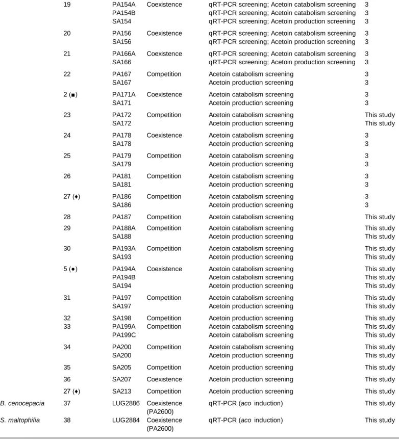
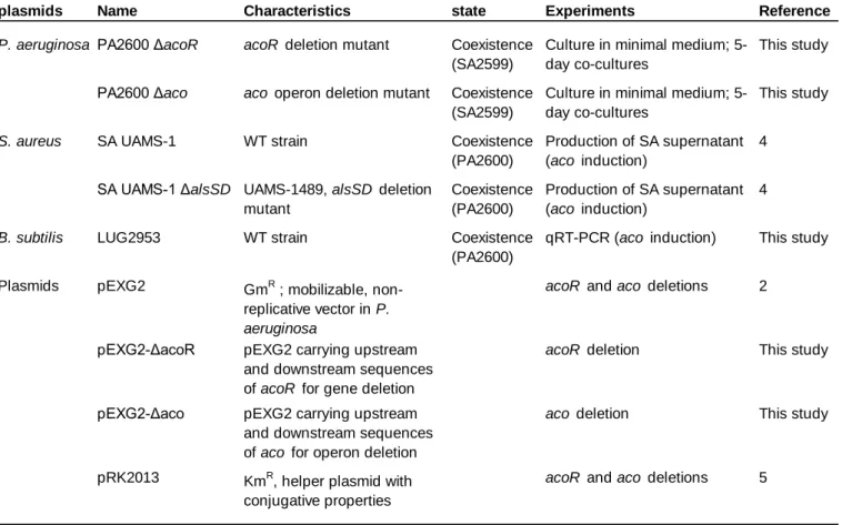
Bacterial strains and plasmids used in this study are listed in Table S1 and S2. CF clinical strains
94
were isolated by the Infectious Agents Institute (IAI) from sputa of patients monitored in the two CF
95
centers of Lyon hospitals (Hospices Civils de Lyon (HCL)). S. aureus and P. aeruginosa strains were
96
isolated from patients co-infected by both bacteria. Each strain pair indicated in Table S1 was recovered
97
from a single sample, obtained from different patients in most cases as indicated in Table S1. All the
98
methods were carried out in accordance with relevant French guidelines and regulations. This study
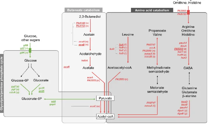
was submitted to the Ethics Committee of the HCL and registered under CNIL No 17-216. All patients
100
were informed of the study and consented to the use of their data.
101
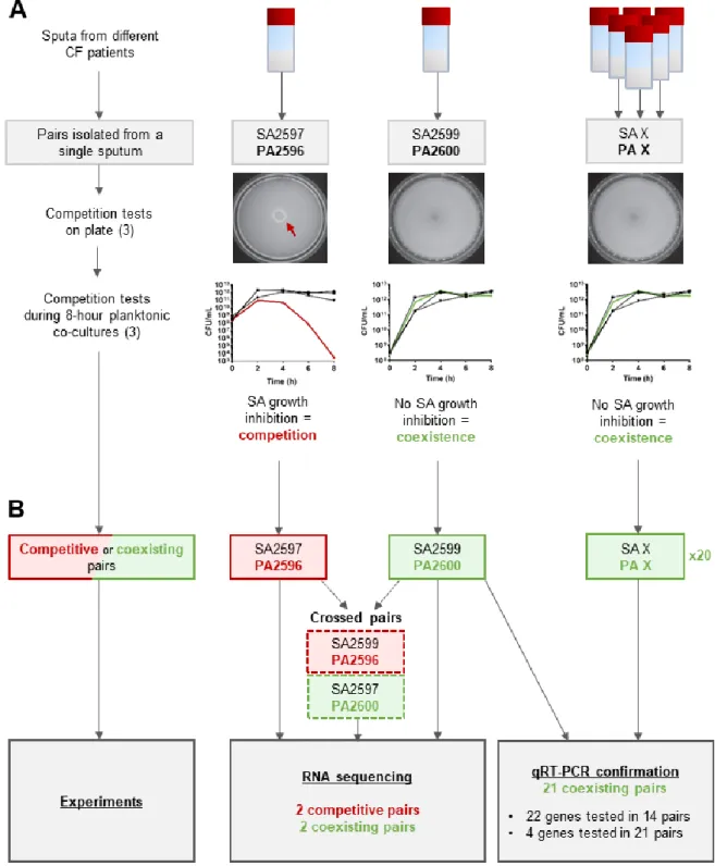
As schematized in Figure S1A, interaction state was determined for each pair by growth
102
inhibition tests on Tryptic Soy Agar (TSA) and in liquid cultures (18). As previously described,
103
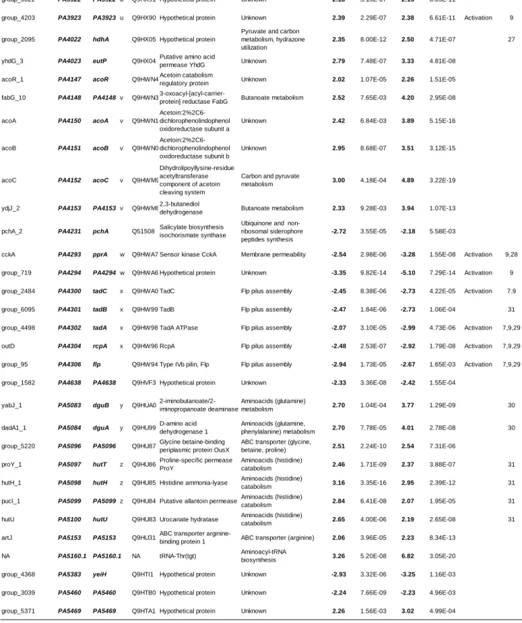
coexistence was characterized by: (i) an absence of inhibition halo on agar tests, and (ii) a similar growth
104
in mono and co-culture during an 8 hours (18).
105
The knock-out ΔacoR and Δaco mutants were generated in P. aeruginosa PA2600 strain via
106
allelic exchange thanks to suicide plasmids pEXG2ΔacoR and pEXG2Δaco constructed as described
107
previously (21, 22). Gentamicin (Euromedex) was used at final concentrations of 50µg/ml. Detailed
108
protocols are given in supplementary data.
109
110
Cultures conditions
111
Strains were grown in monoculture or co-culture in Brain-Heart Infusion (BHI) as described by
112
Briaud et al. (18). Briefly, S. aureus, P. aeruginosa, Burkholderia cenocepacia, Stenotrophomonas
113
maltophilia and Bacillus subtilis overnight precultures were diluted to OD600 of 0.1in BHIand grown 2h30
114
at 37°C and 200rpm. Cultures were then diluted to OD600 of 2 in fresh medium and 10ml were mixed
115
with 10ml BHI for monocultures. Co-cultures were performed by mixing 10ml of P. aeruginosa
116
suspension with 10ml of S. aureus, B. cenocepacia, S. maltophilia or B. subtilis suspension. For
117
supernatant exposure, 10ml of S. aureus supernatant from a 4-hours culture were filtered on 0.22µM
118
filter and added to 10ml of P. aeruginosa suspension. Cultures were grown for 8 hours at 200rpm and
119
37°C for transcriptomic studies. Long-term survival assays were performed by extending incubation time
120
of S. aureus and P. aeruginosa mono- and co-cultures up to five days. Plating at day 0, 3 and 5 were
121
performed on mannitol salt agar (MSA, BBLTM Difco) and cetrimide (DifcoTM) for S. aureus or
122
P. aeruginosa counts, respectively. For growth monitoring in presence of acetoin, minimal medium M63
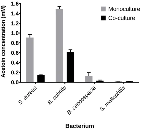
123
(76mM (NH4)2SO4, 500mM KH2PO4, 9µM Fe.SO4.7H2O, 1mM MgSO4.7H2O) was inoculated with
124
P. aeruginosa to OD600 of 0.1 and incubated for 25h at 37°C and 200rpm. Every two hours during 10
125
hours and at t=24h, acetoin was added to obtain a final concentration of 1.5mM in the culture. Plating
126
on TSA (Tryptic Soy Agar) was performed at t=0, one hour after each acetoin addition and at t=24h.
127
128
Genome sequencing and annotation
129
Genome sequencing and annotation of PA2596 and PA2600 strains were performed as
130
previously described (18). In order to compare CDS from PA2596 and PA2600, PAO1 strain
131
(NC_002745.2) was used as a reference. Protein sequences were compared and grouped using a
132
similarity threshold of 95% through Roary (V3.8.2) (23). Gene names and numbers were gathered from
133
PAO1 and used as ID tag for common genes. For non-common genes, CDS from PA2596 and PA2600
134
were tagged with a specific name (gene number and name recovered from UniprotKB database).
135
Functional classification was performed using KEGG database and by manual literature check of each
136
gene function. The complete genome sequences for the PA2596 and PA2600 strains have been
137
deposited in GenBank under the accession number GCA_009650455.1 and GCA_009650545.1.
138
139
Transcriptomic analysis
140
Figure S1 schematizes the global methodology used for transcriptomic analysis. RNAseq
141
analysis was performed on four pairs: the patient-specific pairs SA2597/PA2596 (competitive) and
142
SA2599/PA2600 (coexisting), and the crossed pairs SA2599/PA2596 (competitive) and
143
SA2597/PA2600 (coexisting). RNA extraction, cDNA libraries preparation and sequencing and reads
144
treatment were conducted as previously described (18). Gene expression was considered as
145
dysregulated when: (i) the fold change between co-culture and monoculture was at least 4-fold with an
146
adjusted P-value<0.05, (ii) the dysregulation was observed in the two pairs of strains with the same
147
interaction state, (iii) the dysregulation was specific to either coexistence or competition state. RNAseq
148
data that support our findings are available in the SRAdatabase under the BioprojectID: PRJNA562449,
149
PRJNA562453, PRJNA554085, PRJNA552786, PRJNA554237, PRJNA554233.
150
151
Confirmation of gene expression was achieved by RT-qPCR as previously described on 14 or
152
21 coexisting pairs including the SA2599/PA2600 couple used in RNAseq experiment (Fig. S1) (18).
153
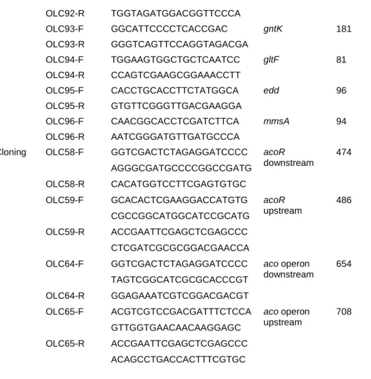
Housekeeping genes gyrB and rpoD were used as endogenous control. Table S3 lists primers used and
154
target genes.
155
156
Acetoin and glucose dosages
157
Acetoin was quantified using modified Voges-Proskauer test (24) optimized in 96-well
158
microplates. To 50µL of culture supernatant, 35µL of creatine (0.5% m/v, Sigma), 50µL of α-naphtol (5%
m/v, Sigma) and 50µL of KOH (40% m/v, Sigma) were sequentially added. The mix was incubated 15
160
minutes at room temperature and optical density at 560nm was read using spectrophometry (Tecan
161
Infinite Pro2000, Tecan-Switzerland). Glucose dosages were performed with glucose (Trinder, GOPOD)
162
assay kit (LIBIOS) in microplates. 185µL of dosage reagent were added to 5µL of culture supernatant
163
and incubated at 37°C for 5 minutes. Optical density at 540nm was measured. Acetoin and glucose
164
standard (Sigma) were performed in water, BHI or M63 according to experiment.
165
166
Statistical analysis
167
Statistical analyses were performed using Prism GraphPad 8.0 software (San Diego, CA).
168
Differences in gene expression fold change and bacterial survival were studied using one-way ANOVA
169
with Dunnett’s or Tukey’s post-test comparisons, as specified in corresponding figures. Median acetoin
170
and glucose concentrations were compared through Mann-Whitney test or Kruskall-Wallis test with
171
Dunn’s correction when appropriate. Differences were considered significant when P-values were lower
172
or equal to 0.05.173
174
Results175
176
P. aeruginosa transcriptome is affected by S. aureus presence
177
We studied the genetic expression of P. aeruginosa in absence or presence of S. aureus in two
178
contexts: when P. aeruginosa and S. aureus were in competition or when they were in coexistence. We
179
thus performed RNAseq analyses using a competitive strain pair (SA2597/PA2596) and a coexisting
180
one (SA2599/PA2600). Each pair was recovered from a single sample of a co-infected CF patient
181
(Table S1). As nature of interaction is solely led by P. aeruginosa (18), the pairs were crossed to study
182
gene expression in additional competitive (SA2599/PA2596) and coexisting (SA2597/PA2600) pairs.
183
Transcriptomic effect was therefore evaluated during co-cultures of two competitive and two coexisting
184
strain pairs (Fig. S1). P. aeruginosa gene expression was considered dysregulated when dysregulation
185
was common to both co-cultures in comparison to monoculture. Each dysregulated gene was then
186
associated to a functional class thanks to a KEGG analysis (Fig. 1A, Tables S4 and S5).
187
188
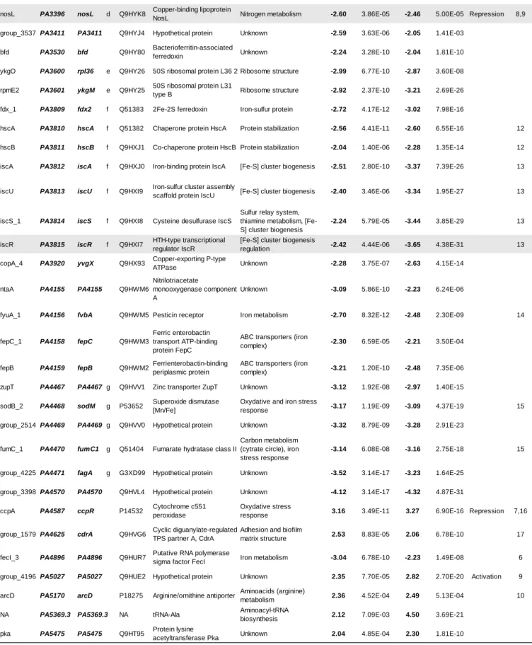
Sixty-eight P. aeruginosa genes were dysregulated in co-culture in the context of competition, with
189
a majority of down-regulated genes (79.4%, Fig. 1A). Fifteen genes involved in nitrogen metabolism
190
and 19 genes involved in iron metabolism were down-regulated, making these two functional classes
191
the most affected in competition. Among these genes, the nir and nos systems involved in denitrification
192
but also genes implicated in iron uptake and transport (isc and fec genes) were down-regulated (Table
193
S4). Other functional classes were less affected, as only 4 genes linked to carbon and amino acids
194
metabolism (bauA, ddaR, gntK, arcD) and 3 genes encoding membrane and virulence factors (rfaD,
195
PA2412, cdrA) were dysregulated in presence of S. aureus.
196
197
More genes were affected during coexistence interaction, as a dysregulation of 105 genes was
198
observed in P. aeruginosa (Fig. 1A). In spite of a trend of up-regulation (56.4% of genes), we could
199
notice the down-regulation of 11 genes involved in nitrogen metabolism. Among these, 8 were also
200
down-regulated in the context of competition, especially genes from the nir system. We thus can
201
presume that the down-regulation of nir genes is not specific to coexistence or competition interaction
202
states. Conversely, the dysregulations of iron metabolism related genes appeared to be specific to
203
competition strains, as only one gene (fhp) of this functional class was down-regulated in coexistence.
204
However, coexistence specifically affected numerous genes belonging to functional classes of carbon
205
and amino acids metabolism (18 and 23 genes) and membrane and virulence factors (15 genes).
206
Concerning this last class, a trend towards lower expression was observed and probably due to the
207
down-regulation of membrane associated factors as the flp-tad system (flp, tad and rcpC genes
208
encoding Flp pilus) and the PA1874-1876 operon (encoding an efflux pump) (Table S5).
209
210
The most affected classes in coexistence with S. aureus were related to P. aeruginosa energetic
211
metabolism (Fig. S2). We observed a down-regulation of genes coding for major pentose phosphate
212
pathway enzymes, as the gluconokinase GntK, its regulator GntR and the operon zwf-edaA
(PA3183-213
PA3181) encoding a glucose-6-phosphate 1-dehydrogenase, a 6-phosphogluconolactonase and a
2-214
keto-3-deoxy-6-phosphogluconate aldolase (25). We also noticed the down-regulation of 5 other genes
215
clustered near to this operon and involved in the same pathway (edd and gapA genes) or glucose
216
transport (gltB, gltF and gltK) (25, 26).
217
218
In contrast, an up-regulation of numerous genes involved in the utilization of alternative carbon
219
sources as butanoate and amino acids was observed. The aco system, comprising the operon
PA4148-220
PA4153 and the gene encoding its transcriptional regulator acoR (PA4147), was thus up-regulated in
221
P. aeruginosa coexisting with S. aureus. This system has been described in P. aeruginosa PAO1 to be
222
responsible for 2,3-butanediol and acetoin catabolism (27). According to KEGG analysis, the
up-223
regulated genes acsA (PA0887), PA2555 (acs family) and hdhA (PA4022) are also involved in the
224
butanoate pathway and energy production from 2,3-butanediol and acetoin, as their products catalyse
225
the production of acetyl-coA from acetaldehyde and acetate (Fig. S2). Finally, 23 genes implicated in
226
amino acids metabolism were up-regulated in P. aeruginosa in presence of S. aureus. Most of them
227
were linked to the catabolism of several amino acids (Fig. S2). We can especially notice the liu operon
228
(PA2015-PA2012), the mmsAB operon (PA3569-PA3570) and the hut gene system (PA5097-PA5100),
229
involved in leucine, valine and histidine catabolism, respectively.
230
231
In order to confirm these transcriptomic effects, we co-cultivated a set of P.
aeruginosa-232
S. aureus coexistence CF pairs and performed RT-qPCR to evaluate P. aeruginosa gene expression in
233
presence of S. aureus (Fig. 1B). Each pair was isolated from a single sputum. Twenty-six genes were
234
tested, including 19 identified as dysregulated during RNAseq analysis and belonging to the most
235
impacted categories, ie. carbon and amino acids metabolism, and membrane and virulence factors.
236
Most of the genes were tested in a total of 14 P. aeruginosa-S. aureus pairs; expression of four of these
237
genes was assessed in seven additional pairs to confirm the observed dysregulations. The different
238
P. aeruginosa strains have shown very different transcriptomic patterns during co-cultivation with
239
S. aureus, especially for membrane-associated and virulence factor genes. We noticed an
over-240
expression of rcpC, tadA, tadG and flp from the flp-tad system from 52.4% to 35.7% of the P. aeruginosa
241
strains. We also tested 7 additional genes encoding virulence factors previously described as involved
242
in P. aeruginosa interaction with S. aureus as las, rhl, pch and pvd genes (7). Regarding these last
243
genes, no clear effect of S. aureus co-cultivation was observed.
244
245
However, clearer transcriptomic patterns were observed for genes linked to carbon and amino
246
acids metabolism, the two most impacted gene classes during RNAseq experiment (Fig. 1B). We
247
confirmed the up-regulation of liuA gene in 78.6% (11/14) of P. aeruginosa strains co-cultivated with
S. aureus. We also confirmed the down-regulation of glucose metabolism genes in a high proportion of
249
strains, ranging from 92.9% (13/14) for zwf, gltF and edd genes to 71.4% (10/14) for gntK gene. Finally,
250
the up-regulation of genes involved in butanoate metabolism was confirmed for acoR (57.2%, 12/21),
251
PA4148 (66.7%, 14/21), acoB (57.1%, 8/14) and PA4153 (64.3%, 9/14), suggesting an impact of
co-252
culture on the whole aco system in P. aeruginosa. In view of these results, we focused our study on four
253
genes: liuA and zwf, respectively involved in leucine and glucose catabolism, PA4148, first gene of the
254
aco operon and acoR, both responsible for acetoin catabolism.
255
256
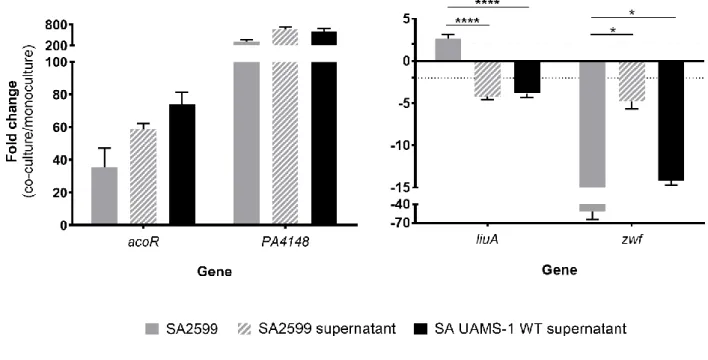
P. aeruginosa aco system is induced by S. aureus acetoin
257
In order to determine if the transcriptomic dysregulations of acoR, PA4148, liuA and zwf in
258
P. aeruginosa PA2600 are specific to interaction with S. aureus, we tested the effect of three other
259
bacterial species: B. cenocepacia and S. maltophilia as they are sometimes associated with
260
P. aeruginosa in CF patients and Bacillus subtilis as it produces high amount of acetoin (28). Dosages
261
in B. cenocepacia and S. maltophilia monocultures confirmed that these bacteria do not produce acetoin
262
(Fig. S3) as previously described (29–31). We observed a down-regulation of P. aeruginosa zwf gene
263
expression in all co-cultures in comparison to monocultures (Fig. 2A). Regarding aco system genes
264
(acoR and PA4148) and liuA gene, only B. subtilis induced similar levels of overexpression than
265
S. aureus SA2599. We thus hypothesized that acetoin, produced by these two species during our
266
experiment (Fig. S3), may be the inductor signal for these genes during co-culture with S. aureus.
267
268
In order to test this hypothesis, we first explored whether an inductor signal was present in the
269
supernatant of S. aureus culture. We indeed observed an overexpression of acoR and PA4148 when
270
P. aeruginosa PA2600 was cultivated in S. aureus SA2599 culture supernatant, as well as the
down-271
expression of zwf but to a lesser extent in comparison to co-culture (Fig. S4). On the contrary, we did
272
not observe overexpression of liuA gene, suggesting that this effect is not due to acetoin and requires
273
the presence of S. aureus cells. Secondly, we cultivated P. aeruginosa PA2600 in presence of S. aureus
274
UAMS-1 WT supernatant or its ΔalsSD derivative defective in acetoin synthesis (32). Supernatant of
275
wild-type UAMS-1 strain induced same transcriptomic patterns on P. aeruginosa as those obtained with
276
CF SA2599 strain (Fig. S4). In presence of UAMS-1 ΔalsSD supernatant, overexpression of aco genes
277
was almost totally abolished (Fig. 2B). However, acetoin addition to this supernatant restored this
overexpression in a dose-dependent manner but with threshold effects between 0.375mM and 1mM of
279
acetoin (Fig. 2B). This indicates that induction through acetoin is one of the mechanisms involved in
280
aco system overexpression in P. aeruginosa. According to this experiment, zwf gene down-regulation
281
did not seem to be mediated by acetoin.
282
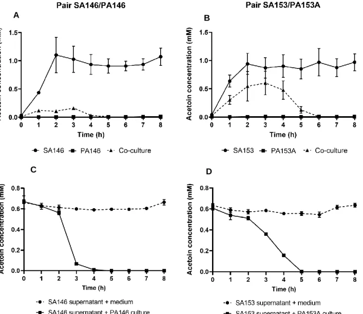
283
Coexisting isolates of S. aureus and P. aeruginosa efficiently metabolize acetoin
284
285
As the aco system is involved in acetoin catabolism in P. aeruginosa (27) and S. aureus
286
produces this molecule (27, 28), we hypothesized that P. aeruginosa could catabolize S. aureus acetoin.
287
To explore this hypothesis, we monitored acetoin concentration during mono- and co-cultures of three
288
pairs of strains: SA2599/PA2600 (Fig. 3A), SA146/PA146 and SA153/PA153A (Fig. S5A and B).
289
P. aeruginosa strains did not produce acetoin, but an acetoin accumulation up to 1.3mM was observed
290
in S. aureus monocultures. Interestingly, a reduction of acetoin accumulation of at least 30% was
291
observed when S. aureus was co-cultivated with P. aeruginosa, in comparison to S. aureus
292
monoculture. The same trend could be observed in CF patient sputa, as a higher acetoin concentration
293
was detected in sputa from S. aureus mono-infected patients (n=9) in comparison to sputa from
294
S. aureus-P. aeruginosa co-infected patients (n=11) (Fig. S6). This effect could be due to a
down-295
regulation of S. aureus acetoin biosynthesis, or an acetoin catabolism in co-culture. Growth of
296
P. aeruginosa PA2600 in S. aureus SA2599 supernatant containing acetoin actually led to a reduction
297
of acetoin concentration, demonstrating the ability of P. aeruginosa to catabolize acetoin (Fig. 3B). This
298
ability was confirmed for the two other strains PA146 and PA153A when grown in supernatant of SA146
299
and SA153, respectively (Fig. S5C and D). We also noted that acetoin catabolism started after glucose
300
depletion in the supernatant and could be delayed by glucose addition (Fig. 3B). This suggests that
301
P. aeruginosa uses acetoin as an alternative carbon source in absence of easily available substrates
302
such as glucose. The early glucose depletion observed during co-culture supports this hypothesis
303
(Fig. 3A).
304
305
In order to test if this acetoin metabolism was specific to interaction state between S. aureus
306
and P. aeruginosa, we evaluated acetoin production and catabolism for 12 couples of competition and
307
12 couples of coexistence (Table S1). Cultivated in P. aeruginosa supernatant, S. aureus strains from
coexisting pairs were able to produce 4-times more acetoin (230µM) than strains from competitive pairs
309
(60µM) (Fig. 4A). Such distinction was not observed during culture in rich medium (Fig. S7). Regarding
310
P. aeruginosa, we cultivated the sets of competitive and coexisting P. aeruginosa strains in SA2599
311
supernatant and monitored acetoin catabolism (Fig. 4B). Both competitive and coexisting strains
312
catabolized acetoin since we observed a decrease in acetoin concentration for both groups. However,
313
coexisting strains showed an increased catabolism efficiency. Indeed, this group catabolized 98.6% of
314
acetoin after 4 hours of culture, while competitive strains catabolized only 47% of acetoin. This increased
315
efficiency of acetoin production and catabolism for coexisting strains could not be explained by a
316
difference in glucose utilization between competition and coexistence strains as both groups catabolized
317
glucose with the same efficiency (Fig. S8). Acetoin production by S. aureus and catabolism by
318
P. aeruginosa therefore seem to be more efficient in isolates from coexisting couples.
319
320
321
Acetoin catabolism by P. aeruginosa increases survival rates of both pathogens in co-culture
322
As P. aeruginosa catabolizes acetoin when medium is glucose-depleted, we tested the effect of
323
acetoin on PA2600 growth in minimal medium M63 (containing no glucose or amino acids)
324
supplemented with 1.5mM acetoin every 2 hours (Fig. 5). P. aeruginosa was able to grow up to 1.6×10⁹
325
CFU/ml after 24h of culture with acetoin as sole carbon source while PA2600 ΔacoR and Δaco mutants
326
grew significantly less, reaching a maximum cell concentration of 1.5×10⁸ UFC/ml at 24h (Fig. 5A). In
327
parallel, we quantified acetoin concentration. We observed an accumulation of acetoin all along the
328
experiment in presence of PA2600 ΔacoR and Δaco mutants. For WT strain, the accumulation of acetoin
329
reached its maximum at 5h of culture and afterwards slowly decreased to reach undetectable values at
330
10h of culture, demonstrating the consumption of acetoin in such conditions (Fig. 5B). A delay of 3h
331
between the start of acetoin catabolism and the growth augmentation of WT strain was noticed, and
332
might be due to metabolism adaptation. Acetoin catabolism nonetheless promoted a 10-fold increased
333
growth of P. aeruginosa during extended culture in glucose depleted medium.
334
335
In order to assess the impact of acetoin catabolism on survival of both pathogens, we
co-336
cultivated P. aeruginosa PA2600, ΔacoR and Δaco mutants with S. aureus SA2599. As S. aureus was
337
not able to grow in M63 poor medium and acetoin affects its long term survival (33, 34), we performed
a long term culture (5 days) in BHI medium. We determined the survival rate of S. aureus co-cultivated
339
with P. aeruginosa in comparison to monoculture (Fig. 6A). Co-culture with the WT strain of
340
P. aeruginosa induced a S. aureus survival rate of 4.7×10⁻¹ after 3 days of culture and of 5.1×10⁻³ after
341
5 days. S. aureus survival thus appears to be highly affected by long-term co-culture with P. aeruginosa,
342
even if the strains stably coexist during shorter spans of culture (18). Interaction state with S. aureus
343
thus seems to rely on nutritional conditions, a fact already observed elsewhere (35, 36). However, the
344
survival rate of S. aureus was even lower during co-culture with P. aeruginosa ΔacoR and Δaco
345
mutants, reaching only 9.7×10⁻² at 3 days of culture and 5.6×10⁻⁴ at 5 days. S. aureus survival was thus
346
4 to 10 times lower when P. aeruginosa was not able to catabolize acetoin, in comparison to co-culture
347
with a strain that efficiently used the molecule. These results suggest that accumulation of acetoin may
348
impact S. aureus survival during co-culture with P. aeruginosa. In parallel, we determined the survival
349
rate of P. aeruginosa (Fig. 6B). We noticed that in such conditions, P. aeruginosa WT strain presented
350
a growth advantage in co-culture, with a maximum of 6.6-fold increase of P. aeruginosa population after
351
5 days in presence of S. aureus in comparison to monoculture (Fig. 6B). The opposite effect was
352
observed for aco mutant strains, as their populations were reduced by 75% for ΔacoR and 32% for Δaco
353
after 3 days of co-culture in comparison to monoculture, confirming the role of acetoin catabolism in
354
favouring P. aeruginosa growth and survival.
355
356
In order to figure out if acetoin accumulation was involved in the decrease of S. aureus survival
357
rate, we monitored acetoin concentration in S. aureus monoculture and co-cultures (Fig. 6C). As
358
expected, we did not detect acetoin in co-culture with PA2600 WT strain over the five days but we
359
observed an accumulation of acetoin with PA2600 ΔacoR and Δaco mutants. The proportion of acetoin
360
in co-culture (1300µM to 1700µM/106 S. aureus) was more than 500 times higher than in monoculture
361
(2.5µM/106 S. aureus) (Fig. 6C). We thus cultivated S. aureus in different acetoin/cells proportions and
362
observed that acetoin had an inhibitory dose-dependent effect on S. aureus growth from 20µM/106
363
S. aureus (Fig. S9). We concluded that acetoin accumulation may be responsible for the decreased
364
S. aureus survival during co-culture with PA2600 ΔacoR and Δaco mutants. P. aeruginosa acetoin
365
catabolism thus allows a greater S. aureus survival during co-culture, in comparison to co-culture with
366
strains unable to catabolize acetoin.
367
368
Taken together, these results demonstrated that acetoin catabolism promotes P. aeruginosa
369
survival as a nutritional alternative carbon source, and improves S. aureus survival during co-culture
370
since a high concentration of acetoin appears to impair S. aureus growth.
371
372
Discussion
373
374
Coinfection with S. aureus and P. aeruginosa is a frequent situation especially in lungs of CF
375
patients, where coinfection accounts for 35% to 50% of cases (17, 18). In this context of coinfection, two
376
states of interaction between the two pathogens have been described: the well-known competitive
377
interaction where P. aeruginosa is able to inhibit the growth of S. aureus and the coexistence state
378
where growth of both species is not affected by each other. The first state has been extensively studied
379
and the leading bacterial determinants of S. aureus growth inhibition have been described (7). On the
380
contrary, little is known about the impact of coexistence state on bacterial physiology. In the present
381
study, we explored the impact of S. aureus on P. aeruginosa gene expression and physiology.
382
Comparing competitive and coexistence states, we observed that the down-regulation of genes
383
involved in iron metabolism was specific to competition. Most of these, such as fec genes or
PA4467-384
PA4471 operon, are involved in ferrous iron uptake and down-regulated during iron-replete conditions
385
(37–39). These conditions are certainly generated by the lysis of S. aureus that provides an iron source
386
to P. aeruginosa during competitive interaction (7, 36), a situation not observed in coexistence. This
387
hypothesis is supported by the work of Tognon et al. (40) that also identified down-regulation of iron
388
metabolism genes during competitive interaction. Interestingly, they also noted typical responses of
389
amino acid starvation including the down-regulation of genes involved in branched-chain amino acid
390
degradation in competitive P. aeruginosa (40). While we did not identify such dysregulation in
391
competition, an overexpression of numerous genes involved in amino acid catabolism was noted during
392
coexistence (Table S5), emphasizing that these dysregulations depend on interaction state.
393
394
More interestingly, we observed that both carbon and amino acids metabolism was specifically
395
affected during coexisting interaction. Many genes involved in glucose catabolism were down-regulated
396
in coexisting isolates during co-culture with S. aureus, especially when the medium was
glucose-397
depleted (Fig. 3). It is worth noting that the zwf gene, down-regulated in almost all P. aeruginosa tested
398
strains, encodes a glucose-phosphate dehydrogenase that converts glucose- phosphate to
6-399
phosphogluconate; the first enzyme in the Entner-Doudoroff pathway, which is central to carbon
400
metabolism in Pseudomonas sp.
In such condition of glucose depletion, we demonstrated that P. aeruginosa was able to use an
402
alternative carbon source provided by S. aureus: acetoin. Acetoin is a four-carbon molecule produced
403
by the decarboxylation of α-acetolactate. Owing to its neutral nature, production and excretion of acetoin
404
during exponential growth prevents over acidification of the cytoplasm and the surrounding medium.
405
When other carbon sources are exhausted, it can constitute an external energy source for fermentive
406
bacteria (28).
407
Acetoin produced by S. aureus was shown to be an inductor of the aco operon and acoR
408
expression in P. aeruginosa (Fig. 2B) (27), allowing acetoin catabolism. This occurred in absence of
409
glucose and was potentially mediated by carbon catabolic repression (Fig. 3), a situation that was
410
already described in other bacteria such as B. subtilis (28). However, threshold effects in
acetoin-411
mediated induction and variability in aco system overexpression in P. aeruginosa strains (Fig. 1B and
412
2B) suggest that other regulatory mechanisms may be involved. Our study may also support the
413
relationship between acetoin and branched-chain amino acid pathways. Indeed, the biosynthesis
414
pathways of acetoin and leucine are co-regulated and share the same precursor α-acetolactate in
415
S. aureus (28). In response to co-culture with S. aureus, P. aeruginosa clinical strains showed
416
overexpression of acetoin and leucine catabolism genes (Fig. 1B and 2), suggesting the presence of
417
both compounds in our co-culture conditions. All of our analyses were performed in vitro. However,
418
using Voges-Proskauer dosage, we were able to confirm the presence of acetoin in CF patient sputa,
419
and in lower concentrations for P. aeruginosa-positive samples (Fig. S6). No direct correlation between
420
P. aeruginosa presence and acetoin quantity can be established as other microorganisms present in
421
sputa may also have an impact on acetoin concentration. Nevertheless, our data support the work of
422
Španěl et al. (41) and suggest that P. aeruginosa may catabolize and use S. aureus acetoin in the lung
423
environment.
424
425
More importantly, we observed that catabolism of acetoin by P. aeruginosa and acetoin
426
production by S. aureus were both more efficient for coexisting isolates, in comparison to competitive
427
ones (Fig. 4). This underlines the adapted metabolic regulation in coexisting isolates in comparison to
428
competitive ones. It is well known that the coexistence phenotype between P. aeruginosa and S. aureus
429
is a consequence of an adaptation process. Indeed, P. aeruginosa strains isolated from early infection
430
outcompete S. aureus while strains isolated from chronic infection are less antagonistic and can be
cultivated with S. aureus (7, 10, 17). It also has been widely described how both pathogens evolve
432
during colonization to evade the immune response and antibiotic treatment (42). Here, for the first time,
433
we suggest that evolution process leads to an adaptation of interspecies metabolic pathways between
434
P. aeruginosa and S. aureus.
435
436
Therefore, we suggestthat acetoin produced by S. aureus could contribute to sputum nutritional
437
richness and be used by P. aeruginosa to survive in this nutritionally competitive environment during
438
chronic infection. This hypothesis is supported by the beneficial effect of acetoin catabolism on
439
P. aeruginosa growth and survival, especially during co-culture with S. aureus (Fig. 5 and 6B).
440
S. aureus survival in presence of P. aeruginosa was also shown to be highly affected by nutrient
441
availability induced by co-culture conditions (Fig. 6A). While coexistence is characterized by an absence
442
of S. aureus growth inhibition during 8-hour co-culture (18), it appears that nutritional competition can
443
still occur under unfavourable conditions and affect S. aureus survival. Therefore, coexistence between
444
the two pathogens is promoted in nutritionally rich environments, in line with previous observations (35,
445
36). Under adverse nutritional conditions induced by long-term culture, although its survival rate is
446
affected, acetoin catabolism benefits its producer, S. aureus (Fig. 6A). Although this effect seems to be
447
linked to acetoin accumulation in the medium as demonstrate by P. aeruginosa ΔacoR and Δaco
448
mutants that do not catabolize acetoin anymore (Fig. 5B and 6C), the precise mechanism remains
449
unclear. In S. aureus, cell death in stationary phase may be induced by acetate production and ensuing
450
intracellular acidification. Thomas et al. showed that acetoin production counters cytoplasmic
451
acidification by consuming protons and promotes S. aureus survival in late-stationary phase (34). We
452
hypothesize that acetoin accumulation in the medium may induce a negative control of acetoin
453
synthesis, affecting S. aureus survival during co-culture conditions.
454
455
Previous studies demonstrated the potential benefits of S. aureus and P. aeruginosa during
456
coinfection. For example, S. aureus facilitates the survival of P. aeruginosa lasR mutants commonly
457
found in CF patients by detoxifying surrounding nitric oxide released by host immune cells (43). On the
458
other hand, 4-hydroxy-2-heptylquinoline-N-oxide (HQNO) produced by P. aeruginosa cells inhibits
459
respiration in S. aureus but also protects S. aureus cells from aminoglycosides (44). Additionally, we
460
recently demonstrated that S. aureus antibiotic resistance and internalization into epithelial cells were
increased in presence of coexisting P. aeruginosa (18). Here, we show that carbon metabolism is largely
462
affected and that P. aeruginosa uses the acetoin produced by S. aureus as an alternative carbon source.
463
This metabolic dialogue between the two pathogens is selected during bacterial adaptation in CF lungs
464
and promotes their survival. Thereby, we highlight for the first time a trophic cooperation between
465
S. aureus and P. aeruginosa during cooperative interaction.
466
467
Acknowledgments
468
This work was supported by the Fondation pour la Recherche Médicale, grant number
469
ECO20170637499 to LC; Finovi foundation to KM; the associations “Vaincre la mucoviscidose” and
470
“Gregory Lemarchal” to KM. We thank Kenneth W. Bayles from University of Nebraska Medical Center
471
(Omaha) for providing S. aureus UAMS-1 WT and mutant strains.
472
473
Conflict of interest
474
All authors declare no competing interests.
475
476
Ethical statement
477
All the strains used in this study were collected as part of the periodic monitoring of patients at the
478
Hospices Civils de Lyon. This study was submitted to the Ethics Committee of the Hospices Civils de
479
Lyon (HCL) and registered under CNIL No 17-216. All patients were informed of the study; however, as
480
the study was non-interventional no written informed consent were required under local regulations.
481
482
483
References
484
485
486
1. Sibley CD, Rabin H, Surette MG. 2006. Cystic fibrosis: a polymicrobial infectious disease. Future
487
Microbiol 1:53–61.
488
2. Guss AM, Roeselers G, Newton ILG, Young CR, Klepac-Ceraj V, Lory S, Cavanaugh CM. 2011.
489
Phylogenetic and metabolic diversity of bacteria associated with cystic fibrosis. ISME J 5:20–29.
490
3. Peters BM, Jabra-Rizk MA, O’May GA, Costerton JW, Shirtliff ME. 2012. Polymicrobial
491
interactions: impact on pathogenesis and human disease. Clin Microbiol Rev 25:193–213.
492
4. Murray JL, Connell JL, Stacy A, Turner KH, Whiteley M. 2014. Mechanisms of synergy in
493
polymicrobial infections. J Microbiol Seoul Korea 52:188–199.
494
5. Abisado RG, Benomar S, Klaus JR, Dandekar AA, Chandler JR. 2018. Bacterial Quorum Sensing
495
and Microbial Community Interactions. mBio 9.
496
6. Tashiro Y, Yawata Y, Toyofuku M, Uchiyama H, Nomura N. 2013. Interspecies interaction
497
between Pseudomonas aeruginosa and other microorganisms. Microbes Environ 28:13–24.
498
7. Hotterbeekx A, Kumar-Singh S, Goossens H, Malhotra-Kumar S. 2017. In vivo and In vitro
499
Interactions between Pseudomonas aeruginosa and Staphylococcus spp. Front Cell Infect
500
Microbiol 7.
501
8. Serra R, Grande R, Butrico L, Rossi A, Settimio UF, Caroleo B, Amato B, Gallelli L, de Franciscis
502
S. 2015. Chronic wound infections: the role of Pseudomonas aeruginosa and Staphylococcus
503
aureus. Expert Rev Anti Infect Ther 13:605–613.
504
9. O’Brien TJ, Welch M. 2019. Recapitulation of polymicrobial communities associated with cystic
505
fibrosis airway infections: a perspective. Future Microbiol 14:1437–1450.
506
10. Baldan R, Cigana C, Testa F, Bianconi I, De Simone M, Pellin D, Di Serio C, Bragonzi A, Cirillo
507
DM. 2014. Adaptation of Pseudomonas aeruginosa in Cystic Fibrosis airways influences virulence
508
of Staphylococcus aureus in vitro and murine models of co-infection. PloS One 9:e89614.
11. Smith EE, Buckley DG, Wu Z, Saenphimmachak C, Hoffman LR, D’Argenio DA, Miller SI, Ramsey
510
BW, Speert DP, Moskowitz SM, Burns JL, Kaul R, Olson MV. 2006. Genetic adaptation by
511
Pseudomonas aeruginosa to the airways of cystic fibrosis patients. Proc Natl Acad Sci U S A
512
103:8487–8492.
513
12. Hoffman LR, Kulasekara HD, Emerson J, Houston LS, Burns JL, Ramsey BW, Miller SI. 2009.
514
Pseudomonas aeruginosa lasR mutants are associated with cystic fibrosis lung disease
515
progression. J Cyst Fibros Off J Eur Cyst Fibros Soc 8:66–70.
516
13. Folkesson A, Jelsbak L, Yang L, Johansen HK, Ciofu O, Høiby N, Molin S. 2012. Adaptation of
517
Pseudomonas aeruginosa to the cystic fibrosis airway: an evolutionary perspective. Nat Rev
518
Microbiol 10:841–851.
519
14. Marvig RL, Sommer LM, Molin S, Johansen HK. 2015. Convergent evolution and adaptation of
520
Pseudomonas aeruginosa within patients with cystic fibrosis. Nat Genet 47:57–64.
521
15. Limoli DH, Whitfield GB, Kitao T, Ivey ML, Davis MR, Grahl N, Hogan DA, Rahme LG, Howell PL,
522
O’Toole GA, Goldberg JB. 2017. Pseudomonas aeruginosa Alginate Overproduction Promotes
523
Coexistence with Staphylococcus aureus in a Model of Cystic Fibrosis Respiratory Infection. mBio
524
8.
525
16. La Rosa R, Johansen HK, Molin S. 2019. Adapting to the Airways: Metabolic Requirements of
526
Pseudomonas aeruginosa during the Infection of Cystic Fibrosis Patients. Metabolites 9.
527
17. Michelsen CF, Christensen A-MJ, Bojer MS, Høiby N, Ingmer H, Jelsbak L. 2014. Staphylococcus
528
aureus alters growth activity, autolysis, and antibiotic tolerance in a human host-adapted
529
Pseudomonas aeruginosa lineage. J Bacteriol 196:3903–3911.
530
18. Briaud P, Camus L, Bastien S, Doléans-Jordheim A, Vandenesch F, Moreau K. 2019.
531
Coexistence with Pseudomonas aeruginosa alters Staphylococcus aureus transcriptome,
532
antibiotic resistance and internalization into epithelial cells. Sci Rep 9.
533
19. Briaud P, Bastien S, Camus L, Boyadijian M, Reix P, Mainguy C, Vandenesch F,
Doléans-534
Jordheim A, Moreau K. 2020. Impact of coexistence phenotype between Staphylococcus aureus
and Pseudomonas aeruginosa isolates on clinical outcomes among Cystic Fibrosis patients. Front
536
Cell Infect Microbiol 10.
537
20. Frydenlund Michelsen C, Hossein Khademi SM, Krogh Johansen H, Ingmer H, Dorrestein PC,
538
Jelsbak L. 2016. Evolution of metabolic divergence in Pseudomonas aeruginosa during long-term
539
infection facilitates a proto-cooperative interspecies interaction. ISME J 10:1323–1336.
540
21. Carriel D, Simon Garcia P, Castelli F, Lamourette P, Fenaille F, Brochier-Armanet C, Elsen S,
541
Gutsche I. 2018. A Novel Subfamily of Bacterial AAT-Fold Basic Amino Acid Decarboxylases and
542
Functional Characterization of Its First Representative: Pseudomonas aeruginosa LdcA. Genome
543
Biol Evol 10:3058–3075.
544
22. Rietsch A, Vallet-Gely I, Dove SL, Mekalanos JJ. 2005. ExsE, a secreted regulator of type III
545
secretion genes in Pseudomonas aeruginosa. Proc Natl Acad Sci U S A 102:8006–8011.
546
23. Page AJ, Cummins CA, Hunt M, Wong VK, Reuter S, Holden MTG, Fookes M, Falush D, Keane
547
JA, Parkhill J. 2015. Roary: rapid large-scale prokaryote pan genome analysis. Bioinforma Oxf
548
Engl 31:3691–3693.
549
24. Nicholson WL. 2008. The Bacillus subtilis ydjL (bdhA) Gene Encodes Acetoin
Reductase/2,3-550
Butanediol Dehydrogenase. Appl Environ Microbiol 74:6832–6838.
551
25. Lessie TG, Phibbs PV. 1984. Alternative pathways of carbohydrate utilization in pseudomonads.
552
Annu Rev Microbiol 38:359–388.
553
26. Chevalier S, Bouffartigues E, Bodilis J, Maillot O, Lesouhaitier O, Feuilloley MGJ, Orange N,
554
Dufour A, Cornelis P. 2017. Structure, function and regulation of Pseudomonas aeruginosa
555
porins. FEMS Microbiol Rev 41:698–722.
556
27. Liu Q, Liu Y, Kang Z, Xiao D, Gao C, Xu P, Ma C. 2018. 2,3-Butanediol catabolism in
557
Pseudomonas aeruginosa PAO1: 2,3-Butanediol catabolism in Pseudomonas aeruginosa.
558
Environ Microbiol 20:3927–3940.
559
28. Xiao Z, Xu P. 2007. Acetoin Metabolism in Bacteria. Crit Rev Microbiol 33:127–140.
29. Gade N, Negi SS, Jindal A, Gaikwad U, Das P, Bhargava A. 2016. Dual Lower Respiratory Tract
561
Infection by Burkholderia cepacia and Acinetobacter baumannii in A Neonate: A Case Report. J
562
Clin Diagn Res JCDR 10:DD01–DD03.
563
30. Amoli RI, Nowroozi J, Sabokbar A, Rajabniya R. 2017. Isolation of Stenotrophomonas maltophilia
564
from clinical samples: An investigation of patterns motility and production of melanin pigment.
565
Asian Pac J Trop Biomed 7:826–830.
566
31. Dryahina K, Sovová K, Nemec A, Španěl P. 2016. Differentiation of pulmonary bacterial
567
pathogens in cystic fibrosis by volatile metabolites emitted by their in vitro cultures: Pseudomonas
568
aeruginosa, Staphylococcus aureus, Stenotrophomonas maltophilia and the Burkholderia cepacia
569
complex. J Breath Res 10:037102.
570
32. Tsang LH, Cassat JE, Shaw LN, Beenken KE, Smeltzer MS. 2008. Factors Contributing to the
571
Biofilm-Deficient Phenotype of Staphylococcus aureus sarA Mutants. PLoS ONE 3:e3361.
572
33. Chaudhari SS, Thomas VC, Sadykov MR, Bose JL, Ahn DJ, Zimmerman MC, Bayles KW. 2016.
573
The LysR-type transcriptional regulator, CidR, regulates stationary phase cell death in
574
Staphylococcus aureus: Metabolic control of cell death in S. aureus. Mol Microbiol 101:942–953.
575
34. Thomas VC, Sadykov MR, Chaudhari SS, Jones J, Endres JL, Widhelm TJ, Ahn J-S, Jawa RS,
576
Zimmerman MC, Bayles KW. 2014. A Central Role for Carbon-Overflow Pathways in the
577
Modulation of Bacterial Cell Death. PLoS Pathog 10:e1004205.
578
35. Miller CL, Van Laar TA, Chen T, Karna SLR, Chen P, You T, Leung KP. 2017. Global
579
transcriptome responses including small RNAs during mixed-species interactions with
methicillin-580
resistant Staphylococcus aureus and Pseudomonas aeruginosa. MicrobiologyOpen 6.
581
36. Mashburn LM, Jett AM, Akins DR, Whiteley M. 2005. Staphylococcus aureus serves as an iron
582
source for Pseudomonas aeruginosa during in vivo coculture. J Bacteriol 187:554–566.
583
37. Visca P, Imperi F. 2018. An essential transcriptional regulator: the case of Pseudomonas
584
aeruginosa Fur. Future Microbiol 13:853–856.
38. Cornelis P, Matthijs S, Van Oeffelen L. 2009. Iron uptake regulation in Pseudomonas aeruginosa.
586
Biometals Int J Role Met Ions Biol Biochem Med 22:15–22.
587
39. Ochsner UA, Wilderman PJ, Vasil AI, Vasil ML. 2002. GeneChip expression analysis of the iron
588
starvation response in Pseudomonas aeruginosa: identification of novel pyoverdine biosynthesis
589
genes. Mol Microbiol 45:1277–1287.
590
40. Tognon M, Köhler T, Luscher A, van Delden C. 2019. Transcriptional profiling of Pseudomonas
591
aeruginosa and Staphylococcus aureus during in vitro co-culture. BMC Genomics 20:30.
592
41. Španěl P, Sovová K, Dryahina K, Doušová T, Dřevínek P, Smith D. 2016. Do linear logistic model
593
analyses of volatile biomarkers in exhaled breath of cystic fibrosis patients reliably indicate
594
Pseudomonas aeruginosa infection? J Breath Res 10:036013.
595
42. Baishya J, Wakeman CA. 2019. Selective pressures during chronic infection drive microbial
596
competition and cooperation. NPJ Biofilms Microbiomes 5:16.
597
43. Hoffman LR, Richardson AR, Houston LS, Kulasekara HD, Martens-Habbena W, Klausen M,
598
Burns JL, Stahl DA, Hassett DJ, Fang FC, Miller SI. 2010. Nutrient availability as a mechanism for
599
selection of antibiotic tolerant Pseudomonas aeruginosa within the CF airway. PLoS Pathog
600
6:e1000712.
601
44. Hoffman LR, Déziel E, D’Argenio DA, Lépine F, Emerson J, McNamara S, Gibson RL, Ramsey
602
BW, Miller SI. 2006. Selection for Staphylococcus aureus small-colony variants due to growth in
603
the presence of Pseudomonas aeruginosa. Proc Natl Acad Sci U S A 103:19890–19895.
604
605
606
607
608
609
610
611
613
Figure legends
614
615
Figure 1: Alteration of P. aeruginosa transcriptome induced by co-culture with S. aureus.
616
A. Number of under-expressed (grey bars) and over-expressed (black bars) genes of
617
P. aeruginosa during co-culture with S. aureus for competitive (left) or coexisting (right) pairs.
618
PA2596 competition and PA2600 coexistence strains were cultivated in absence or presence of SA2597
619
or SA2599 as described in Fig. S1. RNAs were extracted after 4 hours of culture and the RNAseq
620
analysis was performed as described in material and methods. A gene was considered as differentially
621
expressed when the Fold Change (FC) was > |2log2| with an adjusted P-value <0.05. Functional
622
classification was performed thanks to KEGG database and literature.
623
B. Fold change of 26 P. aeruginosa gene expression induced by co-culture with S. aureus during
624
coexistence interaction. Twenty-one S. aureus-P. aeruginosa coexisting pairs were isolated from
625
separate CF sputa recovered from 20 different patients. Each P. aeruginosa strain was cultivated in
626
absence or presence of its co-isolated S. aureus strain. RNAs were extracted after 4 hours of culture
627
and gene expression was assayed by RT-qPCR. A gene was considered as differentially expressed
628
when the Fold Change (FC) was > |2|, indicated by dotted lines. Black lines indicate the median. Genes
629
were tested in 14 (regular genes) or 21 (bold genes) strains. List of used strains is shown in Table S1.
630
Genes annotated with (*) were not identified as dysregulated during the RNAseq experiment.
631
632
Figure 2: Fold changes of P. aeruginosa acoR, PA4148, liuA and zwf induced by culture
633
conditions.
634
A. Fold changes induced by co-culture with S. aureus (black bars), B. subtilis (grey bars), B.
635
cenocepacia (hatched white bars) or S. maltophilia (hatched black bars). P. aeruginosa PA2600
636
strain was cultivated in absence or presence of S. aureus SA2599, B. subtilis, B. cenocepacia or S.
637
maltophilia. RNAs were extracted after 4 hours of culture and gene expression was assayed by
RT-638
qPCR. Bars represent the mean fold change + SEM from three independent experiments. Dotted lines
639
indicate a fold change = |2|. *Padj<0.05, **Padj<0.01, ***Padj<0.001 ANOVA with Dunnett’s correction
640
(S. aureus vs. condition).