HAL Id: tel-02971506
https://tel.archives-ouvertes.fr/tel-02971506
Submitted on 19 Oct 2020
HAL is a multi-disciplinary open access
archive for the deposit and dissemination of sci-entific research documents, whether they are pub-lished or not. The documents may come from teaching and research institutions in France or abroad, or from public or private research centers.
L’archive ouverte pluridisciplinaire HAL, est destinée au dépôt et à la diffusion de documents scientifiques de niveau recherche, publiés ou non, émanant des établissements d’enseignement et de recherche français ou étrangers, des laboratoires publics ou privés.
Deciphering the role of oncofetal fibronectin isoforms in
matrix assembly and cellular function
Georgios Efthymiou
To cite this version:
Georgios Efthymiou. Deciphering the role of oncofetal fibronectin isoforms in matrix assembly and cellular function. Molecular biology. COMUE Université Côte d’Azur (2015 - 2019), 2019. English. �NNT : 2019AZUR6024�. �tel-02971506�
Etude d’assemblage et des fonctions des
isoformes oncofoetales de la fibronectine
Georgios EFTHYMIOU
Institut de Biologie Valrose
Présentée en vue de l’obtention
du grade de docteur en Biologie d’ Université
Côte d’Azur
Dirigée par : Ellen Van Obberghen-Schilling Soutenue le : 19 Décembre 2019 à Nice
Devant le jury, composé de :
Corinne Albigès-Rizo, DR, IAB, Université Grenoble Alpes
Robert Arkowitz, DR, iBV, Université Côte d’Azur
Patricia Rousselle, DR, LBTI, Université Claude Bernard Lyon 1
Ellen Van Obberghen-Schilling, DR, iBV, Université Côte d’Azur
i
Etude d’assemblage et des fonctions
des isoformes oncofoetales de la
fibronectine
Jury :
Président du jury
Robert Arkowitz, CNRS DR, iBV, Université Côte d’Azur Rapporteurs
Corinne Albigès-Rizo, DR, IAB, Université Grenoble Alpes Patricia Rousselle, DR, LBTI, Université Claude Bernard Lyon 1 Directrice de thèse
ii
Titre
Etude d’assemblage et des fonctions des isoformes oncofoetales de la fibronectine
Resumé
La matrice extracellulaire (MEC) constitue une plateforme fibrillaire intégrant l'action des signaux chimiques et mécaniques de l'environnement. La fibronectine (FN), un composant majeur de la MEC, est au centre de cette plateforme de biorégulation et module de nombreuses actions biologiques telles que l’adhésion et la motilité cellulaires, la prolifération et la différenciation, ainsi que la structure et le dépôt de la MEC. La FN se présente sous deux formes : la FN plasmatique (pFN) et la FN cellulaire (cFN), dite « oncofoetale ». Cette dernière est nommée ainsi pour son expression uniquement au cours du développement et dans certaines conditions physiopathologiques (réparation tissulaire, inflammation et cancer). La différence entre les deux est l’existence dans la cFN des domaines supplémentaires EDB et EDA, qui résultent d’un épissage alternatif. Comment la présence de ces « extra-domaines », EDA et EDB, régit l'assemblage des FN et comment les variantes assemblées régulent le comportement des cellules est en grande partie inconnu. Des études de délétion ciblées d’un seul domaine chez la souris ont révélé le rôle de l'EDA dans des phénomènes très variés dont la morphogenèse des valves lymphatiques, l'athérosclérose et la cicatrisation / fibrose. Au niveau moléculaire, l'inclusion de l'EDA élargit le répertoire des récepteurs cellulaires de la FN (intégrines α4β1, α7β1 et TLR4). A ce jour, aucun récepteur n'a été rapporté pour l’EDB. De ce fait il a été proposé que sa présence modifierait la conformation du site de liaison de FN aux cellules et faciliterait ainsi l’assemblage fibrillaire induit par les intégrines via des mécanismes qui restent à établir.
Le but des travaux de cette thèse était de décoder les rôles de l'EDA et de l'EDB de la FN, et plus précisément de :
1) étudier l'impact de la présence des domaines EDA et EDB sur l'assemblage fibrillaire de la FN à la surface de cellules compétentes pour l'assemblage, et
iii 2) déterminer comment la présence de l’EDB et de l’EDA influence les propriétés biochimiques, physiques et fonctionnelles de la matrice, qui à leur tour affectent le comportement des cellules qui y sont attachées.
Dans un premier temps l’équipe a développé un ensemble d'outils biologiques composé de : i) vecteurs lentiviraux hébergeant la séquence codante complète du gène de la FN humaine contenant l’un, les deux ou aucun des « extra-domaines » alternativement épissés, ii) des variants de FN recombinants purifiés, iii) des fibroblastes embryonnaires de souris FN -/- (Fn1 -/- MEF), et iv) des MEF exprimant des variantes de FN. Cette batterie outils uniques a été utilisée pour étudier l'assemblage spécifique de variantes par les fibroblastes et les effets de réseaux de cFN homogène sur le comportement cellulaire.
Nos résultats ont montré que les « extra-domaines « de la FN sont responsables au niveau des fibroblastes du réglage fin de l’amplitude de plusieurs réponses dont l’assemblage de la matrice, la croissance et le métabolisme énergétique. En utilisant une approche informatique « non-biaisée », nous avons démontré que la présence des « extra-domaines » modifie la structure de la matrice de la FN déposée par les fibroblastes. Ceci témoigne d’événements de signalisation cellulaire différents, qui sont susceptibles de modifier aussi bien les réponses précoces que tardives induites par la FN. Les matrices variant-spécifiques que nous avons développées représentent des outils très puissants pour décrypter les fonctions des « extra-domaines » de la FN dans les multiples types de cellules impliquées non seulement dans des réponses physiologiques mais également dans des situations pathologiques.
Mots clés : matrice extracellulaire, fibronectine, extra-domaines, EDB, EDA,
iv
Title
Deciphering the role of oncofetal fibronectin isoforms in matrix assembly and cellular function
Abstract
The Extracellular Matrix (ECM) constitutes a fibrillar platform that integrates the action of chemical and mechanical cues from the environment. Fibronectin (FN), a major component of the ECM, is at the center of this bioregulatory stage, modulating numerous biological procedures such as cell adhesion and motility, cell proliferation and differentiation, as well as ECM deposition and structure. FN is found in two forms: plasma FN (pFN) and cellular FN (cFN). cFN differs from pFN by the presence of alternatively spliced Extra Domains, namely EDB and EDA. Each of these alternatively spliced regions is encoded by a single exon, the regulation of which is strictly regulated and limited to embryonic tissues, as well as pathophysiological conditions such as wound healing, inflammation, and cancer. The term “oncofetal” was coined in order to describe FN isoforms harboring either of the Extra Domains, that are normally absent in adult tissues.
How the presence of EDA and EDB regulates FN assembly and how the assembled variants regulate cell behavior is largely unknown. Single Extra Domain-targeted deletion studies in the mouse have revealed roles for EDA in the morphogenesis of lymphatic valves, atherosclerosis, and wound healing/fibrosis. Mechanistically, the inclusion of EDA expands the repertoire of cellular FN receptors (α4β1, α7β1 integrins and TLR4). To date, no ligand has been reported for EDB. Rather, its presence has been postulated to alter the conformation of the cell-binding domain of FN and to facilitate integrin-driven fibrillar assembly by mechanisms that remain to be established.
The aim of this dissertation is to provide more insight regarding the roles of the FN Extra Domains EDA and EDB, and more specifically:
1) To investigate how the presence of alternatively spliced EDA and EDB domains affect the fibrillar assembly of FN at the surface of assembly-competent cells, and
v 2) To determining how the presence of EDB and EDA influences the biochemical, physical, and functional properties of the matrix that in turn affect the behavior of cells attached to it.
Here we present a biological toolset composed of : i) lentiviral vectors harboring the full-length coding sequence of human cFN containing one, both, or none of the alternatively spliced Extra Domains, ii) purified recombinant FN variants, iii) FN-null mouse embryo fibroblasts (MEFs) and iv) FN variant-expressing MEFs. These unique tools were used to study variant-specific assembly by fibroblasts and the effects of homogeneous cFN networks on cell behavior.
Utilizing an unbiased computational approach, we document that the presence of the Extra Domains results in a distinct pattern of FN deposition and assembly that in turn influences both early and late signaling events that control cell proliferation as well as cell contractility. Finally, we present a series of novel data that point towards a FN-impacted control of cell energetics.
We conclude that FN Extra Domains are responsible for the fine-tuning of the extent of several cellular processes that can reflect strict regulation in pathophysiological procedures such as tissue repair, fibrosis, and tumor progression.
Keywords: extracellular matrix, fibronectin, extra domains, EDB, EDA, integrins,
vi To all those who helped me get where I am now. In their own personal way.
vii
Acknowledgments
At the time that these lines are being written, it has been almost four years since I started my PhD back in 2015, just two months after being recruited in the highly competitive SIGNALIFE PhD program. Four years are definitely not a long period, but it is long enough for knowledge and experience to be obtained, and bonds to be formed among people, the gratitude to whom is hard for me to put into words.
I would like to start by thanking all the people involved in the Labex SIGNALIFE Program. A large family who has worked together in order to create an outstanding platform that has functioned as a springboard for me and other young researchers, providing the funds needed for the smooth completion of our studies. I would like to acknowledge the efforts of Konstanze Beck who has been the guide and companion for me and so many other SIGNALIFE Students since our arrival in France, as well as Martine Avella, the Labex SIGNALIFE Project Officer, and Anna Bliznyuk, the Labex SIGNALIFE Assistant.
At this point, it is also of major importance for me to thank the ARC Scientific Committee who deemed me worthy of one extra year of funding, which has proved both useful and necessary.
My greatest thanks goes to a group of people that have been my second family for the last four years: the team “Adhesion Signaling and Stromal Reprogramming in the Tumor Microenvironment” led by Dr Ellen Van Obberghen-Schilling. My words can in no way describe how grateful I am to Ellen. First for accepting me in the group and secondly for trusting me throughout all these years. You have been a real mentor and an inspiring scientist, who considered me a colleague. I thank you for always being available to discuss, search, troubleshoot, and for always hearing my opinions and proposals. The outcome of those four years is the result of fruitful communication and real teamwork between us. For that, I truly thank you, and I hope that all my future professional partnerships will be like this.
A big “merci” to Dominique Grall. As I used to say “Sans labo, Dominique existe. Sans Dominique, labo n’existe pas”! Do, you were the first person who came in direct
viii contact with my initial ignorance and clumsiness. Thanks to you, I learned so much in such a short time. Thank you for all your technical help in the lab, your patience, and your support. Thank you for our discussions and for believing in me.
Delphine Ciais has played a special part in my life in the lab. You have always been the toughest to convince and through this, you taught me how to be perseverant, coherent, concise, having a critical mind, and distinguish the difference between “doing experiments” and “finding the right approach to answer a question”. We have not really worked together, but our interaction has been of catalytic importance to me.
I feel more than grateful for having met and worked side by side with so many people that have been members of this beautiful family. Primarily, Agata Radwanska, who was the initiator of the project that eventually became my own. I owe you so much. Special thanks to Angélique Saint, the “médecin traitant du labo”, as well as Michael Ruff, “notre petit Michael”, for their help, support, and insight, but also for making the everyday routine more pleasant! I would also like to thank all the “younger” members of the lab: young students, master students, interns. Each one of them has contributed, even in some small way, to “surviving the thesis”!
During those four years, I have been lucky to work and interact with people from different laboratories as well. I greatly appreciate the invaluable insight, help, and sharing of knowledge and expertise provided to me by Laurent Counillon and all the members of the team “Ionic Transport Pathophysiology” at the Laboratory of Molecular PhysioMedicine (LP2M), with a great emphasis to the contribution of Mallorie Poet. Additionally, I am indebted to Didier Pisani for his priceless help with the Seahorse analyzer, and Sabrina Pisano for all the AFM microscopy sessions and analysis she has performed for me.
It would be a serious omission if I did not include our collaborators in the i3S (Laboratoire d'Informatique, Signaux et Systèmes de Sophia Antipolis), namely Laure Blanc Feraud and Xavier Descombes, as well as the SIGNALIFE student Anca Ioana Grapa. Thank you very much for our nice collaboration. Many of the results in this manuscript would not have been possible without your aid and effort.
ix Of great importance has been the help I received from the different core facilities of the institute. Thus, I would like to thank all the people that took part in my efforts, directly and indirectly. More specifically, I would like to express my thanks to Maurice Hattab and the Biochemistry and Molecular Biology Facility for taking care of the purification of the proteins I have been working with all these years. To the Imaging facility and its personnel for all the trainings, assistance, analyses, and troubleshooting, especially Magali Mondin, Sébastien Schaub, and Simon Lachambre. I also thank in the Histopathology facility, Samah Rekima and Belinda Desrues, as well as Agnès Loubat for her assistance in the Cytomertry facility. Finally, I would like to thank all the members of the institute, students, researchers, engineers, administration personnel, and the wonderful ladies in the common services who have collectively made my lab life and work so much easier. Thank you all!
A special thanks to the members of the scientific committee, for accepting to review and evaluate my work, Dr. Corinne Albigès-Rizo and Dr. Patricia Rousselle, for taking the time to come to Nice for my thesis defense. I also express my thanks to Dr. Robert Arkowitz for being my internal advisor during these four years, and for presiding the thesis committee.
This endeavor would not have been successful if not for several people who have been and have become part of my life outside of the lab. I would like to thank my close friends, my friends and my acquaintances in Nice and anywhere else in the world for all the moments we have shared together. You have formed me in so many ways.
Finally, I would like to thank my family. Εσάς, που με ενθαρρύνετε πάντα και δεν έχετε σταματήσει λεπτό να πιστεύετε σε μένα και στις δυνατότητές μου. Σας ευχαριστώ για την υποστήριξή σας σε όλες τις στιγμές της ζωής μου έως τώρα, και για την παρουσία σας σε κάθε στιγμή, ευχάριστη ή λιγότερο ευχάριστη. Ό,τι έχω καταφέρει έως τώρα το οφείλω εξίσου σε σας. Ακόμη κι αν δεν το δείχνω, σας ευχαριστώ.
x
Abbreviations
67LR: 67 kDa laminin receptor
ADMIDAS: adjacent to metal-ion-dependent adhesion site AFM: atomic force microscopy
AMPK: 5’ adenosine monophosphate-activated protein kinase B-CAM: basal cell adhesion molecule
BM: basal membrane cFN: cellular fibronectin CS: chondroitin sulfate
CTGF: connective tissue growth factor DDR: discoidin domain receptor
DGC: dystrophin glycoprotein complex DS: dermatan sulfate
ECM: extracellular matrix EGF: epidermal growth factor
EGFR: epidermal growth factor receptor ERK: extracellular signal-regulated kinase ESE: exonic splicing enhancer
FACITs: fibril associated collagens with interrupted triple helix FAK: focal adhesion kinase
FERM: protein 4.1, ezrin, radixin, moesin FKBP12: FK506 binding protein 12
FN: fibronectin
GAGs: glycosaminoglycans
GARP: glycoprotein A repetitions predominant GPVI: glycoprotein VI
GS domain: Gly-Ser-rich domain HA: hyaluronic acid
xi HIF1: hypoxia inducible factor 1
HRS: hepatic arginine-serine rich protein HS: heparan sulfate
IAP: integrin-associated protein ILK: integrin linked kinase ISE: intronic splicing enhancers
JAK-STAT: Janus kinases – Signal transducer and activator of transcription proteins JNK: c-Jun N-terminal kinase
KS: keratan sulfate
LAIR-1: leukocyte-associated immunoglobulin-like receptor-1 LETS: large, external transformation-sensitive
LGD: laminin globular domain LLC: large latent complex LOX: lysil oxidase
LRRC32: leucine-rich repeat-containing protein 32 LTBP: latent TGF-β binding protein
Lu: Lutheran
MACITs: membrane associated collagens with interrupted triple helices MAPK: mitogen activated protein kinase
MIDAS: metal-ion-dependent adhesion site
mTOR: mechanistic (or mammalian) target of rapamycin PAI1: plasminogen activator inhibitor 1
PDGF: platelet derived growth factor
PDGFR: platelet derived growth factor receptor pFN: plasma fibronectin
PGs: proteoglycans
PI3K: phosphoinositide 3-kinase
PIPKIγ: phosphatidylinositol phosphate kinase type Iγ PKB: protein kinase B, also known as Akt
xii PTB: phospho-tyrosine binding
RIAM: Rap1-GTP-interacting adaptor molecule ROS: reactive oxygen species
RSV: Rous Sarcoma Virus RTK: receptor tyrosine kinase SDS: sodium dodecyl sulfate SF-A: fibroblast surface antigen SLC: small latent complex
SMAD: SMA (small work phenotype) and MAD (mothers against decapentaplegic) family of proteins
SRSF5: serine and arginine rich splicing factor 5 TGFBI: TGF-β inducible gene
TGFβRI: transforming growth factor receptor I TGFβRII: transforming growth factor receptor II TGF-β: transforming growth factor β
TIAM1: T-cell lymphoma invasion and metastasis 1 TMD: transmembrane domain
uPARAP: urokinase-type plasminogen activator associated protein VBS: vinculin binding site
VEFGR: vascular endothelial growth factor receptor α-SMA: alpha smooth muscle actin
xiii
Table of Contents
Acknowledgments ... vii
Abbreviations ... x
Table of Contents ... xiii
Prologue ... xv
1. Introduction ... 1
1.1 The Extracellular Matrix ... 1
1.1.1 ECM Types ... 2
1.1.2 ECM Components ... 3
1.1.2.1 Glycosaminoglycans and Proteoglycans ... 3
1.1.2.2 Collagens and Glycoproteins ... 5
1.1.2.3 Other Matrix-Associated Components ... 11
1.1.3 Fibronectin ... 12
1.1.3.1 The Early Days ... 12
1.1.3.2 Fibronectin: A Major ECM Building Block ... 13
1.1.3.3 FN Structure ... 14
1.1.3.4 Fibronectin Interactions and Function ... 18
1.1.3.5 Oncofetal FN Variants ... 22
1.1.3.6 Functional Roles of the Extra Domains ... 24
1.2 Cell – ECM Interactions ... 27
1.2.1 The Integrins ... 29
1.2.1.1 Integrin Structure ... 30
1.2.1.2 Integrin Activation ... 33
1.2.1.3 Biological Processes Involving Integrins ... 35
1.2.1.4 Fibronectin Fibrillogenesis ... 37
1.3 The TGF-β Pathway ... 40
1.3.1 TGF-β Signaling, Receptors and Effectors ... 41
1.3.2 TGF-β Crosstalk with the ECM and the Role of Integrins ... 44
2. Aim of the Thesis ... 48
xiv
4. Complementary Results ... 102
5. Conclusions and Perspectives ... 110
6. References ... 112
7. Annex 1: Fibronectin Timeline ... 141
8. Annex 2: Classification of the fibronectin variants with
curvelets ... 151
xv
Prologue
It has been almost 300 years since Carl von Linné coined the term biologi in his work Bibliotheca botanica in 1736. A term that has evolved as much as the scientific field it describes: “The objects of our research will be the different forms and manifestations of life, the conditions and laws under which these phenomena occur, and the causes through which they have been effected. The science that concerns itself with these objects we will indicate by the name biology or the doctrine of life” – Gottfried Reinhold Treviranus.
Biology (βιολογία), the study of (-λογία > λόγος) life (βίος), is a relatively recent scientific field despite its ancient roots. It was Antonie van Leeuwenhoek’s improvement of the microscope that led biology to its dramatic development. From the mere observation of unicellular organisms to protein purification, and from the discovery of the genetic material to gene manipulation, biology has been by far one of the most rapidly developing scientific fields.
Biological research initiated as an approach to simplify and elucidate the complex phenomena that govern life. Nowadays, biology is a scientific field that feeds from several domains (mathematics, physics, informatics) providing us with an explosive amount of information and new data on the origins of life, the function of complex intertwined cell communication, and the building of entire organisms and ecosystems. The simultaneous study of all known biological systems is impracticable. This could have possibly been the case 150 years ago. That is why it is important to break down the different biological systems in order to be able to extract useful information, but always bearing in mind that no oversimplified system makes sense except in the light of its entirety, paraphrasing the title of the well-known essay by Theodosius Dobzhansky (Dobzhansky 1973).
In this work, I will try to compile known data, as well as to enrich current knowledge with novel results regarding a major aspect of molecular cell biology in multicellular organisms (and more specifically in vertebrates, mainly focusing on mammals) that is “Cells in their Social Context”. Tissues in multicellular organisms are not made up solely of cells. They are also composed of a remarkably complex
xvi network of macromolecules, collectively known as the extracellular matrix (ECM). These macromolecules, secreted by the cells, provide structural and functional cues to the adjacent cells, resulting in the formation of the highly organized multicellular structure. The target ECM component of this dissertation is fibronectin, which will be discussed extensively in terms of structure, function, expression patterns in health and disease, and how its different forms affect cell behavior.
The following pages consist of an introduction on the nature of the extracellular matrix, and the key players that mediate cell-ECM interactions: the integrins. It is beyond the scope of this manuscript to present all the knowledge obtained about the ECM, the various ECM components, and the integrin structure and function. Basic background information will be presented instead, in order to set the stage before proceeding to a detailed description of “fibronectin, the extracellular glue” (Zollinger and Smith 2017).
1
1. Introduction
In the macroscopic scale, no organism or population can be viewed as an individual entity. On the contrary, it has to be considered as a part of a dynamic ecosystem composed of other individuals and/or populations, as well as the surrounding environment. This notion, initially described by Plato in his Dialogues, is also reflected in the microscale: each cell is part of a tightly regulated community of cells and extracellular material, the extracellular matrix or ECM.
1.1 The Extracellular Matrix
The ECM is an ensemble of high molecular weight macromolecules expressed by the underlying cells that forms an astonishingly elaborate and dynamic structure that constantly interacts with cells in the vicinity. This cell-derived biomaterial had not been fairly characterized until recently when a set of 1056 genes was identified and described by Naba and colleagues (Naba et al. 2012).
The extracellular matrix is a major component of all tissues in every organ, and it provides mechanical support and physical scaffolding, as well as a wide range of crucial biochemical and biomechanical cues regulating the behavior of the residing cells. This way, the ECM influences their survival, development, migration, proliferation, shape, and function, thus guiding multiple facets of different processes such as morphogenesis and tissue homeostasis (Frantz, Stewart, and Weaver 2010; Shaohe Wang et al. 2017; Trapani, Bonaldo, and Corallo 2017).
Depending on the tissue, the extracellular matrix can possess different inherent properties, characteristic of that tissue. It can become calcified to form the rock-hard structures of bone and teeth, it can form the transparent substance of the cornea, or it can adopt the ropelike organization that gives tendons their enormous tensile strength. This extensive variety of different matrix types is reflected in the numerous interactions among the components of the matrix (collectively known as the matrisome) and the components that are not necessarily part of the matrix but plausibly do associate with it. Such components are growth and secreted factors, ECM-modifying enzymes and other proteins (R. O. Hynes and Naba 2012). This interplay as
2 well as with the cells themselves, mainly through a family of cell receptors knows as integrins (Richard O Hynes 2002), results in the ECM diversity across tissues.
1.1.1 ECM Types
Just as the same genome gives rise to different cell types in an organism, the same matrisome generates different matrix types depending on cell type and localization, even within the same tissue. In a broad sense, two types of ECMs exist varying in composition and structure: i) the interstitial matrix and ii) basement membranes (BMs) or basal laminae (Figure 1). Interstitial matrices surround cells, while BMs lie in the interphase between the epithelial cells of the parenchyma and the mesenchymal tissue, the latter being characterized
by the presence of interstitial matrix.
The BM is a thin (40-120nm), tough, flexible sheet of matrix molecules that lines all epithelia, but also surrounds individual muscle fibers, fat cells, and Schwann cells. Although small in volume, it plays a critical role in body architecture. The basal lamina separates cells
and epithelia from the underlying connective tissue and forms the mechanical connections between them. In a different setting, a basal lamina can serve as a filtering barrier, as is the case for the kidney glomerulus. Basal membranes can also serve to determine cell polarity, influence cell metabolism, organize the proteins in adjacent plasma membranes, promote cell survival, proliferation and differentiation, and serve as highways for cell migration (Alberts 2015).
Figure 1 Epithelial cells reside on a rigid basement membrane
(BM) that separates them from the mesenchymal part of the tissue where mesenchymal cells, like fibroblasts, produce and deposit the interstitial matrix. Each matrix type is composed of a set of different components that influence their physical properties and functions. Adapted from Bonnans et al. 2014.
3
1.1.2 ECM Components
Any typical matrix is composed of a large variety of macromolecules that share characteristic modular domains. Based on this feature, Naba and colleagues bioinformatically defined ECM components by screening for proteins containing domains characteristic of ECM proteins, ECM-affiliated proteins, ECM modifiers, and secreted factors. At the same time, they eliminated proteins that shared one or more of the defining domains without being part of the ECM (Figure 2).
In silico results were supported by biochemical analyses on normal and tumor tissues resulting in the establishment of the core matrisome, comprising 278 genes giving rise to proteoglycans (PGs), collagens, and glycoproteins. Furthermore, a second gene set, denoted as matrisome-associated components was established, containing 778 genes that produce ECM-affiliated components, ECM regulators, and secreted factors (Naba et al. 2012). The matrisome is mainly produced locally by cells in the matrix, which also help to organize the matrix through forces generated by the cytoskeleton influencing the orientation of the matrix components outside of the cell.
1.1.2.1 Glycosaminoglycans and Proteoglycans
Glycosaminoglycans (GAGs) or mucopolysaccharides are composed of several repeats of a disaccharide unit, which is characteristic of each GAG. One of the two sugars in the repeating unit is always an amino sugar (acetylglucosamine or N-acetylgalactosamine), while the second is an uronic acid (glucuronic or iduronic) or
Figure 2 In silico definition of ECM components identified 1056 genes comprising two distinct
groups. The core matrisome, composed of 278 genes, is responsible for the generation of the structural components of the ECM. The second set is composed of matrisome-associated genes that give rise to proteins linked to the ECM, proteins that regulate ECM form and function, and soluble factors that are known or believed to bind to the ECM.
4 galactose. GAGs are subject to extensive modification: sulfation, carboxylation, deacetylation, and epimerization (Townley and Bülow 2018), and they form stiff, highly hydrophobic chains that cannot compact to form globular structures. Thus, GAGs adopt extended conformations with a very high volume to mass ratio, attracting Na+ and other cations that, in turn, capture large amounts of water forming highly hydrated gel-like structures with high viscosity and elasticity. These structures fill most of the extracellular space, and can withstand massive compressive forces (Alberts 2015; Sasarman et al. 2016). A characteristic example of such matrix is the cartilage that lines the knee joint.
Six classes of GAGs are distinguished by their sugars, the type of oligosaccharide linker they contain, and the number and location of sulfate groups: chondroitin sulfate (CS), dermatan sulfate (DS), heparan sulfate (HS), heparin sulfate (Hep), keratan sulfate (KS), and hyaluronan or hyaluronic acid (HA) (Sasarman et al. 2016). Two of the aforementioned classes, CS and DS, contain N-acetylgalactosamine as one of the repeating sugars, while the second sugar in the unit is either glucuronic acid or iduronic acid. HS and HA are copolymers of N-acetyglucosamine and glucuronic acid. The disaccharide unit in Hep is N-acetylglucosamine and iduronic acid, while KS is composed of units containing N-acetylglucosamine and galactose.
As mentioned earlier, all GAGs are characterized by long and unbranched, extensively modified sugar chains, and by the covalent attachment to a core protein, giving rise to proteoglycans (PGs). The only exception is HA. Its sugar chains are not sulfated, and it is not covalently linked to any core protein. Instead, it interacts with PGs via hyaluronan-binding motifs in a non-covalent fashion (Sasarman et al. 2016).
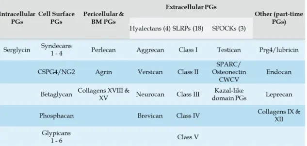
PGs can be classified based on localization, gene/protein homology, and the utilization of specific protein modules within their respective protein cores (Table 1). These criteria result in the generation of four major families with distinct forms and functions: the intracellular, cell-surface, pericellular and extracellular PGs (Iozzo and Schaefer 2015).
PGs interact with numerous growth factors, cytokines and chemokines, cell surface receptors and ECM components through either their core proteins, or their
5 GAG chains, thus participating in key regulatory cell processes such as signaling, differentiation and proliferation, adhesion and migration, and apoptosis (Iozzo and Schaefer 2015; Theocharis et al. 2016). Additionally, they can interact both with cells and with other components of the ECM, playing an important role in ECM scaffolding and remodeling, and in cell distribution within it.
Thus, they affect normal physiology as well as the development of various diseases [reviewed in detail in (Townley and Bülow 2018; Sasarman et al. 2016; Iozzo and Schaefer 2015; Schaefer and Schaefer 2010; Theocharis et al. 2010; Afratis et al. 2012; Theocharis et al. 2014)].
1.1.2.2 Collagens and Glycoproteins
Apart from the GAGs (connected to a core protein or not), there are almost 250 proteins, 43 of which are distinct collagen subunits, and around 200 glycoproteins constituting the matrisome (R. O. Hynes and Naba 2012). An extensive description of every ECM constituent is beyond the scope of this dissertation, so only a brief overview of the major collagens and glycoproteins follows ahead.
Collagens
The collagens are a family of fibrous and non-fibrous proteins found in all metazoa (Table 2). Collagens can form fibrils or networks, they can be laterally associated to other fibrils or serve as core proteins for proteoglycans, or they can be found docked through the plasma membrane. They are secreted in large quantities by connective-tissue cells, and they constitute the main structural element of the ECM.
6
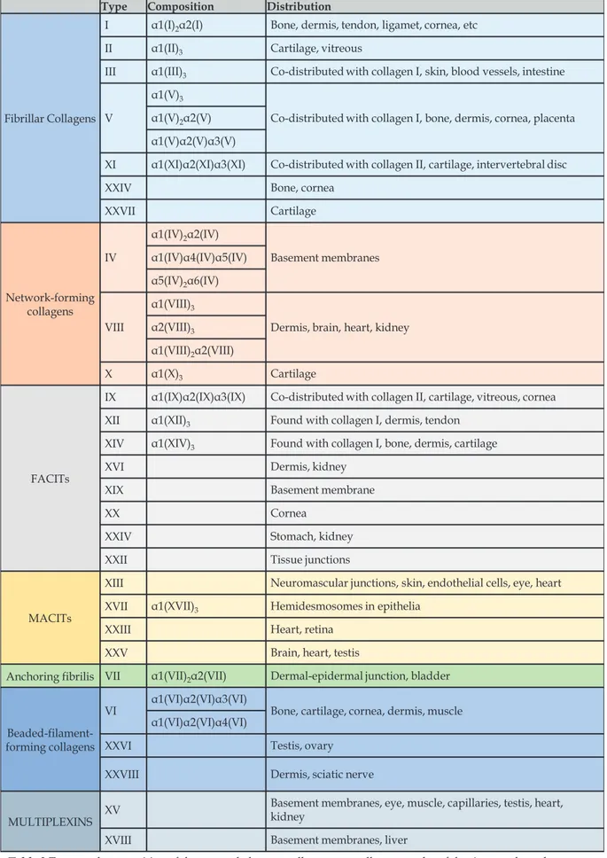
Type Composition Distribution
Fibrillar Collagens
I α1(Ι)2α2(Ι) Bone, dermis, tendon, ligamet, cornea, etc II α1(ΙΙ)3 Cartilage, vitreous
III α1(ΙΙΙ)3 Co-distributed with collagen I, skin, blood vessels, intestine
V
α1(V)3
Co-distributed with collagen I, bone, dermis, cornea, placenta α1(V)2α2(V)
α1(V)α2(V)α3(V)
XI α1(XI)α2(XI)α3(XI) Co-distributed with collagen II, cartilage, intervertebral disc
XXIV Bone, cornea
XXVII Cartilage Network-forming collagens IV α1(IV)2α2(IV) Basement membranes α1(IV)α4(IV)α5(IV) α5(IV)2α6(IV) VIII α1(VIII)3
Dermis, brain, heart, kidney α2(VIII)3
α1(VIII)2α2(VIII)
X α1(X)3 Cartilage
FACITs
IX α1(IX)α2(IX)α3(IX) Co-distributed with collagen II, cartilage, vitreous, cornea XII α1(XII)3 Found with collagen I, dermis, tendon
XIV α1(XIV)3 Found with collagen I, bone, dermis, cartilage
XVI Dermis, kidney
XIX Basement membrane
XX Cornea
XXIV Stomach, kidney
XXII Tissue junctions
MACITs
XIII Neuromascular junctions, skin, endothelial cells, eye, heart XVII α1(XVII)3 Hemidesmosomes in epithelia
XXIII Heart, retina
XXV Brain, heart, testis
Anchoring fibrilis VII α1(VII)2α2(VII) Dermal-epidermal junction, bladder
Beaded-filament-forming collagens
VI α1(VI)α2(VI)α3(VI) Bone, cartilage, cornea, dermis, muscle α1(VI)α2(VI)α4(VI)
XXVI Testis, ovary
XXVIII Dermis, sciatic nerve
MULTIPLEXINS XV
Basement membranes, eye, muscle, capillaries, testis, heart, kidney
XVIII Basement membranes, liver
Table 2 Types and composition of the currently known collagens, as well as examples of the tissues where they are most commonly found. Table adapted from Theocharis et al, 2016.
7 Collagens are found in the fibers of tendons, in the matrices of bone and cartilage, in the laminar sheets of basement membranes, in the viscous matrix of the vitreous humor, and in the interstitial ECM of the dermis and of capsules around the organs. They are the most abundant protein family representing 25 – 30% of the total protein mass (Frantz, Stewart, and Weaver 2010; R. O. Hynes and Naba 2012; Alberts 2015).
The collagen superfamily comprises 28 members designated with Roman numerals in vertebrates [I – XXVIII, reviewed in (Ricard-Blum 2011)]. The primary typifying feature of a collagen molecule is the presence of the repeating triplet Gly-X-Y, where X is frequently proline and Y is frequently 4-hydroxyproline, though they can be any of the 20 amino acids in proteins (Heino 2007). This repeating unit gives rise to stiff, trimeric coiled coils, in which three collagen polypeptide chains, also known as α chains, are wound around one another in a rod-like superhelix.
The length of the superhelix can vary significantly ranging from most of the molecule structure (96% in collagen I) to less than 10% of it (collagen XII), and it correlates with the localization within the tissue, as well as with ECM organization and structure [see (Ricard-Blum 2011) and references therein]. For example, the original type I collagen of bones and tendons consists almost entirely of a long and rigid uninterrupted collagen triple helix that spans approximately 1000 amino acids (R. O. Hynes and Naba 2012; Heino 2007). Each trimer assembles into higher-order fibrils and fibers after being crosslinked by various enzymatic and non-enzymatic reactions, and it is found in all interstitial matrices of the organism.
Other collagen types have interruptions in the repeating sequence (Gly-X-Y), thus adding flexibility into the molecules. An extra level of complexity is added because of the existence of several molecular isoforms for the same collagen type, and of hybrid isoforms comprised of α chains belonging to two different collagen types, distinguished by the use of Arabic numerals (Ricard-Blum 2011). For example, collagen XI is comprised of three α chains assembled into a heterotrimer, but the α (XI) chain forms type V/XI hybrid collagen molecules by assembling with the α1 (V) chain in the vitreous humor (Mayne et al. 1993) and cartilage (J.-J. Wu et al. 2009).
8 The criteria to name a protein collagen are not well defined. Many proteins contain the triple helical domain but do not belong to the collagen family. At the same time, all collagen genes encode additional non-collagenous domains, some of which are characteristic collagen N and C pro-domains, while others are domains shared with other ECM proteins.
Fibrillar collagens are the most abundant collagens in vertebrates where they play a structural role by contributing to the molecular architecture, shape, and mechanical properties of tissues such as the tensile strength in skin and the resistance to traction in ligaments, as also mentioned previously. However, as stated before for the ECM, the various matrix components are “not just pretty fibrils” (Richard O. Hynes 2009).
Indeed, it is now well documented that fibrous collagens, as well as fibril-associated collagens, alongside many other ECM constituents, play important roles in tissue homeostasis, growth factor signaling, differentiation, development, and disease.
Table 3 displays a non-exhaustive list of functions that depend on various collagens
and their receptors. For details, reader can refer to the following reviews and references therein (Frantz, Stewart, and Weaver 2010; Ricard-Blum 2011; Heino 2007;
Table 3 Role of collagens in various biological processes exerted by collagen receptors (see also
9 Richard O. Hynes 2009; Daley and Yamada 2013; Watt and Huck 2013; Bonnans, Chou, and Werb 2014; Pickup, Mouw, and Weaver 2014; Boyd and Thomas 2017).
Elastin and elastic fibers
Fibrillar collagens, as described above, are responsible for providing tissues with tensile strength. Indeed, a collagen fiber can withstand 25 times stronger tensile strain compared to a steel fiber of the same diameter before fracture occurs (Buehler 2006). However, organ functionality depends also on the extendibility of the underlying tissue. Skin, blood vessels, lung, heart, and bladder tissues need not only be strong, but also elastic in order to function properly. This is where the network of elastic fibers comes into play, giving tissues the resilience to recoil after transient stretch.
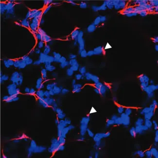
Elastic fibers are the largest structures in the ECM and consist of two morphologically distinct components [reviewed in (Wagenseil and Mecham 2007)]. The principal component is elastin, a crosslinked polymer of the monomeric secreted form of the protein tropoelastin (Sandberg, Weissman, and Gray 1971), a 60-72kDa precursor protein characterized by short segments that alternate along the polypeptide chain (Alberts 2015; Theocharis et al. 2016; Wagenseil and Mecham 2007; Wise and Weiss 2009). Highly hydrophobic segments are responsible for the elastic properties of the molecule (Muiznieks, Weiss, and Keeley 2010), and alanine- and lysine-rich α-helical segments are crosslinked to adjacent molecules by covalent attachment of lysine residues (Wagenseil and Mecham 2007). This covalent modification is performed by lysil oxidase (LOX) or LOX-like molecules (Lucero and Kagan 2006), eventually stabilizing the polymer rendering the network insoluble (Wagenseil and Mecham 2007; Muiznieks, Weiss, and Keeley 2010). Figure 3 shows the organizations of elastin in the developing mouse lung.
While elastin arises from a single gene, the second component of elastic fibers, the microfibrils, is characterized by higher complexity. Microfibrils are, as their name states, small, 10-15nm fibrils that localize to the periphery of the elastic fiber in the adult tissues (Wagenseil and Mecham 2007). The major structural trait of microfibrils is provided by a set of proteins termed fibrillins, large (350kDa), cysteine-rich glycoproteins (L. Y. Sakai 1986; Kumra and Reinhardt 2018), that in combination with
10 other associated proteins (Kielty, Sherratt, and Shuttleworth 2002) perform structural and regulatory roles (Kumra and Reinhardt 2018; Sengle and Sakai 2015).
After secretion, fibrillin monomers go through several interactions with other molecules to form short microfibrils that are subsequently transferred onto FN fibers for stabilization (Kinsey et al. 2008). Elongation then occurs through interaction with other microfibril proteins (Sabatier et al. 2009).
Furthermore, fibrillins regulate transforming growth factor β (TGF-β) localization and activity by modulating the binding of latent TGF-β binding proteins [LTBPs (Dallas et al. 2000; Isogai et al. 2003)], that in turn regulate TGF-β latency (duscussed in Section 1.3). More specifically, LTBP-1 is assembled in the ECM through the presence of FN (Dallas et al. 2005), but it is finally transferred to microfibrils as the ECM matures (Ono et al. 2009). Additionally, different LTBPs show preferential binding for ECM components with various resistance to strain (Zilberberg et al. 2012), thus reflecting a differential
TGF-β activation depending on the ECM composition and state.
Laminins
As described earlier, BMs are thin-layered surfaces responsible for tissue architecture providing protection against stress, and serving as an interactive platform between cells and the microenvironment. BMs are quite heterogeneous in terms of composition and structure, and this heterogeneity depends on the tissue, but also on the localization within the same tissue. Typically, a mature BM consists of laminin, type IV collagen, nidogen, agrin, and perlecan (Alberts 2015; Theocharis et al. 2016; Durbeej 2010).
Figure 3 Immunofluorescence staining of
elastin (magenta) in P7 mouse lung, showing elastin network and patches of elastin present at the tip of alveolar protrusions (arrowheads). Nuclei staining (blue) was used to visualize the structure of the lung tissue. Adapted from Luo et al. 2018.
11 Laminins are large (400 – 900kDa), trimeric, cross or T-shaped molecules with two or three short arms and one long arm (Aumailley et al. 2005). Short arms are composed of parts of one chain, while the long arm is formed by parts of each of the three chains. The three different polypetide chains are termed α, β, and γ. Five α, three β, and three γ chains have been identified so far both for mouse and for human (Theocharis et al. 2016; Aumailley et al. 2005). All chains are glycosylated, while some of them have been shown to contain glycosaminoglycan side chains (Aumailley et al. 2005).
The members of the laminin family are generally characterized by the presence of an α-helical coiled coil domain, but they contain additional domains that contribute to laminin-cell and laminin-ECM interactions. Such domains comprise a large globular N-terminal domain, and five laminin globular (LG) domains at the C-terminal end (Theocharis et al. 2016; Durbeej 2010). Via their various domains, laminins can self-assemble to form networks in close association with the cells. This tight interaction is mediated via cell receptors like integrins or syndecans (See also Section 1.2).
1.1.2.3 Other Matrix-Associated Components
So far, only a small fraction of the elements that contribute to the formation of the ECM has been described. Many other components, members of the aforementioned matrisome, take an active part in matrix generation and assembly. Major examples are the glycoproteins fibronectin (the focus of this work, to be discussed shortly), and tenascins. Other members of the matrisome comprise non-structural extracellular modulators of cellular functions that facilitate cell-cell and cell-ECM interactions (Frantz, Stewart, and Weaver 2010; R. O. Hynes and Naba 2012; Theocharis et al. 2016).
There are also ECM-associated components such as modifiers of the ECM structure and function (transglutaminases, lysyl oxidases, hydrolases, metalloproteinases, ADAMs, etc.), and ECM-bound growth and secreted factors (VEGF, HGF, PDGF, TGF). This way the ECM acts as a reservoir of factors the bioavailability and/or activity of which is subject to the regulation of the cells surrounded by or assembling it (Naba et al. 2012; Frantz, Stewart, and Weaver 2010).
12
1.1.3 Fibronectin 1.1.3.1 The Early Days
In 1948 at Harvard University, Peter Morrison and colleagues were working on the identification and the properties of serum and plasma proteins. Their main interest was fibrinogen and its ability to form clots under the action of thrombin, reflecting a clinical importance considering blood coagulation. In their effort to optimize their fibrinogen isolation technique, they came to realize that they were able to obtain high amounts of fibrinogen from two distinct fractions. The first was separated from the bulk of non-clottable proteins by precipitation at low pH, while the second was obtained after centrifugation and the presence of ethanol at higher pH and low temperature (0oC). This second fraction, however (Fraction I-1) contained an unknown material of lower solubility than fibrinogen, which precipitated. The researchers introduced the term cold-insoluble globulin to denote the insoluble component, in the absence of a specific name (Morrison, Edsall, and Miller 1948).
It took more than 20 years to successfully purify to homogeneity the “cold-insoluble globulin” from plasma cryoprecipitate (Mosesson and Umfleet 1970). In the meantime, other researchers, independently, identified new features in a plasma component that had similar properties with the cold-insoluble globulin, without though being able to assign the described properties to a defined protein (Smith and Korff 1957; Wolff et al. 1967).
It was during the 1970s when a series of studies described a large glycoprotein found on the surface of fibroblasts [large, external, transformation-sensitive (LETS) protein or galactoprotein a, or cell surface protein (CSP)] that was lost after virus-induced transformation (Gahmberg and Hakomori 1973; R. O. Hynes 1973; Yamada and Weston 1974). Additionally, its presence varied depending on the growth state of normal cells (Richard O. Hynes 1974). During the same period, an antiserum was generated that recognized a fibroblast surface antigen [FS-A (Erkki Ruoslahti and Vaheri 1974)], that was also found to react with a serum antigen, later found to be the cold insoluble globulin (Erkki Ruoslahti and Vaheri 1975). This cell surface antigen
13 was recognized as the previously described transformation-sensitive protein LETS (Keski-Oja, Vaheri, and Ruoslahti 1976).
Despite the doubts generated by the limitations of the methods used at the time (not well-defined immunogens, not highly specific antiserum), several data converged towards the opinion that the cell surface glycoprotein and the cold-insoluble globulin were indeed the same protein. In the following years, several studies showed clearly the similarities between the cell-associated protein and the one purified from plasma. The use of antibodies to visualize stain patterns in cultured cells, the use of gel affinity chromatography to simply and rapidly purify the protein, as well as other biochemical approaches (solubility assays, SDS-polyacrylamide gel electrophoresis) resulted in the acceptance that the described proteins comprised a family of related proteins (Richard O Hynes 1990).
Those early studies, even if the researchers at the time could not fully explain their findings, provided information about the function of the molecules that are now known as fibronectins. The term was coined around the mid-1970s (from the latin fibra for fiber and the verb nectere which means to link, to connect), and was later adopted by all scientists in the field to describe the different forms of the protein fibronectin (FN).
1.1.3.2 Fibronectin: A Major ECM Building Block
Since its discovery, FN has been the subject of extensive studies that have importantly contributed to our knowledge regarding its structure, and the wide range of different functions it exerts. Product of a single gene, FN is expressed in two distinct forms with both additive and overlapping functions. The main source of FN is the liver, which secretes plasma FN (pFN) in a soluble form circulating through the blood plasma. Furthermore, cellular FN (cFN) is produced mainly by fibroblasts, as well as endothelial cells, chondrocytes, macrophages, platelets, and other cell types (Richard O Hynes 1990) both in vivo and in culture. cFN is mainly found in an insoluble form around the cells secreting it, where it is assembled into fibrils and fibers, resulting eventually in the formation of the underlying ECM.
14 Only a handful of human disease cases linked to FN mutations have been documented so far, and they concern either glomerulopathy with FN deposits (Castelletti et al. 2008; dos Reis Monteiro et al. 2019) or skeletal dysplasias (Costantini et al. 2019). This suggests that even point mutations can lead to deleterious or lethal effects. Indeed, a gene knockout study in the mouse has shown that inactivation of both FN alleles leads in early embryonic lethality resulting from severe defects in mesodermal migration, reflecting anomalies in cell adhesion, proliferation, and differentiation (George et al. 1993). Additionally, the highly conserved modular structure of the protein reinforces the notion that intact FN is indispensable for normal development and tissue homeostasis (Ni et al. 2003; J. Xu and Mosher 2011; Richard O Hynes 1990), functions that are mainly mediated via direct interactions with cell surface receptors, such as integrins (Richard O Hynes 1990; Richard O. Hynes 2009).
The most important property of FN is the fact that it drives the assembly of the ECM. Being among the very first components of the ECM (if not the first), FN is deposited and polymerized by assembly-competent cells, such as fibroblasts, in a provisional matrix that drives the deposition of other ECM components, such as collagens and other proteoglycans (Richard O. Hynes 2009). Hindering FN assembly in vivo results in delayed embryonic development (Darribère et al. 1990), while absence of FN in cultured fibroblasts impedes the formation of ECM (Cseh et al. 2010; Gopal et al. 2017). FN assembly will be discussed in more detail in Section 1.2.1.4.
1.1.3.3 FN Structure
FN is a high molecular weight glycoprotein composed of two similar subunits of 220-250 kDa (Engel et al. 1981; Erickson, Carrell, and McDonagh 1981). Every FN subunit has two carboxy-terminal cysteine residues that form two disulfide bonds with the cysteine residues of another FN subunit, giving rise to a functional FN dimer. FN is highly produced and secreted by the liver, or expressed locally by fibroblasts and other cell lines. In the first case, FN circulates in the blood plasma in a soluble form (plasma FN – pFN), while in the latter case FN forms an insoluble mesh that gives rise to the extracellular matrix (cellular FN, cFN). The linear structure of the subunits is highly repetitive, characterized by the presence of three distinct types of domains that
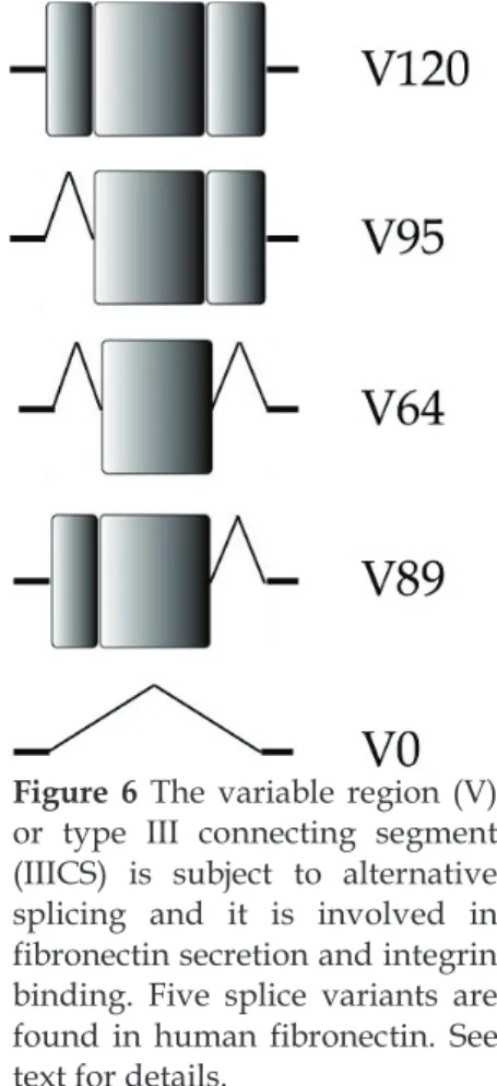
15 together constitute approximately 90% of the sequence [reviewed in (J. Xu and Mosher 2011)]. There are 12 type I repeats (hereafter FNIx, where subscript x denotes the number of the repeat), 2 type II repeats (hereafter FNIIx), and 15 – 17 type III repeats (hereafter FNIIIx) (Richard O Hynes 1990). Apart from the repetitive domains, there is also a variable region (V or IIICS, type III connecting segment) that lies between FNIII14 and FNIII15, which is subject to high alternative splicing, as well as short linker segments between different modules across the protein [reviewed in (J. Xu and Mosher 2011) and Figure 4].
The FNI repeat is composed of 45 amino acids, four of which are cysteine residues that give rise to two disulfide bonds within the repeat. 3D structure studies of the FNI repeat have shown that a scaffold of hydrophilic amino acids forms two compact β-sheets, with two and three strands, that enclose a hydrophobic core of aromatic residues (Baron et al. 1990; Potts et al. 1999).
Figure 4 Graphic representation of the linear structure of FN showing the different FN modules. FN
forms dimers linked in their C-terminal ends by two disulfide bonds. Each dimer is composed of 12 FNI repeats (blue rectangles), two FNII repeats (purple rectangles), and 15-17 FNIII repeats (green and red rectangles) two of which are subject to alternative splicing (EDB and EDA), a Variable region (gray), and short peptide segments that do not constitute parts of the modular domains (black lines). Different segments of FN result after proteolytic cleavage (70 kDa, 40 kDa, and 27 kDa segments). Glycosylation sites are indicated with pink triangles. The highly modular structure of the molecule permits an enhanced degree of folding resulting in the formation of cryptic sites (yellow arches) exposed by addition of extra domains, strain, and proteolysis. FN has multiple binding sites for cells (FNIII9-10 repeats) and other molecules. Adapted from Xu and Mosher 2011, and Van-Obberghen
16 The FNII repeats have a somewhat similar structure with the FNI repeats. They are about 60 amino acids long and have two intra-domain disulfide bonds. Like FNI repeats, they consist of two antiparallel β-sheets each containing two strands, while the amino acid sequence is mainly characterized by the presence of aromatic residues (Constantine et al. 1992; Pickford et al. 1997). Unlike FNI repeats that are only present in vertebrates (Tucker and Chiquet-Ehrismann 2009), FNII repeats have also been identified in other, less complex organisms (Ozhogina and Trexler 2001), as well as in matrix metalloproteinases (Collier et al. 1988).
The FNIII repeat is a functionally conserved domain found in many other proteins including extracellular matrix components, cell surface receptors, and cytoskeletal proteins both in vertebrates and in invertebrates (Bork and Doolittle 1992). There are 15 standard FNIII domains present in every FN monomer, and two additional domains termed Extra Domains (EDB and EDA) that are included in the molecule under specific circumstances.
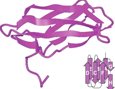
Each FNIII repeat is composed of 90 amino acids (though slight variations do exist across proteins and species) organized in seven β-strands [A, B, C, D, E, F, and G, (Figure 5)], further folding into two antiparallel β-sheets (like the other two types of FN repeats). One β-sheet is composed of three β-strands, while the other is composed of four, and they fold in a sandwich-like conformation the center of which is highly hydrophobic (Dickinson, Veerapandian, et al. 1994; Dickinson, Gay, et al. 1994; Leahy, Aukhil, and Erickson 1996; Main et al. 1992). Surprisingly, FNIII repeats lack any cysteine residues, thus they contain no intra-chain disulfide bonds. This affects the form of the entire molecule, as the separate FNIII domains are prone to conformational changes resulting from mechanical forces (Krammer et al. 1999). These changes can greatly influence the folding state of the FNIII repeat while maintaining its structural integrity. Also the inclusion of additional type III Figure 5 Linear representation of the structure of
the FNIII10 repeat. FNIII repeats are composed of
seven β strands organized in two β sheets that fold in a sandwich-like structure with a highly hydrophobic core. In FNIII10, between strands F
and G, lies the cell binding sequence (RGD), shown here in red. See text for details.
17 repeats, through alternative splicing, can impact FN conformation (Barbara Carnemolla et al. 1992).
The variable region (V) is not homologous to any other part of the protein. Several functional and structural studies have shown that the variable region is 120 amino acids long and it can be partly present or completely
absent from the molecule, while most FN dimers naturally form with distinct variable regions in each subunit [see Figure 4, Figure 6 and (J. E. Schwarzbauer 1989)]. The different forms of the V region arise from alternative splicing, which appears to be regulated in a tissue-dependent manner, and it changes throughout development and lifetime of the organism (Chauhan et al. 2004). The inclusion of different parts of the V region determines the repertoire of cell receptors that FN can interact with in order to promote cell attachment and spreading (Kocher, Kennedy, and Madri 1990), and controls FN dimer secretion and incorporation into fibrin clots and ECM in tissues [reviewed in (J. E. Schwarzbauer and DeSimone 2011)].
FN in solution is found in a compact conformation, which is mainly due to non-covalent interactions established within the FNIII domains (Ohashi and Erickson 2005), as well as an intramolecular interaction between FNI4 and FNIII3 leading in the bending of the N-terminal region of both subunits [reviewed in (J. Xu and Mosher 2011)]. Furthermore, the ability of FN to self-associate through other regions has been documented, namely FNI1-5 (McKeown-Longo 1985), FNIII1-3 (Aguirre, McCormick, and Schwarzbauer 1994; Hocking, Sottile, and McKeown-Longo 1994; Johnson et al. 1999), FNIII12-14 (Bultmann, Santas, and Peters 1998), and FNIII7 (Ingham et al. 1997). Another factor that largely contributes to FN conformation is the variety of post-translation modifications. FN can be phosphorylated, sulfated, and (as every
Figure 6 The variable region (V)
or type III connecting segment (IIICS) is subject to alternative splicing and it is involved in fibronectin secretion and integrin binding. Five splice variants are found in human fibronectin. See text for details.
18 glycoprotein) glycosylated (Paul and Hynes 1984). The degree of glycosylation often depends on the tissue, though generally FN contains approximately 5% of carbohydrate chains either N-linked, or O-linked in distinct sites of both subunits [see Figure 4 and (J. Xu and Mosher 2011)]. Addition of glycans affects FN sensitivity to proteolysis, thermal stability, and binding affinity to other proteins such as collagen, thus modulating cell adhesion and migration (Olden, Pratt, and Yamada 1979; Bernard, Yamada, and Olden 1982; Jones 1986). Phosphorylation has been described in serine residues (Etheredge et al. 1985) enhancing cell attachment and mechanical cell functions (Yalak et al. 2019), while sulfation has been suggested to take place in the V region (Paul and Hynes 1984). Finally, citrulination, the conversion of arginine residues to citruline, was recently described as another type of post-translational modification of FN. Twenty four sites of varied degree of citrulination were identified, impacting the FN attachment to both α5β1 and αvβ3 integrins, and reflecting a refined regulation of integrin clustering and cell contractility (Stefanelli et al. 2019).
1.1.3.4 Fibronectin Interactions and Function
The modular structure of FN, as well as its multiple post-translational modifications result in numerous interactions with a variety of proteins mediating cell attachment, ECM deposition and assembly, cell motility, cytoskeleton contractility, and host-pathogen interactions, to name just a few. The major family of proteins fibronectin interacts with is the family of integrins through which it exerts multiple roles in health and disease.
Many integrins have been shown to interact with FN. The “classic” FN receptor is integrin α5β1 that binds FN through the tripeptide cell-binding site Arg-Gly-Asp (RGD) located within the flexible loop formed between the F and G strands in the FNIII10 repeat [see Figure 5 and (Main et al. 1992; Pytela, Pierschbacher, and Ruoslahti 1985; Pierschbacher and Ruoslahti 1984; Pankov and Yamada 2002)]. This interaction is facilitated and further stabilized by the synergistic effect of the PHSRN site that is located in the adjacent FNIII9 (Pierschbacher and Ruoslahti 1984). Binding of FN to α5β1 results in activation of the integrin, subsequently leading in Rho-mediated contractility that in turn promotes assembly of fibronectin into a fibrillar matrix
19 (Danen et al. 2002; Zhong et al. 1998; Wennerberg et al. 1996). Apart from α5β1, αvβ3 also binds the RGD site as well as the integrins α3β1, α8β1, αvβ1, αIIbβ3, αvβ5, and αvβ6 [reviewed in (J. Xu and Mosher 2011)]. The importance of the RGD sequence has been demonstrated in mice expressing FN with a mutated cell-binding site. These mice die before birth and exhibit anomalies similar to those observed in mice with integrin α5 deletion (Takahashi et al. 2007).
Other integrins that have been demonstrated to interact with FN are integrins α4β1 and α4β7. These two integrin dimers can bind to the dedicated sequences KLDAPT found in FNIII5 (Moyano et al. 1997), and IDAPS in FNIII14 (Pankov and Yamada 2002). FN interaction with integrins results in the transmission of extracellular information to the cell interior inducing cytoskeletal re-organization, and activation of signaling pathways that regulate cell morphology, proliferation, and growth (Richard O Hynes 2002; Assoian and Schwartz 2001; Ginsberg 2014). Additionally, integrin-mediated cell contractility results in integrin-applied tension forces upon FN, which is subsequently stretched to an open conformation that reveals cryptic sites, further promoting the binding of other molecules or the assembly of FN into fibrils [reviewed in (Richard O Hynes 1990; J. Xu and Mosher 2011; Mao and Schwarzbauer 2005)].
Much effort has also been invested in the identification of integrins that bind the alternatively spliced regions of FN. More specifically, the sequence of EDA has been shown to contain the EDHIGEL site that was subsequently identified as a binding site for integrins α4β1, α4β7, and α9β1 (Liao et al. 2002; Shinde et al. 2008). EDA has also been recognized as a ligand and activator of TLR4 (Okamura et al. 2001). No receptor has been identified so far for EDB, though a role has been suggested in osteoblast differentiation involving integrin β3 (Sens et al. 2017). The V region presents an interesting integrin binding site, bearing the sequences LDV and REDV, both recognized by α4β1 and α4β7 (Komoriya et al. 1991; Mould et al. 1991; J. D. Humphries 2006).
A major regulatory domain of fibronectin is the collagen and gelatin-binding region that spans FNI6 through FNI9 repeats, including the two FNII repeats. FN binds locally unfolded collagen in vivo, mediating cell adhesion to denatured collagen and
20 contributing to the maturation of the extracellular matrix, as well as the blood vessel morphogenesis during embryonic development and pathological angiogenesis [reviewed in(J. Xu and Mosher 2011; Van Obberghen-Schilling et al. 2011)]. Interestingly, the collagen-binding domain is also a binding site for other ECM components such as fibrillin and thrombospondin (Dallas, Chen, and Sivakumar 2006).
One of the early-identified functions of FN was its role in wound healing and its incorporation in fibrin clots [reviewed in (Cho and Mosher 2006)]. This procedure is mediated via the three fibrin-binding domains of fibronectin. One comprises the first five FNI repeats (FNI1 through FNI5) and overlaps with another fibrin-binding site composed of FNI4 and FN5 (Williams et al. 1994; J. Xu and Mosher 2011). The third fibrin-binding site is located near the carboxy-terminus and spans FNI10 through FNI12 (J. Xu and Mosher 2011). Fibrin is covalently attached to FN via the crosslinking activity of Factor XIII transglutaminase, thus incorporating FN into the fibrin clot, subsequently regulating platelet growth and cell migration within the clot during the wound healing process (Cho and Mosher 2006).
FN interacts with several heparin sulfate proteoglycans via three heparin-binding domains (Hep I, II, and III). FNI1 through FNI5 constitute a weak Hep domain [Hep I, (Yamada et al. 1980; Richard O Hynes 1990), while Hep II spans FNIII12 – FNIII14 and presents the strongest heparin-binding site (Barkalow and Schwarzbauer 1991). Hep III lies within the region covered by FNIII4 – FNIII6 (Moyano et al. 1999), while a fourth heparin-binding domain has been identified within the V region (Mostafavi-Pour et al. 2001). Heparin binding domains are thought to co-operate with integrin-binding domains enhancing adhesion and spreading. It has been demonstrated, for example, that syndecan-4, among other proteoglycans, synergizes with α5β1 binding to the RGD sequence in order to promote FN fibrillogenesis through the Hep II domain (Saoncella et al. 1999; Woods 2001; Cheng et al. 2016).
The ECM functions as a reservoir of growth factors, facilitating their bioavailability and activity in the cells of the underlying tissue (Frantz, Stewart, and Weaver 2010; R. O. Hynes and Naba 2012). FN interacts with several growth factors (see Figure 4)
21 regulating their function, such as members of the TGF-β superfamily (namely TGF-β1, BMP-2, and BMP-7), platelet-derived growth factor (PDGF), hepatocyte growth factor (HGF), vascular endothelial growth factor (VEGF), and fibroblast growth factor (FGF). Apart from HGF that binds a region lying within the FNI1 – FNI9 repeats, all other growth factors bind the Hep II domain of FN (Sawicka et al. 2015). Direct interaction of FN with growth factors may result in enhanced cell migration, cell proliferation, survival signals, and angiogenesis, as downstream results of the activation of the different growth factors through mechanical or enzymatic activation (Richard O. Hynes 2009).
Finally, FN interacts with ECM components other than the ones mentioned above, such as tenascin-C (TNC), and ECM-associated enzymes, like tissue transglutaminase and lysyl oxidase (LOX) (Naba et al. 2012; R. O. Hynes and Naba 2012). Especially for TNC there are two distinct binding sites, one in the amino-terminal region of the molecule (FNI1 – FNI5) and another in the central part of the molecule (FNIII4 – FNIII6), and a crosstalk between FN and TNC has been described that fine-tunes cell adhesion and motility during angiogenesis, and tumor progression [reviewed in (Van Obberghen-Schilling et al. 2011)]. Additionally, FN can self-associate at the N-terminal domain between FNI4 and FNIII3 probably impacting assembly procedures (Aguirre, McCormick, and Schwarzbauer 1994).
We will close this section by referring to the role of FN as a binding partner of bacterial proteins. The amino-terminal region (FNI1 – FNI5) has been shown to be binding site for bacteria like Staphylococus aureus and Streptococus pyogenes [reviewed in (J. Xu and Mosher 2011)], while several bacterial proteins have been identified (FnBPs – fibronectin binding proteins) that bind that region facilitating FN-mediated cell adhesion and entry of the bacteria into host cells (Schwarz-Linek, Höök, and Potts 2004). Beside the N-terminal region, Borrelia borgdorgeri protein BBK32 binds FNIII1 – FNIII3 (Harris et al. 2014), Clostridium perfringens binds FNIII9 – FNIII10 (Katayama et al. 2015), Yersinia pestis protein Ail binds to FNIII9 (Tsang et al. 2012), Pasteurella
multocida protein PM1665 to FNIII9 – FNIII10 (Mullen et al. 2008), and the Salmonella
22
1.1.3.5 Oncofetal FN Variants
As previously mentioned, many different FN isoforms arise from a single gene through alternative splicing. In this section, we will focus on current knowledge on the oncofetal fibronectin isoforms and their role in health and disease. The term “oncofetal” was introduced to describe the reappearance of an embryonic FN splicing pattern during wound healing in adult tissues (Ffrench-Constant 1989; Jarnagin 1994). This splicing pattern involves the inclusion of Extra Domains B and A (Figure 7 and
Figure 8), that are under normal conditions only observed in embryonic development
and are absent from the circulating plasma FN (pFN). Later it was documented that this embryonic splicing pattern not only reappeared during wound healing, but it also constituted a signature of tumorigenic transformation (B. Carnemolla 1989; P. Castellani 1986), with EDB and EDA FNs being markers of tumor angiogenesis and metastasis (Rybak et al. 2007; Patrizia Castellani et al. 1994; Ventura et al. 2010).
FN splicing is regulated by proteins of the SR family1 (Serine- and Arginine-rich proteins), and depends on regulatory sequences that significantly vary between EDB and EDA. In the case of EDB, specific sequence repeats found in the downstream intron result in the inclusion of Extra Domain B in the FN mRNA. These intronic splicing enhancers (ISE) contain the sequence [TGCATG]n, and are recognized by SRSF5 (Huh and Hynes 1993; 1994; Lim and Sharp 1998). An exonic splicing enhancer (ESE) has been found within the EDB sequence of rat regenerating liver that comprises a purine-rich stretch (Du et al. 1997). This sequence is recognized by HRS (hepatic arginine-serine rich protein), which is the rat homolog of SRSF5 (Screaton et al. 1995).
Extra Domain A is characterized by the presence of an exonic splicing enhancer (ESE) and an exonic splicing silencer (ESS) that form a stem-loop structure (Muro et al. 1999) that influence the binding of the splicing machinery by exposing the regulatory elements (Buratti et al. 2004). The ESE is comprised of the sequence GAAGAAGA and its absence results in constitutive exclusion of EDA in the FN mRNA (Caputi et al. 1994). On the other hand, the ESS comprises the sequence CAAGG, absence of which
1 The nomenclature of the SR protein family members follows the scheme proposed by Manley and