HAL Id: inserm-00613591
https://www.hal.inserm.fr/inserm-00613591
Submitted on 1 Dec 2011
HAL is a multi-disciplinary open access archive for the deposit and dissemination of sci-entific research documents, whether they are pub-lished or not. The documents may come from teaching and research institutions in France or abroad, or from public or private research centers.
L’archive ouverte pluridisciplinaire HAL, est destinée au dépôt et à la diffusion de documents scientifiques de niveau recherche, publiés ou non, émanant des établissements d’enseignement et de recherche français ou étrangers, des laboratoires publics ou privés.
Skeletal muscle lipase content and activity in obesity
and type 2 diabetes.
Johan Jocken, Cedric Moro, Gijs Goossens, Dominique Hansen, Aline Mairal,
Matthijs Hesselink, Dominique Langin, Luc van Loon, Ellen Blaak
To cite this version:
Johan Jocken, Cedric Moro, Gijs Goossens, Dominique Hansen, Aline Mairal, et al.. Skeletal muscle lipase content and activity in obesity and type 2 diabetes.. Journal of Clinical Endocrinology and Metabolism, Endocrine Society, 2010, 95 (12), pp.5449-53. �10.1210/jc.2010-0776�. �inserm-00613591�
Skeletal Muscle Lipase Content and Activity in
Obesity and Type 2 Diabetes
Johan W. E. Jocken, Cedric Moro, Gijs H. Goossens, Dominique Hansen, Aline Mairal, Matthijs K. C. Hesselink, Dominique Langin, Luc J. C. van Loon, and Ellen E. Blaak
Departments of Human Biology (J.W.E.J., G.H.G., E.E.B.) and Human Movement Sciences (M.K.C.H., L.J.C.v.L.), Nutrition and Toxicology Research Institute Maastricht School for Nutrition, Toxicology, and Metabolism, Maastricht University Medical Centre, 6200 MD Maastricht, The Netherlands; Obesity Research Unit (C.M., A.M., D.L.), National Institute of Health and Medical Research, Unit 858-I2MR-Team 4, Institut Fe´de´ratif de Recherche 31 Institut Louis Bugnard, 31432 Toulouse, France; and Jessa Hospital and Rehabilitation and Healthcare Research Center (D.H.), Provinciale Hogeschool Limburg University College, Department of Healthcare, B-3500 Hasselt, Belgium
Context: The obese insulin-resistant state is characterized by elevated lipid storage in skeletal
muscle tissue.
Objective: We tested whether differences in muscle triacylglycerol (TAG) and diacylglycerol (DAG)
lipase content and activity are associated with incomplete in vivo lipolysis and lipid accumulation.
Design and Patients: Two case-control studies were conducted on skeletal muscle biopsies from
lean (n⫽ 13) and obese (n ⫽ 10) men (study 1) and from 11 nonobese type 2 diabetic (T2D), obese T2D, and healthy normoglycemic men (study 2).
Main Outcome Measures: Skeletal muscle lipase protein content and activity and muscle lipid
content (TAG and DAG) were determined.
Results: Skeletal muscle hormone-sensitive lipase protein content was lower (0.39⫾ 0.07 vs. 1.00 ⫾
0.19 arbitrary units; P⫽ 0.004) and adipose triglyceride lipase protein content was higher in obese men compared with lean controls (2.17⫾ 0.40 vs. 0.42 ⫾ 0.23 arbitrary units; P ⫽ 0.008). This apparent difference in lipase content was accompanied by a 60% lower ratio of DAG to TAG hydrolase activity in the obese men (11.4⫾ 2.3 vs. 26.5 ⫾ 7.3 nmol/h 䡠 mg; P ⫽ 0.045), implying incomplete lipolysis. Lower hormone-sensitive lipase and higher adipose triglyceride lipase content was confined to obesity per se, because it was observed solely in obese T2D men but not in healthy normoglycemic controls and nonobese T2D men. Muscle total DAG content was not higher in obese men but was even lower (6.2⫾ 0.7 vs. 9.4 ⫾ 0.9mol/mg dry weight; P ⫽ 0.017). TAG content did not differ between groups (84.7⫾ 18.9 vs. 70.4 ⫾ 12.4mol/mg dry weight; P ⫽ 0.543).
Conclusions: Our data do not support an important role of total muscle DAG content in the
development of insulin resistance in obese men. (J Clin Endocrinol Metab 95: 5449 –5453, 2010)
T
he obese insulin-resistant state is characterized by dis-turbed lipid metabolism including increased circu-lating fatty acid levels (i.e. lipid overflow), resulting in an elevated fatty acid supply to peripheral tissues like skeletalmuscle (1). As a result, ectopic fat deposition may occur in skeletal muscle, causing lipotoxicity. There is a strong link between increased im triacylglycerol (TAG) storage and skeletal muscle insulin resistance in obesity and type 2
ISSN Print 0021-972X ISSN Online 1945-7197 Printed in U.S.A.
Copyright © 2010 by The Endocrine Society
doi: 10.1210/jc.2010-0776 Received April 1, 2010. Accepted August 23, 2010. First Published Online September 15, 2010
Abbreviations: ATGL, Adipose triglyceride lipase; AU, arbitrary units; BMI, body mass index; CGI-58, comparative gene interaction-58; DAG, diacylglycerol; DAGH, DAG hydrolase; DGAT-1, DAG acyltransferase-1; DGK␦, DAG kinase ␦; HbA1c, glycosylated hemoglobin; HOMAir, homeostasis model assessment for insulin resistance; HSL, hormone-sensitive lipase; MUFA, monounsaturated fatty acid; PLIN1, perilipin 1; PUFA, polyunsaturated fatty acid; SFA, saturated fatty acid; TAG, triacylglycerol; TAGH, TAG hydrolase; T2D, type 2 diabetic.
E n d o c r i n e R e s e a r c h
diabetes (2). However, it has become increasingly appar-ent that im accumulation of less abundant lipid metabo-lites, like diacylglycerol (DAG), rather than TAG per se may be causally related to skeletal muscle insulin resis-tance (3). Previously, we showed that under fasting con-ditions, total glycerol release was reduced across the fore-arm muscle of obese vs. lean men, indicating that obesity may be accompanied by disturbances in skeletal muscle lipolysis (4).
The first step in skeletal muscle TAG hydrolysis is cat-alyzed by adipose triglyceride lipase (ATGL), which is ac-tivated by its coactivator comparative gene interaction-58 (CGI-58), as reviewed previously (5). ATGL provides DAG substrate for the subsequent action of hormone-sen-sitive lipase (HSL), resulting in the conversion of DAG into monoacylglycerol. In contrast to HSL, ATGL exhibits high substrate specificity for the hydrolysis of TAG (6). HSL has also TAG hydrolase (TAGH) activity, although lower than its DAG hydrolase (DAGH) activity (7). ATGL-deficient animals display improved glucose toler-ance and enhtoler-anced insulin sensitivity despite TAG accu-mulation in multiple tissues (e.g. adipose tissue and skel-etal muscle) (8), suggesting TAG storage is rather protective for the development of insulin resistance. In contrast, HSL-deficient mice show increased DAG storage in adipose tissue and skeletal muscle and signs of impaired insulin sensitivity (9).
In the present study, we hypothesized that differences in lipase expression, and TAGH relative to DAGH activity, might be associated with incomplete lipolysis and storage of lipid metabolites in skeletal muscle tissue of obese as opposed to nonobese, insulin-resistant men. Therefore, lipase protein content and activity and lipid content (TAG and DAG) were measured in muscle biopsies obtained from male obese and nonobese type 2 diabetic (T2D) sub-jects as well as from a lean normoglycemic control group.
Subjects and Methods
Study 1 is a follow-up on a recent stable isotope study by our group in which we showed a blunted glycerol release across the forearm muscle of obese insulin-resistant men, suggesting im-paired lipolysis (4). Biopsies from these subjects were used to determine muscle lipase protein content, activity, and lipid con-tent (TAG and DAG). In study 2, we wanted to elucidate whether possible differences in lipase content are related to insulin sen-sitivity or obesity per se. Therefore, we took muscle biopsies from male obese T2D [age 60.6⫾ 1.1 yr, body mass index (BMI) 34.9 ⫾ 0.8 kg/m2
, homeostasis model assessment for insulin resistance (HOMAir) 7.8 ⫾ 0.8, glycosylated hemoglobin (HbA1c) 7.9⫾ 0.5%] and nonobese T2D (age 61.0 ⫾ 2.0 yr, BMI 26.1⫾ 0.4 kg/m2
, HOMAir 6.2⫾ 1.1, HbA1c 7.4 ⫾ 0.2%) subjects and a normoglycemic healthy control group (age 56.9⫾ 1.2 yr, BMI 25.8⫾ 0.9 kg/m2
, HOMAir 2.3⫾ 0.4, HbA1c 5.4 ⫾
0.2%), 11 each. All subjects were matched for age, and the nono-bese T2D men were matched for BMI with the normoglycemic control group. Biopsies were used to determine lipase content using Western blot analysis. The Medical Ethical Review Com-mittee of Maastricht University and Virga Jessa Hospital Hasselt approved the study protocol, and clinical investigations were performed according to the declaration of Helsinki.
Skeletal muscle biopsies were taken from the vastus lateralis muscle under local anesthesia of the skin and fascia (xylocaine; AstraZeneca, Zoetermeer, The Netherlands) using the needle biopsy technique (10). Muscle biopsies were immediately frozen in liquid nitrogen and stored at⫺80 C until further analysis.
Muscle tissue was freeze-dried and dissected free of all visible adipose tissue, connective tissue, and blood under a microscope and was subsequently homogenized (1:80 by volume), as de-scribed previously (4). Solubilized muscle tissue protein were separated on a 12% SDS-PAGE, transferred to a nitrocellulose membrane (Hybond-ECL; Amersham Biosciences, Freiburg, Germany), and incubated with primary antibodies rabbit anti-ATGL (catalog no. 2138; Cell Signaling Technology, Beverly, MA) and rabbit anti-CGI-58 (catalog no. NB110-41576; Novus Biologicals, Littleton, CO), and the rabbit anti-HSL antibody was a kind gift from Prof. Dr. Cecilia Holm, Lund University, Sweden, as previously described (4). In a pilot experiment, an adipose tissue sample was loaded as positive control showing a clear band at the expected height for perilipin 1 (PLIN1) (⬃58 kDa); no band was detected in our muscle samples. This indicates that the guinea pig anti-PLIN1 antibody (catalog no. GP33; Pro-gen Biotechnik GmbH, Heidelberg, Germany) is sensitive and useful for assessing potential adipocyte contamination in our muscle samples.
TAGH and DAGH activities were measured on muscle tissue homogenates, as described previously (11). Total lipids were extracted from freeze-dried muscle tissue using the method of Folch et al. (12). TAG and DAG fatty acid methyl esters were separated by capillary gas liquid chromatography using a 50-m⫻ 0.25-mm CP-sil 88 silica column (Varian, Middelburg, The Netherlands) with helium as carrier gas at a flow of 130 kPa. The column oven was maintained at 165 C for 10 min and increased at a rate of 5 C/min to 190 C. This temperature was maintained for 15 min. The temperature was increased to 230 C with a flow rate of 2 C/min for DAG and 5 C/min for TAG analysis. For DAG, this temperature was maintained for 22 min, whereas for TAG, this temperature was maintained for 7 min. All statistical analyses were performed using SPSS for Macintosh (version 16.0; SPSS Inc., Chicago, IL).
Results
Study 1
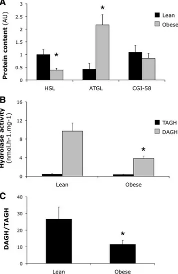
Muscle HSL protein content was significantly lower in obese compared with lean individuals [0.39 ⫾ 0.07 vs. 1.00⫾ 0.19 arbitrary units (AU); ANOVA group-effect
P⫽ 0.004, Fig. 1A]. In contrast to a lower HSL content,
muscle ATGL protein content was significantly higher in obese individuals (2.17 ⫾ 0.40 vs. 0.42 ⫾ 0.23 AU; ANOVA group-effect P⫽ 0.008; Fig. 1A). CGI-58 protein expression was comparable between groups (0.85⫾ 0.19
vs. 1.09⫾ 0.27 AU; P ⫽ 0.484). PLIN was not detectable
in our muscle samples, which excludes potential contam-ination via adipocyte infiltration (data not shown).
In line with a lower HSL protein content, muscle DAGH activity, presumably for a large part reflecting HSL activity, was significantly lower in obese compared with lean men (3.85⫾ 0.48 vs. 9.71 ⫾ 1.73 nmol/h 䡠 mg; group-effect P⫽ 0.004; Fig. 1B). Moreover, DAGH activity was positively correlated with HSL content in skeletal muscle of lean and obese men (r⫽ 0.45 and r ⫽ 0.36, respectively;
P⬍ 0.05). In contrast to a reduced DAGH activity,
skel-etal muscle total TAGH activity was not significantly af-fected in our obese men (obese vs. lean, 0.40⫾ 0.08 vs. 0.50⫾ 0.12 nmol/h 䡠 mg; P ⫽ 0.468; Fig. 1B). Also, HSL-independent TAGH activity, measured after addition of the selective HSL inhibitor BAY [4-isopropyl-3-methyl-
2-[1-(3-(S)-methyl-piperidin-1-yl)-methanoyl]-2H-isoxazol-5-1], was comparable between groups (data not shown). A strong positive correlation between TAGH activity and ATGL content was observed in lean (r⫽ 0.63; P ⬍ 0.01) but not in obese men. The ratio of DAGH to TAGH activity, a marker for complete TAG hydrolysis, was 60% lower in obese individuals (11.4⫾ 2.3 vs. 26.5⫾ 7.3; P ⫽ 0.04; Fig. 1C).
Total muscle TAG content did not differ between lean and obese men (84.7⫾ 18.9 vs. 70.3 ⫾ 12.4mol/mg dry weight; P⫽ 0.543) and correlated positively with TAGH activity (r ⫽ 0.55; P ⬍ 0.05). Moreover, there was no significant difference in total and percentage of saturated fatty acid (SFA), monounsaturated fatty acid (MUFA), and polyunsaturated fatty acid (PUFA) species in TAG across groups. The TAG C16:1 n-7 to C16:0 ratio (esti-mated⌬9-desaturase activity) was greater in obese com-pared with lean men (0.172⫾ 0.017 vs. 0.117 ⫾ 0.009;
P⫽ 0.025). Surprisingly, total DAG (obese vs. lean, 6.2 ⫾
0.7 vs. 9.4 ⫾ 0.9mol/mg dry weight; P ⫽ 0.017) and absolute values of SFA, MUFA, and PUFA species in DAG were lower in muscle from obese men by more than 30%, despite their lower DAGH activity. The percentage of SFA (P⫽ 0.700), MUFA (P ⫽ 0.963), and PUFA (P ⫽ 0.437) in total DAG was comparable between groups. In line, skeletal muscle DAGH activity showed no significant cor-relation with total DAG content in lean and obese men.
Study 2
Obese T2D subjects showed the highest muscle ATGL protein content (0.53 ⫾ 0.06 AU; Fig. 2), whereas nonobese normoglycemic controls showed an intermediate value (0.28 ⫾ 0.05 AU), and the lowest ATGL protein content was observed in nonobese T2D men (0.13 ⫾ 0.04 AU) (ANOVA group-effect P ⫽ 0.021; Fig. 2). Skeletal muscle HSL protein content showed the opposite pattern; post hoc analysis showed
FIG. 1. Skeletal muscle lipase protein content and activity in lean vs.
obese insulin-resistant men (study 1). A, HSL (ANOVA group-effect P⫽ 0.004), ATGL (ANOVA group-effect P⫽ 0.008), and CGI-58 protein content in lean vs. obese men participating in study 1.␣-Actin was used to correct for differences in protein loading. B, TAGH and DAGH activity in skeletal muscle of lean and obese men participating in study 1. C, Ratio of DAGH to TAGH activity was calculated as marker for complete TAG hydrolysis. Values are mean⫾SEM. Post hoc unpaired t test statistics: *, P⬍ 0.05 obese compared with lean.
FIG. 2. Skeletal muscle lipase protein content in obese T2D and
nonobese T2D men (study 2). The graph depicts ATGL (black bars) and HSL (gray bars) protein content in nonobese T2D, healthy normoglycemic controls, and obese T2D (ANOVA group-effect P⫽ 0.021).␣-Actin was used to correct for differences in protein loading. Values are mean⫾SEM. Post hoc unpaired t test statistics: *, P⬍ 0.05, ATGL compared with lean healthy control; **, P⬍ 0.05, HSL compared with lean healthy control.
that HSL content was significantly lower in obese T2D compared with nonobese T2D and normoglycemic con-trols (0.13⫾ 0.05 vs. 0.21 ⫾ 0.03 AU; P ⬍ 0.05; Fig. 2). PLIN was not detectable in our muscle samples, which excludes potential contamination via adipocyte infil-tration (data not shown).
Discussion
The present study shows a lower HSL and higher ATGL protein content in skeletal muscle of obese insulin-resis-tant men. This apparent difference in lipase protein con-tent in skeletal muscle of obese men extends our previous results, which showed a lower HSL content in skeletal muscle of obese men (4). Interestingly, we showed for the first time that this apparent difference in lipase content is related to the obese phenotype because it was observed solely in obese subjects and not in nonobese normoglyce-mic and T2D controls. Moreover, given the close relation-ship between mRNA expression of these two lipases, it might be possible that there is some posttranslational reg-ulatory difference that results in their different relative protein expression, which should be explored in more de-tail in future research (13).
This marked difference in muscle lipase content in obese insulin-resistant men was accompanied by a lower DAGH activity, resulting in a 60% lower ratio of DAGH to TAGH activity, suggesting incomplete TAG hydrolysis (14). This is consistent with our previous observed reduc-tion in HSL serine phosphorylareduc-tion (presumably reflecting lower HSL activity) and lower forearm muscle glycerol release after an overnight fast in obese insulin-resistant compared with lean men (4). Surprisingly, in contrast to a lower DAGH activity, we paradoxically observed no DAG accumulation in skeletal muscle of obese insulin-resistant men compared with the lean group, but rather a 30% reduction. Although numerous studies have shown elevated DAG levels in skeletal muscle from insulin-resis-tant rodents and humans (3, 15), there is actually no con-sensus as to whether this is relevant to the development of skeletal muscle insulin resistance in vivo in humans. Our paradoxical results could be partially attributed by a rapid conversion of DAG back to TAG by DAG acyltrans-ferase-1 (DGAT-1) activity in skeletal muscle of obese in-sulin-resistant men. It has been shown that DGAT-1 over-expression in skeletal muscle decreases DAG and increases TAG content and protects against high-fat diet-induced insulin resistance in rodents (16, 17). However, no differ-ence in muscle DGAT-1 protein content is observed be-tween lean and obese women (18). Alternatively, DAG kinase␦ (DGK␦) can phosphorylate DAG to form phos-phatidic acid, a lipid second messenger. In contrast to an
elevated DGK activity, however, reduced DGK␦ expres-sion and activity has been observed in skeletal muscle from obese T2D patients (19). Our results do not fully preclude the possibility that alterations in DAG species could con-tribute to insulin resistance. However, we also observed lower SFA, MUFA, and PUFA species in DAG in muscle from obese men. Future research should elucidate whether specific DAG species or stereoisomers and its cellular lo-calization are related to the development of skeletal mus-cle insulin resistance.
Because our present result largely judges against DAG, it might be speculated that ceramide accumulation plays a more significant role in human obesity and the develop-ment of insulin resistance (20). Increased skeletal muscle ceramide content, however, has been observed in some (21–23) but not all human studies with obese insulin-re-sistant subjects (24). Mechanistically, funneling off the lipid precursors from the ATGL TAGH step to ceramide formation, or alternatively de novo ceramide synthesis by increasing serine palmitoyl transferase activity are two possibilities that should be investigated in more detail in future research.
In summary, skeletal muscle HSL protein content is lower and ATGL protein content is higher in the obese state. This difference in lipase content is accompanied by a lower DAGH and a normal TAGH activity, implying incomplete lipolysis. Lower HSL and higher ATGL muscle content is confined to obesity per se, because it is solely observed in obese diabetic men but not in nonobese dia-betic and healthy normoglycemic men. Importantly, total DAG content is not elevated in skeletal muscle of obese insulin-resistant subjects and was even lower when com-pared with lean men. Our data do not support the pro-posed key role of muscle total DAG content in the devel-opment of human insulin resistance and suggest that other lipid species (e.g. ceramides) or possibly specific DAG spe-cies and stereoisomers might be of greater importance, at least in obese insulin-resistant men.
Acknowledgments
We greatly appreciate the technical support of Joan Senden and Yvonne Essers.
Address all correspondence and requests for reprints to: Dr. Johan W. E. Jocken, Ph.D., Department of Human Biology, P.O. Box 616, 6200 MD Maastricht, The Netherlands. E-mail: J.Jocken@HB.unimaas.nl.
This work was supported by grants from The Netherlands Organization for Scientific Research.
Disclosure Summary: The authors have nothing to disclose.
References
1. Blaak EE 2003 Fatty acid metabolism in obesity and type 2 diabetes mellitus. Proc Nutr Soc 62:753–760
2. Krssak M, Falk Petersen K, Dresner A, DiPietro L, Vogel SM,
Roth-man DL, Roden M, ShulRoth-man GI 1999 Intramyocellular lipid
con-centrations are correlated with insulin sensitivity in humans: a1H
NMR spectroscopy study. Diabetologia 42:113–116
3. Itani SI, Ruderman NB, Schmieder F, Boden G 2002 Lipid-induced insulin resistance in human muscle is associated with changes in diacylglycerol, protein kinase C, and IB-␣. Diabetes 51:2005–2011 4. Jocken JW, Roepstorff C, Goossens GH, van der Baan P, van Baak
M, Saris WH, Kiens B, Blaak EE 2008 Hormone-sensitive lipase
serine phosphorylation and glycerol exchange across skeletal muscle in lean and obese subjects: effect of-adrenergic stimulation. Dia-betes 57:1834 –1841
5. Jocken JW, Blaak EE 2008 Catecholamine-induced lipolysis in adipose tissue and skeletal muscle in obesity. Physiol Behav 94: 219 –230
6. Zimmermann R, Strauss JG, Haemmerle G, Schoiswohl G,
Birner-Gruenberger R, Riederer M, Lass A, Neuberger G, Eisenhaber F, Hermetter A, Zechner R 2004 Fat mobilization in adipose tissue is
promoted by adipose triglyceride lipase. Science 306:1383–1386 7. Fredrikson G, Belfrage P 1983 Positional specificity of
hormone-sensitive lipase from rat adipose tissue. J Biol Chem 258:14253– 14256
8. Kienesberger PC, Lee D, Pulinilkunnil T, Brenner DS, Cai L, Magnes
C, Koefeler HC, Streith IE, Rechberger GN, Haemmerle G, Flier JS, Zechner R, Kim YB, Kershaw EE 2009 Adipose triglyceride lipase
deficiency causes tissue-specific changes in insulin signaling. J Biol Chem 284:30218 –30229
9. Haemmerle G, Zimmermann R, Hayn M, Theussl C, Waeg G, Wagner
E, Sattler W, Magin TM, Wagner EF, Zechner R 2002
Hormone-sensitive lipase deficiency in mice causes diglyceride accumulation in adipose tissue, muscle, and testis. J Biol Chem 277:4806–4815 10. Bergstro¨m J, Hermansen L, Hultman E, Saltin B 1967 Diet,
mus-cle glycogen and physical performance. Acta Physiol Scand 71: 140 –150
11. Langin D, Dicker A, Tavernier G, Hoffstedt J, Mairal A, Ryde´n M,
Arner E, Sicard A, Jenkins CM, Viguerie N, van Harmelen V, Gross RW, Holm C, Arner P 2005 Adipocyte lipases and defect of lipolysis
in human obesity. Diabetes 54:3190 –3197
12. Folch J, Lees M, Sloane Stanley GH 1957 A simple method for the isolation and purification of total lipides from animal tissues. J Biol Chem 226:497–509
13. Jocken JW, Langin D, Smit E, Saris WH, Valle C, Hul GB, Holm C,
Arner P, Blaak EE 2007 Adipose triglyceride lipase (ATGL) and
hormone-sensitive lipase (HSL) protein expression is decreased in
the obese insulin-resistant state. J Clin Endocrinol Metab 92:2292– 2299
14. Moro C, Galgani JE, Luu L, Pasarica M, Mairal A, Bajpeyi S,
Schmitz G, Langin D, Liebisch G, Smith SR 2009 Influence of
gen-der, obesity, and muscle lipase activity on intramyocellular lipids in sedentary individuals. J Clin Endocrinol Metab 94:3440 –3447 15. Kraegen EW, Saha AK, Preston E, Wilks D, Hoy AJ, Cooney GJ,
Ruderman NB 2006 Increased malonyl-CoA and diacylglycerol
content and reduced AMPK activity accompany insulin resistance induced by glucose infusion in muscle and liver of rats. Am J Physiol 290:E471–E479
16. Liu L, Zhang Y, Chen N, Shi X, Tsang B, Yu YH 2007 Upregulation of myocellular DGAT1 augments triglyceride synthesis in skeletal muscle and protects against fat-induced insulin resistance. J Clin Invest 117:1679 –1689
17. Roorda BD, Hesselink MK, Schaart G, Moonen-Kornips E,
Martínez-Martínez P, Losen M, De Baets MH, Mensink RP, Schrauwen P 2005 DGAT1 overexpression in muscle by in vivo
DNA electroporation increases intramyocellular lipid content. J Lipid Res 46:230 –236
18. Thrush AB, Brindley DN, Chabowski A, Heigenhauser GJ, Dyck DJ 2009 Skeletal muscle lipogenic protein expression is not different between lean and obese individuals: a potential factor in ceramide accumulation. J Clin Endocrinol Metab 94:5053–5061
19. Chibalin AV, Leng Y, Vieira E, Krook A, Bjo¨rnholm M, Long YC,
Kotova O, Zhong Z, Sakane F, Steiler T, Nyle´n C, Wang J, Laakso M, Topham MK, Gilbert M, Wallberg-Henriksson H, Zierath JR
2008 Downregulation of diacylglycerol kinase delta contributes to hyperglycemia-induced insulin resistance. Cell 132:375–386 20. Boden G 2008 Ceramide: a contributor to insulin resistance or an
innocent bystander? Diabetologia 51:1095–1096
21. Adams 2nd JM, Pratipanawatr T, Berria R, Wang E, DeFronzo RA,
Sullards MC, Mandarino LJ 2004 Ceramide content is increased in
skeletal muscle from obese insulin-resistant humans. Diabetes 53: 25–31
22. Straczkowski M, Kowalska I, Baranowski M, Nikolajuk A,
Otziomek E, Zabielski P, Adamska A, Blachnio A, Gorski J, Gorska M 2007 Increased skeletal muscle ceramide level in men at risk of
developing type 2 diabetes. Diabetologia 50:2366 –2373
23. Coen PM, Dube´ JJ, Amati F, Stefanovic-Racic M, Ferrell RE, Toledo
FG, Goodpaster BH 2010 Insulin resistance is associated with higher
intramyocellular triglycerides in type I but not type II myocytes con-comitant with higher ceramide content. Diabetes 59:80 – 88 24. Skovbro M, Baranowski M, Skov-Jensen C, Flint A, Dela F, Gorski
J, Helge JW 2008 Human skeletal muscle ceramide content is not a
major factor in muscle insulin sensitivity. Diabetologia 51:1253– 1260