HAL Id: hal-02292379
https://hal.archives-ouvertes.fr/hal-02292379
Submitted on 16 Nov 2020
HAL is a multi-disciplinary open access
archive for the deposit and dissemination of sci-entific research documents, whether they are pub-lished or not. The documents may come from teaching and research institutions in France or abroad, or from public or private research centers.
L’archive ouverte pluridisciplinaire HAL, est destinée au dépôt et à la diffusion de documents scientifiques de niveau recherche, publiés ou non, émanant des établissements d’enseignement et de recherche français ou étrangers, des laboratoires publics ou privés.
Understanding Active Sites in Pyrolyzed Fe–N–C
Catalysts for Fuel Cell Cathodes by Bridging Density
Functional Theory Calculations and 57 Fe Mössbauer
Spectroscopy
Tzonka Mineva, Ivana Matanovic, Plamen Atanassov, Moulay-Tahar Sougrati,
Lorenzo Stievano, Martin Clémancey, Amélie Kochem, Jean-Marc Latour,
Frédéric Jaouen
To cite this version:
Tzonka Mineva, Ivana Matanovic, Plamen Atanassov, Moulay-Tahar Sougrati, Lorenzo Stievano, et al.. Understanding Active Sites in Pyrolyzed Fe–N–C Catalysts for Fuel Cell Cathodes by Bridging Density Functional Theory Calculations and 57 Fe Mössbauer Spectroscopy. ACS Catalysis, American Chemical Society, 2019, 9 (10), pp.9359-9371. �10.1021/acscatal.9b02586�. �hal-02292379�
1
Understanding active sites in pyrolyzed Fe-N-C catalysts for fuel cell
cathodes by bridging density functional theory calculations and
57Fe
Mössbauer Spectroscopy
Tzonka Minevaa*, Ivana Matanovicb,c, Plamen Atanassovb,d, Moulay-Tahar Sougratia, Lorenzo Stievanoa, Martin Clémanceye, Amélie Kochem,e Jean-Marc Latoure, Frédéric Jaouena*
a
Institut Charles Gerhardt Montpellier, UMR 5253, CNRS, Université Montpellier, ENSCM, 34090 Montpellier, France
b
The Department of Chemical and Biological Engineering, Center for Micro-Engineered Materials (CMEM), University of New Mexico, Albuquerque, NM 87131, USA.
c
Theoretical Division, Los Alamos National Laboratory, Los Alamos, NM 87545, USA
d
Chemical & Biomolecular Engineering and National Fuel Cell Research Center, University of California, Irvine, CA 92697-2580, USA
e
Université Grenoble Alpes CNRS, CEA, DRF/IRIG/LCBM/pmb, 17 rue des Martyrs, 38000 Grenoble, France
emails : tzonka.mineva@enscm.fr, igonzales@unm.edu, moulay-tahar.sougrati@umontpellier.fr , lorenzo.stievano@umontpellier.fr ,
plamen.atanassov@uci.edu, martin.clemancey@cea.fr, amelie.kochem@cea.fr , jean-marc.latour@cea.fr, frederic.jaouen@umontpellier.fr
*
2
Abstract
Pyrolyzed Fe-N-C materials are promising platinum-group-metal free catalysts for proton-exchange membrane fuel cell cathodes. However, the detailed structure, oxidation and spin states of their active sites is still undetermined. 57Fe Mössbauer spectroscopy has identified FeNx moieties as the most active sites, with their fingerprint being a doublet in
low-temperature Mössbauer spectra. However, the interpretation of the doublets for such materials has lacked theoretical basis. Here, we applied density functional theory to calculate the quadrupole splitting energy of doublets (EQS) for a range of FeNx structures in different
oxidation and spin states. The calculated and experimental values are then compared for a reference Fe-N-C catalyst, while further information on the Fe oxidation and spin states was obtained from electron paramagnetic resonance, superconducting quantum interference device and 57Fe Mössbauer spectroscopy under external magnetic field. The combined theoretical and experimental results identify the main presence of FeNx moieties in Fe(II) low-spin and
Fe(III) high-spin states while a minor fraction of sites could exist in Fe(II) S = 1 state. From the analysis of the 57Fe Mössbauer spectrum under external magnetic field and the comparison of calculated and measured EQS values, we assign the experimental doublet D1 with mean
EQS value around 0.9 mm·s-1 to Fe(III)N4C12 moieties in high spin and the experimental
doublet D2 with mean EQS value around 2.3 mm·s-1 to Fe(II)N4C10 moieties in low and
medium spin. These conclusions indicate that D1 corresponds to surface-exposed sites while D2 may correspond either to bulk sites that are inaccessible to O2 or to surface sites that bind
O2 weaker than D1.
Keywords
Density functional theory, Mössbauer spectroscopy, quadrupole energy splitting, FeN4,
3 1. Introduction
57
Fe Mössbauer spectroscopy, based on the recoil-free emission and resonant absorption of -rays, has been extensively used to fingerprint the local electronic structure, magnetic properties and coordination of iron nuclei in different classes of important materials ranging from (extra) terrestrial minerals and biomolecules containing Fe in their active site, to industrial catalysts such as those used for Fischer-Tropsch or ammonia synthesis.1-4 More recently, it has gained growing importance in the characterization of novel platinum-free catalysts for application in fuel cell cathodes for the identification of Fe-based species present in pyrolyzed Fe-N-C catalysts.5-17. Due to their synthetic approach involving a high temperature treatment, such catalysts often comprise a plurality of Fe-based structures, including not only different coordination geometries of atomically-dispersed Fe ions covalently bound to the nitrogen-doped carbon matrix (FeNxCy moieties), but also metallic
iron and iron carbides with long-range order. Compared to other spectroscopic methods and to X-ray absorption in particular, 57Fe Mössbauer spectroscopy allows an easier separation of the signals arising from the different Fe environments. Namely, for spectra recorded in the absence of a strong external magnetic field, metallic iron and iron carbides result in sextets (except for the paramagnetic γ-Fe structure, leading to a singlet) while the fingerprint of FeNxCy moieties is a quadrupole doublet. Its two Mössbauer parameters are the isomer shift
() and the quadrupole splitting energy (EQS).18-20 These two parameters originate from
hyperfine interactions between the iron nucleus and the surrounding electronic environment, and vary therefore with the iron oxidation state, spin state, and its chemical surrounding.21 Due to the clear different shape and Mössbauer parameters of the spectral signals arising from different Fe structures, the fitting of the experimental spectrum of any Fe-N-C catalyst with a number of spectral components (singlets, doublets and sextets) is usually relatively straightforward and gives direct quantitative information on the fraction of Fe present in different forms. However, while the spectral parameters found for the singlet and sextet components identified in the spectra of pyrolyzed Fe-N-C catalysts perfectly match with those known for the well-established Fe crystalline structures (metallic Fe, Fe carbides, Fe nitrides), leading to unambiguous assignments, the interpretation of the spectral parameters fitted for the doublets in such materials have so far lacked strong bases, both from the experimental and theoretical sides. The lack of appropriate reference materials (materials with a single type of FeNxCy moieties in a carbon matrix, with the local structure around Fe atoms being
4 independently determined) prohibits making unambiguous assignments on the experimental side. The Mössbauer spectral parameters of some Fe macrocycles with known structure (iron phthalocyanines and porphyrins, pure or adsorbed on carbon surfaces) have hitherto been used to make assignments of the various doublets typically observed in pyrolyzed Fe-N-C materials to ferrous or ferric iron in different spin states.6 The assumption that such macrocycles may serve as proper models for all or some of the FeNxCy moieties present in
pyrolyzed Fe-N-C materials is however too speculative, for several reasons. First, the size of these reference molecules is much smaller than the average in-plane size of graphitic crystallites in pyrolyzed Fe-N-C materials (typically 2 nm and above). Second, such macrocycles with FeN4 core moiety involve pyrrolic nitrogen atoms engaged in
five-membered rings, while perfectly graphitized materials comprise only six-five-membered hexagonal rings. Experimental proof for the existence of atomically-dispersed Fe atoms in a nitrogen-doped carbon matrix has recently been confirmed by high-resolution electron microscopy, both in amorphous carbons (comprising a significant fraction of non-hexagonal rings) and in perfectly ordered graphene sheets (comprising only hexagonal rings).22-23 Moreover, even for an Fe-N-C model catalyst with amorphous carbon matrix and comprising only atomically-dispersed Fe atoms and whose XANES and EXAFS spectra could be fitted with a single porphyrinic (FeN4C12) structure, the fitting of its Mössbauer spectrum still
required two distinct doublets, at least.10 Third, the FeNxCy moieties in pyrolyzed materials
are integrated in a conductive carbon matrix, which may completely change the electron density at the Fe nucleus relative to an adsorbed Fe macrocycle, even for a similar local coordination. As discussed below, the electron-density at the Fe nucleus is a key in determining the spectral Mössbauer parameters. Hitherto, all pyrolyzed Fe-N-C catalysts comprising FeNxCy moieties have shown at least two distinct doublets in their Mössbauer
spectra, often labeled D1 and D2. Their reported values of EQS at room temperature are in
the range of 0.90-1.25 and 2.0-2.8 mm·s-1, respectively, while their isomer shifts are comparable and non-discriminating (0.30-0.45 mm·s-1).24 The doublet D1 has, most frequently, been empirically assigned to an Fe(II) zero spin (S=0) moiety,6, 9 but also sometime to Fe(III) in high spin (S=5/2).5, 8 The doublet D2 has been empirically assigned either to an distorted Fe(II) moiety in low spin, Fe(II) medium spin (S=1), Fe(III) low spin (S=1/2) or Fe(III) medium spin (S=3/2) moiety.6, 8-9 These empirical attributions are however not yet supported by any theory or independent experimental method. This uncertainty casts a doubt on past interpretation of the 57Fe Mössbauer spectra of such materials and on
structure-5 property correlations, such as activity or selectivity with respect to the fraction or absolute content of a given doublet component.
For all the reasons discussed above, it appears obvious that the detailed analysis of the 57Fe Mössbauer spectra of Fe-N-C materials cannot lean only on the experimental spectra of known macrocycles and that a complementary theoretical approach is required to interpret the Mössbauer spectra of such materials. No theoretical study has hitherto focused on predicting the two Mössbauer spectral parameters of FeNxCy moieties in such materials. The first
parameter, the isomer shift, , is in the specific case of 57Fe inversely proportional to the density of s-electrons at the absorbing nucleus. It can be computed from a molecular orbital method and reported relative to the isomer shift of a reference compound25-28. In this work, we focused our DFT efforts in calculating EQS, while methodologies to calculate were not
investigated, due to the non-discriminating values of for the experimental doublets D1 and D2 in Fe-N-C catalysts. The second parameter, EQS, can be computed from the nuclear
quadrupole moment and the electric field gradient at the absorbing 57Fe nucleus. In recent years, calculation of the and EQS values for several Fe coordination geometries have
become straightforward with computer programs based on the Quantum Chemistry Density Functional Theory (DFT), using either localized molecular orbital (cluster)27-38 or periodic approaches39-41. Despite the apparent easiness of calculating values of and EQS, the use of
DFT as a tool for experimental spectral interpretation is hampered by the strong dependence of the calculated values for and, in particular, for EQS on (i) the DFT exchange-correlation
(XC) functional and (ii) the chosen parameterized set of wave functions (basis set)29-38, 40. Recent DFT studies proposed different combinations of functionals and bases in order to achieve good agreement between experimental and computed Mössbauer parameters in ferrocene29, Fe-porphyrin32, -diketiminate31 derivatives and other iron complexes.33, 35-36, 38 Failures of DFT to reproduce the experimental EQS values were reported for other
compounds.33-35, 38 A study on computed results obtained for a unique Fe coordination but resorting to eight different XC functionals and two basis sets revealed that the mean error between computed and experimental EQS was 0.2 mm·s-1 for EQS < 2 mm·s-1, and 0.4 mm·s -1
for EQS > 2mm·s-1, excluding obvious outliers cases30. While no particular issues arise with
zero-spin iron entities (S=0), more difficulties may be found in the case of degenerate ground states, or closely spaced low-lying energy states of medium (S=1) or high spin (S=2) iron complexes with square-planar coordination30, 33. Other sources of error that have been pointed
6 out are, e.g. the adequacy of the geometrical model or the quality of the optimized structures as cluster or extended periodic structures.31, 33, 40
The first object of this study is to explore the ability of DFT methodologies to predict EQS
values for a range of hypothetical FeN4 structures with different cluster size, metal-oxidation
and spin state. For this purpose, the two most accepted sub-families of ORR-active FeN4
-moieties present in pyrolyzed Fe-N-C materials were modeled, namely the FeN4C10 and
FeN4C12 sub-groups10, 22, 42. The results of this theoretical study are then combined with the
experimental Mössbauer data recorded for a well-defined Fe-N-C catalyst containing only atomically-dispersed iron species. To better discriminate the spin state and oxidation state of iron moieties, the catalyst was characterized also by Mössbauer spectroscopy in the presence of an external magnetic field, superconducting quantum interference device (SQUID) and electron paramagnetic resonance (EPR).
Mössbauer spectroscopy in the presence of an external magnetic field unambiguously reveals the major presence of low-spin ferrous centers accompanied by high-spin ferric centers in the Fe-N-C catalyst. The presence of high-spin ferric centers is independently supported by EPR measurements. Combined with the theoretical insights, we are able to conclude that the identified high-spin ferric signal can be assigned to Fe(III)N4C12 porphyrinic moieties,
corresponding to the experimental doublet D1 (EQS in the range 0.6-1.3 mm·s-1). The
low-spin ferrous signal is assigned to Fe(II)N4C10 pyridinic moieties, corresponding to the
experimental doublet D2 (EQS in the range 1.7-3.1 mm·s-1). A minor fraction of the
Mössbauer signal with EQS values falling in the intermediate region situated between 1.3 and
1.7 mm·s-1 is ambiguous and may be assigned either to high-spin ferric moieties (Fe(III)N4C10) or low-spin ferrous moieties (Fe(II)N4C12). The combination of the present
DFT study with experimental 57Fe Mössbauer spectroscopy under external magnetic field and other independent methods therefore leads to a detailed and rational interpretation of the spectroscopic signatures and electronic states of FeNxCy moieties in Fe-N-C materials.
7
2. Computational and Experimental Methods 2.1 Computational Method
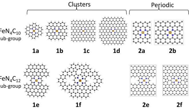
DFT calculations were carried out with the cluster and periodic approaches using, respectively, deMon2k43 and VASP44-47 computer programs on graphene sheets integrating various moieties from the FeN4C10 and FeN4C12 sub-groups (Figure 1).
Figure 1.Cluster and periodic ferrous models of FeN4Cy (y = 10 or 12) moieties considered in this work as
representative Fe sites in Fe-N-C materials. The optimized structures at PBE/DZVP2 (1a-1f) and PBE-PBC (2a,b,e,f) level of theory are presented. See text for the differences in the optimized geometry parameters with the other methods. The atomic color code is the following: brown for Fe; blue for N; dark grey for C, light grey for H.
The ferrous FeN4C10 and FeN4C12 clusters are modeled, respectively, with 1a - 1d and 1e -1f
in Figure 1. The dangling bonds in all clusters and also in the periodic 2e system were saturated with hydrogen atoms. To model the respective ferric moieties, OH- is adsorbed in end-on mode on the ferric site in order to preserve the electric-charge neutrality. The resulting OH-Fe(III)N4Cy moieties (labeled e.g. as 1a-OH) are exemplified in Figure S1. The electrons
in the C, N and H atoms are described by triple-basis sets48. For the iron electrons we examined the performance of the double- plus polarization48 and the original Wachter's49 bases, contracted to (62111111/331211/3111). Three generalized gradient corrected (GGA) exchange-correlation functionals were tested: (1) the revised Perdew−Burke−Ernzerhof’s
1a
1b
1c
1d
2a
2b
Clusters
Periodic
1e
1f
2e
2f
FeN
4C
10 sub-groupFeN
4C
12 sub-group8 exchange50-51 and the Lee, Yang and Parr correlation52 (rPBE-LYP); (2) the Perdew−Burke−Ernzerhof’s exchange-correlation (PBE) and (3) the OPTX exchange of Handy and Cohen53 coupled to the Lee, Yang and Parr correlation52 (OLYP). The geometries of the clusters 1a and 1e were fully optimized at every level of theory. Larger models (clusters
1b-1d, 1f) up to the size of the experimental graphitic crystallites in high surface-area Fe-N-C
materials (~2 nm) were fully optimized with rPBE-LYP/DZVP2, PBE/DZVP2 and PBE/Wachter's. No symmetry constraints were imposed. In deMon2k code, automatically generated auxiliary functions up to l = 2 (for the metal atom) and 3 (for H, C and O atoms) were used for fitting the density with the GGA functionals.54 The GGA functionals were also coupled to an empirical dispersion (D) term.55 A quasi-Newton method in internal redundant coordinates with analytical energy gradients was used for structure optimization. For the numerical integrations of the XC energy and potential, we used an adaptive grid with tighten threshold (10-8 a.u.).56 The convergence was based on the Cartesian gradient and displacement vectors with thresholds of 10-3 a.u. and the energy convergence was set to 10-7 a.u.
DFT calculations with periodic boundary conditions, carried out with VASP code, used the models 1a and 1e to build the unit cells. In the case of model 1a, two unit cells were considered; one with the size of 9.94 Å x 12.64 Å and the other 14.78 Å x 12.80 Å (models 2a and 2b in Figure 1, respectively). In the case of the cluster model 1e, the corresponding periodic structure was modeled using either a cell size 17.18 Å x 20.87 Å (model 2e) or 17.17 Å x 21.0Å (model 2f). Vacuum region of 15 Å was introduced in the z-direction in order to eliminate interactions between the graphene sheet and its periodic images. All the DFT calculations with periodic boundary conditions (PBC) were performed using generalized gradient approximation (GGA) with PBE exchange-correlation functional50 and VASP 5.2 recommended GW projector augmented-wave pseudopotentials.57-58 For Fe pseudopotential that treats 3s and 3p states as valence states was used. Electric field gradients at the positions of the Fe nuclei were calculated using the method reported in reference39 as implemented in VASP. To obtain electric field gradients cut-off energy for the plane wave basis set of 800 eV, break condition for electronic SC-loop of 10-6 eV and 8x8x1 gamma centered mesh were used for the model 2a and 4x4x1 mesh for the model 2b and 2e-2f. In all cases, the Fermi-Dirac smearing method with sigma set to 0.03 was used. In addition, all the calculations included support grid for the evaluation of the augmentation charges.
9 The quadrupole splitting energy is computed as the coupling between the nuclear quadrupole moment (Q) of the non-spherical nucleus and the principal components Vii(i = x, y, z) of the
electric field gradient (EFG) tensor at 57Fe nucleus using the equation:
(1)
In the EQ equation, e is the charge of the electron and the asymmetry parameter is
computed as where . The nuclear quadrupole moment, Q, for the J=3/2 state is taken to be 0.16 barn. Computation of EQ and therefore
becomes a question of computing the EFG tensor, which is readily obtained as an expectation
value of the EFG operator, 5 0 2 0 3 r r ij
Vij , for the electronic ground state 0 and i, j =
x,y,z being the components of the electron radius vector r. In the remainder, the calculated value of EQ are reported in units of mm·s-1 (labeled as EQS), for a direct comparison to
experimentally reported values.
2.2 Experimental Methods
Synthesis of the Fe0.5 -NC catalyst
The Fe-N-C catalyst, labelled Fe0.5-NC, was prepared from FeII acetate, 1,10-phenanthroline
and a ZnII zeolitic imidazolate framework (ZIF-8 with formula ZnN4C8H12, purchased from
BASF under trademark Basolite Z1200). One gram of a catalyst precursor containing 0.5 wt% Fe with a mass-ratio phen/ZIF-8 of 20/80 was prepared by dry ball-milling. The milling was carried out in a ZrO2 crucible with 100 ZrO2 balls of 5 mm diameter using a planetary
ball-miller (FRITSCH Pulverisette 7 Premium) for 4 cycles of 30 min at 400 rpm. The resulting catalyst precursor was then pyrolyzed at 1050 °C in Ar for 1 h, leading to Fe0.5-NC. This
synthesis has been previously shown by Mössbauer spectroscopy and XANES-EXAFS to result in an ORR-active catalyst that comprise only atomically-dispersed FeNxCy moieties.10
A reference sample free of iron or only with trace iron, labelled NC, was prepared identically as Fe0.5-NC except that no FeII acetate was added in the catalyst precursor.
Measurement of the Mössbauer spectra
57
Fe Mössbauer spectra of Fe0.5-NC were measured with a 57Co:Rh source kept at ambient
10 absorber at variable temperatures in the range 5.5-296 K using a Janis CCR 5 K cryostat. Additional measurements were performed in the presence of an external magnetic field applied either parallel or perpendicular to the -rays with the absorber at 4.5 K on a strong-field Mössbauer spectrometer equipped with an Oxford Instruments Spectromag 4000 cryostat containing an 8 T split-pair superconducting magnet. Both spectrometers were operated with a triangular velocity waveform, in a constant acceleration mode in transmission geometry, and were calibrated at 295 K with an α-iron foil. The isomer shift scale is referred to metallic α-Fe at room temperature.
Analysis of the Mössbauer spectra
The broadened quadrupole components of the Mössbauer spectra measured in the absence of a magnetic field were fitted with appropriate superposition of Lorentzian lines (fixed linewidth of 0.25 mm·s-1), assuming Gaussian distributions of the electric quadrupole interaction for each spectral component. Up to three independent components were sometimes necessary to fit coherently the series of spectra acquired on Fe0.5-NC at different temperatures.
In order to fit the complex experimental spectra observed in the presence of an external magnetic field (shown later), simple histogram distributions of magnetic sextets with hyperfine fields up to 50 T were employed. Mössbauer spectra of the same Fe-N-C sample measured with a magnetic field applied parallel or perpendicular to the gamma rays were fitted simultaneously with a single distribution of hyperfine fields. For each hyperfine field considered in the distribution, the only difference being theoretically expected when changing from a parallel to a perpendicular external magnetic field is the relative intensity of the six Mössbauer lines, which is 3:0:1:1:0:3 or 3:4:1:1:4:3, respectively, for the lines 1 to 6, as labelled from the lowest to the highest position of the lines on the x-axis velocity range of Mössbauer spectra. Therefore, during the fitting, only the relative intensity of the lines 2 and 5 were fitted as a function of the direction of the external magnetic field, since it is known that the relative intensity of the lines 1, 3, 4 and 6 do not vary with the direction of the external magnetic field. As will be shown later, this fitting approach can however not result in a perfect fit in all regions of the spectra, since species characterized by low magnetic fields and high quadrupole splittings, the quadrupole interaction cannot be physically treated as a perturbation of the Zeeman magnetic interaction, and thus the Mössbauer components cannot be rigorously fitted simply using magnetic sextets.59
11 The molar magnetic susceptibility χm was measured from 2 to 300 K for Fe0.5-NC and NC
with a Superconducting QUantum Interference Device (SQUID) (MPMS XL-7T, Quantum Design) at a magnetic field of 5,000 Oe. A mass of 16 mg of powder sample was introduced in a polymer straw. The average effective magnetic moment of iron atoms (µeff) was then
obtained by fitting the plot of 1/χm (in mol of iron atoms per emu) vs. 1/T with a linear law.
Electron paramagnetic resonance (EPR)
EPR spectra were acquired at 9.65 GHz. A few mg of the sample Fe0.5-NC was introduced in
a glass capillary. The spectra were acquired at 10 K for different power/amplitude ratios. The optimum power and amplitude were found to be 3 mW and 0.3 mT, respectively.
3. Results
3.1. Theoretical study
We first discuss the impact of various exchange correlation functionals and basis sets on calculated values of EQS. This analysis is carried out for two ferrous moieties representative
for the FeN4C10 and FeN4C12 sub-groups. Subsequently, the effects of the cluster size and iron
oxidation state on EQS are investigated and discussed.
Effect of exchange-correlation functionals and basis sets on calculated values ofEQS
In this section, we focus on the smallest 1a and 1e clusters and their periodic 2a and 2e,f analogs, with Fe in oxidation state II, in low or medium spin state. The high spin state S = 2 was not considered since it is well known to result in an isomer shift ≥ 0.9 mm∙s-1,60 a value that has never been unambiguously experimentally observed in pyrolyzed Fe-N-C catalysts hitherto6. Note that the geometries were optimized without imposing any broken D4h
symmetry.10 All the DFT methods reveal that the electronic state with spin S = 0 is slightly less favored from an energy viewpoint, as follows from the positive values of the relative energy, ΔES=0, in Table 1. In the cluster calculations of EQS, the closed-shell electronic
structures (S = 0) have a relatively low sensitivity toward a particular DFT method. For the 1a cluster, EQS is in the range 2.4-3.1 mm·s-1 (Table 1, row No. 1-6) and for cluster 1e in the
range 1.6-2.0 mm·s-1 (row No. 8-13). The differences between EQS values obtained with the
three functionals for a given basis set is within 0.4 mm·s-1, and between the two bases for a given functional is within 0.3 mm·s-1. Accepting that differences of 0.4 mm·s-1 for EQS ≥ 2
12 mm·s-1 and 0.2 mm·s-1 for EQS ≤ 2 mm·s-1 are within the computational error,30 we conclude
on a good agreement between the different methods in calculated EQS valuesfor the low-spin
clusters. A good agreement is also observed between the low-spin cluster and periodic (PBE/PBC) approaches (e.g. for structure 2a, compare row No. 7 to rows 1-6).
In contrast, for the open-shell cluster structures (S = 1), the distribution of EQS values is
significantly larger. In the 1a cluster, EQS varies between 1.5 and 3.3 mm·s-1 (Table 1, row
No. 1-6) while EQS in the corresponding periodic structure 2a is 3.3 mm·s-1 (row 7). In the
medium-spin 1e cluster (FeN4C12), EQS varies between 0.1 and 1.6 mm·s-1 (rows 8-13) while
EQS in the periodic structures 2e-2f is 1.8 mm·s-1 (rows 14-15). The main outliers are
however obtained with rPBE-LYP functionals (rows 1-2; 8-9), whereas the other two functionals (PBE and OLYP) result in much closer EQS values (row No. 3-6 and 10-13). The
PBE and OLYP functionals with the DZVP2 basis set result in an excellent agreement of
EQS with the periodic approach, in the range of 2.9-3.3 mm·s-1 for models 1a and 2a (row
No. 3, 5, 7) and in the range of 1.6-1.8 mm·s-1 for models 1e and 2e-2f (row No. 10, 12, 14-15).
13
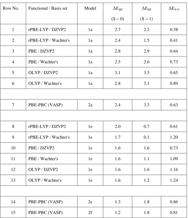
Table 1. Quadrupole splitting energy EQS (in mm·s
-1) and relative energy ΔE (in eV) computed with three DFT-GGA functionals and two basis sets for Fe(II)N4C10cluster(1a) and periodic (2a) models, and Fe(II)N4C12 cluster (1e) and periodic (2e and 2f) models. The models structures are shown in Figure 1. Low (S=0) and medium (S=1) spin states are considered. The relative energies are computed from the total energies (Etot) as ΔES=0 =
Etot(S=0) - Etot(S=1).
Row No. Functional / Basis set Model ΔEQS
(S = 0) ΔEQS (S = 1) ΔES=0 1 rPBE-LYP / DZVP2 1a 2.7 2.2 0.38 2 rPBE-LYP / Wachter's 1a 2.4 1.5 0.41 3 PBE / DZVP2 1a 2.8 2.9 0.64 4 PBE / Wachter's 1a 2.5 2.6 0.73 5 OLYP / DZVP2 1a 3.1 3.3 0.65 6 OLYP / Wachter's 1a 2.8 3.1 0.89 7 PBE-PBC (VASP) 2a 2.4 3.3 0.63 8 rPBE-LYP / DZVP2 1e 2.0 0.7 0.61 9 rPBE-LYP / Wachter's 1e 1.7 0.1 1.20 10 PBE / DZVP2 1e 1.6 1.6 0.73 11 PBE / Wachter's 1e 1.6 1.1 1.09 12 OLYP / DZVP2 1e 1.6 1.6 1.16 13 OLYP / Wachter's 1e 1.6 1.2 1.24 14 PBE-PBC (VASP) 2e 1.3 1.8 0.86 15 PBE-PBC (VASP) 2f 1.2 1.8 0.81
The larger discrepancy in the S = 1 state relative to low-spin state is not unexpected, since the EFG values depend on the ground-state electronic structure, namely on the occupation of the 3d orbitals of Fe33. The large EQS differences seen for DFT methods in the medium-spin
14 electronic structure in iron compounds29, 32-33. When the symmetry is not imposed, as in our calculations, the degeneracy of 3d-spin-orbitals is removed and the occupation of the 3d orbitals becomes highly dependent on the particular XC functional and on the chosen basis sets9,12. In the present study, this general finding is evidenced through the comparison of the occupation of the 3d orbitals below the Fermi level in cluster 1a and periodic 2a models (see Table S1). A significantly lower occupation of the dz2 orbital in cluster 1a with rPBE-LYP/Wachters than that with all other functionals/basis sets is observed. On the other hand, these different Fe electronic states are in geometrically-equivalent structures (see supporting Tables S2-S3 and related text). Thus, we conclude that the larger discrepancy for S = 1 spin state is due to the difficult description of the occupation of the 3d orbitals, in agreement with previous theoretical studies29-38.
Influence of cluster size on calculated values ofEQS
To gain understanding in the importance of the edge or in-plane location of the iron center in a graphene plane, we calculated EQS for larger cluster sizes than those of structures 1a and
1e, up to 2.5 nm (models 1b,c,d and 1f in Figure 1). This value resembles the typical size of
graphene crystallites in Fe-N-C materials61.Periodic boundary conditions were also imposed to the corresponding larger unit cells (models 2b and 2f in Figure 1) in order to evaluate the effect of the distance between two adjacent iron sites. The PBE functional was chosen because it performs similarly to OLYP and is also used in the periodic computations. The changes of EQS values with cluster size are depicted in Figure 2 while the exact values are
15 0,0 0,5 1,0 1,5 2,0 2,5 3,0 3,5 DZVP2 Wachters PBC 1f 1e 1d 1c 1a
E
QS(
mm
s
-1)
1b 1a 1b 1c1d 2a 2b 1e 1f 2e 2f PBC Wachters DZVP2a
0,0 0,5 1,0 1,5 2,0 2,5 3,0 3,5 DZVP2 Wachters PBC 1f 1e 1d 1c 1a
E
QS(
mm
s
-1)
1b 1a 1b 1c1d 2a 2b 1e 1f 2e 2f PBC Wachters DZVP2b
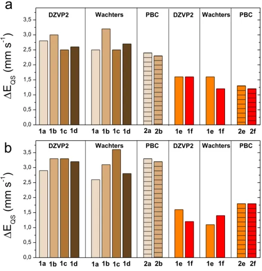
Figure 2. Quadrupole splitting energies, EQS, as a function of cluster size, 1a-1f, and unit cells, 2a-2b,2f, of
FeN4C10 and FeN4C12 ferrous models with low (S = 0) spin (Fig. 2a) and medium (S = 1) spin (Fig. 2b). The results are obtained with PBE functional in the periodic (PBC) and cluster (with DZVP2 or Wachters basis) approaches. Columns for periodic approaches are hatched.
The magnitude of changes in EQS with cluster size is close to the computational error bar for
the FeN4C10 (0.4 mm·s-1) and FeN4C12 groups (0.2 mm·s-1), and only slightly above the error
bar for few cases. This finding correlates well with the very small changes in the Fe-N bond distances and N-Fe-N bond angles in models 1a to 1d (Table S5). Indeed, the fluctuation of the bond distances with cluster size is restrained to a maximum of 0.05 Å for all considered methods and spin states. Regarding bond angles, for S = 0, the trends with cluster size from
1a to 1d are similar with the PBE/Wachters and PBE/DZVP2 methods, showing a reduced
spreading of the four Ni-Fe-Nj bond angles relative to 90°. Thus, as the cluster size is
increased, the FeN4 core becomes closer to a D4h symmetry group. We also note that FeN4C12
16 a nearly identical EQS (see Figure 2, two last columns). Therefore one may conclude that
EQS is mainly sensitive to the spatial arrangement of nitrogen and carbon atoms in the two
first coordination spheres, while the ordering of atoms at longer distances negligibly impacts
EQS.
Effect of Fe oxidation state on calculated values ofEQS
Operando X-ray absorption spectroscopy studies systematically evidenced that pyrolyzed Fe-N-C catalysts contain a significant fraction of ferric moieties in their resting state,14 including the model Fe-N-C catalyst further experimentally characterized in the present study10. Therefore, ferric clusters were considered, and modeled by adsorbing an hydroxide anion to Fe(III). The models are presented in Figure S1, labeled 1a-OH and 1d-OH (FeN4C10) and
1e-OH and 1f-1e-OH clusters (FeN4C12). The quadrupole splitting energies are computed with the
PBE and rPBE-LYP functionals. The latter was included because it resulted in the main outliers for the ferrous clusters 1a and 1e in medium spin (Table 1) and we wanted to examine its performance in the ferric compounds. Table 2 reports EQS values and relative energies for
these four clusters and two periodic models (2a-OH, and 2e-OH), at three different spin states. For each cluster, the agreement between the DFT methods is excellent (see rows 1-3; 4-5; 7-9; 10-11). The rPBE-LYP and PBE functionals, therefore, lead to similar results for ferric models, in contrast to the case with ferrous models. For the low-spin ferric structures, the three DFT methods agree well and give EQS between 2.1-2.5 mm·s-1 in 1a-OH, 1d-OH,
2a-OH (FeN4C10 sub-group) and EQS between 1.8-2.1 mm·s-1 in 1e-OH, 1f-OH and 2e-OH
(FeN4C12 sub-group). However, the spread of the results is very large in the medium-spin
state. EQS values are between 0.9-2.5 mm·s-1 in 1a-OH, 1d-OH, 2a-OH and between 0.7-2.0
mm·s-1 in 1e-OH, 1f-OH and 2e-OH. In particular, the significant difference in the calculated
EQS value for the large 1d cluster (2.5 or 2.3 mm·s-1) and the periodic 2a model (1.1 mm·s-1)
is surprising. For the high-spin state, the agreement for the 1a-OH and 1d-OH clusters is excellent but a discrepancy again occurs with the periodic 2a-OH model. While more restrained compared to the case with the medium spin-state, some spreading of the results remains among 1e-OH, 1f-OH and 2e-OH (porphyrinic sub-group), with EQS values in the
17
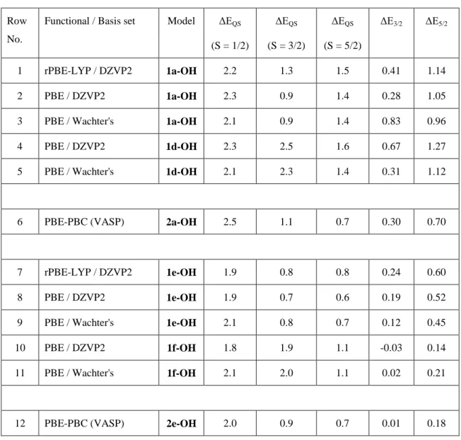
Table 2. Quadrupole splitting energy EQS (in mm·s
-1) and relative energy ΔE (in eV) computed with two DFT-GGA functionals and two basis sets for Fe(III)N4C10 and Fe(III)N4C12 cluster and periodic models. The model structures are shown in Figure S1. Low (S=1/2), medium (S=3/2) and high (S=5/2) spin states are considered. The relative energies are computed from the total energies (Etot) as ΔEn/2 =Etot(S=n/2) - Etot(S=1/2) with n=3/2 and 5/2.
Row No.
Functional / Basis set Model ΔEQS (S = 1/2) ΔEQS (S = 3/2) ΔEQS (S = 5/2) ΔE3/2 ΔE5/2 1 rPBE-LYP / DZVP2 1a-OH 2.2 1.3 1.5 0.41 1.14 2 PBE / DZVP2 1a-OH 2.3 0.9 1.4 0.28 1.05
3 PBE / Wachter's 1a-OH 2.1 0.9 1.4 0.83 0.96
4 PBE / DZVP2 1d-OH 2.3 2.5 1.6 0.67 1.27
5 PBE / Wachter's 1d-OH 2.1 2.3 1.4 0.31 1.12
6 PBE-PBC (VASP) 2a-OH 2.5 1.1 0.7 0.30 0.70
7 rPBE-LYP / DZVP2 1e-OH 1.9 0.8 0.8 0.24 0.60
8 PBE / DZVP2 1e-OH 1.9 0.7 0.6 0.19 0.52
9 PBE / Wachter's 1e-OH 2.1 0.8 0.7 0.12 0.45
10 PBE / DZVP2 1f-OH 1.8 1.9 1.1 -0.03 0.14
11 PBE / Wachter's 1f-OH 2.1 2.0 1.1 0.02 0.21
12 PBE-PBC (VASP) 2e-OH 2.0 0.9 0.7 0.01 0.18
We therefore examined if OH-adsorption led to distinct geometric changes as a function of the structural models and functional/basis set chosen, in particular for the ferric models in medium spin state. It is first noted that OH- adsorption on Fe leads to a non-planar geometry. In all cases, Fe(III) is situated above the graphene plane by 0.2-0.3 Å (calculated, but not reported in Tables). The geometric parameters of the ferric FeN4C10 and FeN4C12 groups are
reported in Tables S6 and S7, respectively. While the spreading of the Fe-N bond distances and N-Fe-N angles in the ferric models is larger than in the corresponding ferrous models, the structural disorder caused by the OH adsorption is comparable for all models (see for example
18
1a-OH and 1d-OH in medium spin state in Table S6), Thus, the large EQS differences
obtained for 1a-OH and 1d-OH on one hand, and 1e-OH and 1f-OH on the other hand, cannot be attributed to the slight structural changes induced by OH-adsorption. In contrast, there might be a distinct OH orientation, depending on the model and spin-state, which could influence the electric field gradient components at the iron nucleus. Analyzing the minimum energy ferric structures, we could distinguish different OH orientations with respect to the N-Fe-N plane. This can be quantified through the dihedral H-O-N-Fe-N angle, , which defines the angle between the H-O-Fe and the N-Fe-N planes (Figure S2). For O-H pointing above the N atom, the H-O-Fe-N dihedral angle is 0° and for O-H pointing above the N-Fe-N bisector, the H-O-Fe-N dihedral angle is ~45°, as exemplified in Figure S2. The calculated values are summarized in Tables S6, S7.
For the ferric FeN4C10 clusters in low-spin, varies only between 8-11°, in line with the
similar EQS values calculated for all those structures. In contrast, in medium spin a large
difference is observed for 1a-OH and 1d-OH models. The values are between 27°-33° in
1a-OH and 9° in 1d-OH. The comparison with trends for EQS values evidences a clear
correlation between the and EQS values for these medium-spin ferric models. For the high
spin models, spans between 17 and 34°. We therefore identify an inverse correlation between and EQS for FeN4C10 models in any spin state. Structures with values between
8-11° have high EQS in the range 2.0-2.5 mm·s-1, whereas those with larger have low EQS
in the range 0.9-1.6 mm·s-1.
A correlation between OH orientation and EQS is established also for the porphyrinic
FeN4C12 moieties (Table S7). In this case, the correlation between and EQS is seemingly
inversed. The structures with low (~1° in 1e-OH, medium- and high-spin states) have low
EQS values between 0.6 - 0.8 mm·s-1 and the structures with high ~9-21°in1e-OH, low
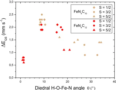
19 0 10 20 30 40 0,0 0,5 1,0 1,5 2,0 2,5 3,0 FeN4C12 S = 1/2 S = 3/2 S = 5/2 S = 1/2 S = 3/2 S = 5/2 E QS (mm s -1 )
Diedral H-O-Fe-N angle
FeN4C10
Figure 3. Calculated EQS values for ferric moieties as a function of the dihedral O-H-Fe-N angle () for the
low, medium and high spin-states in the ferric FeN4C10 (1a-OH and 1d-OH) and FeN4C12 (1e-OH and 1f-OH) cluster models. The rPBE-LYP / DZVP2, PBE/DZVP2 and PBE/Wachters methods were used for cluster models, and PBE/PBC used for periodic models.
The trend of EQS as a function of OH orientation for the three spin-states and all considered
ferric structures is presented in Figure 3. Three distinct regions appear: (1) for ≈ 1, EQS < 1
mm·s-1; (2) for ~8 < ~12°, EQS > 1.8 mm·s-1 and (3) for > ~12°, EQS linearly
decreases from ~2 to 1 mm·s-1 with increasing from 12° to 35°. This graph unifies all the results for the ferric FeN4C10 and FeN4C12 models, demonstrating that the calculated EQS
value rather depends on the orientation of the OH group, and not much on the model geometry or spin state. For the spin 5/2, the calculated is however always outside the 8 -12° region, leading to EQS values in restricted ranges of 1.4-1.6 mm·s-1 for FeN4C10 clusters and
0.6-1.1 mm·s-1 for FeN4C12 clusters (Table 2). As we will see below, experimentally and for
the ferric species, only the high-spin is unambiguously identified in Fe0.5-NC, which eases the
assignments.
3.2. Experimental results
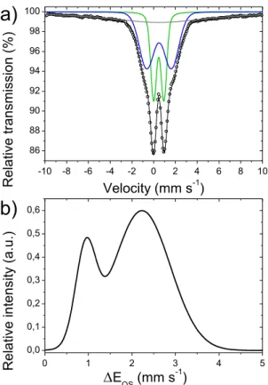
The experimental Mössbauer spectrum of Fe0.5-NC recorded at 80 K, in the absence of
external magnetic field, is shown in Figure 4a. The fitting was performed using two broad doublets with a Gaussian distribution of quadrupole splitting, and with an additional relaxed
20 component to take into account the large background signal. We initially suspected this broad signal to come from Fe oxide nanoparticles, but it was almost unmodified at low and high temperature (5 K and 296 K, Figure S3). This excludes its assignment as Fe oxide clusters, since such phases would show a sextet signal at 5 K and a doublet signal at 296 K. While the fitting shown in Figure 4a could also have been performed without Gaussian distribution of the quadrupole splitting, this would have led to unusually high linewidths for D1 and D2, typically 0.7 and 1.3 mm·s-1, respectively, as reported by us for a similar catalyst, see Figure 1 and Table S1 in Ref. 10. We believe these high linewidths previously reported with such a simpler fitting approach arise from the electronic, chemical and structural disorder existing in pyrolyzed Fe-N-C catalysts. Here, we therefore opted for a fitting with a Gaussian distribution of quadrupole splittings, in order to more accurately compare all quadrupole splitting values experimentally observed to those calculated for a broad family of active sites. The results of the fitting are shown in Table S8. The two quadrupole doublet distributions have similar isomer shifts of 0.46 and 0.49 mm·s-1, but are distinguished by their average quadrupole splittings, centered at 0.94 and 2.25 mm·s-1 for D1 and D2, respectively. Such values are typically observed in Fe-N-C catalysts.5-10, 14-15, 24 While these two doublets in pyrolyzed Fe-N-C materials were clearly correlated to FeN4-type moieties with the help of X-ray absorption
spectroscopy and HR-TEM10, 14-15, 22-23, 62 their experimental values of isomer shift and quadrupole splittings do not allow an unambiguous assignment to a specific oxidation state or spin state of iron in such moieties. The isomer shift is in the range expected for both Fe(II) moieties in low and medium spin state, and for Fe(III) moieties in any spin state. EQS values
in the range 0.8 to 1.5 mm·s-1 can be assigned either to Fe(II) S= 0 or Fe(III) S = 5/2 species (Figure 2.32 in Ref 24). From this result, only Fe in high oxidation states above +III ( value ≤ 0.3 mm s-1) and Fe(II) in high spin state (S = 2) can be excluded ( > 0.9 mm s-1).21 The exclusion of Fe in +IV or higher oxidation state is also and more clearly supported by XPS, showing only Fe in +II or +III oxidation states for atomically-dispersed Fe-N-C catalysts.
21 -10 -8 -6 -4 -2 0 2 4 6 8 10 86 88 90 92 94 96 98 100 0 1 2 3 4 5 0,0 0,1 0,2 0,3 0,4 0,5 0,6
b)
R e la tiv e tr a n sm is si o n ( % ) Velocity (mm s-1) R e la tiv e in te n si ty (a .u .) EQS (mm s-1)a)
Figure 4. 57Fe Mössbauer spectrum at 80 K of Fe0.5-NC measured in air in the absence of external magnetic field
(Fig. 4a) and corresponding distribution of the quadrupole splitting (Fig. 4b). The doublets D1 and D2 (and their fitted distribution of EQS values) are highlighted in green and blue, respectively.
In order to gain more insights on the spin state of moieties present in this catalyst, we first characterized Fe0.5-NC with EPR. Figure S4 shows the X-band EPR spectra for Fe0.5-NC and
for the corresponding blank measurement (no catalyst in the tube). The signal at circa 160 mT (g=4.28) in Figure S4b can be unambiguously assigned to a rhombic S = 5/2 Fe(III) center.63 The six lines at 300-370 mT are assigned to Mn impurities. No specific signal is observed for Fe(III) in other spin states, nor for Fe(II), which is a non-Kramer ion and is normally EPR-silent. This information facilitates the further analysis of the Mössbauer spectrum, since we now can assume that FeN4 moieties are mainly present either as Fe(III) S = 5/2 high spin
state, or as Fe(II) in the S = 0 or S = 1 spin states. The Fe(II) S = 2 state can be excluded, based on the above mentioned high isomer shift value expected for such a configuration, not experimentally observed.
The average molecular susceptibility of Fe0.5-NC was then measured by SQUID as a function
of temperature (Figure S5a). The data could be fitted with a Curie-Weiss law, Eq. 2 (Figure S5b) with slightly negative C-value, corresponding to a weak antiferromagnetic coupling.
22 The constant C can be then further analyzed assuming two different types of Fe centers in Fe0.5-NC, knowing that their values are additive, using Eqs. 2 and 3:
m
-1
= (T-C)/C (2)
µeff2 = µB2 [ a1(s12 + s1) + a2(s22 + s2) ] (3)
where a1 and a2 are the molar fractions of Fe in the two types of sites, s1 and s2 are their
corresponding spin states, µeff the average effective magnetic moment of iron atoms. With the
fitted value of constant C (2.566), the second term in Eq. 3 should be equal to 20.4 to match the experimental fitting. Assuming Fe0.5-NC contains a mix of iron centers with only Fe(II) in
S = 0 state and Fe(III) in S = 5/2 state, the value of 20.4 is obtained with a relative fraction of 58% of high spin Fe(III). Assuming a mix of only Fe(II) in S = 1 state and Fe(III) S = 5/2 state, a match is obtained with a relative fraction of 46% high spin Fe(III). These results are consistent with the relative fraction from Mössbauer (Table S8), if one assumes that both D1 and the broad relaxation background are in high-spin ferric state, and D2 a ferrous center in zero-spin state.
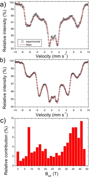
To obtain further information on the spin state of the doublets D1 and D2, Mössbauer spectroscopy measurements were then performed in the presence of an external magnetic field. The behavior of the Mössbauer nuclei under an applied field is strongly dependent on their spin state. In the case of diamagnetic nuclei, i.e. with S = 0, the measured field is equal to the applied external field, whereas in the case of paramagnetic ions the effective measured field is equal to the applied field plus the hyperfine field which is created by the magnetization.59 In the case of high spin Fe(III), due to the large magnetic moment corresponding to the S = 5/2 state, very high effective fields exceeding 50 T can be obtained. Figure 5 shows the spectra of Fe0.5-NC measured with an external magnetic field of 7 T
applied either parallel or perpendicular to the -rays as well as the corresponding distribution of hyperfine magnetic fields obtained by the common fit of the two spectra. Interestingly, two maxima centered at about 7 T and 45 T are observed in the distribution. The second value unambiguously underlines the presence of high spin Fe(III), confirming both SQUID and EPR results. While hyperfine fields exceeding 50 T are commonly measured for slowly relaxing Fe(III) ions, the external magnetic field usually couples antiferromagnetically to the paramagnetic relaxation field, thus decreasing the observed value of the hyperfine field. This
23 explains the strong signal observed at 45 T. On the other hand, the component centered around 7 T could derive from the presence of diamagnetic low spin Fe(II). In fact, a value of the hyperfine field equaling the value of the applied field is a rather clear sign of the presence of a diamagnetic ion, since no additional fields caused by the magnetization of unpaired electrons are experienced by the Fe ions.
It must also be said that the fit shown in Figure 5, obtained using a distribution of magnetic sextets, does not describe precisely the structure of the two spectra acquired at low values of external magnetic fields (not shown). This is not unexpected since, for low values of hyperfine field and relatively high values of quadrupole splitting, the quadrupole interaction cannot be treated anymore as a perturbation of the Zeeman effect caused by the magnetic field, and the full Hamiltonian including both interactions must be solved.64 In this case, the experimental spectrum strongly depends upon the mutual orientation of the Vzz component of
the EFG and of the magnetic field, which is virtually impossible to determine in such broadened multicomponent spectra. However, in the absence of a local magnetic moment on the absorbing ions, which are randomly oriented in the sample, one can assume a random orientation of the magnetic field with respect to the EFG.64 The theoretical spectrum of such a system, calculated assuming a hyperfine field of 7 T and a EFG of 2.3 mm·s-1 (Vzz = 2.3 mm·s-1, = 0, simulating doublet D2 in the spectrum without applied external
magnetic field), is shown in Figure S6. As it can be clearly seen, the features of the central part of the spectrum (central peak, slight asymmetry and intensity of the two central lines) correspond rather well to the calculated spectrum.
This detailed analysis of the Mössbauer spectrum under external magnetic field supports i) the presence of a substantial fraction of diamagnetic Fe(II) ions in the sample, with a EFG close to 2.3 mm·s-1 and ii) its assignment to the experimentally observed D2 signal in the spectrum without applied external field. This in turn implies that D1 can, on the other hand, be assigned in majority to Fe(III)N4 sites with high spin. In spite of these main attributions, the presence
of other Fe sites in minor content cannot be excluded. If no additional signal typical of other Fe(III) sites in S = 3/2 or ½ states is detected by EPR, a minor amount of Fe(II) S =1 species cannot be experimentally excluded.
24 -10 -8 -6 -4 -2 0 2 4 6 8 10 93 94 95 96 97 98 99 100 -10 -8 -6 -4 -2 0 2 4 6 8 10 94 95 96 97 98 99 100
b)
experimental fitted R e la tiv e in te n sity ( % ) Velocity (mm s-1)c)
R e la tiv e in te n sity ( % ) Velocity (mm s-1) 0 5 10 15 20 25 30 35 40 45 50 0 2 4 6 8 10 R e la tiv e c o n tr ib u tio n ( % ) BHF (T)a)
Figure 5. 57Fe Mössbauer spectra of Fe0.5-NC measured at 4.5 K with an external magnetic field of 7 T applied
in the direction a) parallel or b) perpendicular to the -rays, and c) the corresponding distribution of hyperfine fields from the common fit of the two spectra using a distribution of magnetic sextets (with a same isomer shift). The fits in a) and b) were obtained with the single distribution of hyperfine magnetic fields shown in c) (the common fitted isomer shift was = 0.47 mm·s-1), and with line intensity ratios of 3:2x:1:1:2x:3 with fitted x-values of 0.29 and 1.90 for the parallel and perpendicular field spectrum, respectively.
25
Discussion
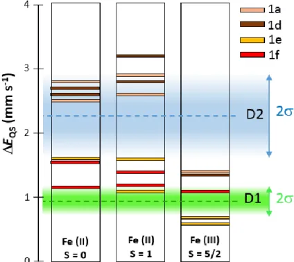
To compare experimental and theoretical insights gained in this study, we focus here on the DFT results obtained on the 1a, 1d, 1e and 1f clusters with the PBE/DZVP2 and PBE/Wachters methods. The main conclusions would not be changed if all DFT results are considered, including those obtained with the periodic approaches. Figure 6 shows the calculated EQS values for the above mentioned combinations of clusters and DFT methods,
with the FeN4 clusters in three possible combinations of oxidation and spin state, namely
Fe(II) S = 0, Fe(II) S = 1 and Fe(III) S = 5/2. These combinations correspond to those identified as major (Fe(II) S = 0, and Fe(III) S = 5/2) or possible minor species (Fe(II) S = 1) for the present Fe-N-C catalyst by experimental Mössbauer spectroscopy, SQUID and EPR.
Figure 6. Comparison between DFT-calculated and measured quadrupole splittings for FeNx sites. EQS values calculated with the PBE/DZVP2 and PBE/Wachters methods for the 1a, 1d, 1e and 1f clusters are presented. The green and blue areas correspond to the experimentally measured distribution of the doublets D1 and D2, and the dashed lines correspond to the center of the fitted Gaussian distribution for each of them (0.94 and 2.25 mm·s-1 respectively). The colored D1 and D2 areas expand (where is their standard deviation, see Table S8) above and below the mean EQS value fitted for D1 and D2, thereby representing 68% of the entire D1 and D2 signal fitted with the broad doublet Gaussian distribution in figure 4.
26 From the comparison of experimental and calculated EQS values only, Figure 6 shows that
the experimental doublet D1 can only be assigned to FeN4C12 sites (1e and 1f clusters,
represented by red and orange horizontal bars), but in any of the three above-mentioned oxidation/spin combinations. The center of the D1 distribution (0.94 mm·s-1, dashed green line) best corresponds however to Fe(III)N4C12 sites (1e and 1f in Figure 6) with high spin.
When considering periodic DFT calculations, it should be noted that high spin Fe(III)N4C10
and Fe(III)N4C12 have identical EQS values (0.7 mm s-1), that can be assigned to the lower
end of the D1 signal. All this supports the main assignment of D1 to Fe(III) S =5/2 moieties earlier in this work, that we derived from the analysis of the experimental Mössbauer spectrum under external magnetic field. The 1a and 1d clusters (FeN4C10 sites) in Fe(III) S =
5/2 state fall in-between the D1 and D2 regions shown in Figure 6 and that correspond to the mean EQS value of D1 and D2 ± , i.e. representing 68% of the total D1 and D2 signal as
fitted in Figure 4a. Thus, they may contribute to the broadening of the D1 and D2 doublets in the region 1.2-1.4 mm·s-1. The overlap in this region is clearly seen in the complete EQS
distributions for D1 and D2 shown in Figure 4b. In contrast, D2 can mostly be assigned to Fe(II)N4C10 sites, in either S = 0 or S = 1 spin state. The center of the D2 distribution (2.25
mm·s-1, dashed blue line) slightly better corresponds however to Fe(II)N4C10 sites with low
spin. This also supports the main assignment of D2 to Fe(II) S =0 moieties earlier in this work, that was derived from the analysis of the experimental Mössbauer spectrum under external magnetic field. We however do not exclude the presence of a minor content of Fe(II) S =1 sites, and such sites would, according to our DFT results, either clearly contribute to the D2 experimental signal (FeN4C10 sites), or to D1 and the intermediate region overlapping D1
and D2 (FeN4C12 sites), see Figure 6, central column.
It can thus be concluded from this comparison between calculated and experimental EQS
values, and from EPR, SQUID and from the analysis of the Mössbauer spectrum under external magnetic field, that the D1 and D2 experimental signals can mostly be assigned to Fe(III)N4C12 sites with high spin and Fe(II)N4C10 sites with low spin, respectively. Because of
the high relative content of Fe(II) S = 0 and Fe(III) S = 5/2 species experimentally identified, and in view of Figure 6, it is difficult to propose other main assignments. For example, while from Figure 6 alone, D1 could be assigned mostly to Fe(II)N4C12 sites with S = 0 and D2
mostly to Fe(II)N4C10 sites with S = 1, there is then no main assignment for an Fe(III) S = 5/2
27 magnetic field and EPR, while SQUID also supports the presence of a high relative fraction of high spin ferric species.
These main assignments of D1 and D2 are important novel insights, and in strong contrast to previous empirical assignments of D1 and D2.5-10, 24 It is here recalled that the ambiguity and lack of evidence of those previous assignments was generally recognized by those involved in the field. It also implies that the D1 signal must be located on the top surface of the carbon matrix, thereby accessible to gas-phase, leading to ferric species as soon as the sample is exposed to air. The D2 signal, assigned to ferrous species, may then be explained by the inaccessibility of such sites to the ambient air, i.e. such sites might be viewed as being buried underneath the surface, or on the surface but with weaker oxygen binding. These assignments are well supported by recent results obtained by Kneebone et al on the effect of NO adsorption on the Mössbauer spectrum of another Fe-N-C catalyst.65 In that study, the doublet with lowest EQS in the pristine catalyst (EQS = 0.83 mm·s-1, = 0.30 mm·s-1 and labelled
D4 in that work but corresponding to D1 label in the present work) was found to explain most of the spectral changes observed upon the electrochemical reduction of the catalyst, and then upon NO adsorption on the surface ferrous sites. That study and the present one agree then in identifying the D1 signal in the Mössbauer spectrum of Fe-N-C catalysts exposed to air as a ferric, surface-located species, which can be reduced to ferrous form when the applied electrochemical potential is lower than that of the Fe(III)/Fe(II) redox couple of such FeN4
sites. The ferric state of D1 sites when exposed to air and their ability to be reduced to ferrous state when the electrochemical potential is lowered in the ORR range indicates that such sites are likely the ORR active sites in Fe-N-C catalysts. Again, this is supported by previous positive correlations between the absolute or relative amount of D1 signal and the ORR activity for different series of Fe-N-C catalysts obtained by varying one single experimental synthesis parameter (e.g. Fe content, pyrolysis duration, pyrolysis temperature, etc).9, 66 The assignment of the D1 signal to high-spin Fe(III)N4C12 moieties is also in agreement with
similar EQS and values as D1 observed for a monolayer coverage of Fe-phthalocyanine
(FePc) on Vulcan carbon black (EQS = 0.96 and = 0.40, Table 1 in Ref 67. In that work, the
author assigned this new doublet (different from that of crystalline FePc) to the binding of FePc to the carbon substrate through an oxygen atom of a carbon functional group. But it appears simpler now to assign this “new” spectral feature of FePc to the fact that all Fe centers in this monolayer coverage configuration are exposed to O2 from ambient air, which is
28 impossible in crystalline FePc. Following our thinking, the D2 signal in pyrolyzed Fe-N-C catalysts has similar Mössbauer parameters to bulk crystalline FePc (EQS = 2.61 and =
0.39, Table 1 in 67 . This supports the idea that D2 may be assigned either i) to similar FeN4
sites as D1, but located in the bulk of the carbon matrix and thus inaccessible to the gas-phase (and the liquid electrolyte) impeding their oxidation by O2, or ii) to FeN4 surface sites with a
Fe(III)/Fe(II) redox potential sufficiently high that their stable form is Fe(II), even when exposed to air in open circuit conditions. Last, the assignment of D1 to, mostly, a ferric high spin moiety is also in line with a recent study from Kramm’s group, involving experimental
57
Fe Mössbauer spectroscopy at 5 K with and without perpendicular applied magnetic field, as well as EPR and nuclear inelastic spectroscopy.68 The major presence of a high-spin ferric moiety (28 % of the absorption signal) was evidenced, and correlated with a doublet with ΔEQS of 1.0 mm s-1 (Table 2 in that work), corresponding to our D1 component, while a
doublet with even smaller ΔEQS value (0.72 mm s-1) was assigned to a ferrous low-spin
moiety, but representing only 12 % of the absorption signal. This is coherent with our Figure 6, showing that porphyrinic moieties (1e-1f clusters) can result in low ΔEQS values of ca 1
mm s-1 either in ferric high-spin or ferrous low spin states.
This work thus identifies D1 as surface sites which, combined with a precise measurement of the Fe bulk content and applying correct Lamb-Mössbauer factors for D1 and D2 and any other Fe species present in a given Fe-N-C catalyst,13 can directly yield the absolute number of Fe(D1) surface sites. Due to the EQS distribution within the broad D1 signal, it may
however be that not all D1 sites are similarly active toward ORR, and some may be unstable, at least in acidic medium. Following the fate of D1 signal during electrochemical operation will be necessary to more closely connect it to the ORR activity after short break-in of the Fe-N-C electrode in electrolyte, and then also after longer operation in liquid-electrolyte or in fuel cell.
The experimental and theoretical methods reported here to study in detail FeNx sites
embedded in graphene for fuel cell application are also of interest for other electrochemical applications such as CO2 electro-reduction69, nitrite and nitric oxide electroreduction70 but
also for the broader field of heterogeneous catalysis, where similar sites have been reported to catalyze a range of interesting reactions for high added-value products.71-73
29
Conclusions
Calculated quadrupole splitting values for ferrous moieties (S = 0 or S = 1 spin state) clearly differentiate FeN4C12 (< 1.8 mm·s-1) from FeN4C10 moieties (> 2.4 mm·s-1)
No significant effect of cluster size on calculated quadrupole splitting value for ferrous FeN4Cy moieties (S = 0 or S = 1 spin state)
Calculated quadrupole splitting value for ferric FeN4Cy moieties in S = 3/2 spin state
mainly depends on OH-orientation, less on model structure
Calculated quadrupole splitting values for ferric moieties in S = 5/2 spin state weakly differentiate FeN4C12 (< 1.2 mm·s-1) from FeN4C10 moieties (> 1.3 mm·s-1)
Experimental identification of the presence of Fe(II) S = 0 and Fe(III) S =5/2 centers in Fe-N-C catalyst with Mössbauer spectroscopy under strong external magnetic field, EPR and SQUID
Assignment of experimental doublet 1 (with mean quadrupole splitting value at 0.94 mm·s-1) to mostly Fe(III)N4C12 sites with S = 5/2 and of the doublet 2 (with mean
quadrupole splitting value at 2.25 mm·s-1) to mostly Fe(II)N4C10 sites with S = 0,
through combined experimental and theoretical results
Identification of experimental doublet 1 as FeNx sites located on top surface and as a
major contributor of ORR activity
Supporting information
Examples of structures optimized with PBE/DZVP2 level of theory (Figure S1), table of population of orbitals for 1a and 2a ferrous models in S = 1 spin state (Table S1) and their structural parameters for S = 0 and S = 1 (Table S2), table of structural parameters for ferrous 1e and 2e models for S = 0 and S =1 optimized with different DFT methods (Table S3), effect of cluster size on quadrupole splitting, relative energies and structural parameters for ferrous models (Table S4-S5), scheme of possible OH orientation on a ferric FeN4Cy model site
(Figure S2), structural parameters of ferric FeN4C10 and FeN4C12 clusters for different spin
states and with different DFT methods (Table S6-S7), results of fitting of experimental Mössbauer spectra (Table S8), effect of temperature on Mössbauer spectra (Figure S3), X-band EPR spectra (Figure S4), SQUID measurement (Figure S5), experimental Mössbauer spectra with external magnetic field applied parallel or perpendicular to -rays and theoretical simulation for randomly oriented Fe(II) ions creating a quadrupole splitting of 2.3 mm s-1 (Figure S6),
30 This work was partially funded by the French National Research Agency (Labex programme CheMISyst, grant agreement ANR-10-LABX-05-01) and the FCH Joint Undertaking (CRESCENDO Project, Grant Agreement n°779366). T.M. acknowledges GENCI-CCRT center (project No. A0050807369) for a generous allocation of high performance computing resources. Corine Reibel (ICGM UMR 5253 CNRS) is acknowledged for her help with SQUID measurements. I.M. thankfully acknowledges the computational resources of the National Energy Research Scientific Computing Center (NERSC), a U.S. Department of Energy Office of Science User Facility operated under Contract No. DE-AC02-05CH11231 and UNM Center for Advanced Research Computing (CARC). This paper has been designated LA-UR-19-27987.