S P E C I A L A R T I C L E
Dif
ficult Airway Society 2015 guidelines for
management of unanticipated dif
ficult intubation
in adults
†
C. Frerk
1,
*, V. S. Mitchell
2
, A. F. McNarry
3
, C. Mendonca
4
, R. Bhagrath
5
, A. Patel
6
,
E. P. O
’Sullivan
7
, N. M. Woodall
8
and I. Ahmad
9
, Dif
ficult Airway Society
intubation guidelines working group
1
Department of Anaesthesia, Northampton General Hospital, Billing Road, Northampton NN1 5BD, UK,
2
Department of Anaesthesia and Perioperative Medicine, University College London Hospitals NHS Foundation
Trust, 235 Euston Road, London NW1 2BU, UK,
3Department of Anaesthesia, NHS Lothian, Crewe Road South,
Edinburgh EH4 2XU, UK,
4Department of Anaesthesia, University Hospitals Coventry & Warwickshire NHS Trust,
Clifford Bridge Road, Coventry CV2 2DX, UK,
5Department of Anaesthesia, Barts Health, West Smith
field, London
EC1A 7BE, UK,
6Department of Anaesthesia, The Royal National Throat Nose and Ear Hospital, 330 Grays Inn Road,
London WC1X 8DA, UK,
7Department of Anaesthesia, St James
’s Hospital, PO Box 580, James’s Street, Dublin 8,
Ireland,
8Department of Anaesthesia, The Norfolk and Norwich University Hospitals NHS Foundation Trust,
Colney Lane, Norwich NR4 7UY, UK, and
9Department of Anaesthesia, Guy
’s and St Thomas’ NHS Foundation
Trust, Great Maze Pond, London SE1 9RT, UK
*Corresponding author. E-mail: chris.frerk@ngh.nhs.ukAbstract
These guidelines provide a strategy to manage unanticipated difficulty with tracheal intubation. They are founded on published
evidence. Where evidence is lacking, they have been directed by feedback from members of the Difficult Airway Society and
based on expert opinion. These guidelines have been informed by advances in the understanding of crisis management; they
emphasize the recognition and declaration of difficulty during airway management. A simplified, single algorithm now covers
unanticipated difficulties in both routine intubation and rapid sequence induction. Planning for failed intubation should form
part of the pre-induction briefing, particularly for urgent surgery. Emphasis is placed on assessment, preparation, positioning,
preoxygenation, maintenance of oxygenation, and minimizing trauma from airway interventions. It is recommended that the number of airway interventions are limited, and blind techniques using a bougie or through supraglottic airway devices have
been superseded by video- orfibre-optically guided intubation. If tracheal intubation fails, supraglottic airway devices are
recommended to provide a route for oxygenation while reviewing how to proceed. Second-generation devices have advantages and are recommended. When both tracheal intubation and supraglottic airway device insertion have failed, waking the patient is the default option. If at this stage, face-mask oxygenation is impossible in the presence of muscle relaxation,
cricothyroidotomy should follow immediately. Scalpel cricothyroidotomy is recommended as the preferred rescue technique and should be practised by all anaesthetists. The plans outlined are designed to be simple and easy to follow. They should be regularly rehearsed and made familiar to the whole theatre team.
† This Article is accompanied by Editorials aev298 and aev404. Accepted: September 28, 2015
© The Author 2015. Published by Oxford University Press on behalf of the British Journal of Anaesthesia. This is an Open Access article distributed under the terms of the Creative Commons Attribution Non-Commercial
License (http://creativecommons.org/licenses/by-nc/4.0/), which permits non-commercial re-use, distribution, and reproduction in any medium, provided the original work is properly cited. For commercial re-use, please contact journals.permissions@oup.com
British Journal of Anaesthesia, 2015, 1–22 doi: 10.1093/bja/aev371 Special Article 1 by guest on December 8, 2015 http://bja.oxfordjournals.org/ Downloaded from
Key words:airway obstruction; complications; intubation; intubation, endotracheal; intubation, transtracheal; ventilation
Clinical practice has changed since the publication of the original Difficult Airway Society (DAS) guidelines for management of
unanticipated difficult intubation in 2004.1The 4th National
Audit Project of the Royal College of Anaesthetists and Difficult Airway Society (NAP4) provided detailed information about the factors contributing to poor outcomes associated with airway management and highlighted deficiencies relating to judgement,
communication, planning, equipment, and training.2 New
pharmacological agents and videolaryngoscopes have been introduced, and further research has focused on extending the duration of apnoea without desaturation by improving preoxy-genation and optimizing patient position.
These updated guidelines provide a sequential series of plans to be used when tracheal intubation fails and are designed to prioritize oxygenation while limiting the number of airway
inter-ventions in order to minimize trauma and complications (Fig1).
The principle that anaesthetists should have back-up plans in place before performing primary techniques still holds true.
Separate guidelines exist for difficult intubation in paediatric
anaesthesia, obstetric anaesthesia, and for extubation.3–5
These guidelines are directed at the unanticipated difficult intubation. Every patient should have an airway assessment
performed before surgery to evaluate all aspects of airway man-agement, including front-of-neck access.
The aim of the guidelines is to provide a structured response to a potentially life-threatening clinical problem. They take into account current practice and recent developments.
Every adverse event is unique, the outcome of which will be influenced by patient co-morbidity, urgency of the procedure,
skill set of the anaesthetist, and available resources.2 6It is
ac-knowledged that anaesthetists do not work in isolation and that the role of the anaesthetic assistant is important in
influen-cing the outcome of an airway crisis.7Decisions about the best
al-ternatives in the event of difficulty should be made and discussed with the anaesthetic assistant before induction of anaesthesia.
These guidelines recognize the difficulties in decision-mak-ing durdecision-mak-ing an unfolddecision-mak-ing emergency. They include steps to assist the anaesthetic team in making the correct decisions, limiting the number of airway intervention attempts, encouraging declar-ation of failure by placing a supraglottic airway device (SAD) even when face-mask ventilation is possible, and explicitly recom-mending a time to stop and think about how to proceed.
An attempt has been made to identify essential skills and techniques with the highest success rate. Anaesthetists and
Fig 1Difficult Airway Society difficult intubation guidelines: overview. Difficult Airway Society, 2015, by permission of the Difficult Airway Society. This image is not covered by the terms of the Creative Commons Licence of this publication. For permission to re-use, please contact the Difficult Airway Society.
CICO, can’t intubate can’t oxygenate; SAD, supraglottic airway device.
by guest on December 8, 2015
http://bja.oxfordjournals.org/
anaesthetic assistants using these guidelines must ensure that they are familiar with the equipment and techniques described. This may require acquisition of new skills and regular practice, even for experienced anaesthetists.
Methods
The Difficult Airway Society commissioned a working group to update the guidelines in April 2012. An initial literature search was conducted for the period January 2002 to June 2012 using da-tabases (Medline, PubMed, Embase, and Ovid) and a search en-gine (Google Scholar). The websites of the American Society of
Anesthesiologists (http://www.asahq.org), Australian and New
Zealand College of Anaesthetists (http://www.anzca.edu.au),
European Society of Anesthesiologists’ (http://www.esahq.org/
euroanaesthesia), Canadian Anesthesiologists’ Society (http:// www.cas.ca), and the Scandinavian Society of Anesthesiology
and Intensive Care Medicine (http://ssai.info/guidelines/) were
also searched for airway guidelines. English language articles and abstract publications were identified using keywords and
fil-ters. The search terms were as follows:‘Aintree intubating
cath-eter’, ‘Airtraq’, ‘airway device’, ‘airway emergency’, ‘airway management’, ‘Ambu aScope’, ‘backward upward rightward pressure’, ‘Bonfils’, ‘Bullard’, ‘bronchoscopy’, ‘BURP manoeuvre’, ‘can’t intubate can’t ventilate’, ‘can’t intubate can’t oxygenate’, ‘C-Mac’, ‘Combitube’, ‘cricoid pressure’, ‘cricothyroidotomy’, ‘cri-cothyrotomy’, ‘C trach’, ‘difficult airway’, ‘difficult intubation’, ‘difficult laryngoscopy’, ‘difficult mask ventilation’, ‘difficult ven-tilation’, ‘endotracheal intubation’, ‘esophageal intubation’, ‘Es-chmann stylet’, ‘failed intubation’, ‘Fastrach’, ‘fiber-optic scope’, ‘fibreoptic intubation’, ‘fiberoptic scope’, ‘fibreoptic stylet’, ‘fibre-scope’ ‘Frova catheter’, ‘Glide‘fibre-scope’, ‘gum elastic bougie’, ‘hyp-oxia’, ‘i-gel’, ‘illuminating stylet’, ‘jet ventilation catheter’, ‘laryngeal mask’, ‘laryngeal mask airway Supreme’, ‘laryngos-copy’, ‘lighted stylet’, ‘light wand’, ‘LMA Supreme’, ‘Manujet’, ‘McCoy’, ‘McGrath’, ‘nasotracheal intubation’, ‘obesity’, ‘oe-sophageal detector device’, ‘oe‘oe-sophageal intubation’, ‘Pentax air-way scope’, ‘Pentax AWS’, ‘ProSeal LMA′, ‘Quicktrach’, ‘ramping’, ‘rapid sequence induction’, ‘Ravussin cannula’, ‘Sanders inject-or’, ‘Shikani stylet’, ‘sugammadex’, ‘supraglottic airway’, ‘suxa-methonium’, ‘tracheal introducer’, ‘tracheal intubation’, ‘Trachview’, ‘Tru view’, ‘tube introducer’, ‘Venner APA’, ‘videolar-yngoscope’, and ‘videolaryngoscopy’.
The initial search retrieved 16 590 abstracts. The searches (using the same terms) were repeated every 6 months. In total, 23 039 abstracts were retrieved and assessed for relevance by the working group; 971 full-text articles were reviewed. Addition-al articles were retrieved by cross-referencing the data and hand-searching. Each of the relevant articles was reviewed by at least two members of the working group. In areas where the evidence was insufficient to recommend particular techniques, expert
opinion was sought and reviewed.8This was most notably the
situation when reviewing rescue techniques for the‘can’t
intub-ate can’t oxygenintub-ate’ (CICO) situation.
Opinions of the DAS membership were sought throughout the process. Presentations were given at the 2013 and 2014 DAS An-nual Scientific meetings, updates were posted on the DAS web-site, and members were invited to complete an online survey about which areas of the existing guidelines needed updating.
Following the methodology used for the extubation guidelines,5
a draft version of the guidelines was circulated to selected mem-bers of DAS and acknowledged international experts for com-ment. All correspondence was reviewed by the working group.
Disclaimer
It is not intended that these guidelines should constitute a min-imum standard of practice, nor are they to be regarded as a sub-stitute for good clinical judgement.
Human factors
Human factors issues were considered to have contributed to ad-verse outcomes in 40% of the instances reported to NAP4; how-ever, a more in-depth analysis of a subset of patients identified
human factor influences in every instance. Flin and colleagues9
identified latent threats (poor communication, poor training and teamwork, deficiencies in equipment, and inadequate sys-tems and processes) predisposing to loss of situation awareness
and subsequent poor decision-making as a precursor tofinal
ac-tion errors.
Adoption of guidelines and a professional willingness to fol-low them are not enough on their own to avoid serious compli-cations of airway management during anaesthesia. All the instances reported to NAP4 occurred despite widespread dissem-ination of the original DAS guidelines, which had been published in 2004. The complexities of difficult airway management cannot be distilled into a single algorithm, and even the best anaesthetic teams supported by the best guidelines will still struggle to
per-form optimally if the systems in which they operate areflawed.10
The 2015 guidelines acknowledge this.
During a crisis, it is common to be presented with more
infor-mation than can be processed.11This cognitive overload impairs
decision-making and can cause clinicians to‘lose sight of the big
picture’ and become fixated on a particular task, such as tracheal intubation or SAD placement. These guidelines provide an
expli-cit instruction for the team to‘stop and think’ to help reduce this
risk.
Poor anaesthetic decision-making secondary to cognitive errors and the impact of human factors in emergency airway
management has recently been discussed.12Cognitive aids are
increasingly being used by clinicians during unfolding
emergen-cies;13for example, the Vortex Approach has been devised to
sup-port decision-making during difficult airway management.14The
algorithms that accompany these guidelines are intended as teaching and learning tools and have not been specifically de-signed to be used as prompts during an airway crisis.
For any plan to work well in an emergency, it must be known to all members of the team and should be rehearsed. For rare events, such as CICO, this rehearsal can be achieved with simula-tion training, as has recently been included in the Australian and New Zealand College of Anaesthetists continuing professional
development requirements.15 16This also provides the
opportun-ity to develop non-technical skills, such as leadership, team co-ordination, communication, and shared understanding of roles, which has been shown to improve performance in intensive
care and trauma teams.17 18
Structured communication between anaesthetists and an-aesthetic assistants could help prepare for and deal with airway difficulties. Talking before every patient, or at least before every list, about the plan to manage difficulties should they develop is good practice. At a minimum, this involves thinking about the challenges that might be encountered and checking that the appropriate equipment is available.
If airway management does become difficult after induction of anaesthesia, a clear declaration of failure at the end of each plan will facilitate progression through the airway strategy. The use of a structured communication tool, such as PACE (Probe,
by guest on December 8, 2015
http://bja.oxfordjournals.org/
Alert, Challenge, Emergency), can aid communication of con-cerns when cognitive overload and hierarchical barriers might
otherwise make this difficult.19
Our profession must continue to acknowledge and address the impact of environmental, technical, psychological, and physiological factors on our performance. Human factors issues at individual, team, and organizational levels all need to be con-sidered to enable these 2015 guidelines to be as effective as possible.
Preoperative assessment and planning
Airway management is safest when potential problems are iden-tified before surgery, enabling the adoption of a strategy, a series
of plans, aimed at reducing the risk of complications.2
Preoperative airway assessment should be performed rou-tinely in order to identify factors that might lead to difficulty with face-mask ventilation, SAD insertion, tracheal intubation, or front-of-neck access.
Prediction of difficulty in airway management is not
com-pletely reliable;20–22the anaesthetist should have a strategy in
place before the induction of anaesthesia, and this should be dis-cussed at the team brief and the sign-in ( pre-induction) phase of
the WHO Surgical Safety Checklist.23 24
Assessment of the risk of aspiration is a key component of planning airway management. Steps should be taken before sur-gery to reduce the volume and pH of gastric contents by fasting and pharmacological means. Mechanical drainage by nasogas-tric tube should be considered in order to reduce residual gasnasogas-tric volume in patients with severely delayed gastric emptying or
in-testinal obstruction.2
Rapid sequence induction
The placement of a cuffed tube in the trachea offers the greatest protection against aspiration. Suxamethonium is the traditional neuromuscular blocking agent of choice because its rapid onset allows early intubation without the need for bag–mask ventilation. Several studies have compared suxamethonium with rocuronium for rapid sequence induction, and although some have shown better intubating conditions with
suxameth-onium, others have found that after rocuronium 1.2 mg kg−1
the speed of onset and intubation conditions are
compar-able.25–30Suxamethonium-induced fasciculation increases
oxy-gen consumption during apnoea, which may become relevant
in the event of airway obstruction.31 32The ability to antagonize
the effect of rocuronium rapidly with sugammadex may be an
advantage,30although it should be remembered that this does
not guarantee airway patency or the return of spontaneous
ven-tilation.33 34If rapid antagonism of rocuronium with
sugamma-dex is part of the failed intubation plan, the correct dose (16 mg
kg−1) must be immediately available.35 36
Cricoid pressure is applied to protect the airway from contam-ination during the period between loss of consciousness and placement of a cuffed tracheal tube. This is a standard
compo-nent of a rapid sequence induction in the UK.37It is often
over-looked that cricoid pressure has been shown to prevent gastric distension during mask ventilation and was originally described
for this purpose.38 39Gentle mask ventilation after the
applica-tion of cricoid pressure and before tracheal intubaapplica-tion prolongs the time to desaturation. This is most useful in those with poor respiratory reserve, sepsis, or high metabolic requirements and also provides an early indication of the ease of ventilation. A force of 30 N provides good airway protection, while
minimizing the risk of airway obstruction, but this is not well
tol-erated by the conscious patient.40
Cricoid pressure should be applied with a force of 10 N when
the patient is awake, increasing to 30 N as consciousness is lost.41 42
Although the application of cricoid pressure creates a physical barrier to the passage of gastric contents, it has also been shown to reduce lower oesophageal sphincter tone, possibly
making regurgitation more likely.43 44Current evidence suggests
that if applied correctly, cricoid pressure may improve the view
on direct laryngoscopy.45However, there are many reports
dem-onstrating that it is often poorly applied, and this may make mask ventilation, direct laryngoscopy, or SAD insertion more
dif-ficult.46–52If initial attempts at laryngoscopy are difficult during
rapid sequence induction, cricoid pressure should be released. This should be done under vision with the laryngoscope in
place and suction available; in the event of regurgitation,41
cri-coid pressure should be immediately reapplied.
Second-generation SADs offer greater protection against
as-piration thanfirst-generation devices and are recommended
should intubation fail during a rapid sequence induction.
Plan A. Mask ventilation and tracheal
intubation
The essence of Plan A (Table1) is to maximize the likelihood of
successful intubation at thefirst attempt or, failing that, to
limit the number and duration of attempts at laryngoscopy in order to prevent airway trauma and progression to a CICO situation.
All patients should be optimally positioned and preoxyge-nated before induction of anaesthesia. Neuromuscular block
fa-cilitates face-mask ventilation53 54 and tracheal intubation.
Every attempt at laryngoscopy and tracheal intubation has the potential to cause trauma. A suboptimal attempt is a wasted at-tempt and having failed, the chance of success declines with
each subsequent attempt.55 56Repeated attempts at tracheal
in-tubation may reduce the likelihood of effective airway rescue
with a SAD.57 These guidelines recommend a maximum of
three attempts at intubation; a fourth attempt by a more experi-enced colleague is permissible. If unsuccessful, a failed intub-ation should be declared and Plan B implemented.
Table 1Key features of Plan A
•Maintenance of oxygenation is the priority
•Advantages of head-up positioning and ramping are highlighted
•Preoxygenation is recommended for all patients
•Apnoeic oxygenation techniques are recommended in high-risk patients
•The importance of neuromuscular block is emphasized •The role of videolaryngoscopy in difficult intubation is
recognized
•All anaesthetists should be skilled in the use of a videolaryngoscope
•A maximum of three attempts at laryngoscopy are recommended (3+1)
•Cricoid pressure should be removed if intubation is difficult
by guest on December 8, 2015
http://bja.oxfordjournals.org/
Position
Good patient positioning maximizes the chance of successful laryngoscopy and tracheal intubation. In most patients, the best position for direct laryngoscopy with a Macintosh-style
blade is achieved with the neckflexed and the head extended
at the atlanto-occipital joint; the classic‘sniffing’ position.58–60
In the obese patient, the‘ramped’ position should be used
rou-tinely to ensure horizontal alignment of the external auditory meatus and the suprasternal notch because this improves the
view during direct laryngoscopy.61–64This position also improves
airway patency and respiratory mechanics and facilitates passive
oxygenation during apnoea.65 66
Preoxygenation and apnoeic techniques to maintain oxygenation
All patients should be preoxygenated before the induction of
gen-eral anaesthesia.67De-nitrogenation can be achieved with an
ap-propriate flow of 100% oxygen into the breathing system,
maintaining an effective face-mask seal68until the end-tidal
oxygen fraction is 0.87–0.9.69Many other preoxygenation
techni-ques have been described.70–79
Preoxygenation increases the oxygen reserve, delays the onset of hypoxia, and allows more time for laryngoscopy, tracheal
intub-ation,65 69and for airway rescue should intubation fail. In healthy
adults, the duration of apnoea without desaturation (defined as the interval between the onset of apnoea and the time peripheral
capillary oxygen saturation reaches a value of≤90%) is limited to
1–2 min whilst breathing room air, but can be extended to 8 min
with preoxygenation.69Preoxygenation using a 20–25° head-up
position80 81and continuous positive airway pressure has been
shown to delay the onset of hypoxia in obese patients.82–84The
duration of apnoea without desaturation can also be prolonged by passive oxygenation during the apnoeic period (apnoeic oxy-genation). This can be achieved by delivering up to 15 litres
min−1of oxygen through nasal cannulae, although this may be
uncomfortable for an awake patient.65 85 86Nasal Oxygenation
During Efforts Of Securing A Tube (NODESAT) has been shown to extend the apnoea time in obese patients and in patients with a
difficult airway.87Transnasal humidified high-flow oxygen (up to
70 litres min−1) via purpose-made nasal cannulae has been
shown to extend the apnoea time in obese patients and in patients
with difficult airways,88although it’s efficacy as a means of
preox-ygenation has not been evaluated fully.89 90Apnoeic oxygenation
is an area of recent research interest about which further evidence is awaited. The administration of oxygen by nasal cannulae in addition to standard preoxygenation and face-mask ventilation is recommended in high-risk patients.
Choice of induction agent
The induction agent should be selected according to the clinical condition of the patient. Propofol, the most commonly used induc-tion agent in the UK, suppresses laryngeal reflexes and provides
better conditions for airway management than other agents.91–93
The 5th National Audit Project of the Royal College of Anaesthe-tists highlighted the relationship between difficult airway
manage-ment and awareness.94It is important to ensure that the patient is
adequately anaesthetized during repeated attempts at intubation.
Neuromuscular block
If intubation is difficult, further attempts should not proceed without full neuromuscular block. Neuromuscular block
abolishes laryngeal reflexes, increases chest compliance, and
fa-cilitates face-mask ventilation.53 54 95Complete neuromuscular
block should be ensured if any difficulty is encountered with
air-way management.96Rocuronium has a rapid onset and can be
antagonized immediately with sugammadex, although the inci-dence of anaphylaxis may be higher than with other
non-de-polarizing neuromuscular blocking agents.97–99
Mask ventilation
Mask ventilation with 100% oxygen should begin as soon as pos-sible after induction of anaesthesia. If difficulty is encountered, the airway position should be optimized and airway manoeuvres such as a chin lift or jaw thrust should be attempted. Oral and nasopharyngeal airways should be considered, and a four-handed technique (two-person or pressure-controlled
mechan-ical ventilation) should be used.100 The ‘sniffing’ position
increases the pharyngeal space and improves mask
ventila-tion.101Inadequate anaesthesia or inadequate neuromuscular
block make mask ventilation more difficult.102 103
Choice of laryngoscope
The choice of laryngoscope influences the chance of successful tracheal intubation. Videolaryngoscopes offer an improved view compared with conventional direct laryngoscopy and are
now the first choice or default device for some
anaesthe-tists.104–113Regular practice is required to ensure that the improved
view translates reliably into successful tracheal intubation.114
All anaesthetists should be trained to use, and have immediate
access to, a videolaryngoscope.115 Theflexible fibrescope or
optical stylets, such as Bonfils (Karl Storz), Shikani (Clarus Med-ical), or Levitan FPS scope™ (Clarus MedMed-ical), may be the
pre-ferred choice for individuals who are expert in their use.116–122
Thefirst and second choice of laryngoscope will be determined
by the anaesthetist’s experience and training.
Tracheal tube selection
Tracheal tubes should be selected according to the nature of the surgical procedure, but their characteristics can influence the ease of intubation. A smaller tube is easier to insert because a bet-ter view of the laryngeal inlet is maintained during passage of the tube between the cords. Smaller tubes are also less likely to cause
trauma.123‘Hold-up’ at the arytenoids is a feature of the left-facing
bevel of most tracheal tubes, and can occur particularly whilst
rail-roading larger tubes over a bougie, stylet, orfibrescope.124 125This
problem can be overcome by rotating the tube anticlockwise to change the orientation of the bevel or by preloading the tube so that the bevel faces posteriorly and by minimizing the gap
be-tween the fibrescope and the tube during fibre-optic
intub-ation.125–127Tubes with hooded, blunted, orflexible tips, such as
the Parker Flex-Tip™ (Parker Medical), and tubes supplied with
the Intubating LMA®(Teleflex Medical Europe Ltd) have been
de-signed to reduce the incidence of this problem.128–132
Laryngoscopy
In these guidelines, an attempt at laryngoscopy is defined asthe in-sertion of a laryngoscope into the oral cavity. Every attempt should be carried out with optimal conditions because repeated attempts at laryngoscopy and airway instrumentation are associated with
poor outcomes and the risk of developing a CICO situation.56 133–
136If difficulty is encountered, help should be summoned early,
re-gardless of the level of experience of the anaesthetist.
by guest on December 8, 2015
http://bja.oxfordjournals.org/
If intubation is difficult, there is little point in repeating the same procedure unless something can be changed to improve the chance of success. This may include the patient’s position, the intubating device or blade, adjuncts such as introducers and stylets, depth of neuromuscular block, and personnel. The number of attempts at laryngoscopy should be limited to three. A fourth at-tempt should be undertaken only by a more experienced colleague.
External laryngeal manipulation
External laryngeal manipulation applied with the anaesthetist’s right hand or backward, upward, and rightward pressure (BURP) on the thyroid cartilage applied by an assistant may improve the
view at laryngoscopy.137–142A benefit of videolaryngoscopy is
that the anaesthetic assistant is also able to see the effects of
laryngeal manipulation.143
Use of a bougie or stylet
The gum elastic bougie is a widely used device for facilitating tra-cheal intubation when a grade 2 or 3a view of the larynx is seen
during direct laryngoscopy.144–146Pre-shaping of the bougie
facil-itates successful intubation.147It can also be helpful during
vi-deolaryngoscopy.148 149 Blind bougie insertion is associated
with trauma and is not recommended in a grade 3b or 4
view.150–155The‘hold-up’ sign may signal the passage of the
bougie as far as small bronchi,156but it is associated with risk
of airway perforation and trauma, especially with single-use
bou-gies.153 157–159Forces as little as 0.8 N can cause airway trauma.153
The characteristics of bougies vary, and this may affect their
per-formance.153 Once the bougie is in the trachea, keeping the
laryngoscope in place enhances the chance of successful
intub-ation.129Non-channelled videolaryngoscopes with angulated
blades necessitate the use of a pre-shaped stylet or bougie to
aid the passage of the tracheal tube through the cords.160–163
When using a videolaryngoscope, the tip of the tube should be in-troduced into the oropharynx under direct vision because failure
to do so has been associated with airway trauma.163–167
Tracheal intubation and confirmation
Difficulty with tracheal intubation is usually the result of a poor laryngeal view, but other factors, such as tube impingement, can hinder the passage of the tube into the trachea.
Once tracheal intubation has been achieved, correct placement of the tube within the trachea must be confirmed. This should in-clude visual confirmation that the tube is between the vocal cords, bilateral chest expansion, and auscultation and capnography. A continuous capnography waveform with appropriate inspired
and end-tidal values of CO2is the gold standard for confirming
ventilation of the lungs. Capnography should be available in
every location where a patient may require anaesthesia.2 168
Absence of exhaled CO2indicates failure to ventilate the
lungs, which may be a result of oesophageal intubation or
com-plete airway obstruction (rarely, comcom-plete bronchospasm).2In
such situations, it is safest to assume oesophageal intubation. Videolaryngoscopy, examination with afibrescope, or ultrasound
can be used to verify that the tube is correctly positioned.169–171
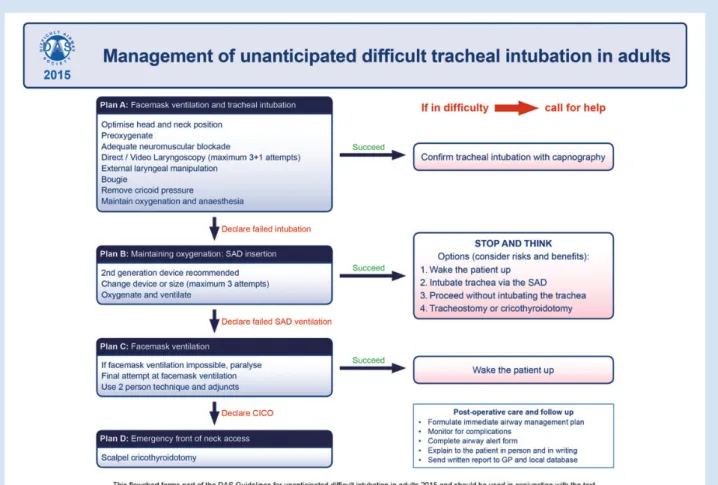
Plan B. Maintaining oxygenation: supraglottic
airway device insertion
In these guidelines (Fig.2), the emphasis of Plan B (Table2) is on
maintaining oxygenation using an SAD.
Successful placement of a SAD creates the opportunity to stop and think about whether to wake the patient up, make a fur-ther attempt at intubation, continue anaesthesia without a tra-cheal tube, or rarely, to proceed directly to a tracheostomy or cricothyroidotomy.
If oxygenation through a SAD cannot be achieved after a max-imum of three attempts, Plan C should be implemented.
Supraglottic airway device selection and placement
As difficulty with intubation cannot always be predicted, every anaesthetist should have a well-thought-through plan for such an eventuality. The decision about which SAD to use for rescue should have been made before induction of anaesthesia, and this choice should be determined by the clinical situation, device availability, and operator experience.
NAP4 identified the potential advantages of second-gener-ation devices in airway rescue and recommended that all hospi-tals have them available for both routine use and rescue airway
management.2Competence and expertise in the insertion of
any SAD requires training and practice.172–176All anaesthetists
should be trained to use and have immediate access to second-generation SADs.
Cricoid pressure and supraglottic airway device insertion
Cricoid pressure decreases hypopharyngeal space177and impedes
SAD insertion and the placement of bothfirst- and
second-gener-ation devices.178–181Cricoid pressure will have been removed
dur-ing Plan A if laryngoscopy was difficult and (in the absence of regurgitation) should remain off during insertion of a SAD.
Second-generation supraglottic airway devices
It has been argued that second-generation SADs should be used routinely because of their efficacy and increased safety when
com-pared withfirst-generation devices.182Several second-generation
SADs have been described,183–191and it is likely that during the
lifetime of these guidelines many similar devices will appear. The ideal attributes of a SAD for airway rescue are reliable first-time placement, high seal pressure, separation of
gastro-intestinal and respiratory tracts, and compatibility with
fibre-op-tically guided tracheal intubation. These attributes are variably
combined in different devices.182Of those currently available,
only the i-gel™ (Intersurgical, Wokingham, UK), the Proseal™
LMA®(PLMA; Teleflex Medical Europe Ltd, Athlone, Ireland),
and the LMA Supreme™ (SLMA; Teleflex Medical Ltd) have
large-scale longitudinal studies,192–195literature reviews,196or
meta-analyses in adults197–200supporting their use. A number
of studies have compared second-generation SADs,201–224but it
is important to recognize that the experience of the operator with the device also influences the chance of successful
insertion.225
Limiting the number of insertion attempts
Repeated attempts at inserting a SAD increases the likelihood of airway trauma and may delay the decision to accept failure and move to an alternative technique to maintain oxygenation.
Successful placement is most likely on thefirst attempt. In
one series, insertion success with the PLMA™ was 84.5% on the
first attempt, decreasing to 36% on the fourth attempt.193In the
series of Goldmann and colleagues,194 only 4.2% of devices
were placed on the third or fourth attempt. Three studies report that a third insertion attempt increased overall success rate by
by guest on December 8, 2015
http://bja.oxfordjournals.org/
more than 5%; however, one was conducted with operators who
had minimal experience, and the other two used the Baska®
mask (Baska Versatile Laryngeal Mask, Pty Ltd, Strathfield, NSW,
Australia).189 214 226Changing to an alternative SAD has been
shown to be successful.192 193 211 216 218 223 224A maximum of
three attempts at SAD insertion is recommended; two with the preferred second-generation device and another attempt with an alternative. An attempt includes changing the size of the SAD.
Even supraglottic airways can fail.227 228If effective
oxygen-ation has not been established after three attempts, Plan C should be implemented.
Guided supraglottic airway device placement
Bougie-aided placement of the PLMA has been described as
im-provingfirst-time placement.229In comparison studies, the
bou-gie-guided technique was 100% effective at achievingfirst-time
placement and more effective than digital insertion or insertion
with the introducer tool.230 231Bougie-aided placement provides
better alignment of the drain port and a betterfibre-optic view of
the cords through the PLMA than the introducer tool method.232
Patients with a history of difficult tracheal intubation or predicted difficulty were excluded from these studies, making it unclear how effective this technique would be in this situation. The tech-nique has been used effectively in a simulated difficult airway
in patients wearing a hard collar,233but again patients with
pre-dicted difficulty were excluded. A comparative study between the i-gel and the PLMA using a guided technique with a
duo-denal tube234showed both devices to have afirst-time insertion
success rate of >97%. An orogastric tube has also been used effectively to facilitate PLMA placement in 3000 obstetric
patients.235Despite the apparent benefit, bougie- and gastric
tube-guided placement of second-generation devices are not
guaranteed to be successful.193 221 The technique requires
Table 2Key features of Plan B. SAD, supraglottic airway device
•Failed intubation should be declared •The emphasis is on oxygenation via a SAD •Second-generation SADs are recommended
•A maximum of three attempts at SAD insertion are recommended
•During rapid sequence induction, cricoid pressure should be removed to facilitate insertion of a SAD
•Blind techniques for intubation through a SAD are not recommended
Fig 2Management of unanticipated difficult tracheal intubation in adults. Difficult Airway Society, 2015, by permission of the Difficult Airway Society. This image is not covered by the terms of the Creative Commons Licence of this publication. For permission to re-use, please contact the Difficult Airway Society.
SAD, supraglottic airway device.
by guest on December 8, 2015
http://bja.oxfordjournals.org/
experience, it may cause trauma,150and it is not listed in the
cur-rent PLMA instruction manual.236
Successful supraglottic airway device insertion and effective oxygenation established:‘stop and think’
Clinical examination and capnography should be used to confirm ventilation. If effective oxygenation has been established through a SAD, it is recommended that the team stop and take the opportunity to review the most appropriate course of action. There are four options to consider: wake the patient up;
at-tempt intubation via the SAD using afibre-optic scope; proceed
with surgery using the supraglottic airway; or (rarely) proceed to tracheostomy or cricothyroidotomy.
Patient factors, the urgency of the surgery, and the skill set of the operator all influence the decision, but the underlying principle is to maintain oxygenation while minimizing the risk of aspiration.
Wake the patient up
If the surgery is not urgent then the safest option is to wake the
patient up, and this should be consideredfirst. This will require
the full antagonism of neuromuscular block. If rocuronium or ve-curonium has been used, sugammadex is an appropriate choice of antagonistic agent. If another non-depolarizing neuromuscu-lar blocking agent has been used then anaesthesia must be main-tained until paralysis can be adequately antagonized. Surgery may then be postponed or may continue after awake intubation or under regional anaesthesia.
If waking the patent up is inappropriate (for example, in the critical care unit, in the emergency department, or where life-saving surgery must proceed immediately), the remaining op-tions should be considered.
Intubation via the supraglottic airway device
Intubation through a SAD is only appropriate if the clinical situ-ation is stable, oxygensitu-ation is possible via the SAD, and the anaes-thetist is trained in the technique. Limiting the number of airway interventions is a core principle of safe airway management; re-peated attempts at intubation through a SAD are inappropriate.
Intubation through an intubating laryngeal mask airway (iLMA™; Teleflex Medical Ltd) was included in the 2004
guide-lines.1Although an overall success rate of 95.7% has been
re-ported in a series of 1100 patients using a blind technique,237
first-attempt success rates are higher using fibre-optic
guid-ance,238 239and a guided technique has been shown to be of
bene-fit in patients with difficult airways.240The potential for serious
adverse outcomes associated with blind techniques remains.241
With the need for repeated insertion attempts to achieve
suc-cess238and a lowfirst-time success rate240 242(even with
se-cond-generation devices243), the blind technique is redundant.
Directfibre-optically guided intubation has been described via a number of SADs, although this may be technically
challen-ging.244–248Fibre-optically guided tracheal intubation through
the i-gel has been reported with a high success rate.249 250
Se-cond-generation SADs specifically designed to facilitate tracheal
intubation have been described,190 251 252but data regarding their
efficacy are limited.
The use of an Aintree Intubation Catheter™ (AIC; Cook
Medic-al, Bloomington, USA) over afibre-optic scope allows guided
in-tubation through a SAD where direct fibre-optically guided
intubation is not possible.248 253The technique is described on
the DAS website.254Descriptions of AIC use include a series of
128 patients with a 93% success rate through a classic Laryngeal
Mask Airway.255The patients in whom the technique was
suc-cessful included 90.8% with a grade 3 or 4 Cormack and Lehane view at direct laryngoscopy and three patients in whom mask ventilation was reported to be impossible.
Aintree Intubation Catheter™-facilitated intubation has also
been described with the PLMA256 257and the i-gel.258Aintree
In-tubation Catheter™-guided inIn-tubation through an LMA
Su-preme™ has been reported,259but it is unreliable260and cannot
be recommended.261
Proceed with surgery using the supraglottic airway device
This should be considered as a high-risk option reserved for spe-cific or immediately life-threatening situations and should in-volve input from a senior clinician. The airway may already be traumatized from several unsuccessful attempts at intubation and may deteriorate during the course of surgery because of de-vice dislodgement, regurgitation, airway swelling, or surgical fac-tors. Rescue options are limited given that tracheal intubation is already known to have failed.
Although waking a patient up after failed intubation is most often in their best interest, this is a difficult decision for an
anaes-thetist to take, especially during a crisis.241 262
Proceed to tracheostomy or cricothyroidotomy
In rare circumstances, even when it is possible to ventilate through a SAD, it may be appropriate to secure the airway with a tracheostomy or cricothyroidotomy.
Plan C. Final attempt at face-mask ventilation
If effective ventilation has not been established after three SADinsertion attempts, Plan C (Table3) follows on directly. A number
of possible scenarios are developing at this stage. During Plans A and B, it will have been determined whether face-mask ventila-tion was easy, difficult, or impossible, but the situaventila-tion may have changed if attempts at intubation and SAD placement have traumatized the airway.
If face-mask ventilation results in adequate oxygenation, the patient should be woken up in all but exceptional circumstances, and this will require full antagonism of neuromuscular block.
If it is not possible to maintain oxygenation using a face mask, ensuring full paralysis before critical hypoxia develops offers a final chance of rescuing the airway without recourse to Plan D.
Table 3Key features of Plan C.
CICO, can’t intubate can’t oxygenate; SAD, supraglottic airway device
•Failed SAD ventilation should be declared •Attempt to oxygenate by face mask
•If face-mask ventilation is impossible, paralyse
•If face-mask ventilation is possible, maintain oxygenation and wake the patient up
•Declare CICO and start Plan D
•Continue attempts to oxygenate by face mask, SAD, and nasal cannulae
by guest on December 8, 2015
http://bja.oxfordjournals.org/
Sugammadex has been used to antagonize neuromuscular block during the CICO situation but does not guarantee a patent
and manageable upper airway.34 263–266Residual anaesthesia,
trauma, oedema, or pre-existing upper airway pathology may
all contribute to airway obstruction.33
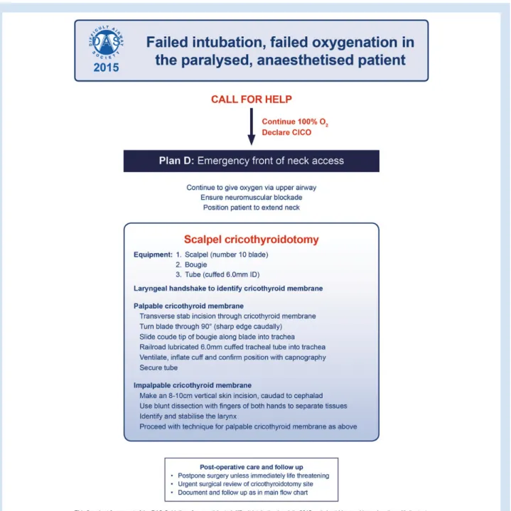
Plan D: Emergency front-of-neck access
A CICO situation arises when attempts to manage the airway by tracheal intubation, face-mask ventilation, and SAD have failed
(Table4). Hypoxic brain damage and death will occur if the
situ-ation is not rapidly resolved.
Current evidence in this area comes either from scenario-based training using manikin, cadaver, or wet lab facilities or from case series, typically in out-of-hospital or emergency
de-partment settings.267–272None of these completely replicates
the situation faced by anaesthetists delivering general anaesthe-sia in a hospital setting.
NAP4 provided commentary on a cohort of emergency surgi-cal airways and cannula cricothyroidotomies performed when other methods of securing the airway during general anaesthesia
had failed.2The report highlighted a number of problems,
includ-ing decision-makinclud-ing (delay in progression to cricothyroidotomy), knowledge gaps (not understanding how available equipment worked), system failures (specific equipment not being available), and technical failures (failure to site a cannula in the airway).
After NAP4, discussion largely focused on the choice of tech-nique and equipment used when airway rescue failed, but the
re-port also highlighted the imre-portance of human factors.2 273–275
Regular training in both technical and non-technical ele-ments is needed in order to reinforce and retain skills. Success depends on decision-making, planning, preparation, and skill ac-quisition, all of which can be developed and refined with
re-peated practice.276 277 Cognitive processing and motor skills
decline under stress. A simple plan to rescue the airway using fa-miliar equipment and rehearsed techniques is likely to increase the chance of a successful outcome. Current evidence indicates
that a surgical technique best meets these criteria.2 269 273 278
A cricothyroidotomy may be performed using either a scalpel or a cannula technique. Anaesthetists must learn a scalpel
tech-nique and have regular training to avoid skill fade.279
Scalpel cricothyroidotomy
Scalpel cricothyroidotomy is the fastest and most reliable
meth-od of securing the airway in the emergency setting.269 278 280A
cuffed tube in the trachea protects the airway from aspiration, provides a secure route for exhalation, allows low-pressure ven-tilation using standard breathing systems, and permits end-tidal
CO2monitoring.
A number of surgical techniques have been described, but there is a lack of evidence of the superiority of one over
an-other.268 281–283The techniques all have steps in common: neck
extension, identification of the cricothyroid membrane, incision through the skin and cricothyroid membrane, and insertion of a cuffed tracheal tube. In some descriptions, the skin and crico-thyroid membrane are cut sequentially; in others, a single inci-sion is recommended. Many include a placeholder to keep the incision open until the tube is in place. Some use specialist equip-ment (cricoid hook, tracheal dilators etc).
A single stab incision through the cricothyroid membrane is appealing in terms of its simplicity, but this approach may fail in the obese patient or if the anatomy is difficult, and a vertical skin incision is recommended in this situation. The approach
recommended in these guidelines is a modification of previously described techniques.
Airway rescue via the front of neck should not be attempted without complete neuromuscular block. If sugammadex has been administered earlier in the strategy, a neuromuscular block-ing agent other than rocuronium or vecuronium will be required. Oxygen (100%) should be applied to the upper airway
through-out, using a SAD, a tightlyfitting face mask, or nasal insufflation.
The use of the‘laryngeal handshake’ as described by
Levi-tan281(Fig.3) is recommended as thefirst step because it
pro-motes confidence in the recognition of the three-dimensional anatomy of the laryngeal structures; the conical cartilaginous cage consisting of the hyoid, thyroid, and cricoid. The laryngeal handshake is performed with the non-dominant hand, identify-ing the hyoid and thyroid laminae, stabilizidentify-ing the larynx between
thumb and middlefinger, and moving down the neck to palpate
the cricothyroid membrane with the indexfinger.
Standardization is useful in rarely encountered crisis situa-tions. It is recommended that the technique described below is adopted. The technique relies on the correct equipment being immediately available. Operator position and stabilization of the hands is important.
Equipment
1. Scalpel with number 10 blade; a broad blade (with the same width as the tracheal tube) is essential.
2. Bougie with coude (angled) tip. 3. Tube, cuffed, size 6.0 mm. Patient positioning
The sniffing position used for routine airway management does not provide optimal conditions for cricothyroidotomy; in this situation, neck extension is required. In an emergency, this may be achieved by pushing a pillow under the shoulders, drop-ping the head of the operating table, or by pulling the patient up so that the head hangs over the top of the trolley.
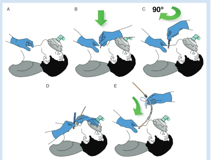
Cricothyroid membrane palpable: scalpel technique (Fig. 4; ‘stab, twist, bougie, tube’)
1. Continue attempts at rescue oxygenation via upper airway (assistant).
2. Stand on the patient’s left-hand side if you are right handed (reverse if left handed).
Table 4Key features of Plan D. CICO, can’t intubate can’t oxygenate
•CICO and progression to front-of-neck access should be declared
•A didactic scalpel technique has been selected to promote standardized training
•Placement of a wide-bore cuffed tube through the cricothyroid membrane facilitates normal minute ventilation with a standard breathing system
•High-pressure oxygenation through a narrow-bore cannula is associated with serious morbidity
•All anaesthetists should be trained to perform a surgical airway
•Training should be repeated at regular intervals to ensure skill retention
by guest on December 8, 2015
http://bja.oxfordjournals.org/
3. Perform a laryngeal handshake to identify the laryngeal anatomy.
4. Stabilize the larynx using the left hand.
5. Use left indexfinger to identify the cricothyroid membrane.
6. Hold the scalpel in your right hand, make a transverse stab in-cision through the skin and cricothyroid membrane with the cutting edge of the blade facing towards you.
7. Keep the scalpel perpendicular to the skin and turn it through 90° so that the sharp edge points caudally (towards the feet). 8. Swap hands; hold the scalpel with your left hand.
9. Maintain gentle traction, pulling the scalpel towards you (lat-erally) with the left hand, keeping the scalpel handle vertical to the skin (not slanted).
10. Pick the bougie up with your right hand.
11. Holding the bougie parallel to thefloor, at a right angle to the
trachea, slide the coude tip of the bougie down the side of the scalpel blade furthest from you into the trachea.
12. Rotate and align the bougie with the patient’s trachea and ad-vance gently up to 10–15 cm.
13. Remove the scalpel.
14. Stabilize trachea and tension skin with left hand.
15. Railroad a lubricated size 6.0 mm cuffed tracheal tube over the bougie.
16. Rotate the tube over the bougie as it is advanced. Avoid exces-sive advancement and endobronchial intubation.
17. Remove the bougie.
18. Inflate the cuff and confirm ventilation with capnography. 19. Secure the tube.
If unsuccessful, proceed to scalpel–finger–bougie technique (below).
Impalpable cricothyroid membrane: scalpel–finger–bougie technique
This approach is indicated when the cricothyroid membrane is impalpable or if other techniques have failed.
Equipment, patient, and operator position are as for the scalpel technique (Fig. 5)
1. Continue attempts at rescue oxygenation via upper airway (assistant).
2. Attempt to identify the laryngeal anatomy using a laryngeal handshake.
3. If an ultrasound machine is immediately available and switched on, it may help to identify the midline and major blood vessels. 4. Tension skin using the left hand.
5. Make an 8–10 cm midline vertical skin incision, caudad to cephalad.
6. Use blunt dissection withfingers of both hands to separate
tissues and identify and stabilize the larynx with left hand.
7. Proceed with‘scalpel technique’ as above.
Note that a smaller cuffed tube (including a Melker) can be used
provided itfits over the bougie. The bougie should be advanced
using gentle pressure; clicks may be felt as the bougie slides
over the tracheal rings.‘Hold-up’ at less than 5 cm may indicate
that the bougie is pre-tracheal.
Cannula techniques
Narrow-bore (<4 mm) cannula
Cannula techniques were included in the 2004 guidelines and have been advocated for a number of reasons, including the fact that anaesthetists are much more familiar with handling cannulae than scalpels. It has been argued that reluctance to use a scalpel may delay decision-making and that choosing a
cannula technique may promote earlier intervention.268
Whilst narrow-bore cannula techniques are effective in the
elective setting, their limitations have been well described.2 284
285Ventilation can be achieved only by using a high-pressure
source, and this is associated with a significant risk of
barotrauma.2 268 286Failure because of kinking, malposition, or
displacement of the cannula can occur even with
purpose-A
B
C
Fig 3The laryngeal handshake. () The index finger and thumb grasp the top of the larynx (the greater cornu of the hyoid bone) and roll it from side to side. The bony and cartilaginous cage of the larynx is a cone, which connects to the trachea. () The fingers and thumb slide down over the thyroid laminae. () Middle finger and thumb rest on the cricoid cartilage, with the indexfinger palpating the cricothyroid membrane.
by guest on December 8, 2015
http://bja.oxfordjournals.org/
designed cannulae, such as the Ravussin™ (VBM, Sulz,
Germany).2 268High-pressure ventilation devices may not be
available in all locations, and most anaesthetists do not use them regularly. Their use in the CICO situation should be limited to experienced clinicians who use them in routine clinical practice. Experience of training protocols carried out using high-fidel-ity simulation with a live animal model (wet lab) suggest that per-formance can be improved by following didactic teaching of
rescue protocols.287Wet lab high-fidelity simulation is unique
be-cause it provides a model that bleeds, generates real-time stress,
and has absolute end-points (end-tidal CO2or hypoxic cardiac
ar-rest) to delineate success or failure. After observation of >10 000 clinicians performing infraglottic access on anaesthetized
sheep,268 288Heard has recommended a standard operating
pro-cedure with a 14 gauge Insyte™ (Becton, Dickinson and Com-pany) cannula technique, with rescue oxygenation delivered via a purpose-designed Y-piece insufflator with a large-bore
ex-haust arm (Rapid-O2™ Meditech Systems Ltd UK). This is
fol-lowed by insertion of a cuffed tracheal tube using the Melker®
wire-guided kit. An algorithm, a structured teaching programme, competency-based assessment tools, and a series of videos have been developed to support this methodology and to promote
standardized training.287
Further evidence of the efficacy of this technique in human practice is needed before widespread adoption can be recommended.
Wide-bore cannula over guidewire
Some wide-bore cannula kits, such as the Cook Melker®
emer-gency cricothyrotomy set, use a wire-guided (Seldinger)
tech-nique.289 This approach is less invasive than a surgical
cricothyroidotomy and avoids the need for specialist equipment for ventilation. The skills required are familiar to anaesthetists and intensivists because they are common to central line inser-tion and percutaneous tracheostomy; however, these techniques
requirefine motor control, making them less suited to stressful
situations. Whilst a wire-guided technique may be a reasonable alternative for anaesthetists who are experienced with this method, the evidence suggests that a surgical cricothyroidotomy
is both faster and more reliable.288
Non-Seldinger wide-bore cannula
A number of non-Seldinger wibore cannula-over-trochar de-vices are available for airway rescue. Although successful use has been reported in CICO, there have been no large studies of
these devices in clinical practice.275 The diversity of
A
B
C
E
D
Fig 4Cricothyroidotomy technique. Cricothyroid membrane palpable: scalpel technique;‘stab, twist, bougie, tube’. () Identify cricothyroid membrane. () Make transverse stab incision through cricothyroid membrane. () Rotate scalpel so that sharp edge points caudally. () Pulling scalpel towards you to open up the incision, slide coude tip of bougie down scalpel blade into trachea. () Railroad tube into trachea.
by guest on December 8, 2015
http://bja.oxfordjournals.org/
commercially available devices also presents a problem because familiarity with equipment that is not universally available chal-lenges standardization of training.
The role of ultrasound
It is good practice to attempt to identify the trachea and the
crico-thyroid membrane during the preoperative assessment.273If this
is not possible with inspection and palpation alone, it can often
be achieved with ultrasonography.171 290The role of ultrasound
in emergency situations is limited. If immediately available and switched on it may help to identify key landmarks but should
not delay airway access.171 291 292Airway evaluation using
ultra-sound is a valuable skill for anaesthetists,292and training in its
use is recommended.273 293
Postoperative care and follow-up
Difficulties with airway management and the implications for postoperative care should be discussed at the end of the
proced-ure during the sign-out section of the WHO checklist.294In
add-ition to a verbal handover, an airway management plan should be documented in the medical record. Many airway guidelines
and airway interest groups169 295 296 (including the DAS
Fig 5Failed intubation, failed oxygenation in the paralysed, anaesthetized patient. Technique for scalpel cricothyroidotomy. Difficult Airway Society, 2015, by permission of the Difficult Airway Society. This image is not covered by the terms of the Creative Commons Licence of this publication. For permission to re-use, please contact the Difficult Airway Society.
by guest on December 8, 2015
http://bja.oxfordjournals.org/
Extubation and Obstetric Guidelines4 5) recommend that patients
should be followed up by the anaesthetist in order to document and communicate difficulties with the airway. There is a close
re-lationship between difficult intubation and airway trauma;297 298
patient follow-up allows complications to be recognized and treated. Any instrumentation of the airway can cause trauma or have adverse effects; this has been reported with
videolaryn-goscopes,163 166second-generation supraglottic devices,192 193 195
andfibre-optic intubation.299The American Society of
Anesthe-siologists closed claims analysis suggests that it is the pharynx and the oesophagous that are damaged most commonly during
difficult intubation.298
Pharyngeal and oesophageal injury are dif-ficult to diagnose, with pneumothorax, pneumomediastinum, or
surgical emphysema present in only 50% of patients.5
Mediastini-tis after airway perforation has a high mortality, and patients should be observed carefully for the triad of pain (severe sore throat, deep cervical pain, chest pain, dysphagia, painful
swallow-ing), fever, and crepitus.297 300They should be warned to seek
med-ical attention should delayed symptoms of airway trauma develop.
Despite these recommendations, communication is often
inadequate.301–304The DAS Difficult Airway Alert Form is a
stand-ard template with prompts for documentation and
communica-tion.305 The desire to provide detailed clinical information
must be balanced against the need for effective communication. At present, there is no UK-wide difficult airway database, al-though national systems such as Medic Alert have been
advo-cated306 and can be accessed for patients with ‘Intubation
Difficulties’.307
Coding is the most effective method of communicating
im-portant information to general practitioners; the code for
‘diffi-cult tracheal intubation’ is Read Code SP2y3303 308
and should be included on discharge summaries. Read Codes in the UK will be replaced by the international SNOMED CT (Systematized Nomenclature of Medicine–Clinical Terms) by 2020.
Every failed intubation, emergency front-of-neck access, and airway-related unplanned admission should be reviewed by de-partmental airway leads and should be discussed at morbidity and mortality meetings.
Discussion
Complications of airway management are infrequent. The NAP4 project estimated that airway management resulted in one ser-ious complication per 22 000 general anaesthetics, with death or brain damage complicating 1:150 000. It is not possible to study such rare events in prospective trials, so our most valuable insights come from the detailed analysis of adverse
events.2 241 262
Guidelines exist to manage complex emergency problems in other areas of clinical practice, with cardiopulmonary resuscita-tion guidelines being an obvious example. Standardized man-agement plans are directly transferable from one hospital to another and make it less likely that team members will encoun-ter unfamiliar techniques and equipment during an unfolding emergency. These guidelines are directed at anaesthetists with a range of airway skills and are not specifically aimed at airway experts. Some anaesthetists may have particular areas of expert-ise, which can be deployed to supplement the techniques described.
The guidelines are directed at the unanticipated difficult airway, where appropriately trained surgeons may not be imme-diately available, so all anaesthetists must be capable of perform-ing a cricothyroidotomy. There are some situations where these
guidelines may be loosely followed in the management of patients with a known or suspected difficult airway, and in these circumstances a suitably experienced surgeon with appro-priate equipment could be immediately available to perform the surgical airway on behalf of the anaesthetist.
Complications related to airway management are not limited to situations where the primary plan has been tracheal intub-ation; 25% of anaesthesia incidents reported to NAP4 started with the intention of managing the airway using a SAD. Whilst the key principles and techniques described in these guidelines are still appropriate in this situation, it is likely that at the point of recognizing serious difficulty the patient may not be well oxy-genated or optimally positioned.
These guidelines have been created for‘unanticipated
diffi-culty’ with airway management, and it is important that what-ever the primary plan may be, a genuine attempt has been made to identify possible difficulties with the generic Plans A, B, C, and D. Assessing mouth opening, neck mobility, and the lo-cation of the cricothyroid membrane before surgery will help to determine whether some rescue techniques are unlikely to be successful.
There are randomized controlled trials and meta-analyses
supporting the use of some airway devices and techniques,197–200
but for others no high-grade evidence is available and
recom-mendations are necessarily based on expert consensus.8In this
manuscript, individual techniques have not been listed against their levels of evidence, although other groups have taken this
approach.309
Implementation of the guidelines does not obviate the need for planning at a local level. The training required to develop and maintain technical skills has been studied in relation to vari-ous aspects of airway management, including
videolaryngo-scopy and cricothyroidotomy.109 276 310–313 To achieve and
maintain competence with devices such as videolaryngoscopes and second-generation SADs and drugs such as sugammadex, they need to be available for regular use, and local training will be necessary. New airway devices will continue to be developed and introduced into clinical practice; their place in these guide-lines will need to be evaluated. Even when no single device or technique has a clear clinical benefit, limiting choice simplifies training and decision-making. In the area of airway rescue by front-of-neck access, feedback from DAS members and inter-national experts suggested that there was a need to unify the
response of anaesthetists to the‘CICO’ emergency and to
recom-mend a single pathway. While UK anaesthetists are required to revalidate every 5 yr and advanced airway management features
in the Royal College of Anaesthetists CPD matrix314(2A01), there
is currently no specific requirement for training or retraining in cricothyroidotomy. A consistent local effort will be required to ensure that all those involved in airway management are trained and familiar with the technique. These guidelines recommend the adoption of scalpel cricothyroidotomy as a technique that should be learned by all anaesthetists. This method was selected because it can be performed using equipment available at almost every location where an anaesthetic is performed and because insertion of a large-bore cuffed tube provides protection against aspiration, an unobstructed route for exhalation and the ability
to monitor end-tidal CO2. There are, however, other valid
techni-ques for front-of-neck access, which may continue to be provided in some hospitals where additional equipment and comprehen-sive training programmes are available. It is incumbent on the anaesthetic community to ensure that data from all front-of-neck access techniques are gathered and are used to inform change when these guidelines are next updated.
by guest on December 8, 2015
http://bja.oxfordjournals.org/