THÈSE PRÉSENTÉE À
L'UNIVERSITÉ DU QUÉBEC À TROIS-RIVIÈRES
COMME EXIGENCE PARTIELLE
DU DOCTORAT EN BIOLOGIE CELLULAIRE ET MOLÉCULAIRE
PAR
JUSTINE RENAUD
LA NEURODÉGÉNÉRESCENCE DOP AMINERGIQUE EN CONDITIONS D'HYPERGLYCÉMIE
DOPAMINERGIC NEURODEGENERATION IN HYPERGLYCAEMIC CONDITIONS
Université du Québec à Trois-Rivières
Service de la bibliothèque
Avertissement
L’auteur de ce mémoire ou de cette thèse a autorisé l’Université du Québec
à Trois-Rivières à diffuser, à des fins non lucratives, une copie de son
mémoire ou de sa thèse.
Cette diffusion n’entraîne pas une renonciation de la part de l’auteur à ses
droits de propriété intellectuelle, incluant le droit d’auteur, sur ce mémoire
ou cette thèse. Notamment, la reproduction ou la publication de la totalité
ou d’une partie importante de ce mémoire ou de cette thèse requiert son
autorisation.
UNIVERSITÉ DU QUÉBEC À TROIS-RIVIÈRES
Cette thèse a été dirigée par:
Maria-Grazia Martinoli, Ph. D. Université du Québec à Trois-Rivières Directrice de recherche Institution à laquelle se rattache l'évaluateur
Jury d'évaluation de la thèse:
Maria-Grazia Martinoli, Ph. D. Université du Québec à Trois-Rivières Prénom et nom, grade Institution à laquelle se rattache l' évaluateur
Michel Cyr, Ph. D. Université du Québec à Trois-Rivières
Prénom et nom, grade Institution à laquelle se rattache l' évaluateur
Gilles Bronchti, Ph. D. Université du Québec à Trois-Rivières Prénom et nom, grade Institution à laquelle se rattache l' évaluateur
Lynda M. Williams, Ph. D. Université d'Aberdeen, Royaume-Uni Prénom et nom, grade Institution à laquelle se rattache l' évaluateur
ACKNOWLEDGMENTS
DearMom,
1 made it. It cornes as no surprise, 1 suspect, as never was there any space to spare for doubt in anyone's mind. Including my own. 1 propelled myself on this joumey, poised and tall and voracious. No space for doubt. Utterly unsuspecting of the obstacle that would lay its livid body on the tracks on my way there. The obstacle was me.
Mom, 1 met many a great demons within myself: marbles rolling to the back of the mind, dark moths fluttering in the peripheral vision, entire veils obstructing the panorama of a long-time goal. 1 did not al ways fight the good fight. At times, 1 would let them flatten me to sheets and redefine my Self. So 1 started buttering the plaster and brai ding the bricks into a fortress to keep me out of me. A Great Wall if there is one. With every single door leading me back inside.
On this joumey inside the outside of myself, 1 was lent a sea of hands. They reached to me and reeled me onto the continent, out of this illusory pit that we're aIl so familiar with. And when my identity eluded me - and this, it did many times -they gathered my pieces for when 1 would retum to reclaim them.
Mom, among these hands, there was my research supervisor, the mightiest of lighthouses, who taught me the art of focus, patience, diligence and perseverance against the stemest of storms. You know, Maria has al ways reminded me of you, in the relentless faith she bore in my aptitude to cut through the crests of waves. Across the ocean that is Research. And during this time in her lab, 1 owe today a great many other people, in tum, for their patience when 1 had none, for their incessant support when 1 was submerged. As you showed me the golden mIe of solidarity, my labmates shaped me in its application.
On my joumey, 1 sometimes shared walls with other friends who were enduring the same transformation. l've shared these on more th an one continent, countless times rescued by the voices on the other side. In Italy, Annesha, kindred spirit with an insatiable mind and the keenest sense of meta-awareness to whom my heartstrings will always be tied, however far apart our shores grow. Here, in my hometown, these voices came from the loves of my life, Simon and Jo, from the canyon of their he arts wherein whatever l've poured has al ways echoed back louder. And 1 know this thing about life now: where hands are outstretched, may the y be themselves freezing and reaching to be rescued, they are the most generous source of warmth and solace.
Looking back now at who the people closest to my heart are, 1 realize that the y share this one thing: authenticity. The ineluctable, unsaid necessity to aIl my relationships, in fact. Because 1 was raised to be true, to see truth, to eamestly trail the footfall of what is, to me, absolute. Because you raised me to embrace authenticity as the gold standard of virtue. This truth, as found in my friends and harboured in your very DNA, has salvaged my mind and kept me lucid, time and again. This truth, 1 have found it in one particular person now, Maude, on whose providential shores 1 washed up. To her, 1 owe health, 1 owe resolution, 1 owe a bewildering encounter with the idea that life may weIl just be a spectacular collision of souls. And 1 would be fine with that.
Mom, 1 made it. 1 tore down my walls, emptied the water in the belly of my boat, opened my sails like the widest eyes, catching ail the colours. 1 think you know why these words are addressed to you, why there was no doubt that 1 would succeed to begin with. Because every dimension of my intelligence, of my heart, of my resolve that allowed me to conquer myself, 1 will fore ver owe them to you. Even in the remotest moments of this strange, strange drift, Mom, your love, your music, your rightfulness have never eluded me, like an inbuilt compass. To you, 1 dedicate this thesis, the thickest, starkest endeavour into my mind and soul that 1 have ever taken on.
To my True North,
PREFACE
The work featured in this thesis represents part of the research 1 perfonned throughout my doctoral studies. These results were harvested from the Laboratory of Cellular Neurobiology at the University of Québec à Trois-Rivières, Canada, but also from a collaborative effort with the Laboratory of Neurophannacology at the University of Cagliari, Italy, where 1 completed six months of research during my doctoral studies. This thesis is presented in the fonn of scientific articles and manuscripts: as of August 2018, two have been published and one has been submitted. It also refers to two reviews and one book chapter pertinent to the core subject and for which 1 am lead author. These can be found in the appendices.
As per the authorization granted by the Cellular and Molecular Biology Graduate Program Committee on April 20th, 2017, this thesis is written entirely in English. A French surnmary of the thesis is provided in the following pages, and French surnrnaries of each of the articles are supplied at the beginning of the corresponding chapters. Please take note that the introduction and discussion sections are written in Canadian English, whereas the articles, manuscripts and reviews are written in American or British English, according to the language requirements specified by the editors of the scientific journals in which they were published or submitted. One of the reviews is also written in French.
AlI abbreviations and acronyrns employed throughout the text or within figures are listed in the table provided for this purpose. Abbreviations in text are defined upon first encounter, except for common biochemical tenns or when the fluidity of the text is hindered. Within figures, abbreviations are described in the legend upon first encounter, regardless of whether the y were first introduced in the text, but are not defined in subsequent legends. It is important to note that each chapter that features an article stands alone within this thesis and may therefore present tenns or abbreviations that are inconsistent with the remainder of the text.
Last, 1 wish to mention that this thesis explores three distinct subject matters integrated in a single discourse: dopaminergic neurons, hyperglycaemia and the polyphenol resveratrol. Granted the broadness of the topics discussed herein, the introduction is accordingly lengthy, as 1 was committed to appropriately ventilate aIl dimensions impinging on the central hypothesis and on future perspectives. Likewise, owing to the multiple techniques and models used to achieve the objectives of this thesis, the methodology is meticulously described in the aim of guiding the reader through the numerous decisions made upstream of the results presented herein.
RÉSUMÉ
Au deuxième rang des maladies neurodégénératives les plus communes après la maladie d'Alzheimer, la maladie de Parkinson atteint quelque 1 % de la population âgée de plus de 60 ans. Cette pathologie est caractérisée par la perte des neurones dopaminergiques logés dans la substance noire pars compacta du mésencéphale, conduisant à une diminution de la dopamine dans le striaturn dorsal et à l'apparition de symptômes moteurs. Malgré d'amples efforts dévoués à l'élucidation des ses mécanismes neuropathologiques, la neurodégénérescence sélective des neurones de la voie nigrostriée demeure incomprise. En effet, la voie dopaminergique mésocorticolimbique, provenant du mésencéphale au niveau de l'aire ventrale tegmentale et innervant le striaturn ventral et le cortex préfrontal, ne semble pas dégénérer. Une explication voudrait que les neurones de la voie nigrostriée expriment un phénotype distinct les rendant plus vulnérables au stress oxydant. Selon cette conjecture, les neurones dopaminergiques de la voie nigrostriée devraient exhiber une plus grande susceptibilité à toute source de stress oxydant.
Dans cette optique, nous avons émis l'hypothèse que les neurones de la voie nigrostriée sont plus vulnérables au stress oxydant engendré par l 'hyperglycémie en comparaison, par exemple, à ceux de la voie mésocorticolimbique. En premier lieu, nous avons vérifié que de fortes concentrations de glucose pouvaient causer un stress oxydant menant à la dégénérescence de neurones dopaminergiques en culture. En effet, chez les cellules PCI2 différenciées en neurones dopaminergiques, ces conditions conduisaient à une hausse des niveaux de l'anion superoxyde, une espèce réactive de l'oxygène produite en excès dans les premiers instants suivant une hyperglycémie. En maintenant ces concentrations élevées sur une période de 96 h, les cellules neuronales PCI2 entraient en apoptose, tel que le confirmaient la fragmentation de l'ADN et l'altération des profils d'expression de plusieurs marqueurs apoptotiques. De plus, l'usage d'un antioxydant éprouvé, le polyphénol resvératrol, abrogeait la hausse des niveaux d'anion superoxyde et la mort des cellules neuronales PCI2.
Suite à la confirmation que de fortes concentrations de glucose pouvaient conduire à la mort de neurones dopaminergiques en culture, nous avons voulu vérifier notre hypothèse centrale dans un modèle rongeur d'hyperglycémie. Nous avons employé un paradigme bien connu qui fait appel à l'administration de streptozotocine, une toxine ciblant les cellules productrices d'insuline, afin de générer un modèle de rat exhibant une hyperglycémie pouvant être maintenue pour une durée de 6 mois. À l'aide de méthodes immunologiques, nous avons démontré qu'une hyperglycémie chronique induisait la dégénérescence des neurones dopaminergiques de la substance noire pars compacta, mais pas de ceux de l'aire ventrale tegmentale. Conséquemment, nous observions une perte des terminaisons dopaminergiques dans le striatum dorsal qui n'était pas perceptible dans le striatum ventral. De plus, par microdialyse intracérébrale, nous avons confirmé une baisse des niveaux de dopamine explicitement dans le striatum dorsal. Cette neurodégénérescence était accompagnée de l'augmentation du nombre
d'astrocytes et de la perte de cellules microgliales au niveau de la substance noire pars compacta et du striatum, mais pas dans l'aire ventrale tegmentale.
En complément, nous avons examiné les aspects comportementaux de notre modèle de rat hyperglycémique à l'aide de tests utilisés chez les rongeurs parkinsoniens. Nos résultats démontrent l'existence de déficits moteurs évoquant ceux retrouvés chez les patients parkinsoniens. Il était intéressant de constater que ces troubles moteurs ainsi que la baisse des niveaux de dopamine se manifestaient avant que ne soit perceptible la neurodégénérescence, indiquant un possible dysfonctionnement du système nigrostrié en amont de la mort neuronale. Puisque la dopamine occupe un rôle prépondérant dans la régulation des comportements sociaux, nous avons également procédé à l'étude des interactions entre paires de rats non-familiers, tout en nous attardant à leurs communications ultrasoniques dont les paramètres sont vraisemblablement modulés par la dopamine. Lors de ces rencontres sociales, nous avons découvert que les rats hyperglycémiques exhibaient une hyper-sociabilité et une agressivité accompagnées de l'accroissement du nombre de vocalisations ultrasoniques émises, suggérant l'existence d'un dysfonctionnement dopaminergique. L'intensité de ces témoignages hyper-réactifs était de plus corrélée au degré de perte d'innervation du striatum dorsal.
La somme de ces résultats nous permet de conclure que les neurones de la voie nigrostriée exhibent une susceptibilité manifeste vis-à-vis de conditions hyperglycémiques soutenues. Ces démonstrations viennent en appui aux études épidémiologiques soulignant un risque accru de développer la maladie de Parkinson chez les patients diabétiques.
Mots-clés: comportements sociaux anormaux, déficits moteurs, diabète, hyperglycémie, maladie de Parkinson, neurones dopaminergiques, stress oxydant, voie mésocorticolimbique, voie nigrostriée.
SUMMARY
Parkinson 's disease affects an estimated 1 % of the population over the age of 60 years, making it the second most cornrnon neurodegenerative disorder after Alzheimer's disease. Fundamentally, this pathology features a progressive loss of dopaminergic neurons harboured in the substantia nigra pars compacta of the midbrain, which leads to decreased dopamine release in the dorsal striatum responsible for the emergence of motor symptoms. Despite arduous efforts deployed to improve our understanding of the neuropathological underpinnings of this disease, researchers are still at loss as to why the nigrostriatal dopaminergic pathway undergoes preferential early degeneration compared, for instance, to the neighbouring mesocorticolimbic pathway originating from the ventral tegmental area in the midbrain and projecting to the ventral striatum and prefrontal cortex. Rapidly gaining momentum is a proposition providing that nigrostriatal dopaminergic neurons possess a distinctive phenotypic liability responsible for their relative susceptibility to oxidative stress. If this holds true, nigrostriatal dopaminergic neurons should be preferentially vulnerable to unspecific oxidative insults.
On these bases, we hypothesized that nigrostriatal dopaminergic neurons are more vulnerable to hyperglycaemia-induced oxidative stress compared to other neuronal populations, expressly ones of the mesocorticolimbic pathway. We began by verifying that high glucose conditions are conducive to the death of dopaminergic neurons in culture and that this degeneration is linked to oxidative stress. When cultured in elevated yet physiological concentrations of glucose, differentiated dopaminergic neuronal PC12 cells promptly exhibited high levels of superoxide anion, a key reactive oxygen species who se overproduction constitutes the earliest event in hyperglycaemia-induced oxidative stress. Sustained for 96 h, high glucose conditions led to the apoptotic death of neuronal PC12 cells substantiated by DNA fragmentation and altered expression profiles of various markers of apoptosis. Treating neuronal dopaminergic PC12 cells with a potent antioxidative polyphenol, resveratrol, attenuated the rise in superoxide anion levels and afforded protection against high glucose-induced apoptosis.
After confirming that high glucose conditions elicit a state of oxidative stress leading to the death of dopaminergic neurons in culture, we set out to verify our central hypothesis in a rat model of long-term hyperglycaemia. We employed a well-known paradigm that utilizes streptozotocin, a toxin that targets insulin-producing pancreatic ~ cells, to generate a model presenting a hyperglycaemic phenotype that could be maintained for up to 6 months. Employing irnrnunohistochemical and immunoblotting techniques, we demonstrated that long-term hyperglycaemia in rats causes the degeneration of dopaminergic neurons in the substantia nigra pars compacta, but not in the ventral tegmental area. Accordingly, dopaminergic terminal fibres were less dense in the dorsal than in the ventral striatum of hyperglycaemic rats. Utilizing the intracerebral microdialysis technique, we also showed that dopamine release was diminished in the dorsal striatum, but not in the ventral striatum or in the prefrontal cortex. We further discovered a noticeable increase in astrocytes that was neuroanatomically coincidental to
a loss of microglial cells in the substantia nigra pars compacta and striatum, but not in the ventral tegrnental area.
Behavioural alterations were assessed in a series of tasks destined to uncover motor deficits in rodent models of Parkinson's disease. Long-term hyperglycaemic rats manifested signs of bradykinesia and gait disturbances reminiscent of parkinsonian motor syrnptoms. Interestingly, motor deficits and dampened dorsostriatal dopamine release were apparent before neurodegeneration could be discerned, suggesting possible functional impairments of the nigrostriatal pathway upstream of neuronal death. Considering dopamine also exerts an important control on social behaviours, we examined interactions between pairs of unacquainted rats and analyzed dopamine-regulated ultrasonic vocalizations known to reflect the subjects' affective state. In particular, hyperglycaemic rats engaged in markedly sociable and hyper-aggressive encounters, while emitting a greater number of ultrasonic vocalizations, evocative of a dopaminergic dysfunction. Remarkably, the intensity of these hyper-reactive phenotypes correlated with the degree of dorsostriatal dopaminergic denervation.
Taken together, our data expose the preferential vulnerability of the nigrostriatal pathway to sustained hyperglycaemia, supporting the physiological significance of their phenotypic liability to oxidative stress. Our findings further strengthen the apparent epidemiological link between pre-existing diabetes and an increased risk of developing Parkinson's disease.
Keywords: abnormal social behaviours, diabetes, dopaminergic neurons, hyperglycaemia, mesocorticolimbic pathway, motor deficits, nigrostrial pathway, oxidative stress, Parkinson' s disease.
TABLE OF CONTENTS
ACKNOWLEDGMENTS ... vii
PREFACE... ix
RÉSUMÉ... xi
SUMMARY ... xiii
LIST OF T ABLES ... xxiii
LIST OF FIGURES ... xxv
LIST OF ABBREVIATIONS AND ACRONYMS ... xxix
CHAPTERI INTRODUCTION ... 1
1.1 Dopaminergic neurons ... 1
1.1.1 Dopamine metabolism and neurotransmission ... ... ... 3
1.1.2 Central dopaminergic systems... 9
1.1.2.1 The nigrostriatal pathway .... ... ... .... ... ... ... ... Il 1.1.2.2 The mesocorticolimbic pathway... 15
1.1.2.3 Converging and diverging functions ... ... ... 16
1.1.3 Parkinson 's disease ... 18
1.1.3.1 Clinical symptomatology and treatments ... 19
1.1.3.2 Prodrome and biomarkers... 24
1.1.3.3 Pathophysiology ... 26
1.1.3.4 Vulnerability of the nigrostriatal pathway... 31
1.2 Hyperglycaemia in the central nervous system... 38 1.2.1 Glucose as a preferential fuel for the brain ... 38 1.2.2 Neuronal glucose transport ... 39
1.2.2.1 Transporters and kinetics ... 39
1.2.2.2 Physiological considerations .... ... .... ... 44
1.2.3 Neuronal glucose metabolism... 46
1.2.3.1 Specifie metabolic fates ... 48 1.2.3.2 CUITent hypotheses in neuroenergetics... ... ... 50
l.2.4 Neuronal oxidative stress in hyperglycaemia ... 54
1.2.4.1 Mitochondrial mechanisms ... 55
1.2.4.2 Rerouting mechanisms: polyol pathway and macromolecule glycation... 60
l.2.5 Vulnerability of the nigrostriatal pathway ... 65
1.2.5.1 Epidemiological basis: Parkinson 's disease in diabetic patients... 65
1.2.5.2 Molecular and cellular bases... 68
1.3 The antioxidative polyphenol resveratrol... ... 72
1.3.1 Background... ... ... ... ... 72
1.3.1.1 Historical context... 72
1.3.1.2 Dietary origins ... 74
1.3.1.3 Structure, chemistry and antioxidative functions ... 76
1.3.2 Protection of dopaminergic neurons against oxidative stress ... 78
1.3.3 Direct putative targets ... ... ... 81
1.3.3.1 Ribosyldihydronicotinamide dehydrogenase (quinone) and oxidative stress... 82
1.3.3.2 Phosphodiesterases and the energy sensing axis ... 83
1.3.3.3 Mammalian target of rapamycin and autophagy ... 85
1.3.3.4 Other targets... 87
1.4 Research aims and hypotheses ... 88
1.4.1 Objective 1: Evaluate the degeneration of cultured dopaminergic neuronal cells in high glucose conditions ... ... 89
1.4.2 Objective 2: Determine the potential of the antioxidative polyphenol resveratrol to hamper the high glucose-induced degeneration of cultured dopaminergic neuronal cells ... 90
1.4.3 Objective 3: Characterize dopaminergic neurodegeneration in a rat model of long-term hyperglycaemia ... 90
1.4.4 Objective 4: Assess the behavioural alterations resulting from nigrostriatal neurodegeneration in a rat model of long-term hyperglycaemia ... 91
1.5 Methodology ... ... .... ... ... .... ... 92
l.5.1 Objective 1: Evaluate the degeneration of cultured dopaminergic neuronal cells in high glucose conditions .... ... ... ... 92
XVll
1.5.1.1 CeU culture... 92
1.5.1.2 High glucose conditions ... 94
1.5.1.3 Superoxide anion quantification ... 96
1.5.1.4 Evaluation of apoptotic death ... 98
1.5.2 Objective 2: Determine the potential of the antioxidative polyphenol resveratrol to hamper the high glucose-induced degeneration of cultured dopaminergic neuronal ceUs ... ... ... ... 105
1.5.2.1 Resveratrol treatments ... 105
1.5.3 Objective 3: Characterize dopaminergic neurodegeneration in a rat model of long-term hyperglycaemia ... 106
1.5.3.1 Rat model of long-term hyperglycaemia ... 106
1.5.3.2 Intracerebral glucose measurements ... 114
1.5.3.3 Assessment of neurodegeneration ... 116
1.5.3.4 Assessment of glial profiles... 117
1.5.3.5 Intracerebral dopamine measurements ... 118 1.5.4 Objective 4: Assess the behavioural alterations resulting from nigrostriatal neurodegeneration in a rat model of long-term hyperglycaemia ... 122
1.5.4.1 Assessment of motor deficits... 122
1.5.4.2 Evaluation of social behaviour ... 125
CHAPTERII RESVERA TROL PROTECTS DOP AMINERGIC PC12 CELLS FROM HIGH GLUCOSE-INDUCED OXIDA TIVE STRESS AND APOPTOSIS: EFFECT ON P53 AND GLUCOSE-REGULATED PROTEIN 75 LOCALIZA TION ... 129
2.1 Author contributions ... 129
2.2 Résumé ... 130
2.3 Full article in English: Resveratrol protects dopaminergic PC12 ce Us from high glucose-induced oxidative stress and apoptosis: effect on p53 and glucose-regulated protein 75 localization ... 131
Abstract... ... ... ... ... ... ... ... ... .... 131
Introduction... 131
Materials and methods... 134
Drugs and chemicals ... 134
Detection of mitochondrial superoxide radical... ... ... ... 136
Immunofluorescence and terminal deoxynucleotidyl transferase dUTP nick end labeling as say ... ... ... .... ... 136
Specific apoptotic DNA denaturation analysis ... 137
Protein extraction... 137 Electrophoresis and Western blotting analysis ... 138
Glucose-regulated prote in 75-p53 colocalization ... 139 Statistical analysis ... 139 Results... 140
Resveratrol rescues high glucose-induced production of superoxide ... 140
Resveratrol reduces high glucose-induced apoptosis ... 141
Resveratrol modulates p53 and glucose-regulated protein 75 subcellular localization and colocalization ... 146
Discussion... 149
Acknowledgments ... ... ... ... .... ... ... ... ... 153 References... 154
CHAPTERIII DOPAMINERGIC NEURODEGENERA TION IN A RA T MODEL OF LONG-TERM HYPERGL YCEMIA: PREFERENTIAL DEGENERA TION OF THE NIGROSTRIATAL MOTOR PATHWAY ... 163
3.1 Author contributions ... 163
3.2 Résumé ... 164
3.3 Full article in English: Dopaminergic neurodegeneration in a rat model of long-term hyperglycemia: preferential degeneration of the nigrostriatal motor pathway ... ... ... ... ... .... .... ... 165
Abstract ... 165
Introduction ... 165
Research design and methods ... 167
Subjects ... 167
Induction of long-term hyperglycemia ... 168
Motor behavior assessments ... 168
Cognitive behavior... 169
X1X
Immunohistochemistry ... ... ... ... ... ... ... 170
Immunoblotting ... 171
Intracerebral microdialysis in freely moving rats ... 171
Brain tissue and microdialysate glucose concentrations... 173
Statistical analyses ... 173
Results ... 173
Glucose concentrations increase in aU brain regions of interest... 173
Long-term hyperglycemia causes preferential degeneration of dopaminergic neurons in the substantia nigra pars compacta... 174
Long-term hyperglycemia causes preferential degeneration of dopaminergic fiber terminaIs in the dorsal striatum ... 176
Long-term hyperglycemia does not cal}se substantial neurodegeneration in the prefrontal cortex or in the hippocampus ... ... 179
Long-term hyperglycemic rats display astrogliosis and loss of microglial cells in degenerated dopaminergic regions ... 182
Long-term hyperglycemic rats show altered motor behaviour ... 183
Discussion ... 185
Acknowledgments ... 188
References ... 189
Supplementary data ... ... ... ... ... ... 195
Metabolic follow-up and disease progression ... 195
CHAPTERIV LONG-TERM HYPERGL YCAEMIA MODIFIES SOCIAL BEHA VIOUR AND EMISSION OF ULTRASONIC VOCALISATIONS IN RATS: A POSSIBLE EXPERIMENTAL MODEL OF AL TE RED SOCIABILITY IN DIABETES ... 203
4.1 Author contributions ... ... ... ... ... ... ... 203
4.2 Résumé .... .... ... ... ... ... ... ... 203
4.3 Full article in English: Altered social behaviour in long-term hyperglycemic rats displaying dopaminergic striatal denervation: an ultrasonic vocalisation study ... 205
Abstract ... 205
Introduction... ... ... ... .... ... ... ... 205
Occurrences of social behaviours and ultrasonic vocalisations ... ... 207
Behavioural covariance profile ... ... ... 209
Behaviour-vocalisation covariance profile ... 211
Magnitude of social interactivity and ultrasonic vocalisations in relation to the degree of striatal denervation, hypoinsulinaemia and glucose intolerance... 212
Discussion ... 217
Methods ... 220
Subjects ... ... ... ... ... ... ... ... 220
Induction ofhyperglycaemia ... 221 Oral glucose intolerance test and baseline insulinemia .. ... 221 Ultrasonic vocalisations and social behaviour ... 222
Immunohistochemistry ... ... ... ... ... ... 223
Statistical analyses ... 224
Acknowledgments ... ... ... ... .... .... ... 225
References... 226 CHAPTERV DISCUSSION ... 235
5.1 Objectives 1 and 2: In vitro, high glucose-induced oxidative stress leads to the death of dopaminergic neurons avertible by resveratrol treatments ... 235
5.1.1 Drawing parallels with Brownlee's theory ... 235 5.1.1.1 From oxidative stress to apoptosis ... ... ... 236
5.1.1.2 Paradoxical poly(adenosine diphosphate-ribose) polyrnerase inactivation ... 238 5.1.1.3 Validating Brownlee's model ... 239
5.1.2 Resveratrol: partial antioxidative effects, but full neuroprotection ... 241
5.1.3 The relevance of glucose-regulated protein 75 in Parkinson's disease .. 243
5.2 Objectives 3 and 4: In vivo, long-term hyperglycaemia causes preferential
nigrostriatal dopaminergique neurodegeneration and consequential
behavioural alterations ... ... ... ... 243
5.2.1 Intracerebral glucose concentrations ... 244
5.2.2 Altered glial profiles as an indicator of oxidative stress ... 245
5.2.3 Nigrostriatal dopaminergic neuronal death: beyond the validation of our hypothesis ... 249
XXI
5.2.3.1 Subtle neurodegeneration and motor deficits ... 249 5.2.3.2 Time course of neurodegeneration ... 251 5.2.4 Hyper-aggressive and hyper-sociable manifestations ... 252
5.2.4.1 A possible relationship with nigrostriatal dopaminergic neurodegeneration... ... ... ... ... 253 5.2.4.2 A possible relationship with phasic and tonic dopaminergic
neurotransmission... ... ... ... ... ... 254 5.2.5 Effects attributable to hypoinsulinaemia ... 256 5.2.5.1 Insulin in neurodegeneration ... 256 5.2.5.2 Insulin in behaviour ... 260 5.2.6 Improving the model ... 265 5.3 Therapeutic perspectives ... 266 5.3.1 Implications for diabetic patients ... 267 5.3.2 Implications for parkinsonian patients ... 268 5.3.3 Employing resveratrol to therapeutic ends ... 271 5.4 Concluding remarks ... 271 REFERENCES ... 273 APPENDIXA
LA NEURO-INFLAMMA TION : DR JEKYLL OU MR HYDE? ... 389 APPENDIXB
OLD MOLECULES, NEW INSIGHTS: THERAPEUTIC
CONSIDERATIONS FOR THE USE OF POL YPHENOLS IN NEURODEGENERATIVE DISEASES ... 411 APPENDIXC
PREVENTION OF NEUROINFLAMMATION BY RESVERA TROL:
FOCUS ON EXPE~ENTAL MO DELS AND MOLECULAR
MECHANISMS ... 461 APPENDIXD
THE LINK BETWEEN PARKINSON'S DISEASE AND ATTENTION-DEFICIT HYPERACTIVITY DISORDER: AN ACCOUNT OF THE EVIDENCE ... 491
LIST OF TABLES
Table Page
1.1 Projected prevalence of select neurological conditions in Canada ... 24 1.2 Markers of prodromal Parkinson' s disease ... ... ... .... 25 1.3 List of monogenic Parkinson 's disease and parkinsonism ... 28 1.4 Overview of the 26 genetic risk variants showing consistent association
with Parkinson's disease in genome-wide association studies ... 33 1.5 Glucose transporter expression sites and substrates . ... ... 40 1.6 Glucose transport capacities of brain cells... 42 1.7 Computed compartment glucose levels for the core (primary) model or
the astrocyte-neuron lactate shuttle (ANLS)... 43 1.8 Recent studies investigating the association between diabetes and
Parkinson's disease ... 68 1.9 Direct putative targets of resveratrol... 82 1.10 Animal models of diabetes... ... ... .... .... ... ... ... ... 108 1.11 Ingredient lists of the rat diets provided by Harlan... 113
LIST OF FIGURES
Figure Page
1.1 The catecholamine biosynthetic pathway ... 5 1.2 Tonic and phasic dopaminergic neurotransmission ... 6
1.3 Dopamine reuptake and catabolism ... ... ... ... ... 8
lA Basal ganglia circuitry in decision-making... ... ... ... 10
1.5 A8-AI0 dopaminergic neuronal clusters in the rat brain ... 12
1.6 Mesocorticolimbic and nigrostriatal projections ... ... ... ... 14
1.7 The various forms ofParkinson's disease ... 20
1.8 CUITent parkinsonian drug therapies ... 22
1.9 Braak staging of the progression of Parkinson's disease-related Lewy body pathology... 30
1.10 Disease mechanisms implicated in Parkinson's disease... 34
1.11 Crosstalk between aging and Parkinson' s disease. ... ... ... ... ... 35
1.12 Glucose and monocarboxylate transporters in the mammalian brain... 41
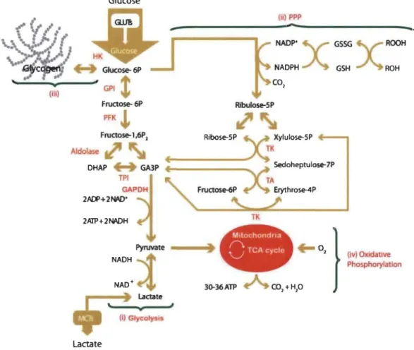
1.13 Schematic representation of glucose metabolism and pertinent connecting pathways in neurons and astrocytes ... 47
1.14 A schematic representation of the astrocyte-neuron lactate shuttle theory.... 53 1.15 Hyperglycaemia-induced generation of superoxide anion at the level of
the mitochondrial electron transport chain ... ... ... .... ... 57
1.16 Detrimental activation of P ARP in response to DNA damage... ... .... ... 58
1.17 NAD in glucose metabolism ... 61 1.18 The chemistry of prote in gl ycation ... 63
1.20 Frequently encountered polyphenols ... 75
1.21 Resveratrol's putative direct scavenging mechanisms... 77
1.22 Molecular mainstays of metabolic homeostasis... 85
1.23 Metabolic regulation of cell growth and autophagy dynamics ... ... ... 87
1.24 Characterization of dopaminergic neuronal cell cultures ... 93
1.25 Time-course and dose-response study of the toxicity of glucose on
dopaminergic cells in culture .. .... ... .... ... ... ... ... 95
1.26 Structure and fluorescent mechanism ofMitoSOXTM Red... 97
1.27 Selectivity ofMitoSOXTM Red ... 98
1.28 The classic intrinsic and extrinsic pathways in apoptosis... 99
1.29 Specificity with respect to temperature kinetics of formamide-induced
DNA denaturation ... 101
1.30 Mitochondrial translocation of Bax followed by the formation of the mitochondrial permeability transition pore ... ... 103
1.31 The multiple roles of GRP75 ... 104
1.32 Mechanisms of pancreatic
B
cel! death in chemical-induced diabetes ... 111 1.33 Metabolic follow-up of nicotinamide-streptozotocin or highfat-streptozotocin treated rats ... 112
1.34 Schematic representation of solute ex changes during microdialysis experiments ... 120
1.35 Experimental setup of social behaviour assessments ... 127
2.1 RESV reduces HG-induced superoxide anion production DAergic PC12 cel!s ... 141
2.2 RESV reduces HG-induced apoptosis in DAergic PC12 cel!s ... 143
2.3 RESV modulates the expression of apoptotic protein markers... 145 2.4 Effect ofRESV on the subcellular localization ofp53 and GRP75 ... 147
XXVll
3.1 Brain glucose concentrations in HG rats compared to CTRL rats... 174
3.2 Analyses ofmidbrain markers ofneurodegeneration in HG rats compared
to CTRL rats ... 175
3.3 Analyses of striatal markers of neurodegeneration in HG rats compared
to CTRL rats... 178
3.4 Analyses of prefrontal markers of neurodegeneration in HG rats
compared to CTRL rats ... 180
3.5 Analyses of hippocampal markers of neurodegeneration in HG rats
compared to CTRL rats ... 181
3.6 Immunohistochemical analyses of astrocytes and microglial cells in the
brains of HG rats compared to CTRL rats ... 183 3.7 Behavioral assessments of HG rats compared to CTRL rats ... ... ... .... 184 4.1 Occurrences of social behaviours and USV s... ... ... ... ... 208
4.2 Affiliative/exploratory and aggression behavioural covariance matrices ... 210
4.3 Behaviour-vocalisation covariance matrix ... ... ... .... 212
4.4 Classification of long-term HG rats based on degree of striatal
denervation, hypoinsulinaemia and glucose Intolerance ... ... 213
4.5 Occurrences of social behaviours in relation to the degree of striatal
denervation, hypoinsulinaemia or glucose Intolerance ... ... ... 215
4.6 Numbers of ultrasonic vocalisations emitted in relation to the degree of
striatal denervation, hypoinsulinaemia or glucose Intolerance. ... .... 216
4.7 Examples of sonograms of ultrasonic vocalisations ... .... .... ... ... 223
5.1 Totallevels ofp53 and GRP75 in neuronal PC12 cells treated with high
glucose concentrations ... 237
5.2 Role ofGRP75 in oxidative stress-induced apoptosis ... 238
5.3 Dose-response curve depicting the quantitative features of hormesis ... 242 5.4 Metabolic activities of astrocytes and neurons ... 247
5.5 Classification of long-term hyperglycaemic rats based on the degree of
5.6 Overview of insu lin and insulin-like peptide signalling in the brain ... 257
5.7 Insulin content and binding in the brain of streptozotocin-treated rats... 259
5.8 Nigrostriatal dopaminergic neurodegeneration in relation to the degree of hypoinsulinaemia or glucose intolerance ... ... ... 260
3-MT 3-0MD 4EBPl 6-0HDA AADC Ac ac Acetyl-CoA ACMSD aCSF AD ADP ADPR Affin Chrom AGE AgRP Akt or AKT-l AMP AMPK AN69
LIST OF ABBREVIA TIONS AND ACRONYMS
3-Methoxytyramine 3-0-Methyldopa
Eukaryotic translation initiation factor 4E (eIF4E)-binding protein 1
6-H ydroxydopamine
Aromatic amino acid decarboxylase Beta-amyloid
Acetyl group
Anterior commissure Acetyl coenzyme A
Aminocarboxymuconate semialdehyde decarboxylase gene
Artificial cerebrospinal fluid (CSF)
Aldehyde dehydrogenase or autosomal dominant or Alzheimer's disease
Adenosine diphosphate
Adenosine diphosphate ribosyl Affinity chromatography
Advanced glycation end-product Agouti-related protein
Protein kinase B
Adenosine monophosphate Adenosine monophosphate kinase Acrylonitrile 69 membrane
ANLS ANOVA AP-1 Apaf-1 APE1/Ref-1 Apo2L1TRAIL aq AR Arc
ARE
Arg ARN Atg13 ATM ATP AUC Bad Bak Bax BBB BCKDK Bel-2 Bel-XL BDNF BHEAstrocyte-neuron lactate shuttle
Anal ysis of variance
Activator prote in 1
Apoptotic protease activating factor 1
Apurinic/apyramidic endonuelease l/redox factor-1
Apoptosis antigen 2 ligand/tumour necrosis factor (TNF)-related apoptosis-inducing ligand
Cerebral aqueduct Autosomal recessive
Arcuate nueleus
Antioxidant response element
Arginine
Acide ribonucléique
Autophagy-related protein 13
Ataxia telangiectasia mutated serine/threonine kinase
Adenosine triphosphate or adénosine triphosphate
Area under curve
B celllymphoma 2-associated death promoter
B celllymphoma 2 homologous antagonist/killer
B celllymphoma 2-associated X protein Blood-brain barrier
Branched chain ketoacid dehydrogenase kinase gene
B celllymphoma 2
B celllymphoma-extra large Brain-derived neurotrophic factor
Bill Bim BSA BSLCR BSTl b.w. c-FLIP C6orfiO C CA CamKKB cAMP CART CB CBR1 cc CCDC62 CCL CCR6 CD
B celllymphoma 2 homology 3 (BH3) interacting-domain death agoni st
B cell Iymphoma 2-like protein 11
Bovine serum albumin
Barrière sang-liquide céphalo-rachidien Bone marrow stromal cell antigen 1 gene
Body weight
Cell
XXXI
First apoptosis signal receptor (Fas)-associated protein with death domain (FADD)-like interleukin-1-beta converting enzyme (lCE) (FLICE/caspase 8)-like inhibitory protein
Chromosome 6 open reading frame 10 gene
Cytoplasmic fraction
Radio-carbon dioxide
Cornu ammonis
Calciurn/calmodulin-dependent protein kinase kinase 2 or beta
Cyc1ic adenosine monophosphate
Cocaine and amphetamine regulated transcript
Cytochalasin B
Carbonyl reductase 1 Corpus callosum
Coiled-coil domain containing 62 gene
Chemokine [C-C motif] ligand
Chemokine [C-C motif] receptor type 6 Cluster of differentiation
CD200R CD95L cGMP CGRP Chem Prot CMH CN CNS CNTF co COMT
cox
CR CSF CSFIR CTC CTLA-4 CTRL or Ctrl CX3CL CX3CR CXCL Cy3 CYP Cytc DlCluster of differentiation 200 receptor
Cluster of differentiation 95 ligand
Cyclic guanosine monophosphate
Calcitonin gene-related peptide
Chemical proteomics
Comp/ex majeur d'histocompatibilité Caudate nucleus
Central nervous system
Ciliary neurotropic factor
Coeruleus/subcoeruleus complex
Catecho 1-0-meth y Itransferase
Cyclooxygenase or cyclooxygénase
Complement receptor
Cerebrospinal fluid
Colony stimulating factor 1 receptor
Cerebellothalamocortical
Cytotoxic T -1 ymphocyte-associated prote in 4
Control
Chemokine [C-X3-C motif] ligand or fractalkine
Chemokine [C-X3-C motif] receptor Chemokine [C-X-C motif) ligand
Cyanine 3
Cytochrome P450
Cytochrome c
Dl-like dopamine (DA) receptor-expressing medium spmy neurons
D2 DIID2 DA-R DA DAB DAergic DAMP DAPI DAQ DAT dc DDC DDRGKI DG DRAP DISC DLS dm DMEM DMS DNA DOPA or L-DOPA DOPAC DOPAL DR4/DR5 XXXlli
D2-like dopamine (DA) receptor-expressing medium spmy neurons
D l-like/D2-like dopamine (DA) receptors Dopamine
3,3'-Diaminobenzidine Dopaminergic
Damage-associated molecular pattern 4',6-Diamidino-2-phen yi indole Dopamine quinones
Dopamine transporter
Dorsal, caudal or dorsocaudal
3,4-Dihydroxyphenylalanine (DOPA) decarboxylase or diethyldithiocarbamate
DDRGK domain containing 1 gene Dentate gyrus
Dihydroxyacetone phosphate Death-inducing signalling complex Dorsolateral striatum
Dorsal IX/X motor nucleus
Dulbecco's Modified Eagle medium Dorsomedial striatum Deoxyribonucleic acid L-3,4-Dihydroxyphenylalanine 3,4-Dihydroxyphenylacetic acid 3,4-D ihydroxypheny lacetaldehyde Death receptor 4/5
DS or dStr dUTP e-EGCG ELKI Enz Inhib Epacl or EPAC
ER
EREERK
Ethidium f FI-ATPase F6P FAD FADD FAM47E Fas FasL FBS fc Dorsal striatum Deoxyuridine triphosphate Electron Epigallocatechin-3-gallateE26 transformation-specifie (ETS)-like transcription factor
Enzyme inhibition assay
Exchange factor directly activated by cyc1ic adenosine monophosphate 1
Estrogen receptor
Estrogen response element
Extracellular signal-related kinase
3,8-Diamino-5-ethyl-6-phenylphenanthridinium Fomix
FI portion of adenosine triphosphatase (ATPase or
A TP synthase) Fructose 6-phosphate
Flavin adenine dinuc1eotide (F AD+ oxidized, F ADH2 reduced)
First apoptosis signal receptor (Fas)-associated death
domain
Family with sequence similarity 47 member E gene First apoptosis signal receptor ligand
First apoptosis signal receptor (Fas) ligand or
ligand Fas
Fetal bovine serum
First order sensory association areas and premotor areas and/or primary sensory and motor fields of the neocortex
FcR~ FGF FIP200 PITC Fluor FM fMRI FOXO Fox03a Freq Fru-2,6-P2 F.U. G6P G6PD GA3P GABA GAK GAPDH GBA GCHl GDNF GFAP GI
Fragment crystallizable region (Fc) receptor beta subunit
Ferrous iron ion (reduced iron 2+)
Ferric iron ion (reduced iron 3+)
Fibroblast growth factor
xxxv
Retinoblastoma 1 (RBl)-inducible coiled-coil protein 1
Fluorescein isothiocyanate
Fluorescence assay
Frequency modulated
Functional magnetic resonance imaging
Forkhead box 0 Forkhead box 03 Frequency Fructose 2,6-bisphosphate Fluorescence units Glucose 6-phosphate
Glucose 6-phosphate dehydrogenase
Glyceraldehyde 3-phosphate
Gamma-aminobutyric acid
Cyclin G associated kinase/auxilin-2
Glyceraldehyde 3-phosphate dehydrogenase Beta-glucocerebrosidase gene
Guanosine triphosphate (GTP) cyclohydrolase 1 gene
Glial cell line-derived neurotrophic factor
Glial fibrillary acidic prote in Gastrointestinal
Glc Glucose
Glo Glyoxalase
Glu Glutamate
GLUT Glucose transporter
GMP Guanosine monophosphate
gp9lphox or Nox-2 Cytochrome b-245 heavy chain
GPe External globus pallidus
GPI Glucose 6-phosphate isomerase
GPi Internai globus pallidus
GPNMB Glycoprotein neuromedin B gene
GRP75 or mtHSP70 or mot-2 Glucose-regulated prote in 75 or mortalin or
mitochondrial heat shock protein 70
GS· Glutathione (GSH) radical
GSH Gamma-L-glutamyl-L-cysteinylglycine or glutathione
GSHR Growth hormone secretagogue receptor or ghrelin
receptor GSK-3 or GS3K GSSG or GSSH GSTPI GTP HAD Hb HbAlc HBSS hc
Glycogen synthase kinase 3
Glutathione disulphide Glutathione S-transferase P Guanosine triphosphate Hydrogen peroxide
Human immunodeficiency virus-associated dementia Hemoglobin
Glycated hemoglobin subunit alpha 1
Hank's balanced salt solution
High order sensory association areas and prefrontal areas of the neocortex
XXXVll
HDAC Histone deacetylase
HDACI Histone deacetylase 1
HDACi Histone deacetylase inhibitor
HF High fat, high-fat diet or high fat-fed
HG Hyperglycaemic or high glucose
hg38 Human genome build 38
HIV-l Human immunodeficiency virus-l
HK Hexokinase
HLA Human leucocyte antigen
HLA-DQBl Major histocompatibility complex, class II, DQ beta 1 gene
HMGBl High mobility group box 1
HMIT H+/myo-inositol transporter
HO-l Herne oxygenase-l
HPC Hippocampus
HPLC High-performance liquid chromatography
HRP Horseradish peroxidase
Hsp or HSP Heat shock protein
HVA Homovanillic acid
IAP Inhibitor of apoptosis proteins
Thal Ionized calcium-binding adapter molecule 1
ICso Half maximal inhibitory concentration
ICAM Intercellular adhesion molecule
IDO Indoleamine 2,3-dioxygenase
IFN Interferon
IGFIR IGF2R IGFBP or IGFBF
mc
IL IL-IR iNOS INPP5F l.p. IPN IR or INSR IRS l.v. JAK JAM JC polyomavirus JNK kcat Keapl KIR KSRPInsulin-like growth factor receptor I
Insulin-like growth factor receptor 2
Insulin-like growth factor binding proteins
Immunohistochemistry
Infralimbic prefrontal cortex or interleukin or
interleukine
lnterleukin-I receptor
Inducible nitric oxide synthase
Inositol polyphosphate-5-phosphatase F gene
Intraperitoneal
Interpeduncular nucleus
Insulin receptor
Insulin receptor substrate
Intravenous
Janus kinase
Junctional adhesion molecules
John Cunningham polyomavirus
c-Jun N-terminal kinases
Maximum turnover rate
Dissociation constant
Kelch-like erythroid cell-derived protein with cap 'n'
collar (CNC) homology (ECH)-associated prote in 1
Inhibition constant
Killer-cell immunoglobulin-like receptor
Maximum affinity or Michaelis constant
LC-MS LCR LepR LFA LHA LKBl Lmxla/b LOPD LPS LRPl Lst8 luc M MAC-l MANN MAO MA PK MAPT Mb mc MC4R MCCC1 Mcl-l
Liquid chromatography-mass spectrometry
Liquide céphalo-rachidien Leptin receptor
Lymphocyte function-associated antigen-l
Lateral hypothalamic area
Liver kinase B 1
Linll, Isl-l and Mec-3 do main (LIM) homeobox transcription factor a/b
Late-onset Parkinson's disease
Lipopolysaccharide
Low-density lipoprotein receptor-related protein 1
Target of rapamycin complex subunit lethal with SEC 13 protein 8 Leukotriene A4 hydrolase Luciferase Mitochondrial fraction Macrophage-l antigen D-Mannitol Monoamine oxidase
Mitogen-activated protein kinase
Microtubule-associated prote in tau gene
Mega base pair
Transentorhinal region and/or ectorhinal region (anteromedial temporal mesocortex)
Melanocortin 4 receptor
Methylcrotonoyl-coenzyme A carboxylase 1 gene XXXIX
MCP-l MCT MDM2 MGor3MG MGO MHC MIR4697HG ml MMP mn-SOD mPGES-l MPO MPP+ MPTP rnRNA a-MSH MT mt mtDNA mTOR mTORCl MTT MVDI N NA
Monocyte chemotactic protein-l Monocarbox ylate transporter Mouse double minute 2 homologue 3 -0-Methylglucose
Methylglyoxal
Major histocompatibility complex
Microribonuc1eic acid (miRNA) 4697 host gene
Medial lemniscus
Matrix metalloproteinases
Manganese superoxide dismutase
lnducible microsomal prostaglandin E synthase-l M yeloperoxidase
I-Methyl-4-phenylpyridinium
I-Methyl-4-phenyl-l,2,3,6-tetrahydropyridine Messenger ribonuc1eic acid
Alpha-melanocyte-stimulating hormone
Mammillothalamic tract Mitochondria
Mitochondrial DNA
Mammalian target of rapamycin
Mammalian target of rapamycin complex 1
3-(4,5-Dimethylthiazol-2-yl)-2,5-diphenyltetrazolium bromide
Diphosphomevalonate decarboxylase Nuc1ear fraction
NAcc orNAc NAD NADP NCAM NE NF-KB NGA neurons NGF NIB
NK
NO NOEL NOR Noxa NPY NQ02 Nr4a2 or NUITl Nrf2 NSERC NT3 NTS Nucleus accumbensNicotinamide adenine dinucleotide (NAD+ oxidized,
NADH reduced)
Nicotinamide adenine dinucleotide phosphate (NADP+ oxidized, NADPH reduced)
Neural cell adhesion molecule
Norepinephrine or noradrenaline
xli
Nuclear factor kappa-light-chain-enhancer of activated B cells
Neuropeptide Y (NPY)/gamma-aminobutyric acid (GABA)/agouti-related protein neurons (AgRP)
Nerve growth factor
National Institute of Health
Natural killer
Nitric oxide
Peroxynitrite
No observed effect level
Novel object recognition
Phorbol-l2-myristate-13-acetate-induced protein 1
Neuropeptide Y
Ribosyldihydronicotinamide dehydrogenase (qui none)
Nuclear receptor 4a2
Nuclear factor erythroid-derived 2 like 2
Natural Sciences and Engineering Research Council of Canada
Neurotrophin-3
NUCKSl OGTT OH ·OH OMIM OR OS OT Ox Phos P Porp p38MAPK or p38 MAPK p47phox p66Shc PAGvl PAMP PARK-A TP13A2 PARK-DJl PARK-DNAJC6 PARK-FBX07 PARK-LRRK2 PARK-parkin or PARK2
Nuclear case in kinase and cyclin dependent kinase
substrate 1 gene Superoxide anion
Oral glucose tolerance test
Hydroxyl radical
Hydroxyl radical
Online Mendelian Inheritance in Man Odds ratio Oxidative stress Olfactory tubercle Oxidative phosphorylation Phosphate group Probability
p38 Mitogen-activated protein kinases
Neutrophil cytosol factor 1
66 kDa Proto-oncogene Src homologous-collagen
homologue (Shc) adaptor protein
Ventrolateral periaqueductal grey
Pathogen-associated mo\ecular pattern
Probable cation-transporting adenosine triphosphatase (ATPase) 13A2 gene
Oncogene DJ-l gene
Auxilin gene
F-box only protein 7 gene
Leucine-rich repeat kinase 2 gene Parkin gene
PARK-PINKl PARK-SNCA PARK-SYNJl PARK-VPS35 PARP or PARP-l PBS PC12 PD PD-l PD-LI PDE PDH PET PFA PFC PFK Pfkfb3 PGC-la PGE2 PGP PI3K PIGD PKA
Phosphatase and tensin homologue (PTEN)-induced putative kinase 1 gene
Alpha-synuclein gene Synaptojanin 1 gene
Vacuolar protein sorting-associated protein 35 gene Poly( adenosine diphosphate-ribose) polymerase Phosphate-buffered saline
Pheochromocytoma cell line 12 Parkinson's disease
Prograrnmed cell death protein 1 Programmed death-ligand 1 Phosphodiesterase
Pyruvate dehydrogenase Positron emission tomography Perifomical area
Prefrontal cortex Phosphofructokinase
xliii
6-Phosphofructo-2-kinase/fructose-2,6-bisphosphatase 3
Peroxisome proliferator-activated receptor gamma coactivator l-alpha
Prostaglandin E2
Proline glycine proline peptide Inorganic phosphate
Phosphatidylinositol 3-kinase or phosphoinositide 3-kinase
Postural instability and gait difficulty Protein kinase A
PKC PKD PKM PL PLC
PMVK
POHEM POMC PPAR PPPPRR
PSG PTP Puma Put PVNQ
RAS RBC REM RESV RlPA RIT2 RNA Protein kinase C Protein kinase DPyruvate kinase isozymes
Prelimbic prefrontal cortex Phospholipase C
Phosphomevalonate kinase gene Population health model
Pro-opiomelanocortin
Peroxisome proliferator-activated receptor
Pentose phosphate pathway
Pattern-recognition receptors
Parkinson's Study Group Penneability transition pore
p53 upregulated modulator of apoptosis Putamen
Paraventricular nucleus Quinone
Free radical Rat sarcoma
Red blood cell
Rapid eye movement
Resveratrol or trans-3,5,4'-trihydroxystilbene Radioimmunoprecipitation as say
Ras-like without CAAX 2 gene
RNS ROH ROOH ROS RPMI RRF RT SCI SDFl SDS-PAGE SE SEM SIPA1L2 SIRPa SIRTI SLC2A SLC50Al Smac/DIABLO SN sn SNC SNc or SNpc SNI
Reactive nitrogen species Alcohol
Hydroperoxide compound Reactive oxygen species
Roswell Park Memorial Institute medium Retrorubral field
Room temperature Spinal cord injuries
Stromal cell-derived factor 1
Sodium dodecyl sulphate-polyacrylamide gel electrophoresis
Status epilepticus
Standard error of the me an
Signal induced proliferation associated 1 like 2 gene Signal-regulatory prote in alpha
Silent mating type information regulation (Sir) 2 homologue 1
Solute carrier family 2 gene
Solute carrier family 50 member 1 gene Second mitochondria-derived activator of
caspases/direct inhibitor of apoptosis (IAP) binding protein with low pl
Substantia nigra
Posterior portion of substantia nigra pars compacta
Système nerveux central
Substantia nigra pars compacta Substantia nigra pars lateralis
SNP SNr SOCS-l SOD Spec SPECT ssDNA STAT STK39 STN STZ TIDM T2DM TA TBHQ TBI tBID TCA TGF-~ TH Th TIMP
TK
TLR
TMEM175Single nucleotide polymorphism Substantia nigra pars reticulata Suppressor of cytokine signalling-l Superoxide dismutase
Spectroscopie assay
Single-photon emission computed tomography Single-stranded DNA
Signal transducer and activator of transcription Serine/threonine kinase 39 gene
Subthalamic nucleus Streptozotocin
Type I diabetes mellitus Type II diabetes mellitus Transaldolase
Tert-butylhydroquinone Traumatic brain injury
Truncated B celllymphoma 2 homology 3 (BH3) interacting-domain death agonist (BID)
Tricarboxylic acid
Transforming growth factor-beta Tyrosine hydroxylase
T helper cell subtype
Tissue inhibitor of metalloproteinase Transketolase or tyrosine kinase Toll-like receptor
TNF-a TPI Treg TREM2 Trp TSB TSC
TUNEL
TyrRS UCP ULKI UPS USCC USV v/v VDAC VEGF VIP VMAT Vmax VPS13C VS VTA WB WBSSH xlviiTumor necrosis factor-alpha Triosephosphate isomerase Regulatory T ceU
Triggering receptor expressed on myeloid ceUs 2 Tryptophane
Thrombospondin
Tuberous sc1erosis complex
Terminal deoxynuc1eotidyl transferase deoxyuridine triphosphate (dUTP) nick end labeling
Tyrosine-transfer RNA ligase Uncoupling protein
Une-51 like autophagy activating kinase Ubiquitin-proteasome system
University of California, Santa Cruz Ultrasonic vocalization
Volume/volume
Voltage-dependent anion channel Vascular endothelial growth factor Vasoactive intestinal peptide Vesicular monoamine transporter Maximum rate of transport
Vacuolar protein sorting 13 homologue C gene Ventral striatum
Ventral tegmental area
Western blotting or immunoblotting White, Bate-Smith, Swain and Haslam
xlviii Xray YIR YOPD Z-DEVD-FMK ZDF ZO
X-ray cocrystal structure Neuropeptide Y receptor YI Young-onset Parkinson 's disease
Fluormeth ylketone-con jugated tetrapeptide (Z-Asp[OMe ]-Glu[OMe]-Val-Asp[OMe ]-FMK) Zucker diabetic fatty
CHAPTERI
INTRODUCTION
Of all the intricate systems that make up the hurnan organism, no other has elicited more debate or has inspired such a vast array of theories regarding its functions than the central nervous system (CNS). An era's worth of research in neuroscience has amounted to our CUITent appreciation of the various temporal, spatial and thermodynamic circumstances that dictate neural outcomes in countless contexts. Nevertheless,
particular modules of the CNS continue to galvanize neuroscientists, in particular the multifarious roles dopaminergic systems fulfil in health and disease. This thesis aims to address one specific aspect of dopaminergic neurons: their selective vulnerability in certain pathological settings, with a keen focus on the nigrostriatal dopaminergic pathway involved in Parkinson's disease.
1.1 Dopaminergic neurons
Possibly the most impressive feature of CNS dopaminergic neurons is their small population size, totalling approximately 200 000 cells per hemisphere in humans (Stark and Pakkenburg, 2004), weighed against the astonishing number of pro cesses the y carry out, for instance motor control, cue-related leaming, arousal, social play,
mood modulation, endocrine regulation and many more. In fact, to sustain these functions, dopaminergic neurons make synapses with no fewer th an 200 million other neurons in the striatum, cortex, amygdala and other structures (Stark and Pakkenburg,
2004). Emerging late in prenatal development, it is possible that dopamine holds a particular position in stabilizing and integrating various other brain circuits (Grace,
2016). Conversely, dopaminergic neurons are phylogenetically ancient, occUITing in all mammals, birds, reptiles and insects, which allows assurnptions on their crucial role in
the adaptation of animal behaviour throughout evolution (Jones and Pilowski, 2002;
Smeets and Gonzalez, 2000; Yamamoto and Vernier, 2011).
Dopaminergic subpopulations were first described in the 1960s upon identification
and classification of groups of catecholaminergic neurons (Dahlstr6m and Fuxe, 1964),
later updated by others (H6kfelt et al., 1984). Of the designated A1-Al7 groups,
it is understood today that only groups A8-All comprise proper dopaminergic neurons
(Bj6rklund and Dunnett, 2007). By definition, dopaminergic neurons utilize the
catecholamine neurotransmitter dopamine (3-hydroxytyramine) to communicate and,
thus, express the rate-limiting enzyme of catecholamine biosynthesis, tyrosine
hydroxylase (TH), as weIl as aromatic amino acid decarboxylase that ultimately
produces dopamine (Bj6rklund and Dunnett, 2007). Conversely, they do not express the
enzymes necessary for the conversion of dopamine to the succeeding catecholamines in
the biosynthetic pathway, namely noradrenaline and adrenaline (Bj6rklund and Dunnett,
2007). Several other ontological hallmarks are also exclusive to dopaminergic neurons,
reviewed in greater detail by Arenas et al. (2015) 1 •
Of equal importance to their role in the CNS, dopaminergic neurons occupy
important functions in the enteric nervous system (Li et al., 2006). They are principally
located in the submucosal plexus, only sparingly so in the myenteric plexus, and are
distributed more densely in the small intestine in comparison to gastric or colonic tissues
(Li et al., 2004). By secreting dopamine onto smooth muscle cells, dopaminergic
neurons inhibit gastrointestinal motility (Li et al., 2006). Although the place he Id by
dopaminergic neurons in the proper operation of the enteric nervous system is
increasingly acknowledged (see for review Mittal et al., 2017), this thesis will focus on
CNS dopaminergic neurons.
1 The presence or absence of developmental factors, such as LIM homeobox transcnptlon factors
(Lmx 1 a/b) or the late transcription factor nuclear receptor 4a2 (N r4a2, better known as NUIT 1),
determines neuronal identity by suppression of lateral fates or activation of dopaminergic ones.
Today, human dopaminergic neurons can be prepared through direct reprogramming of pluripotent stem cells or somatic cells, for the purpose of disease modeling or regenerative therapies (Arenas et al.,
3
1.1.1 Dopamine metabolism and neurotransmission
Dopamine was first described in the hum an brain in the 1950s by Kathleen Montagu (Montagu, 1957) and named after its previously achieved chemical synthe sis utilizing the precursor L-3,4-dihydroxyphenylalanine (L-DOPA) (Fahn, 2008). Arvid Carlsson and Nils-Âke Hillarp confirmed its identity as a neurotransmitter in the CNS (Carlsson et al., 1958), the former receiving the 2000 Nobel Prize in Physiology and Medicine for this demonstration (Benes, 2001). A host of researchers later confirmed dopamine's implication in Parkinson's disease2 (Bazelton et al., 1967;
Ehringer and Homykiewicz, 1960; Homykiewicz, 1966) and behavioural disorders, such as psychoses and addiction (Baldessarini, 1985). Advances in lesion models and dopamine visualization techniques further permitted the discovery of its role in a plethora of brain functions upon which were built entire fields still evolving rapidly today.
Alongside noradrenaline and adrenaline, dopamine belongs to the catecholamine subcategory of monoamine neurotransmitters, aIl produced from amino acid metabolism and collectively important in various aspects of behaviour. The non-essential amino acid L-tyrosine is the precursor for catecholamines, easily obtained in a balanced diet or derived from L-phenylalanine hydroxylation in the liver, but not in the brain. Tyrosine must gain entry to the CNS via the large neutral amino acid transporter by competing with other amino acids (Duelli et al., 2000). Once inside neurons, the first step consists in the hydroxylation of L-tyrosine to L-DOPA by the rate-limiting enzyme TH. Aromatic amino acid decarboxylase3 then promptly decarboxylates L-DOPA to dopamine. In adrenergic neurons, dopamine is further hydroxylated into noradrenaline, which is in tum methylated to yield adrenaline (see for review of catecholamine biosynthetic pathways Daubner et al., 2011) (Figure 1.1). Whereas dopamine is produced in the cytosol of neurons, noradrenaline and adrenaline are mainly synthesized in synaptic vesicles. In order to access these vesicles for transformation into other
2 Parkinson's disease is later described in section 1.1.3.
catecholamines or for storage until its release, dopamine must use vesicular monoamine
transporter 2 (VMAT2) (Cartier et al., 2010; Eiden et al., 2004). To reduce dopamine's
presence in the cytosol, TH and aromatic amino acid decarboxylase are accordingly
complexed to the cytosolic portion of VMAT2, ensuring rapid vesicular uptake and stabilization of dopamine by the acidic environment (Guillot and Miller, 2009). This mechanism is important considering that lingering cytosolic dopamine undergoes
deleterious auto-oxidation reactions (see for review Segura-Aguilar et al., 2014), which yield dopamine-derived quinones known to react with nucleophilic components in
cells like cysteine residues of proteins (Belluzzi et al., 2012). Quinones also participate
in the synthesis of neuromelanin, a pigmented melanin analogue responsible for the dark
coloration of certain catecholamine-rich structures4 (Sulzer et al., 2000).
Dopaminergic neurotransmission occurs by phasic or tonic release (Grace, 2000) (Figure 1.2). Phasic firing involves quick spiking activity produced in dopaminergic
neurons in response to a proper stimulus-mediated action potential (Grace and Bunney,
1984a) or to presynaptic receptor activation by neighbouring neurons (Rice et al., 20 Il).
Phasic bursts of dopamine carry proper information to targets and provide them with spiking currents necessary for long-term potentiation (Wickens et al., 1996). In opposition, tonic firing represents the slower baseline pacemaker activity of
dopaminergic neurons (Grace and Bunney, 1984b) and rather fulfil a modulatory role
by regulating postsynaptic targets that are innervated by other afferents. Once secreted, dopamine binds G-protein linked metabotropic receptors that exist in five types (Missale et al., 1998) and in heterocomplexes with other dopamine or non-dopamine
receptors (Borroto-Escuela et al., 2017), allowing for a great number of outcomes at the
postsynaptic membrane. Dopamine receptors can be conveniently separated into the stimulatory, low-affinity Dl-like (Dl and D5) and inhibitory, high-affinity D2-like (D2-D4) families. It is commonly accepted that D2-like receptors are more sensitive to
tonic neurotransmission than D l-like receptors that rather transduce phasic bursts
4 Neuromelanin's role in health and disease is unclear. On the one hand, it chelates transition metals like
iron ions and may therefore protect catecholaminergic neurons from Fenton reactions that produce
reactive oxygen species (ROS). However, under certain circumstances, neuromelanin also departs itself
5
(Dreyer et al., 2010), but paradigms have been shifting in recent years (Yapo et al., 2017). Figure 1.1 HO
n
°
OH L-Tyrosine 1 # NH2 ~ Tetrahydro- ~ biopterin Tyrosine hydroxylase ~o. Di hydro-biopterinHoX){~
a OH
L-Dihydroxyphenylalanine # NH2 (L-DOPA) HO~
DOPA decarboxylase1
Aromatic L-amino acid decarboxylase HO~~
NH
2 HO Dopamine acid~Ascorbic ~
Dopamine J3-hydroxylase ~. Dehydro -ascorbic acidHO~
N
~H2
HO Norepinephrine S-adenosyl- ~ methiomne Homocysteine Phenylethanolamine N-methyltransferase EpinephrineThe catecholamine biosynthetic pathway.
(From Wikimedia Commons: https://commons.wikimedia.org/wikilFile:
Figure 1.2
Tonie dopamine release
low, irregular firing
~
Dopamine neuron1
Glutamate receptor Dopamine-;,e•
•
Phasie dopamine release
Dopamine release in synapse Burst firing
WrU
1
••
•
•
Extra-synaptic dopamine release Escape From synapse curtailed bydopamine transporter Dopamine tran porterTonie and phasie dopaminergic neurotransmission.
Top: Constant low levels of extracellular dopamine are ensured by
tonic release, dependent on slow irregular firing and modulated by
glutamatergic afferents. Release mainly occurs extrasynaptically where
catechol-O-methyltransferase ensures dopamine degradation. Bottom: Phasic dopamine neurotransmission is triggered by burst firing of
dopaminergic neurons, which release very high levels of dopamine into
the synaptic c1eft that stimulate postsynaptic dopamine receptors.
Phasic dopamine is rapidly inactivated by removal from the synaptic c1eft