THÈSE PRÉSENTÉE À
L'UNIVERSITÉ DU QUÉBEC À TROIS-RIVIÈRES
COMME EXIGENCE PARTIELLE
DU DOCTORAT EN BIOLOGIE CELLULAIRE ET MOLÉCULAIRE
PAR
MD BULBUL AHMED
SHAM TO DECEIVE (STD), A FUNGAL EFFECTOR AND ITS FUNCTIONAL STUDIES
Université du Québec à Trois-Rivières
Service de la bibliothèque
Avertissement
L’auteur de ce mémoire ou de cette thèse a autorisé l’Université du Québec
à Trois-Rivières à diffuser, à des fins non lucratives, une copie de son
mémoire ou de sa thèse.
Cette diffusion n’entraîne pas une renonciation de la part de l’auteur à ses
droits de propriété intellectuelle, incluant le droit d’auteur, sur ce mémoire
ou cette thèse. Notamment, la reproduction ou la publication de la totalité
ou d’une partie importante de ce mémoire ou de cette thèse requiert son
autorisation.
Cette thèse a été dirigée par :
Hugo Germain, Ph. D. Université du Québec à Trois-Rivières Directeur de recherche Institution à laquelle se rattache l'évaluateur
Isabel Desgagné-Penix, Ph. D. Université du Québec à Trois-Rivières Codirectrice de recherche Institution à laquelle se rattache l' évaluateur
Jury d'évaluation de la thèse:
Hugo Germain, Ph. D. Université du Québec à Trois-Rivières Prénom et nom, grade Institution à laquelle se rattache l'évaluateur
Isabel Desgagné-Penix, Ph. D. Université du Québec à Trois-Rivières Prénom et nom, grade Institution à laquelle se rattache l'évaluateur
Guy Samson, Ph. D. Université du Québec à Trois-Rivières
Prénom et nom, grade Institution à laquelle se rattache l'évaluateur
Vincent Maire, Ph. D. Université du Québec à Trois-Rivières Prénom et nom, grade Institution à laquelle se rattache l'évaluateur
David Joly, Ph. D. Université de Moncton
Prénom et nom, grade Institution à laquelle se rattache l'évaluateur
The following thesis focuses on the functional studies of Mlp124478, a poplar leaf rust effector. The study described here will contribute to the functional genornic studies to unravel the interaction between effectors and their targets.
The mam body of the thesis consists of six different chapters: introduction, materials and methods, results, discussion, conclusion and references. The frrst chapter starts with the general introduction of poplar leaf rust M larici-populina, its sequenced genome, prelirninary studies on genornics and transcriptornics, candidate effector discovery, importance of heterologous model systems in plant-pathogen interaction studies, and subcellular localization of effectors inside plant tissues.
Since Duplessis et al (2011) published the whole genome sequence of M larici-populina, it gave an access to further study the candidate effector proteins at molecular
level and investigate their role in pathogenesis. We chose an effector, Mlp124478 for the functional genornic studies. To this end, we used in planta pathogen assays, genotyping, live-cell imaging, comparative transcriptomics, protein-nucleic acid interaction and yeast two-hybrid assay to infer the functional nature of Mlp124478. The detailed materials and methods are discussed in chapter two. In the third chapter, we explained our fmdings, and discuss on those fmdings in chapter four. To our knowledge this is the frrst attempt at using transcriptornics of transgenic Arabidopsis plants expressing rust fungal effector (M larici populina effector) to understand the expression pattern of genes differentially expressed in presence of an effector.
We published a review on relationship between virulence function of effectors and their subcellular accumulation in the host cells in the journal "Virulence" which is presented in Annex B. l carried out two additional research projects, (1) localization of Arabidopsis TAF15b and its role in plant irnmunity, and (II) nuclear protein components participating in MAMP-triggered irnmunity. The manuscripts were published in
Molecular Plant-Microbe Interactions (MPMI) and Plant Signaling and Behavior, respectively, which are presented in Annex C and D. The manuscript describing my doctoral research on Mlp124478 is to be submitted to ''Nature Scientific Reports" and is presented in Annex E.
In conclusion, the studies presented in the following thesis provide novel insights into Mlp124478 functions in the plant, such as target in the host cell, remodelling host transcription process to alter plant gene expression for the benefit of the pathogen.
First, 1 would like to thank my research director Hugo Germain for including me in his team, day to day support and guidance, and introducing me to the amazing research field of molecular interactions of plants and pathogens. 1 would like to thank my co-director, Isabel Desgagné-Penix for her ideas and support to my research. 1 am grateful for the opportunities, support and advices they have offered.
Secondly, 1 would like to express my thanks to the Fonds de la Recherche sur la Nature et les Technologies du Québec (FRQ-NT) for providing me the scholarship for my Ph.D. Thanks to the Conseil Franco-Québécois de Coopération Universitaire (CFQCU) for providing a travel grant for laboratory training at INRA, UMR 1136 Interactions ArbreslMicroorganismes, INRAIUniversité de Lorraine, Nancy, France. 1 express my gratitude to NSERC, since our laboratory was partially funded by NSERC Discovery Grants.
1 would like to express my gratitude towards Benjamin Petre (The Sainsbury Laboratory, Norwich Research Park, UK) for providing his constant support, advices and friendship. 1 would also like to thank Sébastien Duplessis and Arnaud Hecker (INRA, UMR 1136 Interactions ArbreslMicroorganismes, INRAlUniversité de Lorraine, Nancy, France) for their technical assistance, advices and great discussions on recombinant protein production. 1 would like to thank David Joly from the University of Moncton for the fruitful discussion. Additionally, 1 would like to acknowledge Prof essor Marc Beauregard and Céline Van Themsche (Université du Québec à Trois-Rivières) for sharing instruments which helped me a lot during my experiments.
1 am very much thankful to Karen Cristine Gonçalves dos Santos and Marie-Ange Massicotte for helping me in my experiments. 1 am thankful to my ex-Iab colleagues, Ouassila Gaouar, Fakih Zainab, Prateek Chaudhari, Bruno Debotz and Catherine Arnireault for their helpful efforts and discussions. 1 would like to thank
Mélodie B. Plourde for assisting in my research and valuable discussions. Thanks to all of my colleagues in the lab, Genevieve Laperriere, Hur Madina, Claire Letanneur, Apama Singh, Narimene Fradj, Andrew Diamond, Tarun Hotchandani, Annabelle St-Pierre, Doriane Blondeau and Nicolas Dufour.
Special thanks to my beloved wife, Saima Sultana for aIl of her supports, encouragement and help to release my stresses during my PhD. 1 would like to thank Laurent Bekale, Ahmad, Ihmed Hasni for sharing their ide as and supports with true friendship in guiding me to settle in Trois-Rivieres. Finally, 1 would like to thank my family and friends at home and abroad for their great support during my studies.
La rouille du peuplier est causée par le basidiomycète Melampsora larici-populi na, qui infecte les tissus des feuilles et y sécrètent, via des structures d'alimentation spécialisées appelées haustoriums, des protéines effectrices, aussi nommés effecteurs. Les mécanismes par lesquels les effecteurs de la rouille du peuplier favorisent la virulence du pathogène sont mal connus. L'effecteur Mlp124478 a été sélectionné comme sujet d'étude de cette thèse parmi un groupe de 1184 petites protéines sécrétées par Ma larici-populina. Le gène codant pour Mlp124478 appartient à la famille CPG2811, qui comporte neuf membres qui sont spécifiques à l'ordre des Pucciniales. Son expression est fortement augmentée lors de l'infection et peut atteindre des niveaux 50 fois plus élevés que le niveau basal 96 h après le début de l'infection, un moment qui correspond, selon la cinétique d'infection par M larici-populina, au stade de croissance biotrophique à l'intérieur des cellules du mésophylle. Mlp124478 est le seul membre de sa famille à posséder une séquence prédite de localisation nucléolaire (NoLS).
Nous avons utilisé les plantes modèles Arabidopsis thaliana et Nicotiana benthamiana pour déterminer que Mlp124478 se retrouve principalement dans les
noyaux et dans les nucléoles et qu'il favorise la croissance de l'oomycète pathogène
H arabidopsidis. L'expression constitutive de Mlp124478 chez A. thaliana altère la
morphologie des feuilles de rosette, qui acquièrent une apparence ondulée, et diminue l'expression des gènes impliqués dans la réponse immunitaire. Nos résultats indiquent en effet que les gènes surexprimés en présence de Mlp124478 ne sont pas des gènes spécifiquement impliqués dans défense des plantes mais que ces derniers sont plutôt parmi les plus sous-exprimés. En somme, nos résultats suggèrent que Mlp124478 manipule les plantes en ciblant les compartiments nucléaires, où il altère la transcription par sa liaison au promoteur TGAla afm de supprimer la réponse transcriptionnelle à l'exposition aux agents pathogènes et d'augmenter l'expression de gènes qui ne sont pas impliqués dans les réponses de défense. Étant donné l'effet trompeur que cet effecteur
exerce sur l'immunité et la réponse aux pathogènes, nous proposons de le renommer .s.HAM IO DE CE IVE (STD) et c'est ce nom qui sera utilisé pour cette thèse. Nos résultats actuels concordent avec la présence de STD dans le noyau, nous ne pouvons cependant pas écarter que cet effecteur ait une fonction distincte dans le nucléole.
À notre connaissance, il s'agit de la première utilisation de données de transcriptomique obtenues à partir de plants d'A. thaliana transgéniques exprimant un effecteur de Melampsora-larici populina pour étudier les patrons d'expression différentielle. Cette étude transcriptomique a le potentiel d'augmenter considérablement nos connaissances sur les patrons de régulation de l'expression des gènes en réponse aux pathogènes biotrophes, et n'aurait pas pu être réalisée en utilisant le modèle d'expression transitoire dans N benthamiana.
The basidiomycete Melampsora larici-populina causes poplar leaf rust, invades leaf tissue and secretes effector proteins through specialized feeding structures known as
haustoria. The mechanisms by which rust effectors promote pathogen virulence are
poody understood. Out of 1184 small secreted proteins, Mlp124478 has been chosen in this study, it belongs to the CPG2811 gene family which has 9 members specific to the
Pucciniales. Mlp124478 expression is strongly enhanced during infection and reaches
50-fold induction at 96 h after infection. Given the kinetics of M larici-populina infection, this corresponds to the biotrophic growth stage in mesophyll cells. Mlp124478 is the on1y one in its family which contained predicted nucleolar localization
sequence (NoLS).
We investigated the model plants Arabidopsis thaliana and Nicotiana
benthamiana and established that Mlp124478 accumulates in the nucleus and nucleolus,
and promotes growth of the oomycete pathogen Hyaloperonospora arabidopsidis.
Stable constitutive expression of Mlp124478 in A. thaliana altered leaf morphology,
observed through increased waviness of rosette leaves and repressed expression of genes involved in immune responses. Our results indicate that the list of up-regulated genes did not specifically contain genes involved in plant defence, but rather genes involved in defence were among the most repressed. Taken together, our results suggest that Mlp124478 manipulates plants by targeting nuclear compartments, and remodeling transcription via binding to TGA1a promoter to suppress the transcriptional response to pathogens, and mislead the host into up-regulating the expression of genes unrelated to defence. Therefore, we suggest to rename (as use thereafter) this gene as S.HAM ID DECEIVE (STD), to reflect its effect on plant immunity. While our CUITent results are consistent with STD in the nucleus, we cannot rule out that it could also have a distinct function in the nucleolus.
To our knowledge, this is the fust attempts at using transcriptomics of transgenic Arabidopsis plants expressing M -larici populina effector to understand the expression pattern of genes differentially expressed in presence of effector. Since Mlp124478 accumulates in the nucleus and nucleolus, the study of transcriptome of transgenic plants over-expressing effector might boost our understanding of the regulatory pattern of gene expression in response to biotrophs, which could not as easily be performed using transient expression in N benthamiana.
Keywords: Mlp124478, effectors, nucleolus, biotroph, transcription, transcription factor-binding sites
PREFACE... üi ACKN"OWLEDGEMENTS... v , , RESUME... vii ABSTRACT ... ix LIST OF FIGURES ... xv
LIST OF TABLES ... xvii
LIST OF ABBREVIATIONS AND ACRONYMS ... xvili CHAPTERI INTRODUCTION ... 1
1.1 Plant Pathogens-constraint to world food supply ... 1
1.2 Plant defence systems... ... 2
1.2.1 Passive defence ... ... ... 3
1.2.2 Active defences ... 4
1.2.2.1 P AMP-Triggered Immunity (PTI) ... 6
1.2.2.2 Effector-triggered immunity (ET!) ... ... 7
1.3 Resistance proteins ... 8
1.4 Molecular models ofplant-pathogen recognition... 13
1.5 Plant pathogens... 15
1.5.1 Viral pathogens ... 15
1.5.2 Bacterial pathogens ... 16
1.5.3 Oomycete pathogens... 18
1.5.4 Fungal pathogens ... 19
1.5.4.1 Rust fungus as pathogen... 21
1.6 Plant pathosystems... 22
1.7 Effectors... 23
1.7.1 Rust effectors ... 23
1.7.2 Effectors and respective helpers and targets... 26
1.9 Post-genomic approaches for CSEPs... 33
1.10 Know-how from the genome and transcriptome of poplar and M larici-populina... ...... 37
1.11 Heterologous model system for effector studies ... 39
1.12 Subcellular localization of effectors ... 42
1.12.1 Subcellular localization of M larici-populina effectors... 44
1.12.2 Nuclear localized effectors ... ... 47
1.13 Effectors suppression of PTI ... 48
1.13.1 Effectors and host cellular reprogramming ... 49
1.14 Specifie problematic ... 51
1.15 Objectives ... 51
CHAPTERII -MATE RIAL AND METHODS... 53
2.1 Plant Materials and growth conditions ... 53
2.2 Growth of P. syringae pv. tomato and infection assay... 53
2.3 H arabidopsidis Noco2 infection ... 54
2.4 Plasmid construction... 54
2.5 Cloning of genes ... 55
2.6 Colony PCR... 56
2.7 Generation of stable transgenic lines... 56
2.8 Transient expression ... ... 56
2.9 Microscopy ... ... ... 57
2.10 Chromatin immunoprecipitation-polymerase chain reaction (ChIP-PCR) assay... 57
2.11 Electrophoretic mobility shift assay (EMSA)... 59
2.12 RNA extraction and transcriptome analysis ... 60
2.13 Yeast Two-Hybrid Assay ... ... ... 62
2.14 Bioinformatics analyses... ... 62
CHAPTERill RE SUL TS ... 64
3.1 The candidate effector Mlp124478 was selected for functional characterization... 64
3.2 Mlp 124478 affects the shape of A. thaliana leaves... 68
3.3 Mlp124478 accumulates in the nuclear area inA. thaliana leaves... 69
3.4 Mlp124478 carries a Nuclear Localization Sequence ... 70
3.5 Mlp124478 augment oomycete pathogen growth ... 73
3.6 Mlp124478 interacts with several proteins in yeast two hybrid... 76
3.7 Mlp124478 effect is revealed by gene expression network... 80
3.8 Mlp124478 alter the pattern of gene expression ... 86
3.9 Mlp124478 reprogram host processes by binding to DNA... 92
3.10 Mlp124478 ChIP interactor has a poplar homolog with sirnilar regulatory sequence... 94
CHAPTERIV DISCUSSION ... 97
4.1 Mlp124478 alters plant phenotype and response to pathogens growth ... 97
4.2 Mlp124478 accumulate in plant nuclear compartment ... 99
4.3 Mlp124478 contains novel NoLS ... 101
4.4 Mlp124478 nuclear accumulation benefit the pathogen ... 102
4.5 Mlp124478 may interacts with multiple proteins in the host ... 103
4.6 Mlp124478 tricks plant defence by suppressing immune regulators ... 103
4.7 Homolog gene in Arabidopsis and poplar ... 107
4.8 Proposed model for STD ... 108
CHAPTERV CONCLUSION ... 110
5.1 Conclusion... 110
5.2 Perspectives ... 113
5.2.1 Short term perspectives... 113
5.2.1.1 Confirmation ofNoLS as a novel NoLS sequence ... 113
5.2.1.2 Assessment of possible interactors of Mlp 124478 ... 114
5.2.2 Long term perspectives ... 114
5.2.2.1 Effect ofNoLS removal on pathogenesis ... 114
5.2.2.2 Confirmation of possible interactors of Mlp 124478 ... 115
ANNEXA
SUPPLEMENTARY DATA FOR CHAPTER II AND III ... 137
ANNEXB
EFFECTOR BIOLOGY DURING BIOTROPIDC INVASION OF PLANT CELLS... 162
ANNEXC
ARABIDOPSIS TAF15B LOCALIZES TO RNA PROCESSING BODIES AND CONTRIBUTES TO SNCI-MEDIATED AUTOIMMUNITY ... 170
ANNEXD
AN UNBIASED NUCLEAR PROTEOMICS APPROACH REVEALS NOVEL NUCLEAR PROTEIN COMPONENTS THAT PARTICIPATES
IN MAMP-TRIGGERED IMM"UNITY ... 181
ANNEXE
A FUNGAL RUST EFFECTOR TARGETS THE PLANT CELL NUCLEUS AND MODULATES TRANSCRIPTION ....... 192
Figure Page
1.1 Disease triangle... 2
1.2 Outline of plant defence systems against different pathogens... ... 3
1.3 The zigzag model ofthe plant immune system ... ... 5
1.4 Model of MAPK signaling pathway and PTI ... 7
1.5 Generalized model of R protein function and activation... ... 10
1.6 R protein pathways inA. thaliana............................... 12
1. 7 Molecular models of pathogen recognition .. ... 14
1.8 Disease symptoms of top ten fungal pathogens... 21
1.9 Cartoon showing haustoria formation... 22
1.10 Effectors and their host-cell helpers and targets ... 28
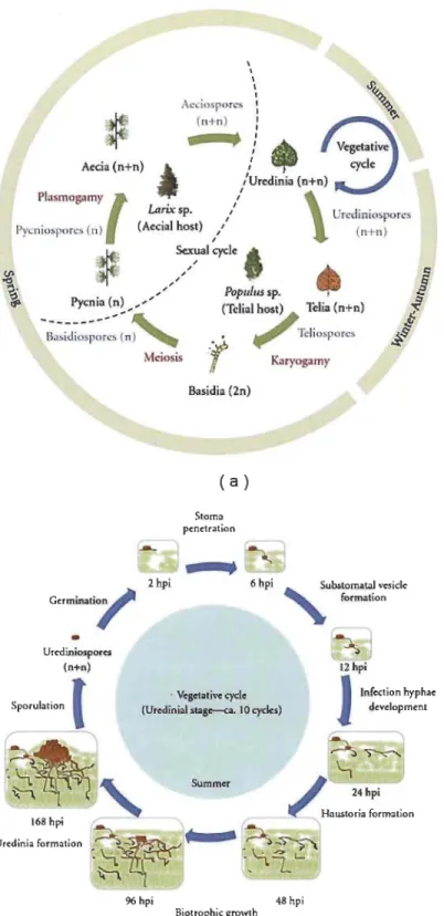
1.11 Life cycle of M larici-populina........... 32
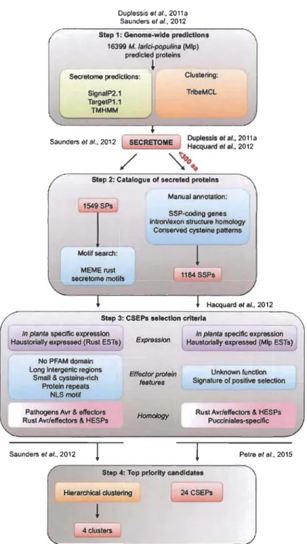
1.12 Pipelines of effector mining of M larici-populina for prioritizing CSEPs ... 36
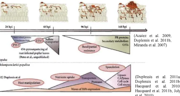
1.13 Major transcriptome regulations in a compatible interaction of Poplar-M larici-populina ... 38
1.14 Arabidopsis plants expressing HaRxLs and response to pathogens ... 41
1.15 Schematic diagram showing host, haustoria and effectors distribution... 43
1.16 Transient expression of tweenty candidate effectors of M larici-populina.. 46
2.1 Gateway recombination reactions... 55
2.2 Sketch of ChIP-PCR ... 59
2.3 Screenshot of green line work:flow ... 61
3.2 Signal peptide and nuclear localization sequences within the peptide
sequence ofMlp124478 ... 65
3.3 Overview offunctional studies of Mlp124478 ... 67
3.4 Phenotype of Mlp124478 inA. thaliana transgenic ... 69
3.5 Subcellular accumulation ofMlp124478 inA. thaliana leaves.... 70
3.6 Transient expression of Mlp124478 showed NLS acts as NoLS... 72
3.7 Defence response of Mlp124478 against bacterial and oomycete pathosystem... 75
3.8 Colocalization of Mlp124478-GFP and RFP-HSPl... 78
3.9 Pathogenic growth response ofHSPI using bacterial pathosystem ... 79
3.10 W ork flow of transcriptomics ... 81
3.11 Gene Ontology (GO) term enrichment network analysis using Cytoscape... 83
3.12 Abundance ofTFBSs among up- and.down-regulated genes... 86
3.13 Regulation of gene expression level ... 91
3.14 ChIP-PCR assay ofTFBSs determining DNA-binding ability... 93
3.15 EMSA of TGAla and DNA-binding domain of Mlp124478 to assess b· d' ID Ing actIvlty ... . 94 ..
3.16 Complete exon and intron structure with TFBSs at the upstream of gene .... 96
Table Page
1.1 Major classes of resistance (R) proteins ... 9
1.2 Top ten viral pathogens in plants ... 16
1.3 Top ten bacterial plant pathogens ... 17
1.4 Top ten oomycete plant pathogens ... 18
1.5 Summary of the top ten fungal pathogens in molecular plant pathology ... 20
1.6 List of rust effector proteins identified so far ... 25
1.7 Subcellular accumulation of candidate effectors of M larici-populina ... 45
3.1 Potential interactors of Mlp124478 identified through Y2H ... :... 77
3.2 GO enrichment of down-regulated (cluster 1) and up-regulated genes of Mlp124478 versus Col-O lines ... 84
3.3 List ofup-regulated genes abundant with TFBSs... 87
ABRC ALSD
ARL
AtML012 AVR bZIP CAZys CC CC-NBS-LRR cfu CgEPl ChIP ChIP-PCR CoIP CRK21 CRN CSEPs CVC DIG Dpi DS EDS1 eds1-1Arabidopsis Biological Resource Center Almond leaf scorch disease
ARGOS-like
MlLDEW RESISTANCE LOCUS 0 12 Avirulent
Basic region/leucine zipper motif Carbohydrate active enzymes Coiled-Coil
Coiled-coil Nucleotide-Binding Site Leucine-Rich Repeat Colony forming unit
Colletotrichum graminicola Effector Protein 1 Chromatin Immunoprecipitation Assay
Chromatin immunoprecipitation-polymerase chain reaction Coimmunoprecipitation
Cysteine-rich receptor-like protein kinase 21 Crinkler
Candidate secreted effector pro teins Citrus variegated chlorosis
Digoxigenin
Days post infiltration Double-stranded
ENHANCED DISEASE SUSCEPTIBILITY 11 / Enhanced disease susceptibility 1-1
EHM EM EMSA ESMI EST ET! ETS GFP GO H a Noco2 HD-ZIP HESPs HMG HR HSPI IBIS IN INo IP K LB LCM LRRs MAMP MAPK Extrahaustorial matrix Extrahaustorial membrane
Electrophoretic mobility shift as say Epithiospecifier modifier 1
Expressed sequence tag
Effector-Triggered Immunity Effector-triggered susceptibility
Green Fluorescent Protein
Gene Ontology
Hyaloperonospora arabidopsidis Noco2
Homeodomains associated with the Leucine Zipper
Haustorially Expressed Secreted Proteins High mobility group
Hypersensitive response
Heat shock protein 20 like protein 1
Institut de Biologie Integrative et des Systems
Nuclear intensity
Nnucleolar intensity
Immunoprecipitated
Lysine
Luria-Bertani
Laser Capture Microdissection
Leucine Rich Repeats
Microbes associated molecular pattern
Mlp
MS N NBS-LRR NDRI NLS No NoLS NPRI NTP OD600 ORF PAD4 PAMP PCD Melampsora larici-populina Mass Spectrometry NnucleusNucleotide-Binding Site Leucine-Rich Repeat
NONRACE SPECIFIe DISEASE RESISTANCE 1 Nuclear Localization Signal
Nucleolus/nucleoli
Nucleolar Localization Sequnce
NONEXPRESSOR OF PR-l
NAC transcription factors Optical density at 600 nm Open reading frame
PHYTOALEXIN DEFICIENT 4
Pathogen associated molecular pattern Programmed cell death
PD Pierce's disease of grapevine
PRR Pathogen recognition receptors
PstDC3000~CEL Pseudomonas syringae pv. tomato strain DC3000~CEL
pth Pathogenicity gene
PTI P AMP-Triggered Immunity
R R RFP RIN4 RLKs Arginine Resistance
Red Fluorescent Protein RPMI Interacting Protein 4 Receptor-Like Kinases
RLPS ROS RT SA SAGIOI SAP11 SLHI snc! SPs SSPs T3SS TAL TCP TFBSs TFs TIR TIR-NBS-LRR TSS Y2H YEP Receptor-Like Proteins Reactive Oxygen Species Room temperature Salicylic acid
SENESCENCE ASSOCIATED GENES 101
Secreted A Y-WB Protein Il
SENSITIVE TO LOW HUMIDITY 1
Suppressor of nprl Secretory pro teins Small secreted protein Type three secretion system Transcription Activator-Like
Teosinte branched 1/CycloideaIProliferating Transcription factor-binding sites
Transcription factors TolVInterleukin-1 Receptor
TolVInterleukin-1 Receptor Nucleotide-Binding Site Leucine-Rich Repeat
Transcription start site Yeast two-hybrid Yeast extract peptone
INTRODUCTION
1.1 Plant Pathogens-constraint to world food supply
Plants provide world's food supply as well as fuel, shelter, medicine and
transportation. Like animaIs, plants are under continuous attack of pathogens impending
threat to food security. Plant pathogens belongs to various taxa such as nematodes (Jones et al. 2013), virus (Scholthof et al. 2011), bacteria (Mansfield et al. 2012),
oomycetes (Kamoun et al. 2015), fungi (Dean et al. 2012), viroids (Owens and
Hammond 2009) and parasitic plants (Hibberd and Dieter Jeschke 2001). Plant diseases
are threat to world's crop production. Among food crops, disease epidemics have driven
to famines and huge migrations over the history, such as Irish potato famine of
1845-1849. Since potato was the main crop in Ireland, Phytophthora infestans the causal
agent of potato blight disease created extensive damages throughout Ireland. As a result of the disaster, approximately 1 million Irish people die d, 1.3 million people migrated and 300,000 births did not take place (Boyle and Grado 1986). However, fundamental
components of plant disease epidemics comprise abundance and susceptibility of crops,
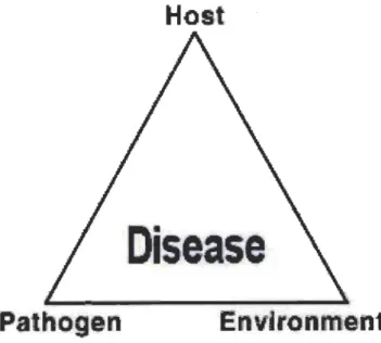
abundance and virulence of pathogen and favorable environments (Fig. 1.1) (Francl
2001). Decline of pathogenic inoculum, suppressing pathogen virulence and increasing
of crop genetic assortment may minimize plant diseases which eventually sustain our
steady food supply (Strange and Scott 2005). Hence, molecular understanding of
pathogenicity, disease resistance and plant-pathogen interactions are indispensable to control plant pathogens and retain plants healthy for food security.
Host
Dis ase
Fig. 1.1 Disease triangle.
Three causal factors are necessary for causing disease: host, pathogen and environment. Successful disease development requires the presenc of a biotic agent with an interaction of a susceptible host, a virulent pathogen and favourable environment. On the other hand, upon the exclusion of interaction of any of these three component plant disease is prevented. (Adapted from Stevens, 1960.)
1.2 Plant defence systems
Plants are constantly challenged by their surrounding environments such as biotic (virus, bacteria, oomycetes, fungi, nematodes, etc.) and abiotic factors ( drought, temperature, nutrient deficiency, lack of oxygen, UV radiation or pollution). Plants have a wide array of defence mechanisms which allows them to fight against diverse biotic and abiotic stresses (de las Mercedes Dana, Pintor-Toro and Cubero 2006, Guest and Brown 1997). Plants have evolved an amazingly rich array of defence mechanisms, which include both passive and active mechanisms, to respond to biotic and abiotic stresses.
Pa ive dcfence
/ Phy (ça! ban! r
~
Chem( al barrter5 ~RaP(d laycd~
ax uU 1 cell wall tomata 1 ntlcel~
utrient deprJvation pHphytoantlcipins
plant defenstns
~
. embrane functionoxtdatlve bur t
cell wall r lnforcement hyper n ltlv c 11 dcath
phytoalextn ac umulatJon
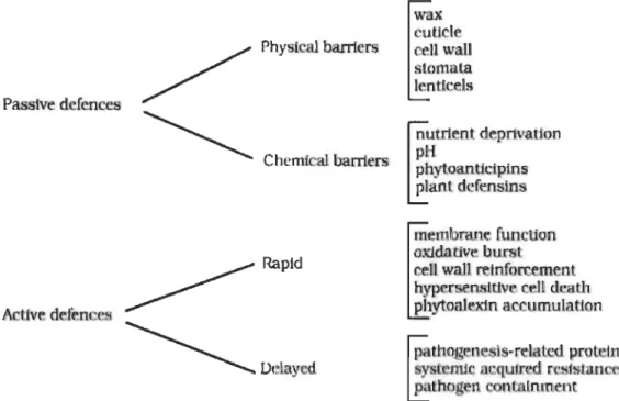
Fig. 1.2 Outline of plant defence systems against different pathogens.
Plant defence systems has been classified into two major systems: passive and active defences. Passive defences include physical and chemical barriers, and active defences includes rapid and delayed mechanisms. (Source: Guest and Brown 1997.)
1.2.1 Passive defence
Constitutive barriers are the paSSIve barrier of plants involving physical and chemical defence. Largely physical barriers involve the specialized plant surface which comprise cell wall, bark, waxy layer, cuticle and stomata. Thickness and chemical composition and molecular mechanism underlying plant cell walls are daunting for sorne pathogens (Malinovsky, Fangel and Willats 2014). Waxy layered cuticular surface on top of epidermal cells covering plant tissues protects plants from many pathogenic atlack (Gahre and Robatzek 2008).
Plants possess sorne physiological access sites, for instance stomata and wounds or hydathodes. Sometimes pathogens can easily enter through these access points, but once the pathogens are inside the host cells, they are antagonized by punitive environments such as uncomfortable pH or antirnicrobial compounds (Gahre and Robatzek 2008).
Plants can respond via the production of chemical compounds or secondary metabolites such as phytoanticipins to the exterior environment which can stop pathogens development. For example, dead cells of brown onion skins excreted quinones catechol and protocatechuic acid preventing spore germination of Botrytis cinerea (neck rot
pa~ogen) and Colletotrichum circinans (smudge pathogen) (Guest and Brown 1997).
Saponins (found abundantly at the surface of quinoa grains) are glycosides based phytoanticipins which are toxic to pathogens having sterols in the membranes, since it binds to sterols in cell membrane of pathogens and subsequent degradation of membrane integrity (Guest and Brown 1997). Plant defensins are another type of chemical compounds produced by sorne plants, for example proteinase- and polygalacturonase-inhibitors or lectins which restrict pathogen nutrient uptake and impede their development. Anti-feeding activity of defensins may provide protection in response to insect transmitted virus (Guest and Brown 1997).
1.2.2 Active defences
Plants may quickly respond to pathogens through varlOUS active defence mechanisms such as changes in membrane functions, oxidative burst (generating Reactive Oxygen Species, ROS), hypersensitive response (HR) and reinforcement of cell wall (Guest and Brown 1997). Upon the attack of pathogens, plants inhibit further growth of infection by rapid death of cells at the infection surroundings visualized as HR, a mechanism sirnilar to programmed ceIl death (PCD) in animals. HR starts with the efflux of potassium and hydroxide ions from the ceIls and influx of hydrogen and calcium ions. As a result, ceIls undergoes rapid response of respiration i.e. oxidative burst by generating ROS, such as hydrogen peroxide, nitrous oxide and super oxide ions (Heath 2000). ROS distresses cell membrane by inducing lipid damage which eventuaIly affects ceIls and is visualised as local cell death or lesions. Sirnultaneously, callose or lignin deposits around the cell walls strengthens the ceIls adjacent to the pathogenic infection.
The defence mechanisms presented above is contingent on pathogen detection and molecular activation of defence pathways. Although plants lack an adaptive immune system like animaIs, they have acquired the ability to detect and protect themselves against infectious microorganisms via a two-layer immune system, r.AMP-Iriggered Immunity (PTI) and Effector-Iriggered Immunity (ETI) (Fig. 1.3) (Jones and Dangl 2006). ... o Q.J "C :J ,'t:: c.. E
PU
ErS Pathogen effectORETS
ETI
Avr-R resho fo H Avr-R•
;::f. -Lm· Fig. 1.3 1 hrashok11o ~ roslstnc-~
. (.()
~O
PAMPSThe zigzag model of the plant immune system.
In phase 1, plants detect microbial/pathogen-associated molecular patterns (MAMPsIPAMPs, red diamonds) via PRRs (Pathogen Recognition Receptors) to trigger P AMP-triggered immunity (PT!). In phase 2, successful pathogens deliver effectors that interfere with PTI, or otherwise enable pathogen nutrition and dispersal, resulting in effector-triggered susceptibility (ETS). In phase 3, one effector (indicated in red) is recognized by an NB-LRR protein, activating effector-triggered immunity (ETI), an amplified version of PTI that often passes a threshold for induction of hypersensitive cell death (I-IR). In phase 4, pathogen isolates are selected that have lost the red effector, and perhaps gained new effectors through horizontal gene flow (in blue) - these can help pathogens to suppress ET!. (Source: Jones and Dangl, Nature, 2006.)
1.2.2.1 P AMP-Triggered Immunity (PTI)
The frrst layer of innate immunity is termed as PIMAMP œathogenlMicrobe-Associated Molecular E.attems) Iriggered Immunity (pTI). PTI is activated by the recognition of P AMP by pathogen recognition receptors (PRR) located at plant cell surface. Plant PRR includes Receptor-Like Kinases (RLKs) or Receptor-Like E.roteins (RLPs) which have an extracellular domain, Leucine Rich Repeats (LRRs) (Shiu and Bleecker 2003). PIMAMPs are conserved microbial features such as bacterial flagellin, Elongation factor-Tu and chitin (Jones and Dangl 2006, Bittel and Robatzek 2007,
BolIer and Felix 2009, G6mez-G6mez and BolIer 2000, Kaku et al. 2006, Zipfel et al. 2006). PIMAMP are integral pathogen protein required for pathogen survival, hence they are under selective pressure and evolve slowly enabling their detection by plants. Thus, PTI is specific to conserved pathogen molecules which means that it is not specific to a bacterial species or genus. Upon the attack of pathogens, PIMAMP activate PRR in the host leading to a downstream activation of a Mitogen-Activated E.rotein Kinases (MAPK) signaling cascade and trigger PTI (Fig. 1.4) (Nitta, Ding and Zhang 2014, Pieterse et al. 2009, Asai et al. 2002). Activation ofMAPK signaling activate the transcriptional rewiring of more than 1200 genes (Zipfel et al. 2004). The importance of MAPK cascade and downstream transcription factors were weIl illustrated in Arabidopsis using bacterial flagellin (Asai et al. 2002). Recognition of flagellin by FLS22 kinase promote the activation of MAPK cascade, MEKK1, MKK41MKK5 and MPK3IMPK6, which activate downstream transcriptional factors WRKY22/WRKY29 for triggering defence resistances (Asai et al. 2002). However, PTI responses are slow and weak because of low specificity of pathogen recognition. So PTI can pause further colonization of pathogens.
Arabidopsis Mammals Drosophila
Kinase Klnase
, ~ ?
,
,
"
AtMEKKl MEKK TAI<
AtMKK4IAtMKKS MEK NtK ?
,
AtMPK3IAIMPK6 JNK IKK JNKERK. p38 p38
, A?
,
' IKB ?,
CactusL
Immune response gene transcription+
O' lsease+
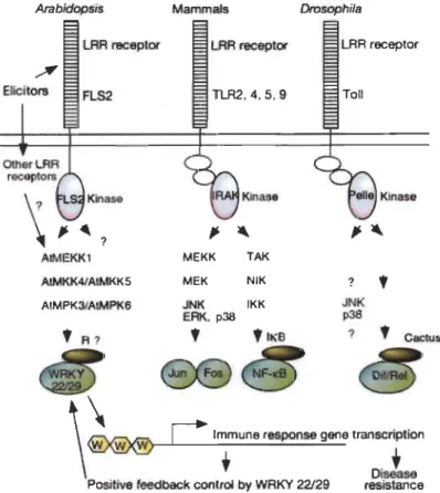
Positive feedbacl< control by WRKY 22/29 resistanceFig. 1.4 Model of MAPK signaling pathway and PTI.
Innate immunity signalling is activated with the recognition of P AMP by LRR receptors in Arabidopsis, mammals and Drosophila. It triggers
MAPK cascades (AtMEKK1, AtMKK4/AtMKK5, AtMPK3/AtMPK6 in
Arabidopsis; MEKK' MEK, JNK, ERK, T AK, NIK and IKK in mammals and JNK, p38 in Drosoplila) which activate transcription factors (WRKY 22/29 in Arabidopsis; Jun Fos and NF-kB in mammals, and DiflRe1 in Drosophila) in the downstream of signaling cascades to trigger disease resistances. But a putative repressor (R) could control the activity of transcription factors WRKY22 and WRKY29 because their overexpression circurnvents the recruitment of elicitors. Figure adapted from Asai et al. 2002.
1.2.2.2 Effector-triggered immunity (ETI)
To suppress PT!, pathogens have deve10ped an arrangement of effectors to evade this defence layer by delivering effectors/virulence factors into the host. Effectors can restrict PT!, resulting in Effector Iriggered .s.usceptibility (ETS) (Fig. 1.3). Effector are detected in plants by a set of intracellular immune receptors, coined resistance
proteins (R). R proteins can directly or indirectly recognize specific effectors (avirulence) ofinvading pathogens leading to activation of Effector-Iriggered Immunity (ETI), the second layer of plant immune system. ETI is extremely specific to a race or strain of pathogen as it recognizes a virulence determinant or its effect, it is fast and a strong defence response leading to a BR to restrict further pathogenic infection (Mur et aL 2008). If the host does not contain suitable receptors (R proteins), then effectors suppress the host defence and subsequent ETS occurs (Fig. 1.3) (Jones and Dangl 2006, Pieterse et aL 2009, AusubeI2005).
1.3 Resistance proteins
Over the course of evolution, plants have evolved copious resistance mechanisms which involves R proteins. Regardless of the diversity of pathogens, genes encoding R proteins belongs to the Nucleotide-Binding Site Leucine-Rich Repeat (NBS-LRR) family (synonyms: NB-ARC-LRR, NB-LRR) , which includes the central nucleotide-binding site (NBINBS) and C-terminal leucine-rich repeat (LRR) domains (Meyers 2003, Tameling and Takken 2008). Furthermore, depending on the N-terminal domain, these NBS-LRR proteins can be subdivided into two subclasses: (i) one class contains significant homology to the cytosolic portion of the Toll/lnterleukin-l Receptor (TIR) domain (i.e. TIR-NBS-LRR); (ii) other class contains non-TIR-NBS-LRR, which has a Coiled-Coil (CC) domain (i.e. CC-NBS-LRR) instead of TIR domain (Dangl and Jones 2001, Tameling and Takken 2008) (Table 1.1). Apart from CC-NBS-LRR, non-TIR-NBS-LRR also comprises other farnilies such as ED-non-TIR-NBS-LRR proteins and NBScc-LRR proteins lacking an additional N-terminus domain. In poplar, another group of protein farnily has been reported, which is called as mixed prote in family containing both TIR and CC domains (TIR-CC-NBS-LRR) (Table 1.1). Dynarnic nuclear trafficking of R pro teins has been demonstrated as important to achieve ETI (Cheng et aL 2009, Liu and Coaker 2008). R proteins localize to different subcellular compartments (from inner plasma membrane to the cytosol and nucleus), but nuclear localization are associated with substantial reprogramming of transcription. It has been reported that some plant R proteins accumulate in the nucleus upon the recognition of
effector (Cheng et al. 2009, Shen et al. 2007, Wirthmueller et al. 2007). In response to effectors, host transcriptional reprogramming include transcription factors (TFs) such as WRKY (Eulgem 2005). For example, MLA10, a powdery mildew effector translocates to the nucleus, interacts with both WRKY (transcriptional repressor) and MYB6 (transcriptional activator) and activate defence responses. Consequent to pathogen effectors invasion, NBS-LRR activate and trigger nuclear associated immune responses that implicates shuttling of proteins through the nuclear membrane; rnRNA export from nucleus and activationlrepression of transcription occur (Shen and Schulze-Lefert 2007).
Table 1.1. Major classes of resistance (R) proteins.
Domain structure Domain Example References
arrangement
TIR-NBS-LRR Mestre and
~ TIR-NBS-LRR N receptor Baulcombe (2006) L6 receptor Howles et al (2005)
~ TIR-NBS-LRR- RRS1-R Deslandes et al (2003)
WRKY receptor Noutoshi et al (2005)
( NBS(TIR)-LRR 2 Arabidopsis* Meyers et al (2003)
cx::::::J-.
CC-NBS-LRR 1-2 Tameling et al (2006)RPS5 Ade et al (2007) non-TIR-NBS-LRR
( NBS(cC)-LRR 4 Arabidopsis* Meyers et al (2003)
-,
) BED-NBS-LRR Poptr_1 :787192 Kohler et al (2008) Mixed~ TIR-CC-NBS- 2 Populus* Kohler et al (2008) LRR
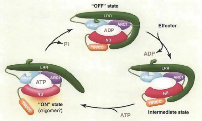
According to the nibbler (named after their central nucleotide-binding, NB and leucine-rich repeat, LRR domains) model of R proteins function (Fig. 1.5), the interaction between effectors and NBS-LRR modifies the configuration of the LRR domain (Takk:en and Tameling 2009). Upon the perception of effector by C-terminus LRR domain, alteration occurs at the edge between ARC2 (NB-ARC) subdomain and N-terminus LRR domain. Hereinafter, nucleotide exchange occurs at NB (sirnilar as NBS domain mentioned earlier section 1.3). However, second confrrmational change occurs due to ADP/ATP convertion which alters the interaction between downstream TIR/CC and LRR domains resulting in formation of an active signalling complex (ON state) (Fig. 1.5). In the "ON" state, NBS subdomain expose to prelirninary defence signalling. In contrast, hydrolysis of ATP rearranges the protein complex to its ADP-bound automated inhibited mode (OFF state) (Fig. 1.5).
"ON" state (oligomer?) "OFF" state
~---~-~-'"
ATP Effector Intermedlate stateFig. 1.5 Generalized model of R protein function and activation.
Biochemical studies revealed that the interaction between effectors and NBS-LRR modifies the configuration of the LRR domain. Upon the preception of effectors by C-terminal part of LRR changes the interface between N-terminal part and the ARC2 domain, thus creating an open confrrmation of R protein which is prone to nucleotide exchange. The exchange between ADP/ATP triggers a second conformational change,
modifying the interaction between the NB-ARC, N-terminal TIR/CC and the C-terminal LRR domains, which results in the ON state of the function.
In absence of pathogen, R proteins (NB-LRR) exist in an autoinhibited,
ADP-bound OFF state which is stabilized by LRR domain. On the other hand, in the activated state, the NB subdomain becomes exposed to initiate defence signaling. ATP hydrolysis resets the prote in into its ADP-bound autoinhibited OFF state. In this figure NB is similar as NBS domain mentioned earlier section 1.3. (Source: Takken and Tameling, 2009.)
Resistance protein pathway is mediated by either TIR-NB-LRR (/TIR-NBS-LRR)
or CC-NB-LRR(lCC-NBS-LRR) (Germain and Seguin 2011) (Fig. 1.6), 93
TIR-NB-LRR and 51 CC-NB-TIR-NB-LRR has been reported in A. thaliana Col-O (Meyers 2003).
TIR-NB-LRR resistance pathway generally signaIs through the gene complex of EDSlIPAD4/SAGIOI (ENHANCED DISEASE SUSCEPTIBILITY lIPHYTOALEXIN
DEFICIENT 4/SENESCENCE ASSOCIATED GENES 101). EDS1 is cytosolic and
forms nuclear prote in complex with PAD4 and SAG101. On the other hand,
CC-NBS-LRR pathway generally signaIs through NDR1 (NONRACE SPECIFIC DISEASE
RESISTANCE 1) gene localizing in plasma membrane. TIR-NBS-LRR and
CC-NBS-LRR pathways mingle for the synthesis of defence responsive hormone salicylic acid
(SA). SA is a necessary signal for Systematic Acquired Resistance (SAR) which is
Resistance protein
((-NB-LRR
NOR1
SA
NPRl
Fig. 1.6 R protein pathways in A. thaliana.Resistance protein
TIR-NB-LRR
EOS1/PA04
/
Different resistance pathways with the requirements of salicylic acid (SA) as signalling molecule, which lead to the induction of defense-related gene have
been identified in A. thaliana. P AD4 and EDSI gene complex functions
upstream of SA signalling pathway in resistance controlled by TIR-NBS-LRR type R genes. In contrary, resistance conditioned by CC-NBS-LRR type R genes activates independently and requires NDRI. Both NDRI and P AD4/EDSl gene complex combined signaIs for SA and resistance triggers via NPRl. EDSl: ENHANCED DISEASE SUSCEPTIBILITY 1; PAD4: PHYTOALEXIN DEFICIENT 4/SENESCENCE ASSOCIATED GENES 101; NDRl: NONRACE SPECIFIC DISEASE RESISTANCE 1; SA: Salicylic acid and NPRl: NONEXPRESSOR OF PR-l. (Source: Germain and Seguin 2011.)
NBS-LRR mediated resistance mechanisms are still poorly understood. Therefore, understanding of the immune activation mechanism mediated by R and its cognate avirulent effector (A VR) is an important topic of research in the field of plant-pathogen interactions.
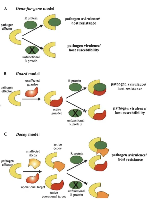
1.4 Molecular models of plant-pathogen recognition
The relationship between effectors and host receptors is defmed by the gene-for-gene model (FIor 1971) which encompasses the direct effect of an unambiguously recognized effector by the receptor i.e. one-on-one relationship between an avirulence protein (Avr) and a resistance protein (R) (Fig. 1.7A). Sometimes support of additional proteins in the host is necessary for the initiation of resistance response, which prompted the second model which is an extension of gene-for-gene model and termed the guard model (Fig. 1.7B). According to the guard model, the target of the effector protein (the guardee) in the host is an essential element which explains indirect perception mechanism of effectors by an appropriate guard prote in, a NBS-LRR receptor
(R protein). This model assumes that R pro teins act by guarding the effectors target and
the modification of this target by the guardee results in activation of the R protein that triggers resistance in the host (Van Der Biezen and Jones 1998, Dangl and Jones 2001).
The guard model was experimentally verified to explain the mechanism of P. syringae
A vrPto perception by the tomato resistance pro teins Pto and Prf (Van Der Biezen and Jones 1998) which were later on generalized for the perception of other effectors (Dangl and Jones 2001). Direct detection of the effectors does not occur in this model, but the R protein detect modification of the guardee; therefore, the guard monitors a host structural and/or functional changes (Jones and Dangl 2006, Tameling and Takk:en 2008).
Depending on the presence or absence of the R gene, the guardees are subjected
to selection forces: (1) to promote the perception of guardees i.e. strong interaction, and (II) to evade manipulation by the guardee i.e. weaker interaction. So these two contradictory selection forces on the same effector interaction surface of the guardee results in an evolutionary unstable situation. This selection force could be eased upon the evolution of a host protein, termed as decoy. Thereafter, it focuses in perception of the effector by the R protein which has no function either in the disease development or resistance. As a result' in the decoy model (Fig. 1.7C), the decoy mimics effectors targets to sham the pathogen into a recognition event without participating to the pathogen fitness in absence of R protein (van der Hoom and Kamoun 2008). The decoy
model was experimentally verified in pepper, tomato an Arabidopsis where pBs3 (Zhou
and Chai 2008), RCR3 (Dixon et al. 2000) and RIN4 (Kim et al. 2005) were identified
as decoy respectively. A Gene-for-gene model R protein
P'~
cs
~
unfu.,.o",1CS
R protein B Guard model pathogen unaffected guardee'ff«
~
_--+
C Decoy model unaffected decoyQ
operational target active guardee active decoy pathogen avirulencel host resistance pathogen virulence! host suscebtibillty unfunctional R protein active unfuncti~naloperational target R protem
Fig. 1.7 Molecular models of pathogen recognition.
pathogen avirulence! host resistance pathogen virulence/ host suscebtibility pathogen avirulence/ host resistance
(A) The gene-for-gene model. It involves the direct effect of a specified
the target protein of the effector (guardee) is guarded by an appropriate guard prote in, NBS-LRR receptor. (C) The decoy model: specific proteins which are similar to the protein targeted by pathogenic effectors are generated by the host plant, whose only function is to bind those effectors and acts as a mediator in the interactions with R proteins. Details are in the text). (Source: Glowacki et al 2011.)
1.5 Plant pathogens
Plants are continuously under the attack from diverse rnicrobial pathogens through multiple ways. Sorne pathogens colonize intra- or inter-cellular spaces of various plant tissues (roots, leaves etc.), settle down on surfaces while sorne others travel through vascular bundles (Yadeta and BP 2013). Pathogens perceive nutritional benefits from the host plants. Sorne pathogens (biotrophic pathogens) depend on living tissues for nutrient uptake. There are also pathogens (necrotrophic) that feed on dead tissues. Regardless of the feeding style, sorne pathogens are devastating for crop plants, and few types of pathogens are being extensively studied to understand the interactions of molecular plant-pathogen interaction.
1.5.1 Viral pathogens
Viruses are very small particles restricted to specific type of host and spread plant diseases causing massive econornic losses. Plant virologists in association with molecular plant pathologists ranked the top ten plant viruses in importance to economy and scientific research (Scholthof et al. 20 Il) enlisted in Table 1.2.
Table 1.2. Top ten viral pathogens in plants.
Rank Virus Common diseases Author of virus
description
1 Tobacco mosaic virus Mosaic and necrotic diseases Karen-Beth G.
(TMV) on tobacco, tomato and other Scholthof
solanaceous plants
2 Tomato spotted wilt Spotted wilt in tomato and Scott Adkins
virus (TSWV) sorne other vegetables
3 Tomato yellow leaf Leaf curl in tomato Henryk Czosnek
curl virus (TYLCV)
4 Cucumber mosaic Mosaic disease on tomato, Peter Palukaitis
virus (CMV) tobacco, pepper
5 Potato virus Y (pVY) Dark mosaic or tuber necrotic Emmanuel Jacquot
ringspot of potato
6 Cauliflower mosaic Mosaic in cauliflower Thomas Hohn and
virus (CaMV) Barbara Hohn
7 African cassava Cassava mosaic disease Keith Saunders
mosaic virus (ACMV)
8 Plum pox virus (PPV) Sharka disease of stone fruits Thierry Candresse
9 Brome mosaic virus Mosaic on barley and other Paul Ahlquist
(BMV) grass family
10 Potato virus X (PVX) Mosaic or necrotic in potato, Cynthia Hemenway
tomato, pepper
Most plant virus are single stranded positive sense RNA. Compatible interaction of host, viral particle and environmental factors may cause severe diseases and plant death. The defence mechanism described above are effective against viruses (Scholthof et al. 2011).
1.5.2 Bacterial pathogens
While sorne bacteria can actually be beneficial to plants, sorne are pathogenic. Pathogenic bacteria cause numerous se rio us diseases in plants, although numbers are fewer than viruses and fungus and cause comparatively less damages and econornic losses (Kennedy and Alcom 1980). Regardless of beneficial bacteria, over 200 bacterial
species are threat to plants. In consonance with economic and scientific importance, recently molecular plant pathologists ranked top ten bacterial plant pathogens (Table 1.3) (Mansfield et al. 2012).
Table 1.3. Top ten bacterial plant pathogens.
Rank Bacterial Common diseases Author of bacterial
pathogen description
1 Pseudomonas Bacterial speck of tomato, bleeding John Mansfield syringae cancer of horse-chestnut, blight
pathovers disease of bean
2 Ralstonia Bacterial wilt of tomato, eggplant, Stephane Genin
solanacearum tobacco and sorne omamental plants, potato brown rot and banana Moko disease
3 Agrobacterium Crown gall tumor Shimpei Magori,
tumefaciens Vitaly Citovsky
4 Xanthomonas Bacterial blight Malinee
oryzae pv. oryzae Sriariyanum, Pamela
Ronald 5 Xanthomonas Black rots of crucifers infecting all Max Dow
campestris cultivated brassicas pathovers
6 Xanthomonas Angular leaf spot of cotton Valerie Verdier axonopodis
7 Erwinia Fire blight disease of several fruit Steven V. Beer
amylovora plants (apple, quince, pear,
blackberry, raspberry) and several wild and cultivated rosaceous omamental plants
8 Xyle lia fastidiosa Citrus variegated chlorosis (CVC), Marcos A. Machado almond leaf scorch disease
(ALSD), Pierce's disease of grapevine (PD)
9 Dickeya (dadantii Necrosis, blight and soft rot of ran Toth and solani) potato tubers, bulbs of vegetables
and omamental crops
10 Pectobacterium Soft rot diseases, potato blackleg George Salmond carotovorum (and disease in the temperate areas
1.5.3 Oomycete pathogens
Because of their similar filamentous structure and feeding habits, oomycetes were once considered within the fungal kingdom. The development of molecular phylogenetic studies and advanced understanding of evolutionary relationships revealed them to constitutes a separate class of organisms, closer to diatoms and brown algae. Now it constitutes an individual class of pathogens, oomycota. Pathogenic effect of oomycetes on plants allowed molecular plant pathologists to identify most devastating oomycetes according to their importance in pathology and economic importance (Kamoun et al. 2015). Based on the recent publication (Kamoun et al. 2015), the top ten oomycete pathogens in plant molecular pathology are summarized in Table 1.4.
Table 1.4. Top ten oomycete plant pathogens.
Rank Species Common diseases
1 Phytophthora infestans Late blight
=2
Hyaloperonospora Downy mildewarabidopsidis
=2
Phytophthora ramorum Sudden oak: death; Ramorum disease4 Phytophthora sojae Stem and root rot
5 Phytophthora capsici Blight; stem and fruit rot; various others
6 Plasmopara viticola Downy mildew
7 Phytophthora cinnamomi Root rot; dieback
=8
Phytophthora parasitica Root and stem rot; various others=8
Pythium ultimum Damping off; root rot10 Albugo candida White rust
The '=' sign before the ranking indicates that the species tied for that position. Number of papers published in 2005-2014 is based on searches of the Scopus database
(http://www.scopus.com) using the species names as a query. For H arabidopsidis, a
search for the alternative name Peronospora parasitica was also performed and the combined number is shown.
Among the top ten, Phytophthora and Pythium are hemibiotrophic and necrotrophic, respectively whereas Hyaloperonospora arabidopsidis is an obligate biotrophic pathogen (Stassen and Van den Ackerveken 20 Il). Complete genome sequencing of H arabidopsidis (Coates and Beynon 2010), P. infestans (Haas et al. 2009), P. sojae, P. ramorium (Tyler et al. 2006), and P. ultimum (Lévesque et al. 2010) unwrapped the identification of huge catalog of effector proteins for enriched understanding of oomycete-plant interactions.
1.5.4 Fungal pathogens
According to the comprehensive phylogenetic classification, fungi are divided into six groups-Ascomycota, Basidiomycota, Glomeromycota, Zygomycota, Chytridiomycota and Microsporidia (Hibbett et al. 2007). Plants and fungi association is primitive and fungi are a vital group of plant pathogens. But less than 10% of all known fungi can inhabit with living plants (Knogge 1996). Mostly fungi are decomposers, living on the relics of plants or other organisms for food supply. Another group of fungi (mycorrhizal fungi) are associated with plant roots and both plants and fungus enjoy the mutual benefits.
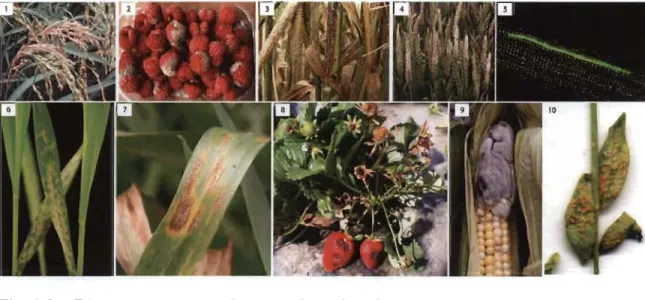
In terms of feeding habit, fungi are divided into biotrophs, necrotrophs and hemibiotrophs. Biotrophic fungi uptakes nutrients from living tissues. Necrotrophs survives on dead tissue of host, whereas hemibiotrophs feeds on both living and dead tissues, which is characterized by usually having an initial biotrophic stage followed by a necrotrophic stage. They feed on living tissues for certain period of time and later on continue on dead tissues. Recently plant pathologists have ranked the top ten fungal plant pathogens according to their economic and scientific importance (Dean et al. 2012). Recently (Dean et al. 2012), the top ten fungal pathogens in plant molecular pathology (Table 1.5) and disease symptoms has been published (enlisted in Fig. 1.8). Magnaporthe oryzaye ranked first in the top ten pathogenic fungus list. M oryzae is a filamentous ascomycetes fungus and causal agent of blast diseases of rice (Ou 1980) which is the primary caloric source for the half of the world' s population (Khush 2005).
Table 1.5. Summary of the top ten fungal pathogens in molecular plant pathology.
Rank Fungal Type Causal agent Author of fungal
pathogen description
1 Magnaporthe Filamentous Rice blast Ralph Dean oryzae ascomycetes
2 Botrytis cinerea Grey mould Soft fruits and JanA. L. van Kan omamentals
3 Puccinia spp. Obligate, Wheat stem rust by Zacharias A.
biotrophic Puccinia graminis f. sp. Pretorius basidiomycetes tritici; stripe rust by
P. striiformis f. sp. Tritici); leaf rust by P. triticina
4 Fusarium Ascomycetes Hexaploid wheat Kim
Harnmond-graminearum Kosack
5 Fusarium Ascomycetes Melon, tomato, cotton and Antonio Di Pietro
oxysporum banana
6 BZumeria Ascomycetes Powdery mildews of Pietro Spanu
graminis grasses
7 Mycosphaerella Ascomycetes Blotch of wheat Jason J. Rudd graminicoZa
8 Colletotrichum Ascomycetes Anthracnose spots and Marty Dickman
spp. blights of aerial plant
parts and post-harvest rots.
9 UstiZago maydis Basidiomycetes, Comsmut Regine Kahrnann biotrophic
10 MeZampsora Basidiomycetes Flux and linseed rust Jeff Ellis fini
Fig. 1.8 Disease symptoms of top ten fungal pathogens.
(1) Rice blast with Magnaporhe oryzae; (2) Gray mould on raspberry with Botrytis cinerea; (3) Stem rust of wheat by Puccinia gramins f. sp. tritici;
(4) Head blight of wheat by Fusarium graminearum; (5) Growth of Fusarium oxysporum hyphae on roots of tomato; (6) Powdery mildew on barly leaves by BZumeria graminis f. sp. Hordei; (7) Blotch of wheat caused by Mycosphaerella graminicoZa; (8) Colletotricum acutatum infected strawberry; (9) Corn smut of maize with UstiZago maydis; (10) Rust of flax with MeZampsora Uni. (Source: Dean et al. 2012.)
1.5.4.1 Rust fungus as pathogen
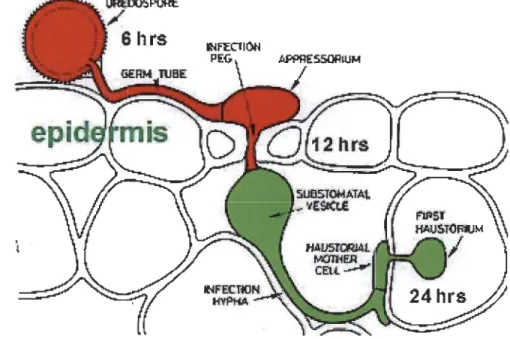
Rust fungi are filamentous biotrophic, eukaryotic plant pathogens belonging to the Pucciniales farnily under the phylum Basidiomycota. Rust fungi cause devastating diseases in native and commercial crop plants inc1uding wheat, maize, bean, pine, poplar and other cereals (Duplessis et al. 2011a, Helfer 2014, Aime et al. 2006). Most pathogenic rust fungi manipulate host physiology and cellular functions according to their own interest which eventually induces a number of diseases. Comparative interaction studies of plant-pathogens have demonstrated the significance of several molecules of pathogens such as effectors, carbohydrate active enzymes (CAZys) and secondary metabolites (Stergiopoulos et al. 2013, O'Connell et al. 2012). Alike oomycetes, rust fungi develop a specialized feeding structure called haustoria (Fig. 1.9). Following contact with the ho st, the urediospore develops a germ tube, forms appressorium intercellularly, penetrates throughout the host cell wall, pushes the plasma membrane, and eventually forms a bubble-like structure, the haustoria (Catanzariti,
Dodds and Ellis 2007, Hahn and Mendgen 2001). In addition to nutrient uptake, rust pathogens secrete effectors proteins inside into the host cell via haustoria and manipulate
host cellular processes and suppress host defences
01
oegele and Mendgen 2003).Fig. 1.9 Cartoon showing haustoria formation.
Following contact with the host, the urediospore develops a germ tube, form
an appressorium intercellularly. Then penetrates throughout the host cell wall
and pushes the plasma membrane, form infection hyphae and haustoria
throughout epidermal cells. (Drawn by K. Mendgen, from Introductory
Mycology from John Wiley & Sons, Inc.)
1.6 Plant pathosystems
A pathosystem is a subsystem of an ecosystem termed by the parasitism phenomenon of a pathogen. In a plant pathosystem, thé pathogen has the ability to colonize within host tissues, and the host may have a resistance or defence system to protect itself from this invasion. (Robinson 1977). It is a multidisciplinary concept that conveys diverse field of plant biology including plant breeding, pathology, nematology and entomology. A good model pathosystem is required in order to improve our understanding of plant-pathogen interactions (Meng et al. 2014); specifically the cellular and molecular understanding of P AMPs perception, PTI, ET!, transport of vesicles,
transcriptional networks and reciprocity between hormone signaling and disease resistance (Nishimura and DangI201O).
1. 7 Effectors
A molecule or secreted protein from a plant associated organisms which colonize and modify cellular structure or functions in the host is termed as "effector" (Hogenhout et al. 2009). Plant pathogens secrete molecules known as effectors into the plant tissues to promote parasitic growth. Gram-negative bacteria (for instance P. syringae) secrete effectors using type three secretion system (thereafter T3SS) for delivery inside the host cells (Abramovitch, Anderson and Martin 2006). The very presence of effector is what defines microbes as pathogenic or not pathogenic (Hacquard et al. 20 16b). Sorne of these secreted proteins can trigger an HR in resistant plants (i.e. avirulence property), while the same proteins were later on found to promote virulence (i.e. virulence property) in susceptible plants (plants lacking the cognate R proteins). Since the same protein display dual activities (avirulence/virulence) in the case of incompatible or compatible interactions, thereafter the term "effector" alleviate the conceptual limitations of avirulence and virulence terminology (Hogenhout et al. 2009). Recently the word effector has been adopted by a broad range of microbiologist/pathologists and now is being preferentially used in the field of fungi and oomycete as well (Hogenhout et al. 2009). Effectors target a variety of host cell compartments and molecules such as proteins and DNA, and modulate their location, stability, or function to the advantage of the pathogen (Chaudhari et al. 2014, Lewis, Guttman and Desveaux 2009, Win et al. 2012). Recently, the term effector has also been used to denote secreted pro teins of microbes that establish symbiotic relationship with plants (plett et al. 20 Il, Kloppholz, Kuhn and Requena 2011).
1. 7.1 Rust effectors
Recently, genome sequences of four rust fungi (two Pucciniaceae and two Melampsoraceae) became available. Genomic and transcriptomic analyse revealed a
wide range of small secreted proteins known as fandidate §ecreted ~ffector 12roteins (CSEPs) (petre, Joly and Duplessis 2014). Haustoria delivers effectors/CSEPs into the host cells and also rewires molecular trafficking between the host and the parasite (Rafiqi et al. 2012). Six effectors have been reported from three rust species to be secreted via haustoria and translocated into host cells (Table 1.6) (Kemen et al. 2005,
Petre et al. 2014, Upadhyaya et al. 2014, Ellis, Catanzariti and Dodds 2006). Nowadays,
plant molecular pathologists utilize effectors as probes to identify and understand plant processes targeted by pathogens in order to develop resistant crop plants.
Effector Aa residues Signal Expression Localization in Avr property protein (mature) peptide infected tissues (immune receptor) AvrM 284-347 Yes Haustoriuma Haustorium, EHMx, Yes (M)
plant cytosola
AvrL567 127 Yes Haustorium Pant cytosol Yes (L5, L6, L7) AvrP123 94 Yes Haustorium Plant nucleus Yes (P, Pl, P2, P3) AvrP4 65 Yes Haustorium Plant cytosol Yes (P4)
RTP1 201 Yes Haustoriuma Haustorium/ nd EHMxlplant cytosol/ plant nucleusa
PGTAUSPE- np np Haustorium nd Yesb 10-1
Role of virulence has not been determined for any one of these effector proteins.
Avr=Avirulence; aa=amino acid; EHMx=extra-haustorial matrix; nd=not determined; np=not published.
a Direct evidence of the presence of the protein acquired by immunolocalization.
b a host-specific toxic effect was detected.
Biochemical function nd nd nd nd Protease inhibitor/ filament-formation nd