Evolution of the Extraglottic Airway: A Review of Its
History, Applications, and Practical Tips for Success
Michael R. Hernandez, MD,* P. Allan Klock, Jr., MD,* and Adranik Ovassapian, MD*†
The development of the laryngeal mask airway in 1981 was an important first step toward widespread use and acceptance of the extraglottic airway (EGA). The term extraglottic is used in this review to encompass those airways that do not violate the larynx, in addition to those with a supraglottic position. Although the term extraglottic may be broad and include airways such as tracheostomy tubes, the term supraglottic does not describe a large number of devices with subglottic components and is too narrow for a discussion of modern devices. EGAs have flourished in practice, and now a wide variety of devices are available for an ever-expanding array of applications. In this review we attempt to clarify the current state of EGA devices new and old, and to illustrate their use in numerous settings. Particular attention is paid to the use of EGAs in special situations such as obstetric, pediatric, prehospital, and nontraditional “out of the operating room” settings. The role of the EGA in difficult airway management is discussed. EGA devices have saved countless lives because they facilitate ventilation when facemask ventilation and tracheal intubation were not possible. Traditionally, difficult airway management focused on successful tracheal intubation. The EGA has allowed a paradigm shift, changing the emphasis of difficult airway management from tracheal intubation to ventilation and oxygenation. EGA devices have proved to be useful adjuncts to tracheal intubation; in particular, the combination of EGA devices and fiberoptic guidance is a powerful technique for difficult airway management.
Despite their utility, EGAs do have disadvantages. For example, they typically do not provide the same protection from pulmonary aspiration of regurgitated gastric material as a cuffed tracheal tube. The risk of aspiration of gastric contents persists despite advances in EGA design that have sought to address the issue. The association between excessive EGA cuff pressure and potential morbidity is becoming increasingly recognized. The widespread success and adoption of the EGA into clinical practice has revolutionized airway management and anesthetic care. Although the role of EGAs is well established, the user must know each device’s particular strengths and limitations and understand that limited data are available for guidance until a new device has been well studied. (Anesth Analg 2012;114:349 –68)
T
he laryngeal mask airway (LMA; LMA North Amer-ica, San Diego, CA) was one of the first extraglottic airways (EGA) invented by Dr. Archie Brain in 1981. It became commercially available in the United Kingdom in 1988 and in the United States in 1991. The LMA Classic (cLMA) received wide recognition in a short time and has had a major impact on anesthesia practice and airway management. Publication of thousands of peer-reviewed articles, book chapters, and textbooks testifies to the success of the LMA as an extraglottic device. Other individuals and manufacturers later introduced similar airway devices.Despite significant literature detailing the use of EGAs, the constant evolution in device design encourages the clinical application of the LMA outside the traditional operating room (OR) suite. This review attempts not only to describe some of the history surrounding EGA devices, but also to understand how EGA use has evolved because of design modifications.
EGAs have become much more than a simple airway device, and now enjoy a wide range of applications and indications, some of which were formerly relative contraindications.
The term supraglottic airway device is often used, but may not accurately describe airways that include periglottic com-ponents. Use of the term extraglottic airway device is perhaps more appropriate for this review, because it encompasses airways that do not violate the larynx. Although airway devices such as a tracheostomy tube may also be considered extraglottic, this review concerns itself only with devices that are inserted via the oropharynx for temporary airway man-agement. A large number of recently introduced devices have complicated efforts to establish a consistent nomenclature and organizational framework for EGA devices1(Table 1).
Despite a multitude of new devices introduced into the market, most devices aim to allow easy placement with predictable ventilation, minimize the chance of pulmonary aspiration, and to serve as an adjunct to tracheal intubation. This review aims to help the reader understand the simi-larities and differences among the numerous EGAs cur-rently available and will present many EGA devices. EGAs are separated by manufacturer and then further subdivided by the chronology of market entry and/or special features.
LMAs
cLMA
One of the first EGAs, the cLMA (LMA North America, San Diego, CA), was studied extensively and used in ⬎7000 patients by Dr. Archie Brain and his associate, Dr. Chandy Verghese, before its release for routine clinical use. The details of the development of LMAs are described by the inventor.2
From the *Department of Anesthesia and Critical Care, University of Chicago Hospitals, Chicago, IL. †Deceased.
Accepted for publication October 4, 2011.
Conflicts of interest: see Disclosures at end of the article.
Supplemental digital content is available for this article. Direct URL citations appear in the printed text and are provided in the HTML and PDF versions of this article on the journal’s Web site www.anesthesia-analgesia.org. Reprints will not be available from the authors.
Address correspondence to Michael R. Hernandez, MD, The University of Chicago Medical Center, Room M252L. Department of Anesthesia and Critical Care, 5841 South Maryland Ave., MC4028, Chicago, IL 60637. Address e-mail to mhernandez@dacc.uchicago.edu.
Copyright © 2012 International Anesthesia Research Society
Initial LMA prototypes were designed based on plaster casts of cadaveric airways. The use of the cLMA in anesthesia encouraged individuals and manufacturing companies to introduce other EGAs with many design modifications.
The cLMA is a reusable device made of silicone. Its disposable version, the LMA Unique (uLMA, LMA North America, Inc. San Diego, CA, USA), is made of polyvinyl-chloride (PVC). Both are latex-free and available in six sizes to fit infants to adults (Table 2; for additional details see Supplementary Table 2A, Supplementary Digital Content 1, http://links.lww.com/AA/A346). Both the cLMA and the uLMA have an elliptically shaped mask attached to a ventilation tube (Fig. 1). The mask has a cuff, a pilot tube, and balloon through which the cuff is inflated and main-tenance of intracuff pressure is monitored. The proximal end of the shaft has a standard 15-mm adapter. The cLMA can be autoclaved and the manufacturer recommendations suggest that it may be reused up to 40 times. Two bars at the junction of the shaft and mask prevent the epiglottis from obstructing the ventilating lumen. In addition to its routine use in the OR during general anesthesia, the laryngeal mask is used for airway management outside of the OR and for management of difficult or failed intuba-tion. It is included in the airway management algorithms of the American Society of Anesthesiologists (ASA) and the Difficult Airway Society of the United Kingdom.3,4
Originally, the cLMA was used in lieu of a facemask in anesthetized patients breathing spontaneously. Over the years, that practice has changed so that now the cLMA is also used with controlled ventilation in complicated opera-tive procedures. Posiopera-tive pressure ventilation via an LMA is possible only if the device provides an adequate airway seal. Quantification of an oropharyngeal seal or “leak” pres-sure is a common maneuver in the evaluation of LMAs, both to confirm suitable device function and for research pur-poses.5
Multiple techniques can be used to evaluate leak pressure.6
One technique uses increasingly positive pressure via the device to determine the level at which an audible leak is heard at the patient’s mouth or during auscultation lateral to the thyroid cartilage. Other techniques include intraoral capnography to detect leakage of carbon dioxide from around
the device at a given pressure, or the use of manometry to measure the pressure at equilibrium with the leak. All 4 techniques successfully measured the “leak” pressure in pe-diatric and adult patients after LMA placement.6,7
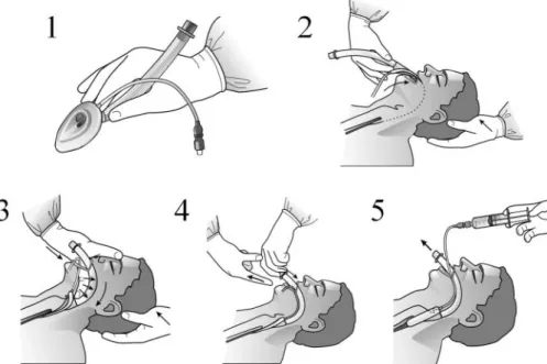
Before inserting the LMA, the cuff is deflated so that the edge is smooth and wrinkle-free with the tip deflected backward toward the convex side of the ventilation shaft. This method encourages the LMA to slide posterior to the epiglottis without deflecting it inferiorly over the glottic opening. The pharyngeal surface is lubricated generously; the lingual surface is lubricated lightly for smooth advance-ment. In the technique described by Dr. Brain (Fig. 2), the shaft of the LMA is held between index finger and thumb with the tip of the index finger at the junction of the mask and the tube. With the patient’s head in the “sniffing” position, the nondominant hand is placed under the oc-ciput, extending the head, while the dominant hand inserts the LMA into the mouth. With optimal technique, the LMA follows a path similar to a bolus of food that is about to be swallowed, traveling from the oropharynx against the hard palate to the soft palate, hypopharynx, and finally the proximal esophagus. The tip of the mask is pressed against the hard palate and advanced toward the larynx until resistance is felt. Manufacturer recommendations call for the mask to be inflated to an intracuff pressure of no⬎60 cm H2O (44 mm Hg). Intracuff pressure should be
moni-tored, especially if nitrous oxide is being used, with its increased risk for expansion of the mask volume and associated injuries.8
When no muscle relaxant is used during insertion or throughout the anesthetic, an adequate depth of anesthesia is essential to prevent the contraction of pharyngeal and laryngeal muscles, which may interfere with proper positioning of the LMA. Occasionally, an LMA may be used to rescue a patient during a rapid sequence induction complicated by an inability to perform tracheal intubation and failed attempts to mask ventilate the pa-tient. In this setting, the operator should remember that cricoid pressure may cause compression of the hypophar-ynx that may prevent the tip of the LMA from reaching a proper final position. As a result, it may be useful to relieve cricoid pressure temporarily to allow proper LMA posi-tioning during insertion.9,10
The technique for placement of the uLMA is the same as the cLMA.
Properly positioned, the cLMA masks the glottis, maintains an open airway, and makes ventilation easy. The tip of the LMA sits posteriorly to the cricoid cartilage engaging the proximal esophageal sphincter, and the proximal end of the mask portion of the LMA lies against the base of the tongue. Satisfactory ventilation through the cLMA does not necessarily indicate proper position-ing. Flexible bronchoscopy has shown that ventilation through a cLMA is acceptable with the device in less than perfect position.11
Proper placement of the LMA may be impeded when the tongue and epiglottis are pushed caudally or when the tonsils are enlarged; in such situations, the tip of the cLMA may press on the glottis causing partial or complete airway obstruction. The proximal part of the mask may obstruct the airway if the cLMA is positioned too deeply, or the mask may fold over on itself if excessive force is used during insertion.
Table 1. Extraglottic Airway
Device Classifications
EGA with an inflatable periglottic cuff Ultra CPV family (AES)
Ambu Aura family (Ambu) ILA/airQ (Cookgas) Vital Seal (GE Healthcare) King LAD family (King Systems) LMA device family (LMA Company) Soft Seal Laryngeal Mask (Portex) Sheridan Laryngeal Mask (Teleflex) EGAs with no inflatable cuff
i-gel (Intersurgical) SLIPA (Slipa Medical) EGAs with 2 inflatable cuffs
Laryngeal Tube family (King Systems) Esophageal Tracheal Combitube (Nellcor) Rusch EasyTube (Teleflex)
EGAs with single pharyngeal inflatable cuff Cobra PLA family (Pulmodyne)
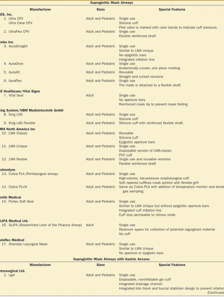
Table 2 Extraglottic Airway Devices at a Glance
Supraglottic Mask Airways
Manufacturer Sizes Special Features
AES, Inc. 1. Ultra CPV
Ultra Clear CPV
Adult and Pediatric Single use Silicone cuff
Pilot valve is marked with color bands to indicate cuff pressure.
2. UltraFlex CPV Adult and Pediatric Single use
Flexible reinforced shaft Ambu Inc.
3. AuraStraight Adult and Pediatric Single use
Similar to LMA Unique No epiglottic bars Integrated inflation line
4. AuraOnce Adult and Pediatric Single use
Anatomically curved, one piece molding
5. Aura40 Adult and Pediatric Reusable
Straight and curved versions
6. AuraFlex Adult and Pediatric Single use
The mask is attached to a flexible shaft GE Healthcare/Vital Signs
7. Vital Seal Adult Single use
No aperture bars
Reinforced mask tip to prevent mask folding King System/VBM Medizintechnik GmbH
8. King LAD Adult and Pediatric Single use
Silicone cuff
9. King LAD Flexible Adult and Pediatric Silicone cuff with reinforced flexible shaft. LMA North America Inc
10. LMA Classic Adult and Pediatric Reusable
Silicone cuff
Epiglottic aperture bars
11. LMA Unique Adult and Pediatric Single use
Disposable version of LMA-classic PVC cuff
12. LMA flexible Adult and Pediatric Single use and reusable versions
Flexible reinforced shaft Pulmodyne
13. Cobra PLA (Perilaryngeal airway) Adult and Pediatric Single use
High-volume, low-pressure oropharyngeal cuff. Soft tapered cuffless mask portion with flexible grill
14. Cobra PLUS Adult and Pediatric Same as Cobra PLA with addition of temperature monitor and distal
gas sampling Smith Medical
15. Portex Soft Seal Adult and Pediatric Single use
Similar to LMA Unique but without epiglottic aperture bars Integrated cuff inflation line
Cuff less permeable to nitrous oxide SLIPA Medical Ltd.
16. SLIPA (Streamlined Liner of the Pharynx Airway) Adult Single use
Reservoir space for collection of potential regurgitant material No cuff
Teleflex Medical
17. Sheridan Laryngeal Mask Adult and Pediatric Single use
Similar to LMA Unique No aperture or epiglotic bars Supraglottic Mask Airways with Gastric Access:
Manufacturer Sizes Special Features
Intersurgical Ltd.
1. i-gel Adult and Pediatric Single use
Disposable, noninflatable gel cuff Integrated drainage channel
Integrated bite block and buccal stabilizer design to prevent rotation (Continued)
Three factors contribute to the failure of proper place-ment: lack of experience of the operator, improper tech-nique, and inadequate depth of anesthesia. Insertion of the cLMA during light anesthesia stimulates contraction of the
pharyngeal wall, cricopharyngeus, and extrinsic laryngeal muscles. The cLMA may also become twisted during placement or if advanced too far when an undersized device is selected.
Table 2. (Continued)
Manufacturer Sizes Special Features
LMA North America, Inc.
2. LMA-ProSeal Adult and Pediatric Reusable
Added posterior cuff for sizes 3 and greater
Integrated bite block (not for size 1) and drainage tube
3. LMA-Supreme Adult and Pediatric Single use
Rigid curved shaft allows easy insertion Integrated drainage lumen
Extraglottic Airways Utilizing Double Cuff System: King System/VBM Medizintechnik GmbH
1. King/VBM LT/LT-D Adult and Pediatric Reusable.
LT-D is single use
Single lumen with dual cuffs pharyngeal and esophageal inflated by single inflation line.
2. King/VBM LTS-D Adult Single use
Similar to LT-D, but adds distal lumen beyond esophageal cuff
3. VBM LTS II Adult and Pediatric Smaller sizes available replaces the VBM LTS
4. VBM G-LT (Gastro-Laryngeal Tube) Adult Reusable
Allows introduction of endoscope through large esophageal lumen for upper gastrointestinal endoscopy.
Nellcor (Covidien) Inc.
5. Esophageal Tracheal Combitube Adult Single use
Dual lumen device, allows ventilation via either lumen after blind insertion
95% of time distal lumen enters esophagus, proximal lumen becomes ventilating lumen
Converse is true if distal lumen enters trachea Separate inflation line for proximal and distal cuff Teleflex Medical
6. Rusch EasyTube Adult Single use
Similar to Combitube
Allows passage of suction catheter, exchange catheter, or fiberoptic via proximal lumen
Latex free
Supraglottic Airway Devices for One-Step Intubation
Manufacturer Sizes Special Features
Ambu Inc.
1. Aura-i Adult and Pediatric Single use
Rigid curved shape No aperture bars
Wide bore ventilating lumen to accommodate standard endotracheal tubes (3.5 mm to 8.0 mm)
Cookgas LLC
2. air-Q/ILA Adult and Pediatric Reusable (ILA) and single use (airQ)
Similar to LMA Classic/Unique but no aperture bars and removable 15 mm connector
Designed to allow standard endotracheal tubes (5.5 mm to 8.0 mm) LMA North America, Inc
3. LMA Fastrach Adult Reusable and single use versions
Rigid curved design with stainless steel handle Epiglottic elevating bar at mask orifice
Available reinforced special Fastrach endotracheal tube, up to 8.0 mm.
4. LMA Classic Excel Adult Reusable
Similar to LMA Classic in shape and rigidity, but has removable 15 mm adapter and epiglottic elevating bar
Advantages of the LMA
A 1995 meta-analysis of 858 publications identified several advantages of the cLMA over tracheal intubation.12 There
were fewer changes in hemodynamic and intraocular pres-sure during placement and removal of the cLMA than during tracheal intubation and extubation. Awakening with a cLMA in place resulted in less coughing, bucking, and hemody-namic changes than awakening with an endotracheal tube in place. Laryngeal competence and mucociliary function were preserved and laryngeal trauma was less.13
The cLMA can be placed in⬍60 seconds after induction of anesthesia without the need for a muscle relaxant and use of a laryngoscope. A meta-analysis that included 3414 patients found a 17% inci-dence of sore throat with the LMA compared with a 39% incidence after endotracheal intubation (P⬍ 0.00001).14
When compared with facemask ventilation, the cLMA is easy to learn and use, it secures the airway better, and decreases OR
pollution from volatile anesthetics. With the cLMA, the anes-thesiologist’s hands are freed for other activities and not fatigued from prolonged holding of a facemask. The cLMA circumvents upper airway obstruction and the need for jaw support by bypassing the tongue and epiglottis. Compared to facemask ventilation, the cLMA may also reduce the risk of injury to the eye and facial nerve.15
Use of the LMA in Nonsupine Patients
The LMA has enjoyed success in routine practice, and enthusiasm for its use has led some to consider an ex-panded range of applications. The need for prone position-ing classically precludes elective LMA placement for most clinicians. Nonetheless, there is experience with LMA use in prone patients. Successful airway rescue of unexpectedly extubated patients in the prone position has been re-ported.16
Prone airway rescue with an LMA is particularly advantageous when a patient is not easily returned to the supine position for tracheal reintubation. Elective use of an LMA in patients who are in the prone position has also garnered some attention. The LMA ProSeal (pLMA; LMA North America, San Diego, CA) was used successfully in 245 patients in the prone position based on a retrospective audit.17
The LMA Supreme (sLMA; LMA North America, San Diego, CA) has also been used successfully in prone patients, as reported in a prospective audit of 205 patients and a prospective study of 40 patients.18,19 Despite
evi-dence to suggest the safety and utility of the elective use of the LMA for airway management of patients in the prone position, data are still insufficient to confidently recom-mend the technique as safe and superior to tracheal intu-bation in this patient population.
Figure 1. LMA Classic™.
Figure 2. Manufacturer’s recommended insertion technique (Courtesy of LMA North America, San Diego, CA): (1) Deflate the laryngeal mask airway (LMA) cuff to a smooth low profile shape. Grasp LMA between thumb and index finger. (2) Extend the patient’s head with the nondominant hand, while the LMA is inserted with thumb and index finger grip toward the hard palate. (3) Advance the LMA into position by applying pressure with the index finger along the palate. Advance until resistance is felt in the orophaynx. (4) Withdraw the dominant hand from the patient’s mouth, while stabilizing the LMA shaft with the nondominant hand. (5) Inflate the LMA cuff, often the device will move slightly out of the patient’s mouth with cuff inflation. Suggested inflation pressure is 60 cm H2O, do not overinflate.
LMA Use for Laparoscopic and Thyroid Procedures
Laparoscopic procedures have grown greatly in popularity and breadth of application over the years. Intraabdominal insufflation with carbon dioxide is a standard technique to allow laparoscopic surgery. Traditionally, tracheal intuba-tion has been performed for airway management during laparoscopic procedures. The cuffed tracheal tube allows positive pressure ventilation and some protection against regurgitant gastric content aspiration in the presence of a pneumoperitoneum. In contrast, EGAs were thought to be less suited for use in laparoscopic procedures. Design modifications of EGA devices have improved the ability to maintain a tight airway seal, and some devices even provide a drainage tube for the evacuation of regurgitant gastric contents. There are reports of success with the LMA for laparoscopic surgery in both pediatric and adult pa-tients.20,21
However, definitive data proving safety and efficacy of LMA during laparoscopic surgery are lacking; large numbers are required to study the safety of this technique, especially because tracheal intubation has a long record of safety in the setting of laparoscopic procedures.
Thyroid surgery presents a special challenge for anes-thetic management. Recurrent laryngeal nerve injury re-mains a devastating complication for patients undergoing thyroidectomy. Surgical manipulation of the airway may induce coughing, laryngeal spasm, or compression and obstruction of the airway. Despite these risks, vocal cord function has been assessed during critical portions of surgical dissection using fiberoptic bronchoscopy and a cLMA.22
Observation of vocal cord movement on stimula-tion of the recurrent laryngeal nerve helps the surgeon locate this vital structure. The use of an EGA that allows visualization of the vocal cords has the potential to aid in the preservation of nerve function.
Additional Nontraditional Applications
of the LMA
Parturients and morbidly obese patients are not typically considered ideal candidates for elective LMA placement. Similarly, the use of an LMA for a long procedure or intensive care unit airway management may not be consid-ered appropriate by many practitioners. Nevertheless, re-ports of the elective use of the LMA in parturients,23
the morbidly obese,24
and for prolonged airway management have emerged.25
Despite reports of nontraditional elective LMA use, it is important to remember that the evidence supporting such applications is scarce.
Complications of LMA Use
Aspiration of gastric contents remains the most serious potential complication of EGA use. The estimated fre-quency of aspiration during a cLMA anesthetic is 0.02%, but reports of aspiration during LMA use are still rare.26
This rate compares favorably to rates of 0.01% to 0.06% for anesthetized patients in general.27
The mortality rate after pulmonary aspiration has been estimated at ap-proximately 5%.28
Gastric distension due to improper positioning of the cLMA, or excessive inspiratory pressure during controlled ventilation, may encourage regurgitation and negatively impact pulmonary mechanics, especially in children.29
A
malpositioned cLMA can partially or completely obstruct an airway when the aryepiglottic fold is displaced anteri-orly toward the larynx. Mechanical kinking or twisting of the cLMA’s ventilating shaft can occur because of torque from the ventilating circuit, impairing ventilation. The possibility of regurgitation and aspiration with the cLMA in high-risk patients, however, should not prevent the emergency use of the device if attempts at tracheal intuba-tion and facemask ventilaintuba-tion have failed.
The inflated mask cuff of the cLMA exerts pressure on surrounding tissue to allow a seal for ventilation, but excessive cuff pressure can result in complications. The pressure transmitted to the pharyngeal mucosa by the cuff of the cLMA can exceed tissue capillary perfusion pres-sure.30
Manufacturer recommendations advise a maximum cuff pressure of 60 cm H2O (44 mm Hg) for LMA products,
and also suggest maximum volumes for air inflation of the cuff. Unfortunately, intracuff pressure is not solely a func-tion of the device design. Cuff pressure can vary from patient to patient, and excessive cuff pressure is possible with minimal cuff inflation, particularly in pediatric pa-tients.31
The degree of tissue trauma from cuff compression is unknown. Compression paralysis of the 12th cranial nerve,32
hypoglossal nerve,33
and bilateral recurrent laryn-geal nerve34
has been reported. Diffusion of nitrous oxide into the cLMA cuff has produced intracuff pressures as high as 110 mm Hg.8
The variability of intracuff pressures for a given volume of air among individual patients supports a need for routine monitoring of intracuff pres-sure during the use of cuffed EGAs. Although prespres-sure monitoring is not universally accepted as necessary, the use of a simple manometer can confirm that cuff pressures are not excessive.
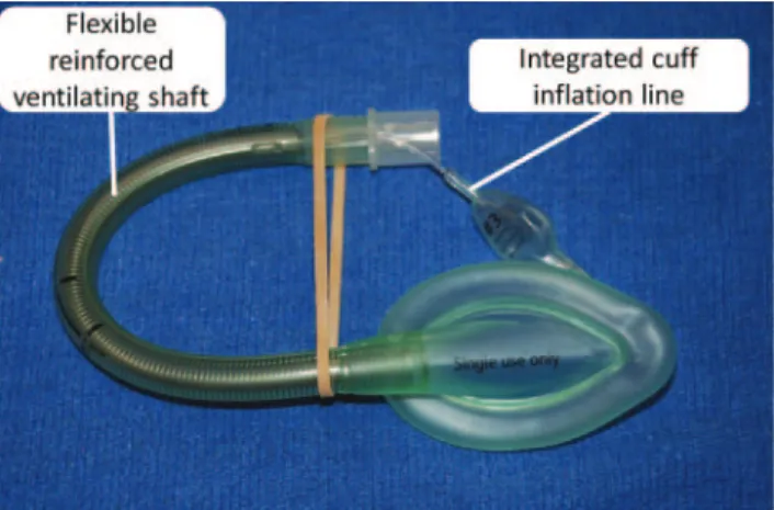
LMA Flexible
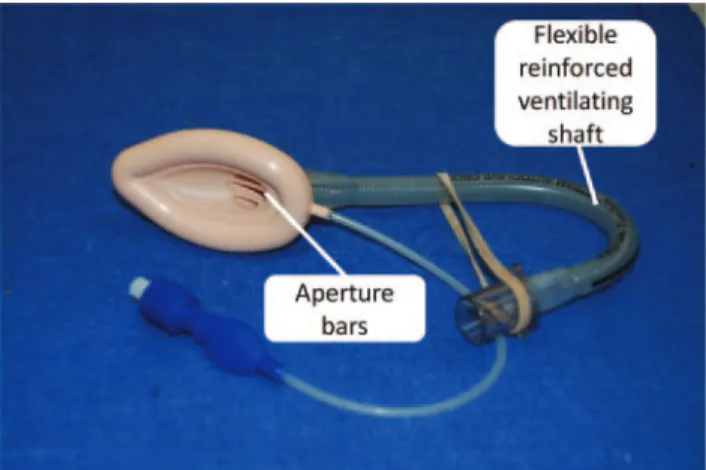
The LMA Flexible (fLMA; LMA North America, San Diego, CA) is designed to be useful for intraoral procedures in which a semirigid ventilation shaft would either obstruct the procedure or kink when displaced for surgical expo-sure. It consists of a flexible ventilating shaft attached to a cuffed mask similar to the cLMA (Fig. 3). The wire-reinforced ventilating shaft of the fLMA offers flexibility but can compromise ventilation if the tube is crimped or damaged from previous use. The ventilating shaft should
be inspected for signs of damage from prior use before the device is reused. It has been suggested that the fLMA is more difficult to insert than the cLMA unless a stylet or introducer tool is used.35
The recommended insertion tech-nique is the same as that for the cLMA. The junction of the mask and the ventilation shaft, a focal area for an index finger insertion technique, is not fundamentally different between the cLMA and fLMA. Despite the theoretical challenge of managing a flexible ventilating shaft during insertion, evidence suggests that insertion success is not different between the cLMA and fLMA.36
LMA ProSeal
The pLMA (LMA North America, San Diego, CA) was introduced in 2000 as the first EGA with two tubes for end-to-end contact with the respiratory and alimentary tracts37
(Fig. 4). A drain tube in the pLMA separates the esophagus from the larynx. If the drainage tube is posi-tioned correctly, it reduces the risk of aspiration if regurgi-tation occurs.38
A second cuff, behind the main body of the mask, increases contact with the posterior pharyngeal wall, increasing oropharyngeal leak pressure to an average of 25 cm H2O.39 Pediatric sizes have a drainage tube but no
posterior cuff. A higher oropharygeal leak pressure is useful for laparoscopic surgery, in patients with reduced lung compliance, or in obese patients who may need higher ventilation pressures.40
As with other EGAs, the pLMA is contraindicated in nonfasting patients, for whom tracheal intubation remains the technique of choice.
There are several pLMA insertion techniques: the index finger technique as recommended with the cLMA; with the help of an introducer tool; or by advancement into the hypopharynx over a gum elastic bougie or gastric tube placed into the esophagus via the drainage tube.41,42
Unlike the cLMA, the pLMA requires strict adherence to the recommended insertion techniques. A multicenter study comparing the pLMA and cLMA in anesthetized patients revealed first attempt insertion success at 92% for the cLMA versus 82% for the pLMA.39
After 3 attempts, success rates were similar (cLMA 100% vs pLMA 98%). Time to establish an airway was quicker with the cLMA (31 seconds) than with the pLMA (41 seconds), though these differences are likely clinically irrelevant. Seal pressure was
better with the pLMA (27 vs 22 cm H2O). Total
intraopera-tive complications and the incidence of postoperaintraopera-tive sore throat were similar.
In addition to decompression of the stomach and re-moval of regurgitant contents, the pLMA drain tube can be used to confirm the proper position of the device. The drainage lumen can be used to confirm proper position using the suprasternal notch test, an air leak test, or orogastric tube placement.37,43,44
In the suprasternal notch test, a water-soluble lubricant or nontoxic soap solution is used to form a meniscus over the end of the gastric drainage tube next to the ventilating lumen. The tracheal rings in the suprasternal notch are gently compressed while the meniscus on the drainage lumen is observed for motion of the lubricant or bubble formation. In a properly posi-tioned pLMA with a patent drainage lumen, the meniscus moves because pressure is transmitted from the proximal esophagus during compression of the suprasternal notch. During the leak test, positive pressure is applied to the ventilating lumen while observing the lubricant meniscus on the drainage lumen for displacement or bubble forma-tion. It is normal to see slight movement of the meniscus with respiration because the drainage port pressure reflects esophageal pressure, which varies during inspiration and exhalation. The leak test verifies separation between venti-lating and drainage lumens. Having a negative leak test, i.e., minimal or no movement of lubricant or soap meniscus with positive airway pressure, is a necessary but not sufficient condition to indicate proper pLMA position. It is possible for the cuff of the pLMA to fold backwards on itself during insertion, yielding a patent airway with an occluded drainage lumen.45
The easy passage of a gastric tube through the drainage lumen should be ascertained to confirm proper pLMA position. Smooth passage of a gas-tric tube into the stomach confirms that the pLMA is not folded and that the lumen is patent and aligned with the esophagus.
Intubating LMA (LMA Fastrach)
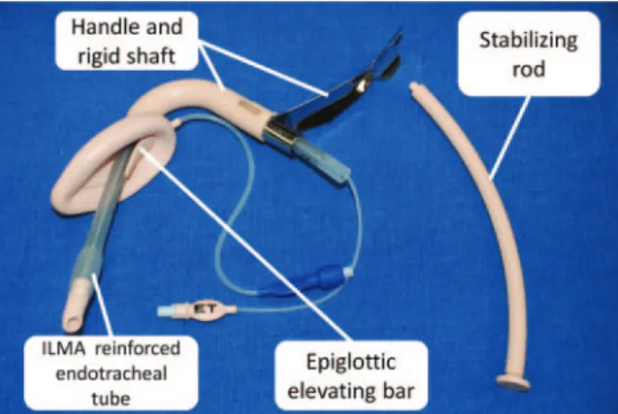
The LMA Fastrach (iLMA, LMA North America, San Diego, CA) was released in 1995. This intubating LMA has a curved metal shaft designed to fit into the oropharyngeal space while the patient’s head and neck are in neutral position (Fig. 5). The iLMA replaces the aperture bars of the
cLMA with a single epiglottic elevating flap attached only at its anterior end. During intubation, the tip of the flap is pushed up by the tracheal tube, deflecting the epiglottis anteriorly for passage of the tracheal tube through the glottis. Because of the rigid handle, one-handed insertion is possible from any position without extension of the pa-tient’s head, a useful feature for patients with limited cervical spine mobility. The shaft is shorter than that of the cLMA, which increases the length of tracheal tube that can be advanced beyond the mask orifice.
In a multicenter study involving placement of the iLMA in 500 patients, ventilation was satisfactory in 95%; it was difficult in 4% and impossible in 1%.46
Blind tracheal intubation through the iLMA was successful in 96.2% of patients after 3 attempts. The efficacy of the iLMA was also demonstrated in patients with abnormal airways47
and in obese patients.48Ferson et al. examined 254 patients with
known or suspected difficult airways. Insertion of the iLMA and successful ventilation were accomplished in all patients within 3 or fewer attempts.47
Similar to prior reports, the success rate for blind intubation was 96.5%. The tracheas of all patients with failed blind intubation via the iLMA were successfully intubated with the aid of a flexible bronchoscope introduced via the iLMA.47The combination
of the iLMA and fiberscope-assisted tracheal intubation via the device is a powerful technique to manage challenging airways. With the iLMA, a stable airway can be established rapidly, particularly in patients with anatomic features that complicate mask ventilation. Thus, the fiberscope operator bypasses most of the soft tissue in the oropharynx so that the fiberscope emerges from the mask near the glottic opening. Ventilation can also continue during the intubation process if the tracheal tube is inserted into the iLMA and a broncho-scope swivel adapter is placed between the anesthesia circuit and the 15 mm adapter on the tracheal tube.
Successful intubation with the iLMA depends on 4 factors: mask size selection, position of the patient, adjunctive maneu-vers, and the type and orientation of the tracheal tube. A number of criteria for size selection have been suggested. The manufacturer’s instructions suggest weight-based sizing, which does not consider variations in airway anatomy among patients of similar size. Sex, height, and nose-to-chin distance have been suggested as a basis for iLMA selection, although some argue that sizes 4 and 5 work better than size 3 for ventilation in men and women.49
An approximate size can be estimated by holding the iLMA next to the patient’s head to determine the eventual position of the iLMA mask relative to the mouth. The iLMA is positioned next to the patient’s head so that the cranial surface of the shaft is positioned at the level of the upper incisors or the hard palate for edentulous patients. If the iLMA is properly sized, the middle of the epiglottic elevating flap is at the level of the thyroid notch. This sizing guideline helps ensure that the tracheal tube will deploy directly into the larynx. If the LMA is too big, the tracheal tube may enter the esophagus. If it is too small, it may get caught into the vallecula.
The iLMA is inserted with the patient’s head and neck in a neutral position. If ventilation or tracheal tube insertion is inadequate or unsuccessful, the “up-down” and “Chandy” maneuvers can be applied.47,50
For the up-down maneuver, the iLMA is pulled out approximately 6 cm with the cuff
inflated and then reinserted into the hypopharynx. This maneuver usually corrects the downfolding of the epiglot-tis, which is often responsible for airway obstruction and difficulty with iLMA-assisted endotracheal intubation. The Chandy maneuver has 2 parts. The first is gentle manipu-lation of the iLMA handle in the horizontal and sagittal planes to adjust positioning during hand ventilation. The astute operator can discern when airway resistance is at a minimum. Usually the position that provides lowest resis-tance and best ventilation is also the position that is best for intubation. The second part of the Chandy maneuver involves lifting the iLMA handle anteriorly to displace the device away from the posterior pharyngeal wall. Both components of the Chandy maneuver attempt to align the iLMA mask orifice to optimize tracheal intubation.
The type of tracheal tube and its orientation within the iLMA also influence the success of intubation. A wire-reinforced silicone tracheal tube designed for the iLMA has a soft, specially shaped tip, has no natural curve or camber, and is more flexible than a standard PVC tube. Despite success with blind iLMA-assisted tracheal intubation using the iLMA silicone tube, success has also been shown with various PVC tracheal tubes.51In addition to the material of
the tracheal tube, the orientation of the tube during inser-tion via the iLMA is also a considerainser-tion. Camber refers to the curved shape of PVC tracheal tubes. During intubation via direct laryngoscopy, the tracheal tube is inserted with forward camber, which directs the concavity of the tube anteriorly. If a PVC tube is inserted through an iLMA with forward camber, the tip of the tube as it emerges from the intubation shaft is not well-aligned with the axis of the trachea. Tracheal intubation with the tube inserted with reverse camber was first reported by Joo and Rose.52
Reverse-camber insertion, i.e., the concavity facing posteriorly, im-proves the angle of emergence of a PVC tube through the iLMA, resulting in a higher first attempt intubation success rate.53 Overall success rates, beyond first attempt,
showed no difference with normal and reverse camber orientation.53A 96% success rate for intubation within 2
attempts was reported with PVC tubes in normal orien-tation and with silicone wire-reinforced tubes.54
LMA Supreme
In 2007, the disposable LMA Supreme (sLMA) became available as an alternative to the reusable pLMA. The
sLMA combines some features of 3 previously introduced LMAs: the uLMA (disposable), the iLMA (rigid curved shaft), and the pLMA (gastric access) (Fig. 6). The sLMA is designed for ease of insertion, it has a gastric drainage tube and provides a high seal pressure. The mask has a narrower transverse diameter than previous LMA models, allowing it to be placed in patients with a smaller mouth opening. The internal webbing of the tip keeps the drain tube open, and the fins within the bowl of the sLMA protect the airway from epiglottic obstruction. Two preliminary stud-ies demonstrated a placement success rate of 96% to 98%.a55
LMA Classic Excel
The LMA Classic Excel (eLMA, LMA North America, San Diego, CA) is designed to aid tracheal intubation. It is similar to the cLMA in appearance, except for an epiglottic elevating bar that replaces the aperture bars in the cLMA (Fig. 7). The 15-mm adapter of the eLMA can be removed from the ventilating lumen. Experience is limited with this device, due to its recent introduction. The eLMA provides an alternative to the intubating LMA for cases requiring an EGA to aid tracheal intubation.
Other EGAs
After the successful introduction of the cLMA, several other EGAs were introduced. Despite the widespread commer-cial availability of alternative devices, none has been stud-ied as extensively as the LMA.
Ambu Laryngeal Masks
The Danish medical device manufacturer Ambu A/S (Ambu, Glen Burnie, MD) launched their version of a laryngeal mask, the AuraOnce, in 2004.67
The Ambu AuraOnce has an ellip-tical, inflatable cuffed mask attached to an airway shaft with a standard connector. It is similar to the cLMA but without aperture bars in the bowl of the mask. The shaft is curved at an almost 90° angle for easy insertion. The AuraOnce cuff, mask, and tube are molded together as a single unit that is latex-free. Indications, contraindications, and disadvan-tages of the AuraOnce are similar to those of the cLMA. In
a multicenter trial involving 118 adult patients, the first-attempt success rate for intubation was 92.4%, ventilation was adequate in all patients, and the vocal cords could be seen with a fiberoptic bronchoscope in 91.5% of patients.68
In another study, the AuraOnce functioned satisfactorily when tested in patients in 5 different head positions.b
This
aFerson DZ, Chi L, Zambare S, Brown D. The effectiveness of the LMA
Supreme in patients with normal and difficult-to-manage airways. (abstract) Anesthesiology 2007;107:A592.
bGenzwuerker HV, Hinkelbein J, Krivosic-Horber R, et al. Performance of
the new single-use Ambu laryngeal mask in different head positions. (abstract) Anesthesiology 2004;101:A1590.
Figure 7. LMA Excel™. ETT indicates LMA ET Tube™. Figure 8. Ambu AuraFlex™.
Figure 9. Ambu AuraStraight™.
feature may be beneficial for patients who undergo head or neck surgery. In 80 patients who had minor gynecologic procedures, the AuraOnce and the uLMA performed equally, with the exception of a slightly higher airway leak pressure with the AuraOnce.57
Other versions include the AuraFlex (Fig. 8) (Ambu, Glen Burnie, MD), which has a flexible airway shaft; the AuraStraight (Fig. 9) (Ambu, Glen Burnie, MD), which has a straight shaft; and the reusable version of the AuraOnce, the Aura40 (Ambu, Glen Burnie, MD). A newer model, the Aura-i (Fig. 10) (Ambu, Glen Burnie, MD), is designed to facilitate 1-step tracheal intu-bation with fiberoptic guidance via the device in pediatric and adult patients. Blind intubation via the Aura-i is not recommended.
Air-Q/Intubating Laryngeal Airway
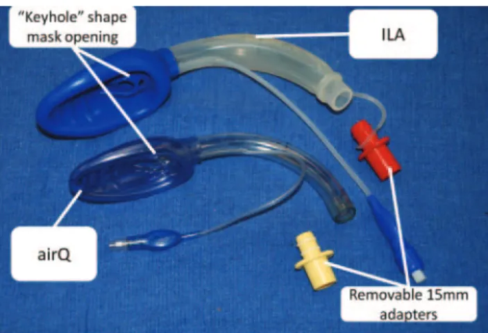
Developed by Dr. Daniel Cook and introduced in 2004, the air-Q/Intubating Laryngeal Airway (air-Q/ILA, Cookgas LLC, St. Louis, MO, USA) is an EGA for use as a primary airway device or as an adjunct to tracheal intubation. The air-Q/ILA is available as a disposable (air-Q) or nondispos-able (ILA) device (Fig. 11). It has an elliptical, inflatnondispos-able, cuffed mask and a slightly curved airway tube with a detachable connector.
The air-Q/ILA has several unique features designed to assist tracheal intubation. In most other EGAs, the length of the shaft, fixed 15-mm adapter, and mask aperture bars complicate blind tracheal intubation. The air-Q/ILA has a shorter shaft than the cLMA, no aperture bars within the mask, a removable connector so that the wide lumen of the shaft can be used for intubation, and a keyhole-shaped distal airway tube to direct a tracheal tube toward the larynx. If ventilation with the device is unsatisfactory, the “Klein maneuver” corrects downfolding of the epiglottis by using a jaw thrust and an up-down maneuver of the ILA.c
The ILA is not recommended for patients at risk for aspiration, those with poor lung compliance, or with le-sions of the oropharynx or epiglottis.
During tracheal intubation through the air-Q/ILA, the tracheal tube is advanced to a depth of 12 to 15 cm so that
the tip of the tube is close to the air-Q/ILA opening. The tube is then advanced into the trachea blindly or with the aid of a fiberoptic bronchoscope. Once tracheal intubation is successful, the ILA can be left in place as a bridge to extubation at the conclusion of the anesthetic. Alternatively, removal of the ILA immediately after tracheal intubation can be assisted by a stylet produced by the manufacturer. Blind intubation success via the air-Q/ILA improves with use of a flexible reinforced tracheal tube.
Cobra Perilaryngeal Airway (Cobra PLA)
The Cobra Perilaryngeal Airway (Cobra PLA; Pulmodyne, Indianapolis, IN) was introduced by Dr. David Alfrey in 1997. It has a large diameter airway tube with a high-volume, low-pressure pharyngeal cuff located proximal to the wide distal Cobra head (Fig. 12). The wide head keeps soft tissues away from the glottis to maintain a patent airway. When inflated, the cuff displaces the base of the tongue to expose the glottis. The Cobra PLA, which does not have a gastric channel, can be used as an adjunct to tracheal intubation. The Cobra head has a soft, flexible grill for passage of a tracheal tube. The Cobra PLUS models allow monitoring of body temperature using an integrated thermistor, and distal CO2 gas sampling in its pediatric
sizes. For successful placement, the patient’s head is placed in the sniffing position, the device is lubricated, the mouth is opened with a scissor motion using the nondominant hand, and the Cobra PLA is advanced straight back be-tween the tongue and hard palate. Modest neck extension and anterior jaw lift aid placement. Properly placed, the tip of the Cobra lies posteriorly to the cricoid cartilage, the ramp grill lifts the epiglottis, and the cuff lies in the hypopharynx at the base of the tongue.
Comparison of the Cobra PLA to the uLMA showed similar ease of insertion and success in obtaining a patent airway.58
Gaitini et al. studied 80 patients, divided evenly between the Cobra PLA and uLMA; airway adequacy, number of adjustments made, and minor complications were the same with both devices, but the cuff pressure required to prevent a leak of the Cobra PLA was more than that of the uLMA.
Comparison of the Cobra PLA to the cLMA found similar results for both spontaneous and positive-pressure ventilation, but oropharyngeal leak pressure was higher
cKlein MT, Jones J: Utility of the intubating laryngeal airway: report of an
observational study. (abstract) Anesthesiology 2005;103:A846.
Figure 11. Cookgas airQ™ (airQ) and Intubating Laryngeal Airway™ (ILA).
with the Cobra airway, 23⫾ 6 cm H2O versus 18⫾ 5 cm
H2O for the cLMA.59 In a series of 110 patients, Cobra
placement was 100% successful with no major complica-tions, and required a mean cuff leak pressure of 34 cm H2O.
60
Esophageal Tracheal Combitube
The design of the Esophageal-Tracheal Combitube (Covidien-Nellcor, Boulder, CO) is a major advance based on earlier esophageal obturator devices (Fig. 13). As an alternative device to secure the airway, it has proven especially useful for prehospital use or when the operator lacks the skills for facemask ventilation or tracheal intuba-tion.61
The device is designed to be inserted blindly and ventilation can be established whether it enters the esoph-agus or the trachea.62
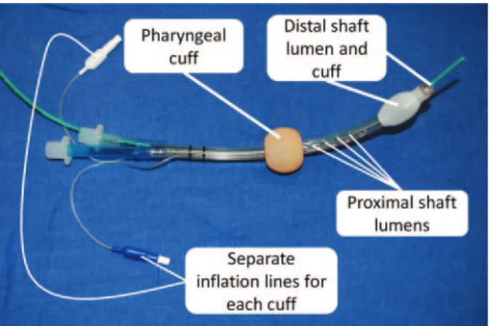
The Combitube requires less training to master, does not require as much continuing use to maintain proficiency as direct laryngoscopy, and is easier to insert than a traditional tracheal tube. It is a disposable double-lumen, double-cuffed device with separate pilot balloons for the proximal and distal cuffs. The distal lumen (known as lumen #2) is clear and terminates at the end of the device. Lumen #1 is colored blue and terminates in several small fenestrations located between the proximal and distal cuffs (Fig. 13). The Combitube is available in 37-Fr and 41-Fr sizes; the 41 Fr is for adults taller than 6 feet.63
When inserted blindly, it typically enters the esoph-agus stabilizing the device and providing access to the stomach for decompression. The proximal tube #1 provides ventilation of the patient’s lungs when the Combitube is placed in the esophagus. Once placed, confirmation of position can be obtained by auscultation for lung sounds during ventilation via tube #1. The absence of lung sounds, or the presence of gastric insufflation during ventilation via lumen #1 indicates esophageal ventilation, and ventilation should be switched to lumen #2.62
Less than 5% of the time, the Combitube enters the trachea; in this position, lumen #2 is used as a tracheal tube for ventilation. If ventilation is not possible via either lumen, it may be due to the Combitube being inserted too deeply. Despite the utility of ausculta-tion for placement confirmaausculta-tion, the addiausculta-tional use of capnometry (i.e., Easycap, Nellcor, Inc., Pleasanton, CA) or
an esophageal detector (i.e., TubeChek, Ambu, Linthicum, MD) is advisable to ensure proper use.62,63
The Combitube may be placed blindly or under direct vision with the help of a rigid laryngoscope. It can be placed in patients with a mouth opening as small as 15 mm and is helpful in “cannot-intubate-cannot-ventilate” situations, especially when other EGAs have failed. The Combitube does not require movement of the patient’s head or neck, an advan-tage for patients with cervical spine pathology. Uncon-sciousness with absence of a gag reflex or a combination of light anesthesia and a muscle relaxant are needed for placement.61
There are several advantages of the Combitube: it re-quires minimal preparation; blind insertion is possible; it can be positioned in the trachea or esophagus; it reduces aspiration risk; it is less invasive than a cricothyrotomy; neck extension of the patient is unnecessary during inser-tion; stomach decompression is possible; and positive pressure ventilation is possible.62
Disadvantages of the Combitube airway include an inability to suction the tra-chea when placed in the esophageal position, except with a redesigned version that has a large opening for suctioning and for airway evaluation through a fiberoptic broncho-scope.64
Because of the tendency of the Combitube to intubate the esophagus when inserted, its use is contrain-dicated in those who have ingested caustic substances, or in patients with known upper esophageal pathology such as Zencker’s diverticulum or esophageal varices. Complica-tions associated with the use of the Combitube include laceration of the esophagus, esophageal rupture, tongue engorgement and venous congestion, and inability to ventilate.65
EasyTube
Similar to the Combitube, the Rusch EasyTube (Teleflex Medical, Research Triangle Park, NC) has the same general shape, applications, and insertion technique. It was intro-duced in 2003 by Frass and colleagues. The latex-free device is available in 2 sizes for use in adults (130 cm or taller) or children (90 to 130 cm tall). The pharyngeal lumen between the 2 balloons of the EasyTube opens into the pharynx via a single open end for passage into the trachea of a fiberoptic bronchoscope, suction catheter, or tube changer. The distal tube is similar to a tracheal tube and has a Murphy eye.
Laryngeal Tube
The Laryngeal Tube (LT; King Systems, Noblesville, IN) was introduced in Europe in 1999 and in the United States in 2003. There are several different LT airways in the LT family, and all these devices are shorter than many other EGAs. The silicone tubes are curved slightly and have two cuffs, a small distal esophageal and a larger proximal pharyngeal cuff (Fig. 14). A single inflation line serves both low-pressure, high-volume cuffs. After inflation, the distal cuff seals the esophagus and the proximal cuff seals the hypopharynx, allowing for ventilation through the larynx via the ventilating lumen between the cuffs. The LT can serve as an adjunct to tracheal intubation because its primary ventilating passage accepts a flexible broncho-scope or tube exchanger. The ventilating lumen is located
Figure 13. Nellcor Puritan Bennett Esophageal Tracheal Combi-tube™. Green exchange catheter in distal lumen is for demonstration purposes.
between the proximal and distal cuffs. The LT is reusable and has a blind tip. Additional models include the dispos-able Laryngeal Tube (LT-D), and the disposdispos-able Laryngeal Tube Suction (LTS-D), which has a suction channel leading to an opening at its distal tip. In 2004 the Laryngeal Tube Suction II (LTS II; VBM, Medizintechnik, Sulz am Neckar, Germany, not available in the United States) was intro-duced; it has a longer shaft than the LTS, as well as a modified tip and cuff shape. The LTS II is available in pediatric sizes and most resembles the King LTS-D.66
Before insertion of the LT, both cuffs are deflated and lubricated. The head of the patient is placed in neutral or sniffing position with jaw lift. The LT is placed in the mouth along the midline of the tongue with the tip against the hard palate and directed in a caudal direction until resistance is felt. The cuffs are inflated up to 60 cm H2O
pressure. Auscultation of breath sounds during ventilation via the ventilating lumen serves to confirm proper position, although the addition of capnometry is advisable as is standard with tracheal intubation. LT models with esoph-ageal ports LTS-D, LTSII, and G-LT (see below) are not designed to allow ventilation should the distal tip of the device enter the trachea. The soft tip and low-pressure cuff are designed to minimize tissue trauma, sore throat, hoarseness, and airway bleeding.
The LT is a reliable device for airway management during elective surgery.67
First attempt insertion success has ranged between 86% to 94%.67,68
Tracheal intubation via the LT was successful in 3 patients after the cLMA proved unsatisfactory because the pharyngeal space was narrowed by enlarged tonsils.69
Leak pressure performance for the LT and LTS-D appears to be similar to that of the pLMA.70,71
The newer LTS II was found to be more difficult to insert and have a lower leak pressure than the pLMA.66
A new version of the LT, the Gastro-Laryngeal Tube (G-LT, VBM Medizintechnik, GmbH, Sulz am Neckar, Germany) is now available. The G-LT is designed for use in gastrointestinal endoscopy cases. It provides a conduit to the esophagus that accommodates a gastroscope. The esophageal lumen is wide and has a distal cuff that can be inflated to seal the esophageal inlet. Ventilation is estab-lished via a separate lumen that terminates distally to the
larger pharyngeal cuff. The G-LT has the theoretical advan-tage of controlled airway management without endotra-cheal intubation while providing easy access to the upper gastrointestinal tract for endoscopic procedures.
I-GEL
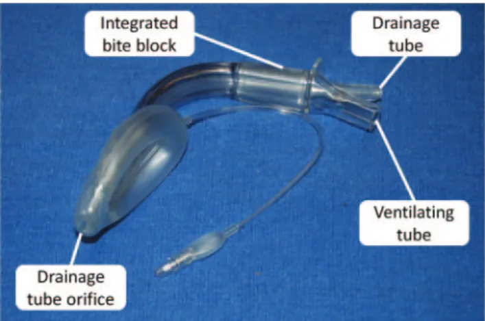
The i-gel (Intersurgical, Wokingham, Berkshire, UK), in-vented by Dr. Muhammed Nasir, is anatomically designed to conform to the hypopharynx without using an inflatable cuff. The mask surface is made of a gel-like thermoplastic elastomer, which is soft and conforms to the larynx (Fig. 15). The mask bowl lacks aperture bars, providing free passage of an endotracheal tube. The shaft has an integral bite block and is flattened to resist rotation. A second lumen runs along the entire length of the device to the distal tip to accommodate a nasogastric tube for drainage of potential regurgitant gastric contents. The i-gel is intended for single use and is available in 7 sizes, pediatric to adult. A comparison of the i-gel, cLMA, and pLMA found that the seal pressures of the i-gel and the pLMA were compa-rable, and higher than the cLMA seal pressure. Success rates of initial insertion were similar among the devices.72
Early published experience with the i-gel suggests ease of use, a high success rate for first attempt insertion, and good fiberoptic visualization of airway structures through the device.
Streamlined Liner of the Pharynx Airway
The Streamlined Liner of the Pharynx Airway (SLIPA; SLIPA Medical, Douglas, Isle of Man) resembles a slipper that lines the pharynx. It was developed by Dr. Donald Miller and introduced in Europe in 2004 (Fig. 16). This disposable, cuffless device has an enlarged cavity for trapping regurgitated fluids. The soft plastic mask includes a gastric port that leads to the hollow cavity within. The airway shaft of the SLIPA is flexible at its attachment to the mask and rigid more proximally to its 15 mm adapter. The SLIPA device is rigid at room temperature but softens once inserted. For placement, the patient’s head is ex-tended, and the SLIPA is angled toward the esophagus until the heel of the device reaches the pharynx.73
The SLIPA is available in 6 adult sizes selected according to patient height. When compared to the cLMA, insertion
Figure 14. King LTS-D™ (Laryngeal Tube Suction Disposable). Green exchange catheter in esophageal lumen is for demonstration purposes.
Figure 15. Intersurgical i-gel™. Green exchange catheter in drainage lumen is for demonstration purposes.
success rates, airway seal pressures, and incidence of sore throat were similar.73–75
Use of EGAs for Special Situations
Prehospital
The prehospital setting poses numerous challenges to suc-cessful airway management. Advanced airway expertise is often lacking, the physical conditions under which care must be provided are suboptimal, and planned preparation is often impossible. Tracheal intubation is considered the “gold standard” for definitively securing an airway, but the skills necessary to accomplish this task are often not available in the prehospital setting. In addition, patients are often not fasted and are at risk for regurgitation of gastric contents and subsequent pulmonary aspiration. Trauma or environmental hazards may make visualization of airway structures during direct laryngoscopy difficult or impos-sible. EGAs have many advantages in the prehospital setting. They are designed for blind insertion, do not require laryngoscopy, and may establish a reliable ventilat-ing airway better than bag mask ventilation. The first use of the cLMA for emergency prehospital airway control was reported by Greene et al. in 1992.76
Because the cLMA is relatively easy to insert, it was soon promoted as an emergency airway device. Nurses, paramedics, and other inexperienced personnel have been highly successful in first attempts at cLMA placement.77,78
The Combitube has been popular for use in trauma and nontrauma patients. In one study, a Combitube was a helpful adjunct for airway management after failed tracheal intubation following rapid sequence induction of 12 pa-tients with facial trauma and fractured mandible.79
The LT has also been used successfully in the prehospi-tal setting. In one study of 157 patients who required prehospital airway management, the success rate for LT insertion and ventilation was 96.8%.80
Patients were man-aged outside of the hospital by paramedics in 70 of the 157 cases, the rest were cared for by emergency physicians in the same setting.
Resuscitation
Airway management is a critical component of cardiopul-monary resuscitation. One of the goals of treating patients in respiratory arrest is to maintain a patent airway for
oxygenation and ventilation. It is often difficult for the nonexpert to establish adequate ventilation with the com-monly used facemask and self-inflating bag. EGAs have been used successfully to overcome this problem by pro-viding a stable ventilating airway that does not require the advanced skill of a practitioner to maintain an adequate seal. In one study, 130 nurses trained in the use of the LMA placed the device within 3 attempts during resuscitation of 164 patients who suffered cardiac arrest.78
The success rate on the first attempt was 71%; for the second attempt, 26%. The overall failure rate was only 12%. Tracheal intubation proved difficult in 11 of these patients when attempts were made to replace the LMA with a tracheal tube. The LMA was properly replaced, and satisfactory ventilation contin-ued in all patients in whom tracheal intubation had failed previously. Thirty-three patients (20%) showed evidence of regurgitation. However, it is important to note that 20 of the 33 patients who regurgitated did so before LMA placement, 3 regurgitated during LMA insertion, and in the remaining 10, regurgitation occurred after LMA removal. Only 1 patient (0.6%) had clinical evidence of pulmonary aspiration. In fact, it appears that the incidence of regurgi-tation of gastric contents during resusciregurgi-tation using an LMA is similar to, if not less than, that during self-inflating bag mask ventilation.81
Several studies have compared the use of the Combitube for resuscitation after cardiac arrest outside or inside the hospital.82,83
In the hospital, the Combitube was as effective as a tracheal tube. When compared to physicians perform-ing tracheal intubation, nurses were able to place a Com-bitube with a similar success rate, but in a shorter period of time.
Obstetrics
The cLMA can be lifesaving in cesarean deliveries when tracheal intubation and mask ventilation have failed. LMAs with a drain tube (such as the ProSeal and the Supreme) theoretically offer better protection against regurgitated stomach contents than the cLMA, but protection is not as reliable as with a cuffed tracheal tube. Repeated attempts at tracheal intubation using different techniques may be more dangerous than placing a pLMA to secure the airway and passing a gastric tube to decompress the stomach. Because of the nature of the surgery and the limited time involved, cesarean delivery can be completed with an EGA in place. Although a rapid sequence induction with tracheal intuba-tion remains common for obstetric general anesthesia, elective use of an EGA for patients undergoing a cesarean delivery has been described.23
In a large cohort study, 1067 parturients with a preference for general over regional anesthesia were scheduled for elective cesarean delivery with an LMA. Patients with a perceived or known difficult airway, a pregravid body mass index ⬎30, ASA physical status⬎2, and with pharyngeal reflux, were excluded. An effective airway using an LMA was established in 99% of patients, 0.7% of patients required intubation, and there was no evidence of regurgitation or pulmonary aspiration in this selected patient group.
Pediatrics
Pediatric patients present special challenges for the design and use of EGAs. The transition from pediatric to adult
Figure 16. SLIPA Medical SLIPA™ Airway (Courtesy of SLIPA Medical, Douglas, Isle of Man).
airway does not occur at a predictable age. Pediatric versions of the cLMA are smaller scale versions of the adult model. Yet, pediatric anatomy is not simply miniaturized adult anatomy. Young children typically have a more cephalad glottis, and a more funnel-shaped than cylindrical airway in comparison with that of an adult.84
As children approach their teen and even preteen years, their airway may become anatomically adult. Not all EGAs can be downsized for pediatric use to maintain good function, and availability of devices suitable for the smallest patients is limited (Table 3). Studies suggest a high incidence of epiglottic downfolding with smaller LMA sizes, as con-firmed by a fiberscope assessment through the LMA.85,86
Despite potential downfolding of the epiglottis, clinically significant airway obstruction was not present in the ma-jority of patients studied. The first pediatric sizes of the cLMA were size 1 (for children weighing⬍6.5 kg) and size 2 (for children weighing 6.5 kg to 25 kg). Two additional sizes, 1.5 and 2.5, have since been added.
Pediatric sizing and anatomy are important consider-ations, but pediatric physiology must also be considered. Children have a limited tolerance for apnea and a tendency toward laryngospasm. Unintended gastric insufflation dur-ing positive pressure ventilation is common in pediatric patients.29
Gastric distension not only makes ventilation more difficult, but may also predispose to regurgitation of gastric contents. Unintended dislodgment of the EGA is possible with relatively minor manipulation of the patient or device, particularly in infants. However, despite differences in airway anatomy and physiology, the overall experience with pedi-atric cLMAs has been favorable, with some notable exceptions, such as increased epiglottic downfolding that may complicate blind intubation techniques.85,87
Indications for EGA use in children generally mirror those for adults. Extraglottic devices can be used as a primary airway, a rescue airway, an adjunct device during difficult intubation, or for special situations in or outside the OR (Table 4). When an unexpected need for pediatric
airway management arises, personnel with airway exper-tise may not be available. In situations where tracheal intubation is not feasible, ventilation may be established with a pediatric EGA.88
Besides emergent resuscitation, an EGA can be used to ventilate a child’s lungs during transport to a location that can provide pediatric personnel and resources.89
Contraindications to use of an EGA in children are similar to those for adults. Among them is the risk of gastric regurgitation and the potential for pulmonary aspiration. There are design modifications meant to lessen the chance of pulmonary aspiration but not to the degree provided by cuffed tracheal intubation. A full stomach, active gastric reflux, intrapharyngeal masses or pathology, and the need for high inspiratory ventilatory pressures because of lung pathology or procedure type are typical contraindications for the use of a pediatric EGA.
Compared to adults, the risk of pressure injury to the airway in children may be greater with the use of EGAs. Of particular concern for pediatric practitioners is the potential for higher cuff pressures from small devices placed in small airways. High pressures may increase the risk of injury to the surrounding mucosa and structures of the pharynx. A review of 400 pediatric patients managed with either the cLMA or the uLMA found median cuff pressures of 90 cm H2O to ⬎120 cm H2O, pressures that were much higher
than the manufacturer’s recommended maximum of 60 cm H2O.90The effect of high cuff pressures on vital structures
is not easy to predict, but mucosal lesions from hypoper-fusion, nerve injury, and sore throat seem likely risks.32–34
In an observational study of 400 children, the likelihood of postoperative sore throat increased with higher cuff pres-sures. Among the children who had either the cLMA or uLMA (n ⫽ 139), 17% had a sore throat; in 56% of sore throat cases, cuff pressures were ⬎100 cm H2O.91 When
cuff pressures were⬍40 cm H2O, children did not
experi-ence sore throat.
Table 3. Devices with Pediatric Sizing for Small Children
Manufacturer of EGAs with sizes appropriate for small children (in alphabetical order)
Device Neonates (<5 kg) 5–10 kg 10–20 kg
AES Ultra CPV/Ultra Clear CPV X X X
UltraFlex CVP X X X
Ambu Aura Straight X X X
Aura Once/Aura40 X X X AuraFlex X Aura-i X X X Cookgas airQ X X X Intersurgical i-gel X X X King LAD X X X LAD Flex X Laryngeal Tube (LT) X
LMA LMA Classic X X X
LMA Unique X X X
LMA Flexible X
LMA ProSeal X X X
LMA Supreme X X X
Excessive cuff pressure is not always a result of over-zealous cuff inflation. A review of 1000 children managed with different sizes and brands of LMAs revealed some interesting results.31
Only the volume of air already present in the device as packaged was used during placement. In 20.5% of cases, cuff pressures were ⱖ60 cm H2O. Of the
smaller size 1 devices, 66.6% had cuff pressures ⱖ60 cm H2O when just the packaged volume of air was present in
the cuff. These results suggest that smaller-sized devices may need to be deflated after placement to achieve pres-sures ⬍60 cm H2O. Mounting evidence warns against
reliance on subjective clinical indicators of appropriate cuff volumes and pressures in pediatric patients. With devices such as a simple manometer, cuff pressures can be monitored to avoid potential morbidity.92
Pediatric versions of the fLMA, pLMA, sLMA, and uLMA are on the market (Table 2). Similar to its adult counterpart, the pediatric pLMA is designed with a cuffed mask bowl and a gastric drainage port but does not have the posterior cuff found on the adult sizes. Multiple studies have compared the cLMA to the pLMA in pediatric pa-tients.93,94
Evidence suggests that the pediatric pLMA
has a high success rate for first pass insertion, and typically provides a higher leak pressure relative to the equivalent pediatric cLMA.95
The pediatric fLMA is useful when the ventilating shaft of a typical EGA device interferes with a procedure. Children receiving radiation therapy to the head may require a special mask that can limit access to the airway, thus complicating airway management. The fLMA often works well for these patients as it does in many patients having tonsillectomies, dental procedures, and other pro-cedures surrounding the mouth. Unlike adults, in whom there may be other options, general anesthesia is frequently required in children who undergo daily radiotherapy, dental procedures, or head or neck operations, creating a niche for the fLMA in pediatric practice.96,97
There is a paucity of literature evaluating newer EGA devices in children.98
Most of the available evidence con-sists of case reports or small trials. Pediatric models of the Cobra PLA have been used successfully in children, but epiglottic downfolding that obstructs the view of the vocal cords was noted by fiberoptic bronchoscopy in children weighing⬍10 kg.99
Other studies of children weighing⬎10
Table 4. Use of Pediatric Extraglottic Airways Outside of the Operating Room
Pediatric applications for extraglottic airway devices outside of the traditional operating room’s location/service
Challenges Potential benefits of EGA* device use
Bronchoscopy suite117 ● Sedation is seldom a viable option ● Endotracheal intubation limits the size of
bronchoscope that can be introduced
● Allows general anesthesia with a larger ventilating lumen than a comparable endotracheal tube
● Upper airway structures are not visible due to the presence of the endotracheal tube
● Larger bronchoscope can be used with an EGA than tracheal tube
● Supraglottic structures can be observed with an EGA in place also with spontaneous ventilation, less likely with endotracheal intubation
Radiology suite ● Noninvasive, but patient may need to be
monitored from a distance
● EGAs can provide a stable airway under general anesthesia
● Patient movement will adversely affect study quality
● Long procedure time may predispose to atelectasis during prolonged spontaneous
● May allow lighter depth of general anesthesia then endotracheal intubation. Helpful in situations with little procedural stimulation
ventilation under general anesthesia
Radiotherapy96 ● Repetitive, often daily, treatments
necessitating patient stillness for treatment efficacy
● EGAs can be used repeatedly without the need for repeated laryngoscopy for tracheal intubation
● Repeated daily endotracheal intubation is traumatic and may require muscle relaxation
● EGAs with a flexible shaft permit the device to be placed without interfering with the accuracy of radiation therapy
Gastroenterology/endoscopy suite118 ● Typically requires general anesthesia with nasal cannula, facemask, or airway intubation
● Prevents upper airway obstruction that can occur with general anesthesia with nasal cannula
● When general anesthesia with tracheal intubation is planned a deeper level of anesthesia may be required
● Lighter plane of general anesthesia may be sufficient to tolerate procedure and an EGA versus tracheal tube anesthetic
● Gastric drainage lumen may be used to facilitate endoscope introduction Interhospital transport86,199 ● Children with congenital airway anomalies
who require nonelective endotracheal intubation, can be very challenging in environments where expertise and resources are lacking
● EGAs can allow a stable method of ventilation during transport of the patient to a center with the appropriate facilities and personnel to definitively secure the airway ● An EGA may be more effective for ventilation
than attempted bag mask ventilation, particularly if the operator is less experienced