Effects of the Beach Chair Position, Positive
End-expiratory Pressure, and Pneumoperitoneum on
Respiratory Function in Morbidly Obese Patients
during Anesthesia and Paralysis
Franco Valenza, M.D.,* Federica Vagginelli, M.D.,† Alberto Tiby, M.D.,† Silvia Francesconi, M.D.,† Giulio Ronzoni, M.D.,† Massimiliano Guglielmi, M.D.,† Marco Zappa, M.D.,‡ Ezio Lattuada, M.D.,‡ Luciano Gattinoni, M.D., F.R.C.P.§
Background: The authors studied the effects of the beach chair (BC) position, 10 cm H2O positive end-expiratory
pres-sure (PEEP), and pneumoperitoneum on respiratory function in morbidly obese patients undergoing laparoscopic gastric banding.
Methods:The authors studied 20 patients (body mass index 42ⴞ 5 kg/m2) during the supine and BC positions, before and
after pneumoperitoneum was instituted (13.6 ⴞ 1.2 mmHg). PEEP was applied during each combination of position and pneumoperitoneum. The authors measured elastance (E,rs) of the respiratory system, end-expiratory lung volume (helium technique), and arterial oxygen tension. Pressure–volume curves were also taken (occlusion technique). Patients were paralyzed during total intravenous anesthesia. Tidal volume (10.5ⴞ 1 ml/kg ideal body weight) and respiratory rate (11 ⴞ 1 breaths/min) were kept constant throughout.
Results:In the supine position, respiratory function was ab-normal: E,rs was 21.71ⴞ 5.26 cm H2O/l, and end-expiratory lung
volume was 0.46ⴞ 0.1 l. Both the BC position and PEEP improved E,rs (P < 0.01). End-expiratory lung volume almost doubled (0.83
ⴞ 0.3 and 0.85 ⴞ 0.3 l, BC and PEEP, respectively; P < 0.01 vs.
supine zero end-expiratory pressure), with no evidence of lung recruitment (0.04ⴞ 0.1 l in the supine and 0.07 ⴞ 0.2 in the BC position). PEEP was associated with higher airway pressures than the BC position (22.1ⴞ 2.01 vs. 13.8 ⴞ 1.8 cm H2O; P < 0.01).
Pneumoperitoneum further worsened E,rs (31.59 ⴞ 6.73; P < 0.01) and end-expiratory lung volume (0.35ⴞ 0.1 l; P < 0.01). Changes of lung volume correlated with changes of oxygenation (linear regression, R2ⴝ 0.524, P < 0.001) so that during
pneumo-peritoneum, only the combination of the BC position and PEEP improved oxygenation.
Conclusions:The BC position and PEEP counteracted the ma-jor derangements of respiratory function produced by anesthe-sia and paralysis. During pneumoperitoneum, only the combi-nation of the two maneuvers improved oxygecombi-nation.
A NUMBER of authors have shown that during anesthesia and paralysis, respiratory function in obese patients is greatly impaired,1– 4and further worsened when laparo-scopic procedures are performed.5– 8
Several strategies have been tested to improve oxygen-ation. Positive end-expiratory pressure (PEEP) has been shown to improve respiratory function9 –18; however, it may cause relative hypotension.15,18 –20Large tidal volumes or a high respiratory rate have been tested with negative results such that the authors concluded that the only way to safely improve oxygenation is to increase the fraction of inspired oxygen (FIO2).8The effects of body positioning on
respiratory mechanics have also been investigated: The head-up position has been shown to be beneficial as op-posed to the head-down position,7,21even if this was not always the case.8
Apart from the investigation of Perilli et al.,15most of the aforementioned studies investigated either position or PEEP, leaving the interested reader with an incom-plete picture, particularly if considering the comparison between the two maneuvers and the effects of pneumo-peritoneum on these maneuvers.
Moreover, the main endpoints of most studies were respiratory system compliance and oxygenation. How-ever, Pelosi et al.22clearly showed that end-expiratory lung volume decreases exponentially with the increase of body mass index, and that oxygenation correlates with lung volume.
Based on the these considerations, we decided to in-vestigate the effects of the beach chair position and PEEP on respiratory function in obese patients during anesthe-sia and paralysis, before and after intraabdominal pres-sure was increased with pneumoperitoneum, giving pe-culiar reference to lung volume, considered as the main outcome variable, and its correlation to oxygenation.
Materials and Methods
The ethics committee of our institution (Fondazione Ospedale Maggiore, Mangiagalli e Regina Elena–IRCCS, Milan, Italy) approved the study. Patients were fully informed that the protocol would imply an extension of the anesthesia time and were aware of the potential implications of the additional time of anesthesia and paralysis. Written informed consent was obtained from each patient.
Study Population
Twenty consecutive morbidly obese patients undergo-ing laparoscopic gastric bandundergo-ing were included. Patients
* Assistant Professor, Staff Anesthesiologist, † Staff Anesthesiologist, § Full Professor, Head of the Department, Istituto di Anestesia e Rianimazione, ‡ Staff Surgeon, Istituto di Chirurgia Generale.
Received from the Fondazione Ospedale Maggiore, Mangiagalli e Regina Elena–IRCCS, Milan, Italy. Submitted for publication January 11, 2007. Accepted for publication July 19, 2007. Support was provided solely from institutional and/or departmental sources. Presented at the annual meetings of the European Society of Anaesthesiology, Gothenburg, Sweden, April 7–10, 2001, and Nice, France, April 6 –9, 2002.
Address correspondence to Dr. Valenza: Universita` degli Studi di Milano, Fondazione Ospedale Maggiore, Mangiagalli e Regina Elena–IRCCS, Via F. Sforza 35, 20122 Milano, Italy. franco.valenza@unimi.it. Information on purchasing reprints may be found at www.anesthesiology.org or on the masthead page at the beginning of this issue. ANESTHESIOLOGY’s articles are made freely accessible to all readers, for personal use only, 6 months from the cover date of the issue.
with heart failure (defined as New York Heart Associa-tion classificaAssocia-tion⬎2) or coronary disease and/or docu-mented obstructive disease currently being treated were excluded from the study.
Anesthesia
On the morning of investigation, patients received midazolam (0.05 mg/kg, per os), atropine (1 mg, per os), metoclopramide (10 mg, endovenous), and nizatidine (100 mg, endovenous). Upon arrival in the theater, each patient was given 100% oxygen. After topical anesthesia was applied to the oropharynx (5% lidocaine), a contin-uous infusion of remifentanil (0.1 g/kg ideal weight/ min) was commenced, and tracheal intubation (cuffed tube 7–7.5; Portex, London, England, United Kingdom) was performed during spontaneous breathing and fiber-scopic vision. Once the correct position of the tube was verified, anesthesia was induced with intravenous mida-zolam (0.154⫾ 0.036 mg/kg ideal weight), and muscle paralysis was induced with 0.1 mg/kg vecuronium bro-mide and maintained with subsequent boluses when needed. Thereafter, remifentanil and midazolam were administered as a continuous infusion (0.2– 0.4 g/kg ideal weight/min and 0.2– 0.3 mg/kg ideal weight/h, respectively). Depth of anesthesia was assessed by clin-ical signs, and paralysis was assessed by ulnar nerve stimulation (train-of-four). At the end of the protocol, general anesthesia was maintained with sevoflurane.
Standard monitoring was used throughout the proce-dure, and the radial artery was cannulated for invasive blood pressure and blood gas monitoring.
Protocol Procedure
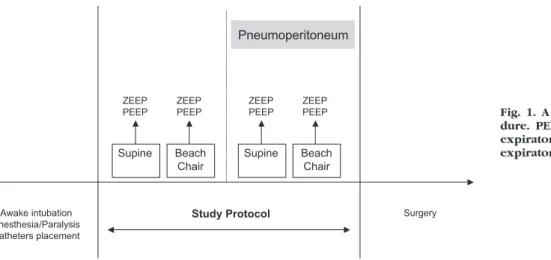
A schema of the protocol is summarized in figure 1. As shown, it included four different steps:
Supine position, before pneumoperitoneum was in-duced (step 1)
Beach chair position, before pneumoperitoneum was induced (step 2)
Supine position, after pneumoperitoneum was induced (step 3)
Beach chair position, after pneumoperitoneum was in-duced (step 4)
The beach chair position was obtained as a reverse Trendelenburg position (30° head-up) with the legs lifted to the abdomen.
At each step, zero end-expiratory pressure (ZEEP) or 10 cm H2O PEEP (PEEP) was applied in a random fashion (closed envelopes).
Throughout the protocol, ventilator settings (Servo 900C; Siemens-Elema, Solna, Sweden) were maintained unchanged as follows: volume control mode with con-stant inspiratory flow (0.700⫾ 0.104 l/s), tidal volume of 10.5⫾ 1 ml/kg ideal body weight, respiratory rate of 11 ⫾ 1 breaths/min, inspiratory time of 33% with no pause, and 60% inspiratory oxygen fraction.
After each combination of position (supine, beach chair position), pneumoperitoneum (without, with), and PEEP (0, 10 cm H2O) was set, a recruitment maneuver was performed (three consecutive inspiratory holds of 5 s at 45 cm H2O airway pressure). Thereafter, 15 min of mechanical ventilation was allowed before the following measurements were taken:
Physiologic Measurements
Intraabdominal Pressure. Intraabdominal pressure
was measured using a transurethral bladder catheter. Using a sterile technique, 100 ml normal saline was infused through the urinary catheter into the bladder. Intraabdominal pressure was recorded as mean pressure at end-expiration. The zero was set at the level of the pubis.
Pneumoperitoneum was generated by insufflating car-bon dioxide into the abdomen (40-l Hight flow insuffla-tor; Stryker Endoscopy, San Jose, CA). Displayed pres-sures were recorded.
End-expiratory Lung Volume. The end-expiratory
lung volume was measured using the closed-circuit he-lium dilution method.
Supine Beach Chair ZEEP PEEP ZEEP PEEP Supine Beach Chair ZEEP PEEP ZEEP PEEP Pneumoperitoneum Awake intubation Anesthesia/Paralysis Catheters placement
Study Protocol Surgery
Fig. 1. A scheme of the protocol proce-dure. PEEP ⴝ 10 cm H2O positive
end-expiratory pressure; ZEEP ⴝ zero end-expiratory pressure.
Respiratory Mechanics. Airway pressure (Paw) and
gas flow were measured at the endotracheal tube open-ing, while esophageal pressure (Pes) was measured from an esophageal balloon (Bicore CP-100; Irvine, CA) in-flated with 0.5–1 ml of air, positioned at the lower third of the esophagus. The validity of Peswas verified accord-ing to Baydur et al.23 Because the patients were para-lyzed, the airway and esophageal pressure were altered by compressing the thorax with the airways occluded at end-expiration. This method was previously used to par-tition respiratory mechanics in obese patients.24 Both flow and pressure signals were recorded on a personal computer via an analog-to-digital converter (Colligo; Ele-kton, Milan, Italy) at a sample rate of 200 Hz and stored for subsequent computer analysis.
We recorded Paw and Pesduring 4 –5 s of airway oc-clusions at end-expiration followed by an occlusion at end-inspiration. Static elastance of the total respiratory system (E,rs) was computed as⌬Paw/VT, where⌬Pawis the difference between plateau inspiratory and end-expiratory airway pressure (corrected for intrinsic PEEP) and VTis the tidal volume. Static elastance of the chest wall (E,cw) was computed as⌬Pes/VT, where⌬Pesis the difference between plateau end-inspiratory and end-ex-piratory esophageal pressure. Static lung elastance (E,l) was calculated as E,l⫽ E,rs ⫺ E,cw.
Resistances of the Respiratory System, Chest Wall, and Lung. Maximum (Rmax,rs) and minimum
(Rmin,rs) resistances of the respiratory system were computed as (Paw,max ⫺ P2)/Flow and (Paw,max ⫺ P1)/ Flow, where Paw,max is the maximal pressure at end-inspiration after the occlusion maneuver, P1is the pres-sure of the airway immediately after the sudden decrease from Paw,max, and P2is the plateau pressure measured at the end of the occlusion. The minimum resistance of the respiratory system represents the so-called ohmic com-ponent, whereas the maximum includes ohmic and re-sistances caused by stress relaxation or rere-sistances caused by time constant inequalities. These are called “additional” resistances (D,rs) and are calculated as Rmax,rs⫺ Rmin,rs.
There was no appreciable decrease of esophageal pres-sure after the occlusion (P1), suggesting that minimum chest wall resistances were negligible, so that chest wall resistances may be considered entirely due to viscoelastic properties of the chest wall (DR,cw). Finally, maximum (R,L), minimum (Rmin,L), and additional (DR,L) resistances of the lung were obtained by using transpulmonary pres-sures, i.e., airway minus esophageal pressures.
Static Inflation Pressure–Volume Curves and Alveolar Recruitment
Static pressure–volume (P-V) curves were measured at 0 and 10 cm H2O PEEP by performing intermittently a series of end-inspiratory airway occlusions at different inflation volumes that ranged between 0.1 and 1.0 l, in
100-ml steps, and recording the Paw and Pes at end-expiration and after each end-inspiration. End-expiratory volume corresponded to the elastic equilibrium volume in each patient, as evidenced by zero flow during expi-ratory pause and absence of changes in Pawafter airway occlusion.
To estimate alveolar recruitment, we first computed the difference of gas volume measured with helium at 10 and 0 cm H2O pressure (actual⌬gas volume). We then computed on the P-V curve, starting at 0 cm H2O PEEP, the volume expected at 10 cm H2O (expected ⌬gas volume), i.e., V ⫽ 10/a(1/b). We defined alveolar
recruitment as the difference between the actual⌬gas volume and the expected ⌬gas volume. Any positive difference implies recruitment (i.e., upward shift of the P-V curve).
Gas Exchange
Arterial blood samples were taken from the radial ar-tery and analyzed for arterial partial pressure of oxygen (PaO2) and arterial partial pressure of carbon dioxide
(PaCO2) (IL 1312 BGM; Instrumentation Laboratory
Com-pany, Lexington, MA). Alveolar–arterial difference was then calculated with standard formulas to correct for hypercapnia.
The physiologic dead space fraction (VD/VT) was also computed according to the following formula: VD/VT⫽ (PaCO2⫺ PECO2)/PaCO2, where PECO2is the mixed expired
carbon dioxide partial pressure. This was obtained by means of continuous expiratory air sampling (CO2SMO PLUS 8100; Novametrix Medical System Inc., Walling-ford, CT).
Hemodynamics
Blood pressure was measured invasively through a radial catheter. Heart rate was derived from the electro-cardiogram.
Statistics
The study was designed to enroll 20 patients, provid-ing a power of 0.8 with a minimum detectable difference of the means of end-expiratory lung volume of 0.2 l, an expected SD of 0.1 l, and an␣ value of 0.01. The mean value of three breaths was used for pressure and flow variables at each experimental condition. We used anal-ysis of variance for repeated measures to test the effects of the beach chair position and PEEP with or without pneumoperitoneum, considering steps (see Materials and Methods, Protocol Procedure) and PEEP as entry into the model. The Bonferroni correction was applied for multiple comparisons. Multiple linear regression anal-ysis was conducted to correlate changes of oxygenation (PaO2) to changes of end-expiratory lung volume.25,26
Statistical significance was accepted as P⬍ 0.05. Analy-sis was performed with the SAS System for Windows version 9.1 (Cary, NC).
Results
Study Population
Twenty of 21 patients approached for consent for this study accepted. The 20 consented patients (5 men, 15 women) were aged 37 ⫾ 10 yr, and their body mass index was 42⫾ 5 kg/m2. The forced vital capacity and forced expired volume in 1 s before surgery were 92⫾ 16.5% and 91 ⫾ 14.6%, respectively. Gas exchange pa-rameters were within normal ranges.
Protocol Procedure
The protocol lasted approximately 160 min. No prob-lems occurred during this time or during the following time needed for surgery. None of the patients experi-enced delayed awakening at the end of the surgical procedure or had to be reintubated postoperatively. The postoperative course was unremarkable.
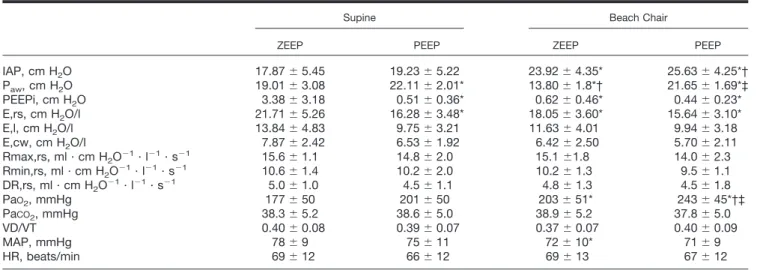
Respiratory Mechanics in the Supine Position
As shown in table 1 (column supine, ZEEP) and figure 2, during sedation and paralysis in the supine position, the obese patients we investigated were characterized by increased intraabdominal pressure, low end-expira-tory lung volumes (0.46⫾ 0.1 l), and increased elastance of both lung and chest wall. Maximum and additional resistances of the respiratory system were high, the increase being mainly due to lung resistances.
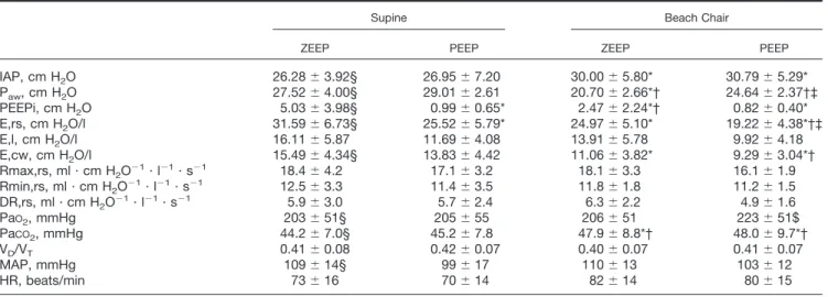
Overall respiratory mechanics worsened after pneu-moperitoneum was induced, as shown in table 2. Lung volume was as low as 0.35 ⫾ 0.1 l; respiratory system elastance further worsened compared with baseline (P ⬍ 0.001), mainly because of chest wall elastance impairment (P ⬍ 0.001). Airway resistance worsened, even if not significantly (P⫽ 0.0745), and intrinsic PEEP
was higher (P ⫽ 0.0075). Oxygenation improved (P ⫽ 0.0057). Carbon dioxide tension (P⬍ 0.001) and mean arterial pressure (P ⬍ 0.001) were significantly higher during pneumoperitoneum. The P-V curve of the respi-ratory system was shifted rightward during pneumoperi-toneum as compared with baseline (fig. 3A).
Effect of the Beach Chair Position and PEEPbefore
Pneumoperitoneum Was Induced
As shown in table 1 and figure 2, the beach chair position compared with the supine position improved lung volume (0.85 ⫾ 0.3 l; P ⬍ 0.001) and respiratory system elastance (P⫽ 0.0122). Airway resistances were not affected (P⫽ 0.1079). Gas exchange improved dur-ing the beach chair position (P⫽ 0.0468). Mean arterial pressure decreased during the beach chair position (P⫽ 0.0134), whereas heart rate (P⫽ 1.00) was unaffected.
Table 1. Effect of Beach Chair Position and PEEP without Pneumoperitoneum
Supine Beach Chair
ZEEP PEEP ZEEP PEEP
IAP, cm H2O 17.87⫾ 5.45 19.23⫾ 5.22 23.92⫾ 4.35* 25.63⫾ 4.25*† Paw, cm H2O 19.01⫾ 3.08 22.11⫾ 2.01* 13.80⫾ 1.8*† 21.65⫾ 1.69*‡ PEEPi, cm H2O 3.38⫾ 3.18 0.51⫾ 0.36* 0.62⫾ 0.46* 0.44⫾ 0.23* E,rs, cm H2O/l 21.71⫾ 5.26 16.28⫾ 3.48* 18.05⫾ 3.60* 15.64⫾ 3.10* E,l, cm H2O/l 13.84⫾ 4.83 9.75⫾ 3.21 11.63⫾ 4.01 9.94⫾ 3.18 E,cw, cm H2O/l 7.87⫾ 2.42 6.53⫾ 1.92 6.42⫾ 2.50 5.70⫾ 2.11 Rmax,rs, ml · cm H2O⫺1· l⫺1· s⫺1 15.6⫾ 1.1 14.8⫾ 2.0 15.1⫾1.8 14.0⫾ 2.3 Rmin,rs, ml · cm H2O⫺1· l⫺1· s⫺1 10.6⫾ 1.4 10.2⫾ 2.0 10.2⫾ 1.3 9.5⫾ 1.1 DR,rs, ml · cm H2O⫺1· l⫺1· s⫺1 5.0⫾ 1.0 4.5⫾ 1.1 4.8⫾ 1.3 4.5⫾ 1.8 PaO2, mmHg 177⫾ 50 201⫾ 50 203⫾ 51* 243⫾ 45*†‡ PaCO2, mmHg 38.3⫾ 5.2 38.6⫾ 5.0 38.9⫾ 5.2 37.8⫾ 5.0 VD/VT 0.40⫾ 0.08 0.39⫾ 0.07 0.37⫾ 0.07 0.40⫾ 0.09 MAP, mmHg 78⫾ 9 75⫾ 11 72⫾ 10* 71⫾ 9 HR, beats/min 69⫾ 12 66⫾ 12 69⫾ 13 67⫾ 12
* P⬍ 0.01 vs. supine ZEEP. † P ⬍ 0.01 vs. supine PEEP. ‡ P ⬍ 0.01 vs. beach chair ZEEP.
DR,rs ⫽ additional airway resistance; E,cw ⫽ chest wall elastance; E,l ⫽ lung elastance; E,rs ⫽ respiratory system elastance; HR ⫽ heart rate; IAP ⫽ intraabdominal pressure; MAP⫽ mean arterial blood pressure; PaCO2⫽ arterial partial pressure of carbon dioxide; PaO2⫽ arterial partial pressure of oxygen; Paw⫽ plateau airway pressure; PEEP ⫽ 10 cm H2O positive end-expiratory pressure; PEEPi⫽ intrinsic PEEP; Rmax,rs ⫽ maximum airway resistance; Rmin,rs ⫽ minimum airway resistance; VD/VT⫽ physiologic dead space; ZEEP ⫽ zero end-expiratory pressure.
Supine Beach Chair
End-expiratory Lung Volume (l)
Supine Beach Chair 0,00 0,25 0,50 0,75 1,00 1,25 1,50 1,75 2,00 Pneumoperitoneum
Fig. 2. The effects of the beach chair position and positive end-expiratory pressure (PEEP) on end-expiratory lung vol-umes. Black columns represent values at zero end-expiratory pressure; white columns represent values at 10 cm H2O PEEP.
The use of 10 cm H2O PEEP improved lung volume (0.83 ⫾ 0.3 l, P ⫽ 0.0014) and respiratory system elas-tance (P ⬍ 0.001). There was no significant effect on airway resistance (P ⫽ 0.1133), oxygenation (P ⫽ 1.000), mean arterial pressure (P⫽ 0.3596), or heart rate (P⫽ 1.00).
Between the beach chair position and PEEP, there were no differences of end-expiratory lung volume (P⫽ 1.00), oxygenation (PaO2; P⫽ 1.00), respiratory system
elastance (P ⫽ 1.00), or mean arterial pressure (P ⫽ 0.2359). Airway pressure was significantly higher during PEEP application (P⬍ 0.001).
The P-V curves of the respiratory system before pneu-moperitoneum was induced are shown in figure 3. The curves at ZEEP and PEEP in both positions were almost superimposed. The alveolar recruitment was 0.04⫾ 0.1 l in the supine and 0.07⫾ 0.2 in the beach chair position; values are not significantly different (supine vs. beach chair position, P⫽ 0.47).
Effect of the Beach Chair Position and PEEPafter
Pneumoperitoneum Was Induced
As shown in table 2 and figure 2, during pneumoperi-toneum, the beach chair position improved lung volume (0.45 ⫾ 0.2 l; P ⫽ 0.0177) and respiratory system elas-tance (P ⬍ 0.001), mainly because of changes in chest wall elastance (P⫽ 0.0002). Mean arterial pressure was unaffected (P⫽ 0.7852). Similarly, PEEP improved lung volume (0.55⫾ 0.1 l; P ⫽ 0.003) and respiratory system elastance (P⫽ 0.0132), with no effect on mean arterial pressure (P⫽ 1.00). Respiratory system resistances were unaffected by both the beach chair position (P⫽ 1.00) and PEEP (P ⫽ 0.6805). During pneumoperitoneum,
neither the beach chair position (P ⫽ 1.00) nor PEEP (P⫽ 1.00) led to significant changes of oxygenation.
The P-V curves of the respiratory system after pneu-moperitoneum was induced are shown in figure 3. As described above, the slopes of the curves were lower than before pneumoperitoneum was induced; however, the behavior was similar. In fact, curves at ZEEP and PEEP in both positions were almost superimposed. The alveolar recruitment was⫺0.014 ⫾ 0.111 l in the supine and 0.04 ⫾ 0.127 l in the beach chair position; values were not significantly different (supine vs. beach chair position, P⫽ 0.273).
End-expiratory Lung Volume and Oxygenation
Changes of oxygenation (calculated with supine ZEEP values as a reference) correlated with changes of end-expiratory lung volume (fig. 4; P⬍ 0.001, R2⫽ 0.524).
Discussion
The aim of this study was to assess the effects of the beach chair position, PEEP, and pneumoperitoneum on respiratory mechanics in morbidly obese patients during anesthesia and paralysis. The main results were that both the beach chair position and PEEP significantly improved lung volumes and respiratory mechanics. Each maneuver individually improved oxygenation at baseline, whereas the combination of the two was needed during pneumo-peritoneum.
Respiratory Mechanics in the Supine Position
Our results confirm previous observations that obese patients sustain a major derangement of respiratory
func-Table 2. Effect of Beach Chair Position and PEEP after Pneumoperitoneum Was Induced
Supine Beach Chair
ZEEP PEEP ZEEP PEEP
IAP, cm H2O 26.28⫾ 3.92§ 26.95⫾ 7.20 30.00⫾ 5.80* 30.79⫾ 5.29* Paw, cm H2O 27.52⫾ 4.00§ 29.01⫾ 2.61 20.70⫾ 2.66*† 24.64⫾ 2.37†‡ PEEPi, cm H2O 5.03⫾ 3.98§ 0.99⫾ 0.65* 2.47⫾ 2.24*† 0.82⫾ 0.40* E,rs, cm H2O/l 31.59⫾ 6.73§ 25.52⫾ 5.79* 24.97⫾ 5.10* 19.22⫾ 4.38*†‡ E,l, cm H2O/l 16.11⫾ 5.87 11.69⫾ 4.08 13.91⫾ 5.78 9.92⫾ 4.18 E,cw, cm H2O/l 15.49⫾ 4.34§ 13.83⫾ 4.42 11.06⫾ 3.82* 9.29⫾ 3.04*† Rmax,rs, ml · cm H2O⫺1· l⫺1· s⫺1 18.4⫾ 4.2 17.1⫾ 3.2 18.1⫾ 3.3 16.1⫾ 1.9 Rmin,rs, ml · cm H2O⫺1· l⫺1· s⫺1 12.5⫾ 3.3 11.4⫾ 3.5 11.8⫾ 1.8 11.2⫾ 1.5 DR,rs, ml · cm H2O⫺1· l⫺1· s⫺1 5.9⫾ 3.0 5.7⫾ 2.4 6.3⫾ 2.2 4.9⫾ 1.6 PaO2, mmHg 203⫾ 51§ 205⫾ 55 206⫾ 51 223⫾ 51$ PaCO2, mmHg 44.2⫾ 7.0§ 45.2⫾ 7.8 47.9⫾ 8.8*† 48.0⫾ 9.7*† VD/VT 0.41⫾ 0.08 0.42⫾ 0.07 0.40⫾ 0.07 0.41⫾ 0.07 MAP, mmHg 109⫾ 14§ 99⫾ 17 110⫾ 13 103⫾ 12 HR, beats/min 73⫾ 16 70⫾ 14 82⫾ 14 80⫾ 15
* P⬍ 0.01 vs. supine ZEEP. † P ⬍ 0.01 vs. supine PEEP. ‡ P ⬍ 0.01 vs. beach chair ZEEP. § P ⬍ 0.01, before vs. after pneumoperitoneum within supine ZEEP.
DR,rs ⫽ additional airway resistance; E,cw ⫽ chest wall elastance; E,l ⫽ lung elastance; E,rs ⫽ respiratory system elastance; HR ⫽ heart rate; IAP ⫽ intraabdominal pressure; MAP⫽ mean arterial blood pressure; PaCO2⫽ arterial partial pressure of carbon dioxide; PaO2⫽ arterial partial pressure of oxygen; Paw⫽ plateau airway pressure; PEEP ⫽ 10 cm H2O positive end-expiratory pressure; PEEPi⫽ intrinsic PEEP; Rmax,rs ⫽ maximum airway resistance; Rmin,rs ⫽ minimum airway resistance; VD/VT⫽ physiologic dead space; ZEEP ⫽ zero end-expiratory pressure.
tion during anesthesia and paralysis.1– 4 In fact, end-expiratory lung volume measured in supine position was as low as 0.5 l, in accordance with the body mass index and end-expiratory lung volume relation described by Pelosi et al.22 Elastance and airway resistance were greater than normal, and there was hypoxemia relative to inspiratory fraction of oxygen.
As suggested by the P-V curve analysis (fig. 3A), we did not find evidence of recruitable lung tissue. This is in contrast to the previous report by Pelosi et al.13 How-ever, the values of body mass index of our patients were lower than those in the study of Pelosi et al. (42⫾ 5 as opposed to 51⫾ 8 kg/m2). We took our measurements soon after induction of anesthesia, whereas Pelosi et al. investigated patients at the end of surgery and after transfer from the operating room to the intensive care unit. Moreover, we performed frequent recruitment ma-neuvers to normalize lung volumetric history. This likely contributed to our results. Whalen et al.18 recently showed that recruitment maneuvers may be an effective
mode of improving intraoperative respiratory mechanics and oxygenation in obese patients.
Therefore, the decrease of lung volume and increase of lung elastance in the supine position may be ex-plained by a prevalent decrease of the size of the alveoli rather than atelectasis. Of note is the fact that in the supine position, some grade of intrinsic PEEP developed. This may be due to flow limitation, as suggested by Pankov et al.27
Pneumoperitoneum further worsened respiratory me-chanics, as previously shown in both normal-weight28 –35 and obese5– 8patients.
Interestingly, oxygenation improved during peritoneum, possibly because of the effects of pneumo-peritoneum on hemodynamics. Pneumopneumo-peritoneum, in fact, activates sympathetic tone and contributes to in-crease arterial blood pressure.5 The fact that pneumo-peritoneum has been shown to greatly affect venous return, particularly in volume-depleted subjects, and the overall hemodynamic stability of our patients may sug-gest that they were not hypovolemic. This notion could not be substantiated in the current investigation.
Effects of the Beach Chair Position and PEEP on Respiratory Mechanics
Several investigations have considered the effects of head-up position on either normal-weight36or obese6 – 8,21 patients. We measured lung volumes and found that these almost doubled during the beach chair position. The effect of bowels sliding under gravity and relieving the diaphragm is possibly relevant, as suggested by the increase of bladder
Airway pressure (cmH Volume (ml) Volume (ml) 2O) 0 5 10 15 20 25 30 35 40 45 0 250 500 750 1000 1250 1500 1750 2000 2250 2500 Airway pressure (cmH2O) 0 5 10 15 20 25 30 35 40 45 0 250 500 750 1000 1250 1500 1750 2000 2250 2500 Supine Beach chair
A
B
Fig. 3. Pressure–volume curves taken in the supine position (A) and in the beach chair position (B). In each panel, values taken at baseline are shown as circles, and those obtained during pneumoperitoneum are shown as squares. Closed symbols in-dicate values at zero end-expiratory pressure, and open symbols indicate those at 10 cm H2O positive end-expiratory pressure.
To compare curves, each one starts on the y-axis from the volume corresponding to the mean value of end-expiratory lung volume measured by helium dilution technique.
Changes of EELV Changes of PaO 2 (mmHg) (L) 0,0 0,2 0,4 0,6 0,8 1,0 1,2 1,4 0 20 40 60 80 100 120
Fig. 4. Correlation between changes of end-expiratory lung vol-ume (EELV) and changes of oxygenation (arterial partial pres-sure of oxygen [PaO2]). Changes were calculated considering
supine zero end-expiratory pressure values as a reference. Open
symbolsrefer to values taken during pneumoperitoneum, and
closed symbols refer to those at baseline. Squares represent values obtained during the beach chair position, circles repre-sent values at 10 cm H2O positive end-expiratory pressure, and diamondsrepresent values obtained with the combination of the beach chair position and 10 cm H2O positive end-expiratory
pressure. Multiple regression analysis was conducted (see Ma-terials and Methods, Statistics).
pressure in the beach chair as compared with the supine position. The results of the effects of PEEP were consistent with previous reports.9 –18
A positive feature of this study was the investigation of the relative effects of the beach chair position and PEEP: It was intriguing to find that the two maneuvers produced similar effects on lung volume, respiratory system elas-tance, or oxygenation. However, similarly to laparotomy surgery,15airway pressures were much lower during the beach chair position than during PEEP application. There-fore, under the perspective of lung-protective ventilation strategies,37the beach chair position would be preferred. One must consider, however, that recruitable atelec-tasis, if any, was negligible (fig. 3): The picture might be different with lung collapse, hence with sicker patients. In this light, the recruitment maneuvers we frequently performed must be strongly considered. Whalen et al.18 have in fact shown that recruitment maneuvers improve oxygenation and respiratory mechanics as long as lungs are ventilated and PEEP is applied.
The combination of the beach chair position and PEEP improved lung volume before pneumoperitoneum was instituted. However, at high lung volumes, the P-V curve began to flatten, suggesting overdistention of the alveoli. Although there was no hemodynamic compromise in that state, we would suggest that this condition is not desirable per se.
On the contrary, during pneumoperitoneum, this did not occur, possibly because of the low starting volumes in this condition. Interestingly, whereas PEEP induced some de-recruitment in the supine position (as evi-denced from a negative value of calculated lung recruit-ment and from P-V curves in fig. 3), if anything, there was some recruitment during the beach chair position. Of note is the fact that during pneumoperitoneum, only the combination of the beach chair position and PEEP led to a significant lung volume increase and oxy-genation improvement, whereas the single maneuvers did not (fig. 4). This is partly in contrast to the results of Sprung et al.,8who found that arterial oxygenation was not affected by body position, pneumoperitoneum, or mode of ventilation. However, the same group of au-thors recently showed that recruitment maneuvers and PEEP are effective on lung mechanics and oxygenation, even if short-lived.18Therefore, it is possible that, given the high abdominal pressure during pneumoperitoneum application, only the combination of the beach chair position and the recruitment maneuver followed by PEEP was able to counteract the detrimental effects of pneumoperitoneum. Accordingly, low PEEP may be in-sufficient to counteract pneumoperitoneum. The opti-mum PEEP is ideally achieved by titrating the “best” PEEP, recognizing that our data and others13suggest that 10 cm H2O may be reasonable.
Of note is the fact that changes of lung volume corre-lated with changes of oxygenation, further underlying
the crucial role of end-expiratory lung volume in these patients.
Limitations of the Study
The sequence of measurements, because of technical difficulties, was randomized only for PEEP application within each step of the protocol (fig. 1). This is a major limitation. However, we do not think that the protocol sequence influenced the results of lung volume, respira-tory mechanics, or resistances. Carbon dioxide tension was of course affected by the protocol design provided respiratory rate was kept constant and pneumoperito-neum generated insufflating carbon dioxide into the ab-domen. However, we corrected oxygenation for this possible bias by calculating the alveolar to arterial differ-ence and did not find significative differdiffer-ences with PaO2.
The rather short time between steps (approximately 15 min) is another limitation. This time was chosen because of the many measurements to be taken. How-ever, we believe that 15 min was enough to reset respi-ratory mechanics at each step of the protocol.
The technique we used to estimate end-expiratory lung volume might have underestimated lung volumes because of air trapping at low lung volumes. However, the same technique has been used in the past.13 More-over, in 10 patients, we also measured end-expiratory lung volume by the release technique (measuring flow while releasing PEEP, then calculating volume as integral of flow tracing): The values obtained with the two tech-niques were correlated (R2 ⫽ 0.793, P ⬍ 0.001), sug-gesting that gas trapping (that would have appeared with the release technique) was negligible (intercept 0.098 l).
Conclusion
We have shown that the beach chair position and PEEP may be used to counteract the major derangements produced by anesthesia and paralysis in morbidly obese patients. The beach chair position and PEEP each simi-larly improved lung volume, oxygenation, and respira-tory mechanics at baseline. However, during pneumo-peritoneum, only the combination of the two improved oxygenation.
References
1. Sharp J, Henry J, Sweany S, Meadows W, Pietras R: Effects of mass loading the respiratory system in man. J Appl Physiol 1964; 19:959–65
2. Waltemath CL, Bergman NA: Respiratory compliance in obese patients. ANESTHESIOLOGY1974; 41:84–5
3. Luce JM: Respiratory complications of obesity. Chest 1980; 78:626–31 4. Damia G, Mascheroni D, Croci M, Tarenzi L: Perioperative changes in functional residual capacity in morbidly obese patients. Br J Anaesth 1988; 60:574–8
5. Dumont L: Changes in pulmonary mechanics during laparoscopic gastro-plasty in morbidly obese patients. Acta Anaesthesiol Scand 1997; 41:408–13
6. Casati , Comotti L, Tommasino C, Leggieri E, Bignami F, Tarantino F, Torri G: Effects of pneumoperitoneum and reverse Trendelenburg position on
cardio-pulmonary function in morbidly obese patients receiving laparoscopic gastric banding. Eur J Anaesth 2000; 17:300–5
7. Perilli V, Sollazzi L, Bozza P, Modesti C, Chierichini A, Tacchino RM, Ranieri R: The effects of the reverse Trendelenburg position on respiratory mechanics and blood gases in morbidly obese patients during bariatric surgery. Anesth Analg 2000; 91:1520–5
8. Sprung J, Whalley D, Falcone T, Wilks W, Navratil J, Bourke D: The effects of tidal volume and respiratory rate on oxygenation and respiratory mechanics during laparoscopy in morbidly obese patients. Anesth Analg 2003; 97:268–74
9. Hedenstierna G, Santesson J: Breathing mechanics, dead space and gas exchange in the extremely obese, breathing spontaneously and during anaesthe-sia with intermittent positive pressure ventilation. Acta Anaesthesiol Scand 1976; 20:248–54
10. Santesson J: Oxygen transport and venous admixture in extremely obese: Influence of anaesthesia and artificial ventilation with and without positive end-expiratory pressure. Acta Anaesthesiol Scand 1976; 20:387–94
11. Eriksen J, Andersen J, Rasmussen J, Sorensen B: Effects of ventilation with large tidal volumes or positive end-expiratory pressure on cardiorespiratory function in anesthetized obese patients. Acta Anaesthesiol Scand 1978; 22:241–8 12. Tokics L, Hedenstierna G, Strandberg A, Brismar B, Lundquist H: Lung collapse and gas exchange during general anesthesia: Effects of spontaneous breathing, muscle paralysis, and positive end-expiratory pressure. ANESTHESIOLOGY 1987; 66:157–67
13. Pelosi P, Ravagnan I, Giurati Panigada M, Bottino N: Positive end-expira-tory pressure improves respiraend-expira-tory function in obese patients but not in normal subjects during anesthesia and paralysis. ANESTHESIOLOGY1999; 91:1221–31
14. Yoshino J, Akata T, Takahashi S: Intraoperative changes in arterial oxygen-ation during volume-controlled mechanical ventiloxygen-ation in modestly obese pa-tients undergoing laparotomies with general anesthesia. Acta Anaesthesiol Scand 2003; 47:742–50
15. Perilli V, Sollazzi L, Modesti C, Annetta M, Sacco T, Bocci M, Tacchino R, Proietti R: Comparison of positive end-expiratory pressure with reverse Tren-delenburg position in morbidly obese patients undergoing bariatric surgery: Effects on hemodynamics and pulmonary gas exchange. Obes Surg 2003; 13: 605–9
16. Chalhoub V, Yazigi A, Sleilaty G, Haddad F, Noun R, Medi-Jebara S, Yazebeck P: Effect of vital capacity manoeuvres on arterial oxygenation in morbidly obese patients undergoing bariatric surgery. Eur J Anaesth 2006; 7:1–6 17. Erlandsson K, Odenstedt H, Lundin S, Stenquist O: Positive end-expiratory pressure optimization using electric impedance tomography in morbidly obese patients during laparoscopic gastric bypass surgery. Acta Anaesthesiol Scand 2006; 50:833–9
18. Whalen FX, Gajic O, Thomson GB, Kendrick ML, Que FL, Williams BA, Joyner MJ, Hubmayr RD, Warner DO, Sprung J: The effects of the alveolar recruitment maneuver and positive end-expiratory pressure on arterial oxygen-ation during laparoscopic bariatric surgery. Anesth Analg 2006; 102:298–305
19. Moffa S, Quinn J, Slotman G: Hemodynamic effects of carbon dioxide pneumoperitoneum during mechanical ventilation and positive end-expiratory pressure. J Trauma 1993; 35:613–7
20. Kraut EJ, Amira S, Barbosa R, Wolf BM: Impairment of cardiac performance
by laparoscopy in patients receiving positive end-expiratory pressure. Arch Surg 1999; 134:76–80
21. Boyce JR, Ness T, Castroman P, Gleysteen JJ: A preliminary study of the optimal anesthesia positioning for the morbidly obese patient. Obes Surg 2003; 13:4–9
22. Pelosi P, Ravagnan I, Croci M, Tredici S, Pedoto A, Lissoni A, Gattinoni L: The effects of body mass on lung volumes, respiratory mechanics, and gas exchange during general anesthesia. Anesth Analg 1998; 87:654–60
23. Baydur A, Behrakis PK, Zin WA, Jaeger M, Milic-Emili L: A simple method for assessing the validity of the esophageal balloon technique. Am Rev Respir Dis 1983; 126:788–91
24. Pelosi P, Croci M, Ravagnan I, Vicardi P, Gattinoni L: Total respiratory system, lung, and chest wall mechanics in sedated-paralyzed postoperative mor-bidly obese patients. Chest 1996; 109:144–51
25. Bland J, Altman D: Statistics notes: Correlation, regression and repeated data. BMJ 1994; 308:896
26. Bland J, Altman D: Statistics notes: Calculating correlation coefficients within repeated observations: 1. Correlation within subjects. BMJ 1995; 310:446 27. Pankow W, Podszus T, Gutheil T, Penzel T, Peter J, Von Wichert P: Expiratory flow limitation and intrinsic positive end-expiratory pressure in obe-sity. J Appl Physiol 1998; 85:1236–43
28. Drummond M: Pressure-volume relationships in the lung during laparos-copy. Br J Anaesth 1978; 50:261–70
29. Erice F, Fox GS, Salib YM, Romano E, Meakins JL, Magder SA: Diaphrag-matic function before and after laparoscopic cholecystectomy. ANESTHESIOLOGY 1993; 79:966–75
30. Fahy BG, Barnas GM, Flowers JL, Nagle SE, Njoku MJ: The effects of increased abdominal pressure on lung and chest wall mechanics during laparo-scopic surgery. Anesth Analg 1995; 81:744–50
31. Oikkonen M, Tallgren M: Changes in respiratory compliance at laparos-copy: Measurements using side stream spirometry. Can J Anaesth 1995; 42:495–7 32. Fahy BG, Barnas GM, Nagle SE, Flowers JL, Njoku MJ, Agarwal M: Changes in lung and chest wall properties with abdominal insufflation of carbon dioxide are immediately reversible. Anesth Analg 1996; 82:501–5
33. Chassard D, Berrada KR, Tournadre JP, Bouletreau P: The effect of increase in end-tidal carbon dioxide on lower esophageal sphincter tone. Anesth Analg 1996; 82:374–6
34. Pelosi P, Foti G, Cereda M, Vicardi P, Gattinoni L: Effects of carbon dioxide insufflation for laparoscopic cholecystectomy on the respiratory system. Anaes-thesia 1996; 51:744–9
35. Carry PY, Gallet D, Francois Y, Perdrix JP, Sayag A, Gilly F, Eberhard A, Banssillon V, Baconnier P: Respiratory mechanics during laparoscopic cholecys-tectomy: The effects of the abdominal wall lift. Anesth Analg 1998; 87:1393–7
36. Odeberg S, Ljungqvist O, Svenberg T, Gannedahl P, Backdahl M, von Rosen A, Sollevi A: Haemodynamic effects of pneumoperitoneum and the influ-ence of posture during anaesthesia for laparoscopic surgery. Acta Anaesthesiol Scand 1994; 38:276–83
37. Gattinoni L, Carlesso E, Cadringer P, Valenza F, Vaginelli F, Chiumello D: Physical and biological triggers of ventilator-induced lung injury and its preven-tion. Eur Respir J Suppl 2003; 43:15s–25s
![Fig. 4. Correlation between changes of end-expiratory lung vol- vol-ume (EELV) and changes of oxygenation (arterial partial pres-sure of oxygen [PaO 2 ])](https://thumb-eu.123doks.com/thumbv2/123doknet/2366127.41381/6.877.97.392.101.596/correlation-changes-expiratory-changes-oxygenation-arterial-partial-oxygen.webp)
