Haemodynamics and cerebral oxygenation during arthroscopic shoulder surgery in beach chair position under general anaesthesia
H. Jeong1,S. H. Lee1,E. A. Jang1,S. S. Chung3,J. Lee2andK. Y. Yoo1
1Department of Anesthesiology and Pain Medicine,Chonnam National University Medical School,Gwangju, South Korea,2Department of Physiology,Chonnam National University Medical School,Gwangju, South Korea and3Department of Anesthesiology,School of Dentistry, Chonnam National University Medical School,Gwangju, South Korea
Background: Patients undergoing surgery in beach chair posi- tion (BCP) are at risk of cerebral ischaemia. We determined the prevalence and risk factors of jugular venous bulb oxygen desaturation (SjvO2<50%) in BCP. It was also examined whether regional cerebral tissue oxygen saturation (SctO2) meas- ured by near-infrared spectroscopy and SjvO2are interchange- able for assessment of cerebral oxygenation.
Methods: Fifty-six consecutive patients undergoing arthro- scopic shoulder surgery in BCP were studied. Anaesthesia was intravenous with propofol and remifentanil (P/R) or inhala- tional with sevoflurane and 50% nitrous oxide (S/N) depending on provider choice. Mean arterial pressure (MAP), heart rate (HR), SjvO2, and SctO2were measured before (baseline; post- induction in supine position) and after the patients assumed BCP. Bland–Altman analysis was performed to measure the agreement between SctO2and SjvO2.
Results: SjvO2, SctO2, MAP, and HR decreased significantly when patients were raised into BCP. Jugular desaturation occurred in 41% of patients (56% with P/R vs. 21% with S/N
anaesthesia, P=0.0077). Risk factors for the desaturation included P/R anaesthesia [adjusted odds ratio (aOR) 4.76, 95% confidence interval (CI) 1.34–16.95, P=0.016] and MAP<50 mmHg (aOR 3.85, 95% CI 1.21–12.25, P=0.023).
Bland–Altman analysis showed a mean difference of-8.9% with 95% limit of agreement between-40.0% and 23.0%. The percent- age error [1.96 standard deviation/mean of the reference method] was 48.5%.
Conclusions: The incidence of jugular desaturation in BCP was 41%, and P/R anaesthesia and hypotension were associated with its occurrence while undergoing surgery under general anaesthesia. SctO2may not replace SjvO2for the determination of cerebral oxygenation.
Accepted for publication 9 April 2012
© 2012 The Authors
Acta Anaesthesiologica Scandinavica
© 2012 The Acta Anaesthesiologica Scandinavica Foundation
The beach chair position (BCP) is commonly used in shoulder arthroscopy.1,2 However, the sitting position is associated with significant reduc- tions in cardiac output, mean arterial pressure (MAP), and cerebral perfusion pressure (CPP), which persists for up to 30 min after the position- ing.3,4 Severe cerebral desaturation events,5,6 brain and spinal cord ischaemia,7,8hemiplegia,9transient visual loss, and opthalmoplegia10have been indeed documented in patients who had undergone shoul- der surgery in the upright position. Although the aetiology of cerebral injury after BCP surgery has not been established, systemic hypotension devel- oped immediately after the positioning has been assumed to compromise cerebral perfusion,
resulting in neurological injury when the episodes are prolonged.
Jugular venous oximetry is used to continuously monitor jugular venous bulb oxygen saturation (SjvO2) in a variety of clinical settings.11However, this modality is invasive and difficult to use. More- over, it may miss focal ischaemia because it provides a more hemispheric assessment of oxygenation.
As an alternative cerebral monitoring entity, near- infrared spectroscopy (NIRS) assessing regional cerebral tissue oxygen saturation (SctO2) offers a noninvasive and less expensive technique, which is easy and quick to apply. It has been indeed success- fully used to assess the adequacy of cerebral per- fusion in patients undergoing procedures at high risk
© 2012 The Acta Anaesthesiologica Scandinavica Foundation ACTA ANAESTHESIOLOGICA SCANDINAVICA
doi: 10.1111/j.1399-6576.2012.02716.x
1
of adverse neurological outcomes (cardiac, vascular, liver transplant, and major abdominal surgery).12,13 Nevertheless, SctO2 monitored by NIRS may not accurately reflect significant changes in SjvO2deter- mined by jugular venous bulb oximetry.14–16
The present study was aimed to determine the prevalence and risk factors of jugular bulb desatura- tion (SjvO2<50%) when patients were raised into BCP. It was also examined whether SctO2 reflects SjvO2for the assessment of the cerebral oxygenation in patients undergoing the surgery in BCP.
Materials and methods
The study protocol was approved by the University Hospital Ethics Committee, and all patients gave informed consent. Fifty-six patients scheduled to undergo elective arthroscopic shoulder surgery under general anaesthesia in BCP were enrolled in the study. Patients were excluded if they had pre-existing cerebrovascular diseases or history of orthostatic hypotension, aged <18 years, or were American Society of Anaesthesiologists physical status IV or V.
All patients were pre-medicated with midazolam (0.1 mg/kg, orally) 60 min before induction of anaesthesia. Support stockings were placed on the lower extremities. Upon arrival in the operating room, a 20-gauge catheter was inserted into a radial artery to continuously monitor blood pressure and to take blood samples. Ringer’s lactate solution was administered at a rate of 10 ml/kg/h throughout the study. Pressure transducer was referenced to the mid-axillary level when patients were in supine and to the external ear canal level when in BCP. A stand- ard bispectral index (BIS) electrode montage (BIS Sensor-Aspect Medical Systems, Natick, MA, USA) was applied to the forehead before induction of anaesthesia. BIS was continuously measured using an Aspect A-2000 BIS® monitor (BIS® XP, software version 3.31, Aspect Medical Systems). SctO2 was determined by NIRS with an INVOS 5100B Cerebral Oximeter (Somanetics Corporation, Troy, MI, USA).
The oximeter probes were placed on the right and left foreheads, with the caudal border~1 cm above the eyebrow with the medial edge at the midline.
SctO2 values from the right and left frontal lobes were averaged to represent cerebral oxygenation.
After recording pre-induction values and full pre- oxygenation, anaesthesia was induced with propofol (2.0–2.5 mg/kg)/remifentanil (1.0–2.0mg/kg) in patients receiving sevoflurane/nitrous oxide (S/N) anaesthesia, or with an effect-site target controlled
infusion (TCI) of propofol (3.0mg/ml)/remifentanil (3.0 ng/ml) in those receiving intravenous (i.v.) propofol/remifentanil (P/R) anaesthesia. Anaes- thetic management was not randomized and depended entirely on provider choice. However, the providers for these cases fall neatly into two camps:
those who always use inhalational agents (S/N) for maintenance and those who always use i.v. anaes- thesia (P/R). After administration of rocuronium (0.8 mg/kg, i.v.), the trachea was intubated, and the lungs were mechanically ventilated with 50%
nitrous oxide in oxygen in patients given S/N, and with air and oxygen mixture (50% oxygen) in those given P/R. Sevoflurane concentrations combined with 50% nitrous oxide in oxygen were then adjusted to maintain MAP within 20% of pre- induction values and BIS values between 40 and 50 throughout the surgery in patients given volatile anaesthetics. TCI effect-site concentrations of propo- fol were also adjusted to achieve BIS readings of 40–50, and those of remifentanil were adjusted to maintain MAP within 20% of the pre-induction value in patients given i.v. anaesthetics. For meas- urement of SjvO2 and blood sampling, a PreSep™
catheter (Edwards Lifesciences, Irvine, CA, USA) connected to a Vigileo™ monitor (Edwards Lifesci- ences) was placed retrogradely in the jugular bulb contralateral to the side of surgery. Proper position- ing of the catheter was verified radiographically.
Approximately 20 min after anaesthesia induction when haemodynamics became stable, the head was secured in a neutral position to ensure that cerebral venous drainage was not impaired. The back of the operating room table was then raised to 65–75° above the horizontal plane. TCI effect-site concentrations for P/R were achieved using the Orchestra Base Primea®infusion pump (Fresenius, Brezins, France).
The surgery was started approximately 20 min after the positioning when vital signs became stable.
Patients were mechanically ventilated to maintain the end-tidal carbon dioxide (CO2) tension between 4.7 and 5.5 kPa. Routine monitoring included inva- sive measurement of systemic blood pressure, heart rate (HR) and rhythm by 5-lead electrocardiogram, and pulse oximetry. The end-tidal concentrations of CO2 and sevoflurane were measured using a gas analyser (Capnomac Ultima, Datex-Ohmeda; Hel- sinki, Finland). Arterial blood gas analysis was determined to match the end-tidal CO2 tension when a stable haemodynamic was achieved before the positioning and more if necessary.
MAP and HR were recorded by an independent investigator before induction of anaesthesia. Simul-
taneously, peripheral arterial oxygen saturation, SctO2, and BIS values were measured in patients while breathing room air. These variables (MAP, HR, BIS, and SctO2) and SjvO2were recorded before (baseline values; post-induction, supine position) and every minute after BCP for 15 min, and then every 5 min for 15 min. In addition, minimum values of SctO2 and SjvO2 were recorded within 15 min after BCP with MAP at their corresponding time points. Baseline SctO2 and SjvO2 values were the means over a 1-min period just before the positioning. Jugular venous bulb desaturation was defined as SjvO2 value less than 50% lasting
>5 min,11and cerebral desaturation was defined as
>20% decrease of SctO2from the baseline value for
>15 s.17,18Hypotension was defined as MAP meas- ured at the level of the external auditory canal of
<50 mmHg and was treated with a bolus of ephe- drine (8 mg) or phenylephrine (100mg), and fluid loading (100~200 ml crystalloid solution). Bradycar- dia, defined as HR<50 beats/min, was treated with i.v. boluses of atropine 0.5 mg as required. Vasopres- sor treatment was repeated every 2 min if hypoten- sion persisted or recurred. The incidences of cerebral and jugular venous desaturation and hypo- tension were recorded.
At the completion of surgery, the anaesthetic was discontinued. Residual neuromuscular block was antagonized with neostigmine and glycopyrrolate.
Estimated blood loss and amount of fluid or blood administered during the surgery were recorded. The post-operative visit was made by the responsible surgeon at the evening of surgery, and the patient was assessed neurologically with a gross motor and sensory neurological evaluation and a gross cogni- tive evaluation (orientation in time and space, recall of name, date of birth, and address). Any side effects were recorded. Management of anaesthesia and haemodynamics were left completely to the discre- tion of the anaesthesiologist responsible, who was blinded to the SjvO2 and SctO2 values. Data were assessed by a person not involved in anaesthetic care.
Statistical analysis
Sample size calculation was based on the assump- tion that a relative decrease in SjvO2 of 20% (the smallest effect to be clinically important11) would be detected. Based on the preliminary study, a mean and standard deviation (SD) of 69% and 15%, respectively, were chosen. For a power of 0.8 and an aof 0.01, a sample size of 41 patients was calculated to be appropriate to detect a clinically relevant decrease in SjvO2.
Data are expressed as number or mean⫾SD.
They were analysed using StatView software version 4.0 (Abacus Concepts, Berkeley, CA, USA) on a Mac- intosh computer. Serial changes in cardiovascular, SctO2, SjvO2, and BIS data were analysed using one- way analysis of variance (ANOVA) with repeated measures. The Scheffé test was used for multiple pairwise comparisons when a significant difference was indicated with ANOVA. Relationship of SjvO2<
50% to anaesthetic methods (S/N vs. P/R), MAP<
50 mmHg, age>60 years, diabetes mellitus, body mass index, haemoglobin, history of hypertension, or antihypertensive drugs were analysed using multiple logistic regression analysis. A Bland–
Altman plot with multiple measurements per sub- ject19 was used to measure the agreement of two measurements of SctO2 and SjvO2, and percentage errors were calculated using MedCalc Version 12.2.1 (MedCalc Software, Mariakerke, Belgium). A mean percentage error not exceeding 30% was defined to indicate clinical useful reliability of the SctO2. A P-value of <0.05 was considered statistically significant.
Results
Demographical and surgical data are presented in Table 1. Of the 58 patients, one patient was excluded because of unsuccessful cannulation of internal jugular vein and another one because of malfunc- tion of the monitoring system. They aged 60 years (ranging 18–77) and underwent shoulder arthro- scopic surgery. Fifteen patients had the fibre-optic catheter placed in the right side and remaining 41 patients in the left side. BIS data remained constant (45.1⫾3.0) throughout the intraoperative period.
Average duration of surgery was 134 min (ranging 40–285). None of them developed gross neurologi- cal or cognitive dysfunction post-operatively.
Haemodynamic data are presented in Table 2 and Fig. 1. MAP decreased after induction of anaesthesia and decreased further from 1 min to 20 min after the positioning (one-way ANOVA, P<0.0001). Thirty- one patients (55%) developed hypotension (69% vs.
38% in patients given P/R and S/N, respectively, P=0.0199), and received ephedrine (n=22, 14⫾ 7 mg) or phenylephrine (n=9, 160⫾100mg). The duration of the hypotensive episode ranged 1–15 min. HR decreased from 3 min to 20 min into BCP (P<0.0001).
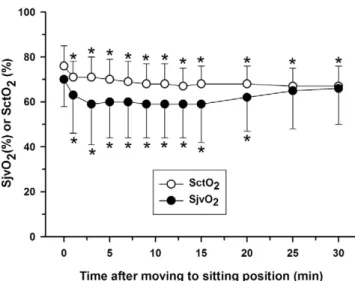
SjvO2 and SctO2 values are presented in Fig. 2.
SjvO2values significantly decreased below baseline (70⫾12%) from 1 min to 20 min into BCP
(P<0.0001). However, the magnitude of decreases of SjvO2 (22⫾12% vs. 14⫾12%, P=0.0103) was more pronounced in patients given P/R along with lower pre-sitting baseline values (65⫾8% vs.
76⫾10%, P=0.0001) than in those given S/N. On the other hand, SctO2values increased after induc- tion of anaesthesia from 69⫾7% (pre-induction) to 76⫾9% (post-induction baseline,P<0.0001). SctO2
decreased significantly from 2 min after BCP throughout the study (P<0.0001). Of 56 patients, 23 (41%) developed episodes of SjvO2<50% (56% vs.
21% in patients given P/R and S/N respectively, P=0.0077) and nine patients (16%) had a SjvO2<40% (25% vs. 4%,P=0.0357).
Risk factors for jugular desaturation included P/R anaesthesia [adjusted odds ratio (aOR) 4.76, 95%
Table 1
Demographical and intraoperative variables in patients undergoing surgery in the beach chair position under general anaesthesia.
All patients (n=56)
Male/female 21/35
Age (years) 60⫾10
Weight (kg) 63⫾11
Haemoglobin (g/dl) 14⫾1
Underlying diseases,n(%)
Hypertension 20 (36%)
Diabetes mellitus 11 (20%)
Preoperative medication,n(%)
b-blockers 5 (9%)
Calcium-channel blockers 11 (20%)
ACEI or angiotensin II antagonist 12 (21%)
Smoking history,n(%) 2 (4%)
Methods of anaesthesia,n(%)
Sevoflurane/nitrous oxide 24 (43%)
Propofol/remifentanil 32 (57%)
Vasopressor administered,n(%)
Ephedrine 22 (39%)
Total ephedrine dose per patient (mg) 14⫾7
Phenylephrine 9 (16%)
Total phenylephrine dose per patient (mg) 160⫾100
Duration of anesthesia (min) 191⫾59
Duration of surgery (min) 134⫾56
Fluid administered (ml) 1577⫾514
Blood loss (ml) 170⫾162
Data are mean⫾standard deviation or numbers (%). ACEI, angiotensin converting enzyme inhibitor.
Table 2
Preoperative haemodynamic and intraoperative blood gas data in patients undergoing surgery in the beach chair position under general anaesthesia.
All patients (n=56) Mean arterial pressure (mmHg) 102⫾15
Heart rate (beats/min) 69⫾16
SpO2(%) 99⫾0
SctO2(% ) 69⫾7
PaCO2(mmHg) 39⫾3
PaO2(mmHg) 212⫾50
Data are mean⫾standard deviation or numbers. SpO2, periph- eral arterial saturation of oxygen; SctO2, regional cerebral tissue oxygen saturation; PaCO2, arterial partial pressure of carbon dioxide; PaO2, arterial partial pressure of oxygen.
Fig. 1. Mean arterial pressure (MAP) and heart rate (HR) before (time 0; baseline, post-induction in supine position) and after the beach chair position in patients (n=56) undergoing surgery under general anaesthesia. Values are means⫾standard deviation.
*P<0.05 vs. baseline.
Fig. 2. Jugular venous bulb oxygen saturation (SjvO2) and regional cerebral tissue oxygen saturation (SctO2) before (time 0;
baseline, post-induction in supine position) and after the beach chair position in patients (n=56) undergoing surgery under general anaesthesia. Values are means⫾standard deviation.
*P<0.05 vs.baseline.
confidence interval (CI) 1.34–16.95, P=0.016] and MAP<50 mmHg (aOR 3.85, 95% CI 1.21–12.25;
P=0.023). In four patients (7%), jugular desaturation episode happened concomitantly with both hypo- tension (MAP<50 mmHg) and >20% decrease of SctO2from the baseline. In another 13 patients (23%), the episode happened concomitantly with a hypo- tensive episode (MAP of<50 mmHg). The incidence of cerebral desaturation (>20% decrease of SctO2
from the baseline) was 27% (28% vs. 25%, patients given P/R and S/N, respectively; P=0.7938). The duration of the episodes ranged from 1 min to longer than 30 min. The sensitivity and specificity of MAP<50 mmHg to detect SjvO2<50% were 73.9%
and 57.6% (area under the curve=0.657,P=0.047), whereas those of cerebral desaturation to detect SjvO2<50% were 30.4% and 75.8% (area under the curve=0.531,P=0.696), respectively.
The Bland–Altman analysis showed a mean dif- ference (bias) between the two measurements of -8.9% and 95% limit of agreement (1.96 SD) of -40.0% to 23.0% (Fig. 3). The percentage error (1.96 SD/mean of reference method) was 48.5%.
Discussion
The present study demonstrated that the jugular venous desaturation (SjvO2<50%), indicative of cerebral hypoperfusion,11 occurred in 41% of
patients undergoing shoulder surgery in BCP under general anaesthesia. The desaturation was related to hypotension and P/R anaesthesia. The agree- ment between SctO2 monitored by NIRS and SjvO2 detected by jugular venous oximetry was unacceptable.
The major adverse haemodynamic consequence after raising patients from the supine to sitting posi- tion is a decrease in venous return, leading to sig- nificant reductions of cardiac output, MAP, and CPP.3,4 A sitting positioning activates the sympa- thetic nervous system and hence baroreceptor reflexes,20but during anaesthesia, the response may be attenuated, resulting in a greater decrease in MAP.21 It has been reported that propofol-based anaesthesia results in lower levels of epinephrine, norepinephrine, and cortisol in response to surgical stimuli than does sevoflurane-based anaesthesia,22 implying that compensatory sympathoadrenal activ- ity is less preserved with the former. Indeed, 55% of patients developed hypotension (MAP<50 mmHg) in BCP with higher frequency in patients given P/R in the present study. Moreover, the hypotensive epi- sodes was causally related to jugular desaturation (P=0.023). This finding is in line with that in a recent study that showed a high prevalence of sig- nificant cerebral oxygen desaturation as determined by NIRS related to decreases of MAP in patients undergoing surgery in BCP.23 Cerebral blood flow (CBF) is tightly controlled via the cerebral autoregu- lation within a range of MAP from 50 to 150 mmHg, and a reduction of MAP below the lower limit of autoregulation is associated with comparable decreases in CBF.24It is likely that a sitting position- ing, especially under P/R anaesthesia, reduces MAP and thus CPP and CBF, leading to jugular desatura- tion. In fact, a reduction in SjvO2represents a reduc- tion in CBF and global cerebral oxygenation in the absence of a change in arterial O2 content, haemo- globin concentration, and cerebral metabolic rate of oxygen (CMRO2).
P/R anaesthesia was associated with jugular desaturation in BCP (P=0.016). Sevoflurane increases CBF in excess relative to the cerebral oxygen demand.25,26In contrast, propofol decreases CBF greater than CMRO2,25,26 implying that the margin of safety against impaired cerebral oxygena- tion is small. Indeed, patients given P/R had signifi- cantly lower pre-sitting baseline values of SjvO2, being consistent with previous observations.25,26 They also showed a more pronounced fall of SjvO2
in BCP than in those given S/N. It is likely that the P/R anaesthesia reduces jugular saturation because Fig. 3. Bland–Altman plot with multiple measurements per
subject of the means of the measurements of the jugular venous oxygen saturation (SjvO2) and the cerebral tissue oxygen satura- tion (SctO2) against the difference between the means of all patients. Mean bias-8.9% (solid line) with the 95% limits of agreement from-40.0 to 23.0% (dotted line) is shown. SD, stand- ard deviation.
of a decreased cerebral oxygen delivery in the wake of an unfavourable cerebral oxygen balance and a greater reduction of MAP and CPP. S/N rather than P/R anaesthesia may be a better choice in BCP, where haemodynamics and cerebral perfusion may be rapidly deteriorated.
Bland–Altman analysis showed a poor agreement between the SjvO2and SctO2in measuring cerebral oxygenation (Fig. 3). In addition, the sensitivity of cerebral desaturation to detect a SjvO2 <50% was low (30.4%,P=0.696). The lack of agreement may be explained by several factors. First, because NIRS technology does not distinguish between arterial and venous haemoglobin saturations, changes in the proportion of cerebral arterial and venous blood volume may confound measurements.27 Sevoflu- rane has intrinsic cerebral vasodilator effect,28,29 leading to an increased arterial blood proportion and oxygen state.30On the contrary, propofol has a cerebral vasoconstrictor effectin vivo,31thus decreas- ing the cerebral blood volume. Moreover, changes in body position may also affect the ratio of arterial to venous compartments through alterations of venous and arterial blood pressures in the cerebral circulation.27 If the ratio changes, the output of the device may be altered without real changes in oxygen availability, resulting in a discordance between the two modalities. Second, cerebral oxi- metry evaluates only a part of the region of the ante- rior cerebral artery distribution (cortical tissue of the frontal lobes), whereas SjvO2reflects a more global oxygen balance as determined by venous blood from the grey as well as the white matter. Thus, any inhomogenous distribution of blood or meta- bolic activity will reduce the agreement. Third, the brain may have the ability to extract more oxygen from the blood despite a decrease in CBF resulting in a decreased SjvO2, but not neces- sarily a reduced SctO2. Finally, NIRS values are contaminated by extracerebral blood flow, haemo- globin concentration, and the layer of cerebro- spinal fluid.32 Moreover, cerebral oximetry values may be affected by arterial CO2 concentrations, inspired oxygen content, and systemic blood pres- sure management.33,34
Despite the frequent occurrence of SjvO2of less than 40% (16%), which may be associated with global ischaemia,35,36 no new major neurological deficits were observed in the early post-operative period in the present study. An association between desaturation (SjvO2<40%) and global ischaemia has been noted in patients with acute brain injury.35,36In addition, the duration of low MAP was relatively
brief (i.e. 1–15 min) in the present study. It is likely that the short duration of hypotension may have resulted in subtle neurocognitive dysfunction and cerebral injury, which cannot be detected easily on routine clinical examination. In fact, the prevalence of cerebrovascular events was exceedingly rare (0.00382–00461%) during shoulder surgery in BCP in a survey of the membership of the American Shoulder and Elbow Surgeons.37Nevertheless, clini- cal outcomes and implications for cognitive func- tion of cerebral oxygen imbalance observed in BCP still need to be determined.
When the sitting position is used, an arithmetic correction of MAP measured at other sites is required to determine the blood pressure at the level of the brain (1 mmHg for each 1.35 cm) because of the hydrostatic gradient within a vertical column of blood.38Instead of arithmetic correction of MAP, in the present study, the blood pressure was monitored at the level of the external ear canal during surgery in BCP, and MAP of 50 mmHg was chosen as a threshold for intervention on the assumption of a normal range of autoregulation in all patients.
However, the lower limits of autoregulation may be much higher, particularly in elderly patients with chronic hypertension, atherosclerosis, diabetes mel- litus, or a cerebral pathology.38 Moreover, 41% of patients developed jugular desaturation with our blood pressure management protocol in the present study. Therefore, we would like to emphasize that when BCP is adopted for surgery, it is highly recommended to raise a threshold of MAP for inter- vention to above 50 mmHg with active fluid man- agement. On the other hand, arterial CO2is closely related to CBF and thus SjvO2values during anaes- thesia.15 Therefore, the end-tidal CO2 tension that matched the concomitant arterial value before the study was kept between 4.7 and 5.5 kPa throughout the study.
One limitation of our study is that the SjvO2cath- eter was inserted into the contralateral side of surgery for better handling. However, most patients have dominant right-sided drainage for the jugular vein, although we did not examine the drainage system by angiography in each patient. The lack of catheterization in the dominant drainage system in every patient may have affected the results.
In conclusion, our study demonstrates that the incidence of jugular desaturation is 41%. Hypoten- sion and P/R anaesthesia increase the risk of its development in patients undergoing shoulder surgery in BCP. It is also shown that NIRS cerebral oximetry does not reflect significant changes in
cerebral oxygenation measured by jugular venous oximetry. NIRS cannot be possibly used as a stand- ard monitoring technique to prevent perioperative cerebral ischaemia from BCP.
Acknowledgements
Financial Support: This study was supported by a grant (#CRI20044-1) from Chonnam National University Hospital Research Institute of Clinical Medicine, 8 Hak-dong, Gwangju 501-757, South Korea.
Conflict of interest: None.
References
1. Peruto CM, Ciccotti MG, Cohen SB. Shoulder arthroscopy positioning: lateral decubitus versus beach chair. Arthros- copy 2009; 25: 891–6.
2. Rains DD, Rooke GA, Wahl CJ. Pathomechanisms and com- plications related to patient positioning and anesthesia during shoulder arthroscopy. Arthroscopy 2011; 27: 532–41.
3. Dalrymple DG, MacGowan SW, MacLeod GF. Cardiorespi- ratory effects of the sitting position in neurosurgery. Br J Anaesth 1979; 51: 1079–82.
4. Buhre W, Weyland A, Buhre K, Kazmaier S, Mursch K, Schmidt M, Sydow M, Sonntag H. Effects of the sitting position on the distribution of blood volume in patients undergoing neurosurgical procedures. Br J Anaesth 2000; 84:
354–7.
5. Dippmann C, Winge S, Nielsen HB. Severe cerebral desatu- ration during shoulder arthroscopy in the beach-chair posi- tion. Arthroscopy 2010; 26 (9 Suppl.): S148–50.
6. Murphy GS, Szokol JW, Marymont JH, Greenberg SB, Avram MJ, Vender JS, Vaughn J, Nisman M. Cerebral oxygen desaturation events assessed by near-infrared spectroscopy during shoulder arthroscopy in the beach chair and lateral decubitus positions. Anesth Analg 2010; 111: 496–505.
7. Pohl A, Cullen DJ. Cerebral ischemia during shoulder surgery in the upright position: a case series. J Clin Anesth 2005; 17: 463–9.
8. Papadonikolakis A, Wiesler E, Olympio M, Poehling G.
Avoiding catastrophic complications of stroke and death related to shoulder surgery in the sitting position. Arthros- copy 2008; 24: 481–2.
9. Drummond JC, Lee RR, Howell JP Jr. Focal cerebral ischemia after surgery in the ‘beach chair’ position: the role of a congenital variation of circle of Willis anatomy. Anesth Analg 2012; (in press).
10. Bhatti MT, Enneking FK. Visual loss and ophthalmoplegia after shoulder surgery. Anesth Analg 2003; 96: 899–902.
11. Schell RM, Cole DJ. Cerebral monitoring: jugular venous oximetry. Anesth Analg 2000; 90: 559–66.
12. Casati A, Spreafico E, Putzu M, Fanelli G. New technology for noninvasive brain monitoring: continuous cerebral oxi- metry. Minerva Anestesiol 2006; 72: 605–25.
13. Fischer GW, Torrillo TM, Weiner MM, Rosenblatt MA. The use of cerebral oximetry as a monitor of the adequacy of cerebral perfusion in a patient undergoing shoulder surgery in the beach chair position. Pain Pract 2009; 9: 304–7.
14. Yoshitani K, Kawaguchi M, Iwata M, Sasaoka N, Inoue S, Kurumatani N, Furuya H. Comparison of changes in jugular venous bulb oxygen saturation and cerebral oxygen satura- tion during variations of haemoglobin concentration under propofol and sevoflurane anaesthesia. Br J Anaesth 2005; 94:
341–6.
15. Ali MS, Harmer M, Vaughan RS, Dunne JA, Latto IP.
Spatially resolved spectroscopy (NIRO-300) does not agree with jugular bulb oxygen saturation in patients undergoing warm bypass surgery. Can J Anaesth 2001; 48: 497–501.
16. Lewis SB, Myburgh JA, Thornton EL, Reilly PL. Cerebral oxygenation monitoring by near-infrared spectroscopy is not clinically useful in patients with severe closed-head injury: a comparison with jugular venous bulb oximetry.
Crit Care Med 1996; 24: 1334–8.
17. Samra SK, Dy EA, Welch K, Dorje P, Zelenock GB, Stanley JC. Evaluation of a cerebral oximeter as a monitor of cerebral ischemia during carotid endarterectomy. Anesthesiology 2000; 93: 964–70.
18. Rigamonti A, Scandroglio M, Minicucci F, Magrin S, Carozzo A, Casati A. A clinical evaluation of near-infrared cerebral oximetry in the awake patient to monitor cerebral perfusion during carotid endarterectomy. J Clin Anesth 2005; 17: 426–30.
19. Bland JM, Altman DG. Calculating correlation coefficients with repeated observations part 1 – correlation within sub- jects. BMJ 1995; 310: 446.
20. Smith JJ, Porth CM, Erickson M. Hemodynamic response to the upright posture. J Clin Pharmacol 1994; 34: 375–86.
21. Porter JM, Pidgeon C, Cunningham AJ. The sitting position in neurosurgery: a critical appraisal. Br J Anaesth 1999; 82:
117–28.
22. Ledowski T, Bein B, Hanss R, Paris A, Fudickar W, Scholz J, Tonner PH. Neuroendocrine stress response and heart rate variability: a comparison of total intravenous versus bal- anced anesthesia. Anesth Analg 2005; 101: 1700–5.
23. Moerman AT, De Hert SG, Jacobs TF, De Wilde LF, Wouters PF. Cerebral oxygen desaturation during beach chair posi- tion. Eur J Anaesthesiol 2012; 29: 82–7.
24. Larsen FS, Olsen KS, Hansen BA, Paulson OB, Knudsen GM.
Transcranial Doppler is valid for determination of the lower limit of cerebral blood flow autoregulation. Stroke 1994; 25:
1985–8.
25. Jansen GF, van Praagh BH, Kedaria MB, Odoom JA. Jugular bulb oxygen saturation during propofol and isoflurane/
nitrous oxide anesthesia in patients undergoing brain tumor surgery. Anesth Analg 1999; 89: 358–63.
26. Iwata M, Inoue S, Kawaguchi M, Takahama M, Tojo T, Tan- iguchi S, Furuya H. Jugular bulb venous oxygen saturation during one-lung ventilation under sevoflurane- or propofol- based anesthesia for lung surgery. J Cardiothorac Vasc Anesth 2008; 22: 71–6.
27. Pollard V, Prough DS, DeMelo AE, Deyo DJ, Uchida T, Widman R. The influence of carbon dioxide and body posi- tion on near-infrared spectroscopic assessment of cerebral hemoglobin oxygen saturation. Anesth Analg 1996; 82: 278–
87.
28. Kaisti KK, Långsjö JW, Aalto S, Oikonen V, Sipilä H, Teräs M, Hinkka S, Metsähonkala L, Scheinin H. Effects of sevoflu- rane, propofol, and adjunct nitrous oxide on regional cer- ebral blood flow, oxygen consumption, and blood volume in humans. Anesthesiology 2003; 99: 603–13.
29. Matta BF, Heath KJ, Tipping K, Summors AC. Direct cerebral vasodilatory effects of sevoflurane and isoflurane. Anesthe- siology 1999; 91: 677–80.
30. Tsypin LE, Prokop’ev GG, Lazarev VV, Shchukin VV, Popova TG, Kochkin VS, Lin’kova TV, Chusov KP. Effect of volatile inhalational anesthetics on cerebral blood volume and oxygen status in children. Anesteziol Reanimatol 2007;
1: 4–7.
31. Watts AD, Eliasziw M, Gelb AW. Propofol and hyperventi- lation for the treatment of increased intracranial pressure in rabbits. Anesth Analg 1998; 87: 564–8.
32. Yoshitani K, Kawaguchi M, Miura N, Okuno T, Kanoda T, Ohnishi Y, Kuro M. Effects of hemoglobin concentration, skull thickness, and the area of the cerebrospinal fluid layer on near infra red spectroscopy measurements. Anesthesiol- ogy 2007; 106: 458–62.
33. Fassoulaki A, Kaliontzi H, Petropoulos G, Tsaroucha A. The effect of desflurane and sevoflurane on cerebral oximetry under steady-state conditions. Anesth Analg 2006; 102:
1830–5.
34. Lovell AT, Owen-Reece H, Elwell CE, Smith M, Goldstone JC. Continuous measurement of cerebral oxygenation by near infrared spectroscopy during induction of anesthesia.
Anesth Analg 1999; 88: 554–8.
35. Gopinath SP, Cormio M, Ziegler J, Raty S, Valadka A, Rob- ertson CS. Intraoperative jugular desaturation during surgery for traumatic intracranial hematomas. Anesth Analg 1996; 83: 1014–21.
36. Dearden NM. SjO2 and critical perfusion pressure after severe brain injury. Br J Intensive Care 1992; 1: 7–11.
37. Friedman DJ, Parnes NZ, Zimmer Z, Higgins LD, Warner JJ.
Prevalence of cerebrovascular events during shoulder surgery and association with patient position. Orthopedics 2009; 32: 256.
38. Drummond JC. The lower limit of autoregulation: time to revise our thinking? Anesthesiology 1997; 86: 1431–3.
Address:
Kyung Yeon Yoo
Department of Anesthesiology and Pain Medicine Chonnam National University Medical School 8 Hak-dong
Gwangju 501-757 South Korea
e-mail: [email protected]