HAL Id: tel-03184571
https://tel.archives-ouvertes.fr/tel-03184571v2
Submitted on 9 Apr 2021
HAL is a multi-disciplinary open access
archive for the deposit and dissemination of sci-entific research documents, whether they are pub-lished or not. The documents may come from teaching and research institutions in France or abroad, or from public or private research centers.
L’archive ouverte pluridisciplinaire HAL, est destinée au dépôt et à la diffusion de documents scientifiques de niveau recherche, publiés ou non, émanant des établissements d’enseignement et de recherche français ou étrangers, des laboratoires publics ou privés.
Role of the Interaction of BRCA2 and DDX5 in the
DNA Damage Response
Gaetana Sessa
To cite this version:
Gaetana Sessa. Role of the Interaction of BRCA2 and DDX5 in the DNA Damage Response. Cancer. Université Paris-Saclay, 2020. English. �NNT : 2020UPASS116�. �tel-03184571v2�
Role of the interaction of BRCA2
and DDX5 in the DNA damage
response
Thèse de doctorat de l'université Paris-Saclay
École doctorale n° 582 : cancérologie : biologie - médecine - santé (CBMS)Sciences de la vie et de la santé Université Paris-Saclay, CNRS, Stress génotoxique et cancer, 75248, Paris 05, France. Faculté de médecine
Thèse présentée et soutenue à Orsay, le 9 Juillet 2020, par
Gaetana SESSA
Composition du Jury
Simon SAULE
Directeur de Recherche, Institut Curie Président
Angelos CONSTANTINOU
Directeur de Recherche, Institut de Génétique Humaine
CNRS
Rapporteur & Examinateur
Gaëlle LEGUBE
Directeur de Recherche, Centre de biologie intégrative
CBI, LBCMCP
Rapportrice & Examinatrice
Stéphan VAGNER
Directeur de Recherche, Institut Curie Examinateur
Aura CARREIRA
Directeur de Recherche, Institut Curie Directrice de thèse
Thès
e de
doctorat
NNT : 2020U PA SS1162 To my aunt Alfonsina,
to her story, my strongest motivation (A mia zia Alfonsina,
3
ACKNOWLEDGEMENTS
Here we are, at the end of this wonderful path that gave me so much during these four years! Of course, I am curious about the next part, the “everything” outside that is waiting for me, but I am sure that the person I will be will be “shaped” by the experience of these amazing years. Therefore, I wish to thank all the people that have contributed to make so beautiful my “PhD” journey.
First, this project would have been not possible without the financial support provided by MESRI-University of PSL for the first three years and “Fondation ARC pour la Recherche sur le cancer” that financed my 4th year of PhD.
I would like to thank all members of my Jury for having accepted to evaluate my thesis work: Simon Saule, Stephan Vagner, Gaëlle Legube and Angelos Constantinou. In particular, my special thanks go to Gaëlle Legube and Angelos Constantinou who read and evaluate my manuscript.
Thanks to Stephan Vagner, who was also member of my “Comité de these” for the half-time evaluation and who gave me special advice and contributions during my whole PhD project. Thanks, in particular, to Sylavin Martineau, from Vagner’s Lab who helped me with “laser microirradiation” experiments.
I would like to thank Andres Aguilera and members of his lab, in particular Belen Gómez-González: their help and experience in this field was a precious contribution to this project. But, of course, this work would have been not possible without my thesis director and supervisor, Aura Carreira, who accepted me in her lab, first for my Erasmus Internship and then for my PhD and who was my guide for these last four years. Thank you for your enthusiasm and your motivation, for showing always devotion for your job. Thank you for teaching me how to work in science, to suggest me how to proceed and face so many “everyday” problems. In particular, thank you for believing in me and increased confidence in myself.
I could never forget, of course, all present and past lab members that contributed to make happier this journey: Asa, for being always kind and helpful, for her always welcomed advices; Juan, who, with patience, guided me through my first years in the lab; Charlotte, for her joy, for being always caring and available for work (or not)-related chats. And still, Isaac, for the laughs and the amazing time, for being always helpful; Romane for being kind, caring and supporting, for her great help on analysis of IFs; Anna, for her support, for having brought a little of my Italy in the lab. And still, Davide, Domi and all the other members that joined us during these years.
4 Thanks to all members of UMR3348 unit, for the support, the advices and the great time spent together, especially during lunch breaks.
I would also like to thank Marie-Noelle Soler and especially Laetitia Besse from Imaging Facility for the great help with image acquisition and analysis, Patricia Duchambon from the recombinant protein platform at I. Curie for the help with protein purification and members of Mass Spectrometry Facility in Curie Paris.
But I would have never been able to get to the end of this experience without the unfailing support of my Family. Thank you Mum and Dad, for being always with me, no matter what, for your help during these years, for being always so proud of me. Thanks to my sisters, for supporting me during this path, for the advices, the laughs, for being my eyes on Mum and Dad during this time. A special thanks to my sweet Irene, the latest arrival in our family, for making this last period happier and easier to face. Thanks to my sweetheart Davide, I could have never done this without you, your unconditional support, your courage in facing these years with me, so far away from each other. I can’t wait to start the next part of my life with you!
Thank you to all my family, my friends, near and far, especially to my little “Italian” family here in Paris: it would have been not the same without you! Thank you for dinners, laughs and your help in the most difficult moments.
And last but absolutely not least, I’d like to thank a person that is not with us anymore but who inspired me during these years; my aunt Alfonsina, to which I have dedicated this work. Her story represented my strongest motivation to continue this work and knowing that, even if to a small extent, our work contributes every day to helping people like her, makes me proud and honoured of the path that led me here today.
5 INDEX
PAGE
CHAPTER 1 - INTRODUCTION 7
1. Importance of the DNA damage response and DNA-RNA hybrids metabolism in maintaining genome integrity 8
1.1 The DNA damage response 8
1.1.1 DNA damage sensing and signalling 9
1.1.2 DNA repair pathways 11
1.1.2.1 Direct repair, BER, NER, mismatch and DNA damage- associated replication stress repair 12
1.1.2.2 Inter-strand crosslink repair 21
1.1.2.3 DNA single-strand break (SSB) repair 23
1.1.2.4 DNA double-strand break (DSB) repair 24
1.2 Homologous recombination-mediated DSB repair 30
1.3 BRCA2: structure and roles as a custodian of genome integrity 34
1.3.1 The tumor suppressor BRCA2 34
1.3.2 Structure and functional domains of BRCA2 37
1.3.3 BRCA2 variants of unknown clinical significance (VUS) 42
1.4 R-loops and genome instability 44
1.4.1. R-loops: physiological roles and causes of genome instability 46
1.4.2. R-loops preventing and processing factors 50
1.4.3. R-loops and DDR 52
1.4.3.1. Interplay between RNA helicases and nucleases involved in RNA metabolism and DNA repair proteins in resolving R-loops 53
1.4.3.2. R-loops and DSB repair 54
1.4.3.3. BRCA2 and R-loop metabolism 57
1.5. DEAD-box (DDX) family of proteins: focus on DDX5 58
1.6. Hypothesis and objectives of this thesis project 62
CHAPTER 2 – RESULTS AND DISCUSSION 66
2. RESULTS 66
2.1. OBJECTIVE 1 66
2.2. OBJECTIVE 2 72
6
3. DISCUSSION 93
4. OUTLOOK AND PERSPECTIVES 99
5. REFERENCES 102
6. MATHERIALS AND METHODS 132
7. APPENDIX 145
8. MANUSCRIPTS AND PUBLICATIONS 152
9. ABSTRACT 269
7
CHAPTER 1 - INTRODUCTION
The genetic information of every living organism is contained within the DNA and its integrity needs to be preserved for the inheritance of traits to their offspring. Therefore, cells have developed a complex DNA repair system to protect genetic information from endogenous and exogenous sources of DNA damage. In this Introduction, I will describe the main DNA damaging insults and their origin. Next, I will introduce general aspects of the DNA damage response (DDR), describing main features of DNA damage sensing, DDR signalling and DNA repair pathways. This thesis project relates to the interplay between DNA repair factors involved in homologous recombination repair pathway (HR), in particular the Breast Cancer susceptibility gene 2 (BRCA2), with RNA binding proteins/ RNA helicases, in this context. Therefore, I will give an extensive and detailed overview on these topics presenting the mechanistic and functional aspects of HR and describing BRCA2 structure, functions and its involvement in tumorigenesis. I will emphasize the link between DNA-RNA structures or R-loops, genomic instability and DDR discussing the interplay of RNA helicases and nucleases and DNA repair proteins in resolving R-loops. Finally, I will focus on RNA helicases involved in RNA metabolism specifically at DNA breaks that is the general context in which I am going to describe and discuss the results of my project regarding BRCA2 and the RNA helicase DDX5.
The role of DNA-RNA hybrids at DSBs has attracted a lot of interest in the last few years and although is being actively studied, there are many aspects of their function that are controversial. Moreover, a growing number of factors have been implicated in DNA-RNA hybrid regulation at DSBs yet fundamental mechanistic aspects of this process remain to be elucidated. This thesis project should contribute to advance our understanding on this recent field.
8
1. Importance of the DNA damage response and DNA-RNA hybrids metabolism in maintaining genome integrity
1.1. The DNA damage response
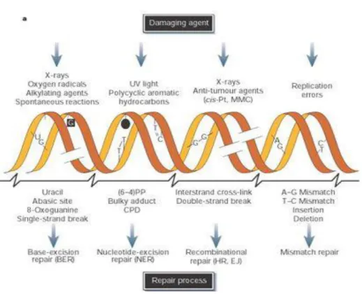
The maintenance of genomic integrity is important for the survival of all organisms. The DNA molecule is highly vulnerable to chemical modification, which can cause numerous lesions. In fact, our genome is constantly exposed to diverse sources of damage (Fig. 1); it is estimated that the DNA can experience up to 105 spontaneous lesions of different origin per day1. These insults can arise from endogenous and exogenous sources: examples of endogenous sources are spontaneous reactions like hydrolysis, leading to the formation of abasic sites and causing deamination or alkylation2. Another example is represented by the reactive oxygen species (ROS) generated by mitochondria, peroxisomes etc., that induce extensive damage to the genome including base oxidation and DNA single-strand breaks (SSBs)3. Common exogenous agents causing lesions in the DNA include ultraviolet (UV) or ionizing radiations (IR): the main consequence of UV rays on the DNA structure is the formation of covalent links between adjacent pyrimidine bases (photodimers), whereas IR induce base modifications, SSBs and DNA double-strand breaks (DSBs) by direct ionization or indirect ROS production. Other exogenous sources of DNA damage are chemical agents used in cancer chemotherapy (alkylating agents, crosslinking agents such as mitomycin C (MMC) and cisplatin) or man-produced chemicals (ex. hidrocarbons or aldehydes in cigarette smoke)4.
To preserve genomic integrity, cells have developed an arsenal of DNA healing strategies- collectively known as the DNA damage response (DDR) that detect different types of damage and initiate the appropriate repair pathway or, if irreparable, induce cell cycle arrest and/or apoptosis. In the case of errors in DNA repair or in DNA replication itself, these lesions can be converted into permanent mutations and therefore passed-on to the next generations of cells. One important consequence of mutations is the loss of tumor-suppressor genes and the improper activation of oncogenes, which can trigger uncontrolled cellular proliferation and the development of malignant cells. Indeed, genome instability is a hallmark of all forms of cancer1,6.
9
1.1.1. DNA damage sensing and signalling
The DDR is a signal transduction pathway consisting of sensors, transducers and effectors that detects DNA damage and induces a specific response to this in order to protect the cell and ensure genome integrity7. In the last 30 years, a conserved set of DNA damage signalling kinases have been identified and widely characterized; they are traditionally categorized in apical and downstream (or effector) kinases (Fig. 2). The apical kinases are ataxia telangiectasia-mutated (ATM), ATM and Rad3-related (ATR), that belong to the PIKK kinase family, and the DNA-dependent protein kinase, catalytic subunit, (DNA-PKcs) 8. Generally, ATR is activated by a wide range of genotoxic stresses, such as replication stress. ATR is recruited via its partner protein ATRIP to extended tracts of ssDNA coated with the ssDNA binding protein replication protein A (RPA) 11. ATRIP-bound ATR interacts directly with ssDNA-coated RPA and promotes ATR localization to sites of replication stress and DNA damage9. Full activation of ATR requires also the presence of activator proteins such as TopBP1, and ssDNA/dsDNA junctions11.
Figure 1. Damage types, DNA lesions and cell fate. Top: most common endogenous and exogenous
sources of DNA damage and resulting DNA lesions. Bottom: according to the amount of DNA damage, cells can activate DNA damage-specific repair processes or induce apoptosis, leading to cell survival or death, respectively. However, in both scenarios mutations can occur engendering malignant transfor-mation (Shiloh, 20035).
10 In contrast to ATR, ATM and DNA-PKcs associate with the ends of DSBs8. In particular, the heterodimer Ku (Ku70/Ku80) binds to the ends of DSBs first and recruit DNA-PKcs in order to start a cascade of phosphorylation events on downstream targets that are involved in DSBs DNA blunt ends processing10. By contrast, ATM is recruited to chromatin in
response to DSBs in a process that requires ATM binding to the C-terminus of NBS1, a component of the MRE11-RAD50-NBS1 (MRN) complex, which contributes also to ATM activation11.
Activated apical kinases transfer stimulatory phosphorylation to the downstream effector
Figure 2. DNA damage response activation. DNA damage sensor proteins (MRN complex, RPA and
ATRIP) are essential for apical kinases activation (ATM, ATR and DNA-PKcs). Once activated, apical kinases phosphorylate and activate downstream kinases, essential for responses to DNA damage, such as cell-cycle arrest, in order to allow DNA damage repair. (Sulli et al. 201113)
11 checkpoint kinases, CHK1 and CHK2, that in turn catalyze phosphorylation events that would coordinate the cellular responses to DNA damage as part of the canonical DNA damage checkpoint. ATM and ATR utilize checkpoint adaptors to mediate the transfer of phosphorylation to effector kinases: ATR relies on Claspin to mediate activation of CHK1, whereas two adaptors, MDC1 and 53BP1, have been linked to the signalling axis ATM-CHK2. MDC1 and 53BP1 possess BRCT domains that directly bind to phosphorylated variant histone H2AX (γH2AX), whose phosphorylation is mediated by ATM or ATR in response to replication stress8. Once activated, DNA damage signalling kinases CHK1 and CHK2 mediate a number of cellular responses such as cell cycle arrest8: CHK1 and CHK2 can promote, in turn, the proteasomal degradation of CDC25, a phosphatase that removes inhibitory modifications from mitotic cyclin-dependent kinases (CDKs) in order to slow down or arrest cell-cycle progression. This prevents the cells from entering mitosis prematurely, providing the time required to repair the DNA or, if the damage is too extensive, for the activation of senescence or apoptotic pathways11. Another critical mediator of cell cycle arrest in humans is the p53 transcription factor, whose classical function is to trigger the apoptotic program. p53-mediated expression of the CDK inhibitor protein p21 represents the primary mechanism by which p53 blocks progression through the cell cycle 8,12. Other cellular processes affected by the activation of DNA damage
signalling kinases are replication fork stability, transcriptional response, inhibition of replication origin firing and dNTP regulation8.
Although the DNA damage checkpoints were originally considered as non-essential regulatory factors, it is now clear that they are one important and integrated part of the DDR. One typical example that illustrates the physiological importance of these checkpoint proteins is the disorder Ataxia Telangiectasia. Individuals carrying two mutated ATM alleles may show loss of motor control owing to Purkinje cell loss, immune deficiencies and increased predisposition to cancer7.
1.1.2. DNA repair pathways
Because of the plethora of possible DNA lesions, cells have evolved a sophisticated and highly regulated set of DNA repair systems specific for almost all types of damage (Fig. 3). In some cases, more than one pathway is required for the repair of one type of DNA damage. In the next section, I will give an overview of the canonical DNA repair pathways focusing my attention on DSBs repair, the HR pathway and on its important mediator BRCA2.
12
1.1.2.1. Direct repair, BER, NER, mismatch and DNA damage-associated replication stress repair
Alkylating agents are a source of DNA damage. They represent a class of reactive chemicals highly abundant in the environment and in living cells. Externally, alkylating agents can be components of air, water, food and pollutants whereas within the cells they can result from oxidative stress. Moreover, due to their cytotoxic properties, many alkylating agents are currently used as chemotherapeutic drugs15. As reactive chemicals,
they are able to transfer alkyl-carbon groups onto the DNA generating a variety of covalent adducts. The pattern of DNA lesions generated by an alkylating agent depends on the number of reactive sites within the alkylating agent (monofunctional versus bifunctional), its particular chemical reactivity, the type of alkyl-group addition (methyl or chloroethyl) and the DNA substrate (double-stranded (ds) or single-stranded (ss)) 15. Among the most common alkylating agents-induced lesions there are N7‑methylguanine (7meG),
Figure 3. DNA damage repair pathways. Top: common DNA damage agents leading
to the formation of different kind of DNA lesions. Bottom: DNA repair pathways
in-duced in response to specific DNA lesions (Adapted from Hoeijmakers, 200114)
Figure 3. DNA damage repair pathways. Top: common DNA damage agents leading to the formation
of different kind of DNA lesions. Bottom: DNA repair pathways induced in response to specific DNA lesions (Adapted from Hoeijmakers, 200114)
13 N3‑methyladenine (3meA) and O6-methylguanine (O6meG). Although 7meG is a relatively
harmless lesion, 3meA and O6meG generate more serious effects and compromise genomic
integrity inducing mutagenesis or blocking essential cellular processes like DNA replication or transcription15. Therefore, the repair of these lesions is essential for the survival of the cell. The main repair mechanisms for alkylation damage include direct DNA repair, base excision repair (BER), nucleotide excision repair (NER) and mismatch repair (MMR).
- Direct DNA repair is mediated by a variety of DNA methyl and alkyltransferase such as
O6meG DNA methyltransferase (MGMT) and O6-alkylguanine DNA alkyltransferase (AGT). In general, their mechanism of action consists in a reaction that transfers the methyl or alkyl group from the O6 position of a guanine to a highly conserved cysteine residue inside AGT or MGMT15, 16, thereby removing the DNA lesion. Another important enzyme involved in DNA direct repair is AlkB homologue (ALKBH) family of α‑ketoglutarate-dependent dioxygenases that catalyse the hydroxylation of aberrant methyl groups15 (Fig.4a).
- Base excision repair (BER) is a highly conserved pathway from bacteria to humans and
is responsible for repairing the vast majority of endogenous DNA damage including
alkylations, oxidations, deaminations and depurinations, as well as SSBs. The initial step in BER is the search for the lesions in DNA by DNA glycosylases17. In humans, there are
currently 11 known DNA glycosylases, alkyladenine-DNA glycosylase (AAG) is one example (see fig. 4b) 15. The glycosylases that recognize uracil, thymine, and alkylated bases remove the damaged base by cleaving the N-glycosyl bond between the base and the sugar. The resulting abasic site is recognized by an apurinic (AP) endonuclease, APE1, that cleaves the abasic site leaving a sugar attached to the 5′ side of the nick. The resulting 3′ hydroxyl is a substrate for the repair polymerase, DNA polymerase β (Pol β). The gap is finally filled in and sealed by a DNA ligase17. Importantly, each step of BER generates intermediates that are highly toxic; therefore, it is essential to prevent the accumulation of these intermediates. The X-Ray Cross Complementing 1 (XRCC1), a key BER protein, coordinates the DNA-processing events of BER to ensure its proper completion15.
- Nucleotide excision repair (NER) is a versatile mechanism that removes helix-distorting
DNA lesions and structures from the genome. NER removes lesions from the entire genome15; however, it partitions in two branches: (i) transcription-coupled repair (TCR) which involves the repair of DNA damage that specifically blocks the progression of RNA polymerase II (RNAP) along the DNA strand during transcription. (ii) Global genomic
14 repair (GGR); a slower and transcription-independent random process that inspects the entire genome for DNA damage18.
The NER process begins with the recognition of a DNA lesion. Then, dual incisions flanking the lesion are generated. The lesion-bearing oligonucleotide is removed, a patch is
Figure 4. DNA damage repair pathways for alkylated bases. (a) Direct repair consists on the
rever-sal of an alkylated base to a normal base without the need of excision and generation of DNA breaks. (b) BER removes simple alkyl and oxidative base lesions. (c) NER is especially implicated in eliminat-ing helix-distorteliminat-ing lesions. (d) MMR mediates the removal of mismatched bases and miss-pairs. See text for details on the mechanisms. (From Fu et al. 201215)
15 synthesized using the undamaged complementary strand as a template, and the patch is ligated to the contiguous strand (Fig. 4c). Although, most of the steps of NER process are common between GGR and TCR, the recognition of the DNA lesion differs: in GGR repair, a helix distorting lesion or structure is directly recognized by XPC, complexed with hRAD23B and Centrin 2 (CETN2). Then, the complex melts the DNA around the lesion and recruits the multiprotein complex TFIIH. By contrast, in TCR-recognized DNA lesions such as bulky adducts, RNAP arrest constitutes the first step for damage recognition. The arrested elongation complex recruits CSB (ERCC6), a transcription elongation factor that translocates along the template DNA together with RNAP. CSB strongly binds the polymerase when it is blocked at a lesion and changes the DNA conformation by wrapping the DNA around itself, altering the interface between RNAP and DNA. CSB recruits the CSA complex and other NER factors whose role is to remove or backtrack RNAP to allow access to TFIIH18. At this step, in both GGR and TCR, XPB and XPD, two components of TFIIH complex, unwind the DNA to create a 20–30-nucleotide bubble18 and then XPA, RPA and XPG are recruited. XPA facilitates the release of the CDK-activating kinase (CAK) sub-complex from TFIIH, possibly promotes lesion verification by TFIIH and binds to the DNA lesion in a single-stranded configuration. RPA binds the ssDNA coding strand and likely has a role in DNA damage signalling by activating the DNA damage response kinase ATR. Together, TFIIH, XPA and RPA promote the recruitment and positioning of the endonucleases ERCC1–XPF and XPG, which incise the DNA 5′ and 3′ sides of the lesion, respectively. Following excision of the damaged DNA, the gap is filled by DNA synthesis and ligation, mediated by proliferating cell nuclear antigen (PCNA), DNA polymerase δ, ε or κ and DNA ligase 1 or XRCC1–DNA ligase 319.
Defects in NER lead to diverse clinical consequences such an extreme predisposition to cancer as it occurs in patients suffering from Xeroderma Pigmentosum (XP), a pathological disorder leading to hypersensitivity to UV radiation, sun-induced cutaneous features such as hypopigmentation and hyperpigmentation, and high risk of skin-cancer among others. Other serious effects derived from NER pathways defects include neurodevelopmental diseases like Cockayne syndrome and the even more severe cerebro-oculo-facio skeletal syndrome (COFS), characterized by microcephaly, mental retardation, retinal degeneration, photosensitivity, among other defects, and a highly reduced life-expectancy20.
- Mismatch repair (MMR) is implicated in correcting error-containing sections of the
16 within repeated-sequence motifs, such as microsatellites, when primer and template strands dissociate and re-anneal incorrectly generating heteroduplex DNA molecules. As a result, the number of repeated units in the template and in the newly synthesized strand is different. These heterogeneities are known as insertion/deletion loops (IDLs) and together with base–base mismatches due to DNA polymerases errors that escape their proofreading function, represent the main target of MMR repair proteins21. In MMR, mismatches are identified and firstly bound by MutSα heterodimer (MSH2–MSH6) forming a sliding clamp. Upon an ATP-dependent conformational change, MutSα recruits and binds MutLα heterodimer (PMS2–MLH1). Diffusion of MUTSα–MUTLα complex leads to nicking of the DNA either upstream or downstream of the mismatch mediated by the endonuclease activity of the PMS2 subunit 15, 22. The nicking serves as an entry point for exonuclease 1 (EXO1) that removes a segment of DNA which is then filled in and repaired by a combination of Pol δ, Pol ε and sealed by DNA ligase22 (Fig. 4d). Loss of MMR activity, due to the lack of function of any of its key players, is associated with tumor development, microsatellite instability (MSI tumors) and triplet repeat expansions, the latter being at the origin of neurological diseases such as Huntington disease and myotonic dystrophy23, 24.
- DNA damage-associated replication stress repair generally arises when replication
fork progression during genome duplication is impeded by obstacles of intracellular or extracellular origin leading to the condition of “replication stress”25, 26. The most common
causes of replication stress include limited nucleotide pool, nicks or gaps in ssDNA, unrepaired DNA lesions, ribonucleotide incorporation, repetitive DNA motifs, DNA secondary structures in the DNA (ex. DNA hairpins, G-quadruplexes) or DNA-RNA hybrids generating transcription-replication conflicts26.
The first signal of replication stress is represented by stalling or slowed progression of the replication fork and/or DNA synthesis. All the obstacles mentioned above can lead to the generation of ssDNA caused by the uncoupling of the polymerase and the DNA helicase that continues to unwind the DNA helix regardless the stalling of the polymerase. Persistence of ssDNA adjacent to the stalled replication fork leads to the coating of ssDNA by RPA, which in turn stimulates the activation of the DNA damage–checkpoint kinases ATR and CHK125, 26 (Fig. 5). ATR-mediated signalling orchestrates different pathways at stalled replication forks allowing cell-cycle arrest and the regulation of intracellular dNTP levels, thus ensuring proper fork repair and restart. In addition, ATR phosphorylates and regulates the activity of several replisome components and fork-remodelling enzymes: for example, ATR promotes the association of the Fanconi anemia (FA) protein FANCD2
17 with the MCM replicative helicase and this interaction slows DNA synthesis and prevents the formation of long ssDNA stretches under conditions of reduced nucleotide pools. Moreover, ATR activation can either prevent new origin firing by inhibiting replication initiation, or promote firing of dormant origins within pre-existing replication factories, thus allowing completion of DNA synthesis in the vicinity of perturbed replication forks25.
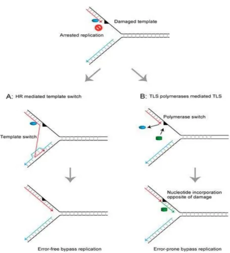
Stalled replication forks and gaps can be restarted by different mechanisms including translesion synthesis (TLS), template switching (TS), fork regression or HR27. TLS is catalyzed by specialized low-fidelity DNA polymerases (Pol η, Pol ι, Pol κ, and Rev1) to bypass DNA lesions. Due to a large active site and the lack of proofreading activity, these polymerases allow the incorporation of a nucleotide opposite to a damaged DNA template. On the contrary, TS is proposed to use a recombination-like mechanism by which the nascent DNA of the sister chromatid is utilized as a temporary template for replication (Fig. 6 a, b). TLS and TS are generally considered as mechanisms evolved to deal with damage encountered during replication, a condition commonly known as DNA damage tolerance (DDT). This pathway allows the replication to continue in the presence of a DNA lesion by promoting damage bypass. The choice between these two pathways of DDT
Figure 5. The ATR-mediated replication stress response. After replication fork stalling, the ssDNA
generated by polymerase–helicase uncoupling is coated by RPA to prevent secondary structure formation. RPA-coated ssDNA is recognized by ATR. ATR kinase targets CHK1 phosphorylation which prevents progression through the cell cycle until replication is completed (Adapted from Liao et al, 201843)
18 (TLS and TS) is important because it can determine an error-prone or error-free outcome and a key player driving this choice is proliferating cell nuclear antigen (PCNA). PCNA regulating role in DDT is mediated by its ubiquitination and SUMOylation28.
HR plays a pivotal role in the repair of ssDNA gaps and DSBs and contributes to the general robustness of DNA replication29, 30. With the help of recombination mediator
proteins such as BRCA2, RAD51 recombinase nucleates onto ssDNA to form a nucleoprotein filament. Subsequently, this filament performs the homology search on the
Figure 6. Schematic representation of the mechanisms restoring the arrested replication fork at the damaged DNA template. (a) Homologous recombination (HR)-mediated template switch restores
the arrested replication fork using intact newly synthesized DNA as the template strand and promotes error-free bypass replication. (b) Translesion DNA synthesis (TLS) polymerases mediate direct bypass replication across the damaged template in an error-prone manner. (From Abe et al., 201833).
19 intact strand and promotes strand invasion within the homologous DNA duplex. This results in the formation of a D-loop structure within which the 3′end of the invading strand primes DNA synthesis30. For a more extensive explanation of HR pathways see section 1.2 (page 29).
Another important contribution of HR proteins at the replication fork is the protection of the fork from aberrant nuclease activity31, 32. Upon the inhibition of fork elongation, the BRCA2, RAD51 and many other factors such as breast cancer type 1 susceptibility protein BRCA1 or the Fanconi anemia (FA) protein FANCD2297 protect newly replicated DNA from unscheduled degradation by nucleases such as MRE1131, 32, 164-166. First evidence of BRCA2 in protection of stalled replication forks came from a study showing that BRCA2 deficiency de-stabilizes the structure of DNA intermediates formed at stalled replication forks induced by exposure to hydroxyurea (a common replication stress inducing agent), subsequently triggering their collapse into DSBs, leading to the proposal that BRCA2 stabilizes the structure of arrested forks to allow their error-free resolution133. Years later, a study using a mutant at the C-terminal RAD51 binding site of BRCA2 showed that this region is dispensable for HR but required to protects nascent DNA strands at stalled forks from degradation by the MRE11 nuclease31. Following this discovery, much efforts have
been devoted to uncovering the nucleases and regulatory factors leading to extensive fork degradation, as well as the structure of the replication fork required for protection and HR proteins recruitment. Indeed, recent studies have identified that in a BRCA-deficient background the structure targeted by nucleases is a reversed replication fork167-170. Fork reversal is a key mechanism that allows replication forks to reverse their course in order to cope with DNA lesions171. These studies have demonstrated that BRCA2, as well as BRCA1, is required to stabilize the RAD51 filament on the regressed arms of the reversed replication fork, thereby protecting the nascent DNA from nucleolytic degradation (Fig. 7). In addition, BRCA2 has been proposed to prevent ssDNA gap accumulation both at replication fork junctions and behind forks by stabilizing RAD51 binding167. In the absence of BRCA2, forks with persistent ssDNA gaps would be converted into reversed forks, leading to extensive degradation. In the context of BRCA-deficient tumors, MRE11 was the first nuclease associated with fork degradation31, 32, 164-166. However, later studies have also identified EXO1 and DNA2 as nucleases contributing to fork degradation in BRCA-deficient tumors168. In addition, CtIP protein is likely required to initiate MRE11-dependent fork degradation168, although a more recent study seems to contradict this conclusion proposing a role for CtIP in protecting stalled replication forks from
20 degradation172.
However, it is important to distinguish this excessive degradation of stalled replication intermediates that underlies the pathological effects of resection from the limited resection of nascent DNA strands which is required for efficient fork restart. For example, controlled DNA2-dependent resection of reversed replication forks is a functionally relevant mechanism mediating reversed-fork restart and providing resistance to prolonged genotoxic treatments25.
DNA replication stress is the major source of spontaneous DSBs in dividing cells. Prolonged fork stalling or failure to resume DNA synthesis by the mechanisms described above lead to fork collapse and formation of one-ended DSBs25. Fork collapse or under-replication of the DNA can contribute to ultrafine bridges, MiDAS (mitotic DNA synthesis) and sister chromatid bridges as a consequence of the excess of ssDNA generated at arrested forks, that can subsequently result in chromosomal instability and diseases298. For example, mutations in the cognate binding partner of ATR, ATRIP lead to Seckel
Figure 7. Role of HR factors BRCA1/2 and RAD51 in protection of stalled replication forks:
Schematic representation of recent findings on the role of BRCA proteins and RAD51 at stalled replica-tion forks. In BRCA proficient cells, in case of replicareplica-tion stress causing the stall of the fork, the fork is reversed and BRCA1/2 are recruited here to stabilize RAD51 nucleoprotein filament preventing ssDNA degradation. This excessive degradation might be occurring in BRCA-deficient tumors. (From Byrum,
21 syndrome, characterized by developmental delay, microcephaly and mental retardation. Loss of proteins that recognize or repair DNA lesions also leads to a number of human diseases. For example, loss of the specialized DDT polymerase Pol η, involved in TLS, results in a variant form of the cancer-susceptibility condition of Xeroderma Pigmentosum26.
1.1.2.2. Inter-strand crosslink repair
Inter-strand crosslinks (ICLs) are lesions that covalently link two bases on the complementary strands of DNA. These lesions are generated by chemicals with two reactive electrophilic groups. The formation of ICLs is highly dependent on the sequence because two nucleophilic groups on opposite strands must be aligned geometrically to allow the cross-link to occur34. The presence of an ICL affects the unwinding of the DNA, an essential step in DNA replication and transcription; thus, ICLs are detrimental for cells, especially those in rapid division. Not surprisingly, several crosslinking agents such as mitomycin C (MMC) and cisplatin are used in the clinic as anti-cancer drugs. ICLs such as aldehydes also form naturally as by-products of cellular metabolism34.
Since ICLs compromise both DNA replication and transcription, it is clear that their repair is of high importance. Several DNA repair pathways are involved in the repair of ICLs including NER, HR and TLS. The pathway involved in repairing them will depend on how the DNA lesion is recognised35, 36, 37. Although in most cases ICLs repair is coupled to replication and triggered when a DNA replication fork collides with the ICL35, in the context of non-replicating DNA, the distortion of the DNA helical structure caused by the lesion attracts proteins involved in the global damage surveillance of DNA. This process has been shown to involve proteins of the NER pathway, as XPC implicated in the initial recognition36.
In replicating cells, the stalled fork at the site of the lesion attracts protein of the Fanconi anemia (FA) pathway (Fig. 8). The FA pathway comprises 22 gene products (FANCA to FANCW). Autosomal biallelic germline inactivation of any of the 22 FA genes (with the exception of FANCB, which is X-chromosomal) causes Fanconi anemia174, a genetic disease that results in sensitivity to ICLs and predisposes patients to bone marrow
22 failure and development of cancer37, 174. The inability to repair DNA ICLs is a key cellular feature of FA and cellular markers for its diagnosis are usually represented by chromosome breakages174. Interestingly, biallelic mutations in breast and ovarian cancers susceptibility genes such as BRCA1/2 and PALB2 are found also in FA patients175. In particular, BRCA2 was found to be identical to FANCD1 as BRCA2 transient transfection could
Figure 8. FA pathway for ICLs repair: Upon fork stalling at ICL sites, FANCM–FAAP24–MHF1/2
complex binding activates ATR signalling and promotes recruitment of the FA core complex. The core complex in turn ubiquitinates the FANCI–FANCD2 heterodimer, which acts to promote the next steps of nucleolytic incision to unhook the ICL, translesion synthesis and, finally, HR pathway to repair the nucleolytic-induced DSB (Adapted from Michl, Zimmer and Tarsounas 201638)
23 restore ICL repair in FANCD1 deficient cells, establishing a role for BRCA2 in this pathway176, 177.
The FA pathway operates mainly during the S phase of the cell cycle and requires converging replication forks. ICL repair is elicited when the replisome is partially dismantled by eviction of MCM replicative helicase subunits from the chromatin, thereby enabling ICL recognition by FANCM and its interacting partners FAAP24 and MHF1/2. FANCM binding adjacent to ICLs leads to recruitment of the FA core complex formed by FANCI–FANCD2 and ATR-dependent checkpoint activation, which stalls the replisome. The binding of the FA core complex to the lesion triggers monoubiquitination of the FANCI–FANCD2 complex, as the central event in the FA pathway. Monoubiquitinated FANCI–FANCD2 is recruited to the damaged chromatin and promote downstream reactions including endonucleolytic incision of the ICL, probably mediated by ERCC4 (NER), translesion synthesis and nucleolytic cleavage-induced DSB repair37, 38. This last step is preferentially mediated by HR, since many of the first identified FA pathway factors were involved in HR175. As in DSB repair, the HR step in ICL repair requires the loading of RAD51 (FANCR) onto the resected DNA followed by strand invasion to resolve the DSB. Therefore, HR proteins such as PALB2 (FANCN), BRCA1 (FANCS), BRCA2 (FANCD1), among others, are essential in this step. Furthermore, RAD51 is also necessary to protect stalled forks at a ICL lession299 and a recent study reported a similar
function for BRCA2300.
1.1.2.3. DNA single-strand break (SSB) repair
DNA single-strand breaks (SSBs) are discontinuities in one strand of the DNA double helix and are usually accompanied by loss of a single nucleotide and by damaged 5’- and/or 3’-termini at the site of the break. SSBs can arise from oxidized nucleotides/bases during oxidative stress, intermediate products of DNA repair pathways (e.g., BER), and aborted activity of cellular enzymes39, 40. It has been estimated that more than 10,000 SSBs are generated per mammalian cell each day representing the most common type of DNA lesion40.
It is generally accepted that SSBs are repaired by various DNA repair mechanisms that, globally involve SSB detection, DNA end processing, DNA gap filling, and DNA ligation steps40. SSB detection is primarily mediated by poly (ADP-ribose) polymerase-1 (PARP1)39,
24 modifies itself and other target proteins through the addition of chains of poly-ADPribose (PAR) using NAD+ as substrate40, 41. PARP1 promotes the recruitment of XRCC1, a critical
scaffold protein that interacts with several enzymatic components of SSB repair and accelerates the process40. The following step of DNA end processing is mediated by protein binding partners necessary for conversion of different types of damaged termini to conventional 3′-hydroxyl and 5′-phosphate termini, including Polβ (to remove 5′- deoxyribose phosphate termini during BER), PNKP (to remove 3′-phosphate and 5′-hydroxyl termini), APTX (to remove 5′-AMP during abortive DNA ligation events), and TDP1 (to remove Top1 peptide from 5′-termini) 42. Once damaged 3’-termini at SSB have been restored to their conventional hydroxyl configuration, gap filling can occur; which often involves insertion of the single nucleotide that is missing at most SSBs (short-patch repair). Other SSBs involve larger DNA gaps (long-patch repair) which require FEN-1 flap-endonuclease to remove the displaced 5’-nucleotides. The gap filling is carried out by Pol β, although other DNA polymerases, such as Pol δ and Pol ε can also perform this role40. Finally, the DNA ligation step is carried out by Lig1 and Lig3α40, 42.
Unrepaired SSBs are implicated in human diseases such as neurodegenerative disorders, cancer, and heart failure. SSB repair has been associated with hereditary genetic diseases including ataxia-oculomotor apraxia 1 (AOA1) and spinocerebellar ataxia with axonal neuropathy 1 (SCAN1). Importantly, all SSB repair proteins currently associated with neurological disease are associated with DNA processing step. Both germline and tumor-associated variants of genes encoding SSB repair proteins (e.g., XRCC1, APE1, and Pol β) have been identified in humans, suggesting SSB repair also as a tumor suppressor mechanism39, 42.
1.1.2.4. DNA double-strand break (DSB) repair
DSBs are generated when the two complementary strands of the DNA double helix are physically dissociated into two separate chains. They are considered as the most cytotoxic DNA lesions and it has been calculated that even one single DSB can trigger the arrest of the cell cycle46. DSBs pose an immediate threat to the stability of the genome because when repaired inappropriately they can lead to chromosome rearrangements, amplification or loss of chromosome material or translocations, thereby disrupting gene structure and function. Indeed, germline mutations in DSB repair genes cause genomic instability in numerous hereditary human diseases such as many forms of cancer, developmental disorders and premature ageing44,45.
25 As many kind of DNA damage lesions, DSBs can form as a result of exposure to either exogenous or endogenous agents. Some well-known exogenous DSBs inducing agents are anticancer chemotherapeutic drugs (cisplatin, MMC, radiomimetic compounds and topoisomerase inhibitors) and ionizing radiation (IR). IR leads to extensive base damage47: generally, the more common outcome of IR exposure are SSBs by producing radiolysis radicals that attack the sugar-phosphate backbone, which can be later converted into DSBs. Frequently, at high doses of irradiation, two such nicks are present in both complementary DNA strands within one helical turn leading to DSBs47, 48. Endogenous sources of DSBs include ROS that trigger both SSBs and DSBs upon DNA base oxidation49or defective telomere metabolism that may originate DSBs at chromosome termini50.
However, most of the endogenously-generated DSBs are associated with DNA replication. SSBs are transformed into DSBs when reached by a replication fork (Fig. 9A). In addition,
Figure 9. Induction of single- and double-ended DSBs (A) In the presence of a SSB, the replication
fork is converted into a single-ended DSB. (B) When replication fork progression is impeded by a blocking DNA lesion, the complementary nascent strands create a so called “chicken foot” structure by reversion of the stalled fork and re-annealing of the parental strands; cleavage of this structure induces a single-ended DSB. (C) Double-ended DSB, generally induced by IR exposure or endonuclease cleav-age. (From So et al. 201751)
26 when a replication fork is blocked, fork regression through annealing of the nascent strands (which are complementary) generates a four-branched structure called chicken foot. Specialized endonucleases can resolve this structure generating a DSB (Fig. 9B). In both of these cases, replication fork stalling leads to the formation of single-ended DSBs, whereas double ended DSBs are generally associated to IR or endonuclease induced DNA damage (Fig. 9C)51. Despite their high toxicity, DSBs may be deliberately generated by the cell for a specific biological purpose, for instance, to initiate recombination between homologous chromosomes during meiosis. Also, DSBs naturally occur as intermediates during developmentally regulated rearrangements, such as V(D)J recombination and immunoglobulin class-switch recombination in B-cells. Recent evidence suggest the coevolution of processes that couple introduction of programmed DSBs to their accurate repair in order to constitute an effective safeguard against genomic instability52.
Repair of DSBs involves four possible pathways (Fig. 10): two of them, classical non homologous end joining (C-NHEJ) and HR have been extensively studied and are considered the two main DSBs repair mechanisms. More recently, two other pathways, alternative end joining (alt-EJ) and single-strand annealing (SSA), have been shown to operate in many different conditions and to contribute to genomic rearrangements and oncogenic transformation45, 53. Among the four, HR and C-NHEJ have evolved as
high-fidelity processes.
C-NHEJ can take place along the entire cell cycle and it does not require a homologous
sequence. The starting step in c-NHEJ consists on the rapid binding of KU proteins, Ku70– Ku80, to both ends of the broken DNA molecule to prevent promiscuous end resection. Once bound, Ku70–Ku80 recruits and activates DNA-PKcs which, in turn, triggers an extensive signalling cascade that orchestrates downstream repair54. Briefly, binding of KU to blunt DNA ends requires minimal DNA processing mediated by the nuclease Artemis, activated upon interaction with DNAPKcs, and specialized DNA polymerases λ and μ, and repair is directly assisted by two scaffold proteins, XRCC4 and the non-homologous end-joining factor 1 (XLF) that bind to DNA ligase 4 (LIG4) responsible for sealing the break
45,54. A number of accessory factors support or regulate c-NHEJ, including the MRN
complex, found involved in the stabilization of distant breaks and in the processing of DNA ends 55 and PARP proteins implicated in the correct ligation of DNA ends56. Several additional positive and negative regulators of Ku70/Ku80 have been identified such as proteins containing a KU binding motif like the Aprataxin-and-polynucleotide kinase/phosphatase-like Factor (APLF) and the exonuclease/helicase mutated in Werner
27 syndrome (WRN) 57.
Despite the mutagenicity of c-NHEJ that may arise in repetitive sequences, the fast kinetics of c-NHEJ has a clear role in protecting genome integrity notably by suppressing chromosomal translocations, at least for the majority of repair events53. Mutations in c-NHEJ pathway components are associated with a variety of human pathological disorders such as diverse immunodeficiency-associated syndromes, observed in carriers of mutated Artemis, XRCC4 and DNA-PKcs54.
HR takes place mainly during the S phase of the cell cycle, where a sister chromatid can be
used as a homologous template to copy and restore the DNA sequence missing on the damaged chromatid. The search for sequence homology as a template for HR requires the presence of ssDNA at the DSB end. This intermediate can be generated by the resection step which consists in the nucleolytic degradation of the 5’strand of a DSB. Some
of the most important players of HR pathway are the proteins BRCA2 and RAD51. As HR-mediated DSBs repair is particularly relevant for this thesis project, I will include an extensive description of it in section 1.2 (page 29).
Figure 10. Four pathways to repair DSBs. The repair of DSBs relies primarily on whether DNA end
resection occurs. When resection is blocked, repair through c-NHEJ is favored (left). However, when DNA resection occurs, three pathways (alt-EJ, SSA and HR) can compete for the repair of DSBs (from left to right). Details of each pathway are described in the text. (From Chang et al. 201754)
28
Alt-EJ is an alternative pathway that mainly operates during the S and G2 phases of the
cell cycle on 3’ ssDNA ends generated by DNA resection when c-NHEJ is compromised53, 54. PARP1 plays the initial role in alt-EJ recognizing and tethering either ssDNA nicks or
blunt dsDNA ends. In addition, PARP1 recruits MRN and CtIP to the DSB end. CtIP enhances the MRN endonuclease activity resulting in an internal single-strand break within the 5′ strand. The short single-strand fragment at the DSB end is then degraded by the MRN exonuclease activity. The loading of the EXO1 or DNA2 generates longer stretches of RPA-coated ssDNA. Following resection, short regions of sequence complementarity ranging from 2 to 20 nucleotides are exposed within the RPA-coated ssDNA regions. Here, PARP1 plays another role in DNA end bridging and alignment, at which point non-complementary 3’ tails are removed. Error-prone gap filling is performed most likely by the polymerase Polθ, and sealing of the nick is carried out by LIG1 or LIGIIIα58, 59. Repair of DSBs by alt-EJ is inherently mutagenic potentially giving rise to chromosomal translocations as well as intra- and inter-chromosomal deletions and insertions45, 53, 58, 59.
SSA is a non-conservative homology-directed repair (HDR) pathway (which, generally,
involve loss of nucleotides), that does not entail the presence of a sister chromatid54, 60. Similarly to alt-EJ and HR, SSA is initiated by end resection mediated by CtIP, thus occurs preferentially during the late S and G2 phases of the cell cycle. However, in contrast to alt-EJ, SSA requires >20bp of homology. SSA joins direct repeat sequences (e.g., tandem repeats) at 3’ssDNA end through annealing at the cost of deletion of the intervening sequence between the repeats. RAD52 is responsible for the annealing of the flanking repeats resulting from the end resection61. The nuclease activity of ERCC1 in complex with ERCC4 then removes the non-homologous 3’ssDNA tails, which is enhanced by RAD52. Polymerases and ligases are in charge of the final steps - gap filling and ligation - although the exact players remain poorly understood60. In order to reveal complementary homologous sequences, SSA requires extensive DNA end resection and RPA displacement; moreover, sequence information can be lost or rearranged if the overlapping ends pairs are unsuitably joined. Therefore, SSA is considered to be an obligatorily error-prone pathway60. Given the various possibilities available for DSBs repair, these four pathways could potentially compete for access to the free DNA ends of a DSB.
29 Thus, as illustrated in Figure 11, the regulation of pathway choice could be depicted as a “decisional tree” where the branch points represent points of commitment to c-NHEJ or HR, points where physiological sub-pathways are selected and steps at which repair intermediates are vulnerable to hijacking by error-prone repair pathways45. One crucial factor that determines pathway choice in this context is DNA end resection: the presence of a ssDNA tail, for instance, can affect Ku70-Ku80 binding and c-NHEJ pathway. Extensive end resection is stimulated in the S/G2 phase of the cell cycle in a manner that depends on CDK activity, which mediates phosphorylation of multiple substrates such as CtIP45, 53.
Figure 11. A decision tree for DNA double-strand break repair. Schematic representation of DSB
repair pathway choice. In blue, the critical aspects influencing the choice of the repair pathway (in vio-let). In red, the activity triggered after pathway choice. In grey, most important mediators of each DSB repair pathway. (From Scully et al. 201945)
30 Thus, the phase of the cell cycle is also a key influencing DSB repair pathway choice. Accessory factors also contribute via the modulation of end resection; for instance, the balance between BRCA1 and 53BP1 regulates pathway choice by either promoting or preventing end resection53. In addition, it is becoming increasingly clear that the native chromatin state of the damaged DNA and its position within the nucleus influences DNA repair kinetics and pathway choice60. Finally, once resection takes place, the formation of a RAD51 nucleoprotein filament becomes the critical step to trigger a conservative repair by sister chromatid recombination instead of other error-prone pathways that also require resection. For instance, RAD51 is negatively controlled at different levels by key factors of other resection-dependent DSB repair pathways such as Polθ, implicated in Alt-EJ53.
1.2. Homologous recombination-mediated DSB repair
HR is an essential process that uses the redundant genetic information existing on the sister chromatids (or homologous chromosomes) when both strands of the DNA double helix are compromised by DNA damage64. HR plays essential roles as DNA replication support, the repair of DSBs in somatic cells and in meiosis, where it is important to generate genetic diversity48, 52. In contrast to SSA that can be mutagenic, HR is a conservative
homology-directed pathway.
Upon DSB induction, the MRN complex rapidly localizes to the damaged DNA. MRE11 binds to CtIP for the initiation of DNA-end resection through endonucleolytic cleavage of the 5′-terminated strand upstream from the DSB end. Starting from the nick, the exonuclease activity of MRE11 degrades DNA in a 3′ to 5′ direction towards the DSB end. The resulting single-stranded DNA (ssDNA) overhang is immediately coated by RPA to protect the ssDNA from degradation or self-annealing. The 5′-recessed end now represents a preferred substrate for the 5′ to 3′ exonuclease EXO1 to carry out extended resection. Alternatively, extended resection is catalyzed by the combined endonuclease and helicase activities of DNA2-BLM in human cells63, 64 (Fig. 12).
Other DNA end resection regulators, both positive and negative, have been described. For example, BRCA1, in complex with BRCA1-associated RING domain protein 1 (BARD1), interacts with CtIP and MRN and is implicated in DNA end resection especially counteracting the NHEJ factor 53BP1 in S/G2 phase of cell cycle66. Another important function of BRCA1 is in a later step of HR; indeed, it allows the recruitment of BRCA2 to
31
DSBs through the bridging protein PALB2. BRCA2 is required to assist RAD51 loading onto ssDNA and displace RPA and thus, to promote RAD51 nucleoprotein filament formation67, 68, 69. In particular, my PhD supervisor and her collaborators showed, using both single molecule visualization of individual RAD51 nucleoprotein filaments and ensemble experiments, that through the BRC motifs, BRCA2 stimulates the ssDNA
Figure 12. DNA end resection upon DSB induction. Representation of the main DNA-end resection
events in human cells. First step is the recruitment of MRN complex; hence, MRE11-CtIP action is essential for an endonucleolytic cleavage and initial short-range DNA end resection. Subsequent step of extensive DNA resection (long-range DNA end resection) is mediated by EXO1 or DNA2/BLM. (From
32 binding activity of RAD51 by modulating its ATPase activity while preventing the assembly of RAD51 onto dsDNA107, 108.
The modulation of RAD51 DNA binding preference then promotes the subsequent steps of recombination. The successful purification of full-length BRCA2 allowed them to confirm these findings with the entire BRCA267, 68 (Fig. 13).
The RAD51-ssDNA nucleoprotein filament searches the entire genome for homology regions in association with other factors, such as RAD54. Five RAD51 paralogs are involved in assisting RAD51-ssDNA nucleoprotein filament stability, optimizing its
Figure 13. Model depicting the function of BRCA2 in HR-mediated DSBs repair. A DSB is
resect-ed to reveal a 3’ ssDNA tail that is immresect-ediately coatresect-ed by RPA. BRCA2 binds to the ssDNA /dsDNA junction and loads RAD51 onto the RPA-coated ssDNA while preventing its unproductive assembly on dsDNA. This allows the formation of a stable pRAD51 nucleoprotein filament, essential for the search of homology on a donor DNA duplex and to promote DNA strand invasion for the repair of the dam-aged chromosome. (From Jensen, Carreira and Kowalczykowski 201067)
33 efficiency and, at the same time, restricting RAD51 function to appropriate DNA substrates. Mutations in any of the RAD51 paralogs lead to defects in RAD51 foci formation and DNA damage sensitivity, thus suggesting that all of them are essential and could have specific function72. During synapsis (physical connection between two DNA regions with homology), the RAD51-ssDNA complex facilitates the base-pairing between the invading DNA substrate and the homologous duplex DNA template resulting on a displaced strand or D-loop. Upon pairing of the 3’ extremity of the broken molecule with the donor sequence, DNA synthesis by a polymerase (usually Polδ or TLS polymerases) restores the sequence information disrupted by the DSB65, 72. Depending on how the D-loop structure is disrupted, it is possible to distinguish different HR sub-pathways (Fig. 14). The double Holliday junction (dHJ) formation and the synthesis dependent strand annealing (SDSA) are the two main HR sub-pathways. dHJ is predominant in meiotic recombination and occurs when the D-loop captures also the second end of the break not involved in strand invasion. Subsequently, the 3’ssDNA overhang forms a dHJ with the homologous chromatid which can be processed by a resolvase complex (composed of MUS81 and EME1 and structure-specific endonucleases)73. The outcome of this process is
Figure 14. HR sub-pathways. Top: first step HR mediated DSBs repair pathway leading to search for
homology, DNA invasion and formation of D-loop heteroduplex. Resolution of D-loop structure can occur in different ways, generating HR sub-pathways: from the left to the right SDSA, formation of dHJ and BIR. (From Piazza and Heyer 201965)
34 mainly crossover products, although, because of the possible gene conversion events, non-crossover products can also be generated65, 71, 73. SDSA is the major HR sub-pathway in
somatic cells; in it, the strand invasion leads to the copying of the template but, in this case, the new strand is displaced from the donor and is captured by the second end of the DSB. The resulting single Holliday junction then slides down the DNA duplex in the same direction in a process called branch migration, displacing the extended strand from the template strand. This displaced strand pops up to form a 3'overhang in the original double-stranded break duplex, which can then anneal to the opposite end of the original break through complementary base pairing. Therefore, SDSA produces only non-crossover products because the flanking markers of the heteroduplex DNA are not exchanged65, 71, 73. Break-induced replication (BIR) occurs when a single end of the DSB acts independently; this may take place when one side of the break fails to engage with a homologous sequence or when the two ends find different homologous templates. In addition, BIR arises in case of single-ended DSBs, which, as described above, can occur due to replication through a DNA lesion that results in fork stalling or collapse, or through telomere erosion that exposes a single-ended DSB. The most relevant difference between BIR and other HR sub-pathways is the DNA synthesis mode; in BIR, after DNA strand invasion and D-loop formation, the invading strand is extended by DNA synthesis (mostly mediated by Polα) concomitant to D-loop migration, driven by a helicase, potentially Pif1 or MCM. In the process, the newly synthesized DNA accumulates unrepaired DNA lesions74. Therefore,
although HR is generally considered an error-free pathway, if not perfectly regulated, it can also lead to the accumulation of lesions and chromosome rearrangements65.
1.3. BRCA2: structure and roles as a custodian of genome integrity
1.3.1. The tumor suppressor BRCA2
BRCA2 was discovered in 1995 as a gene implicated in the predisposition to breast and ovarian cancer75, 301. In addition, inherited mutations affecting a single copy of BRCA2 also significantly elevate the risk of cancers in the pancreas, male breast, prostate, and other tissues70, 76, 77. Furthermore, biallelic mutations in BRCA2 are also found in Fanconi anemia patients leading to predisposition to various types of cancer at early age175.
The earliest clues about the importance of BRCA2 in maintaining genome integrity came from observations in mice showing that Brca2-mutated mice exhibited early embryonic
35 lethality and DNA repair defects similar to Rad51-defective mice78, 79. At the same time,
BRCA2 was found to physically interact with RAD51 in vitro80. In addition,
Brca2-defective mouse cells where shown to exhibit chromosome rearrangements such as translocations and chromosome breakages81 making the hypothesis of a direct role of BRCA2 in DNA repair even stronger. Later on, the role of BRCA2 in HR was directly measured using a recombination reporter assay in CAPAN-1 cells (pancreatic BRCA2 deficient cancer cells)82 which could be rescued by transient transfection of BRCA2130. Afterwards, based on the established interactions between the BRC motifs of BRCA2 and RAD51, together with the preferential binding of BRCA2 to ss DNA112, it was proposed that BRCA2 could facilitate recruitment of RAD51 to sites of processed DSBs requiring repair and enhance RAD51-promoted strand invasion111. In support of this hypothesis, Brh2 (BRCA2 ortholog in U. maydis), was shown to stimulate the ATPase activity of Rad51 on RPA-coated DNA containing a single-stranded gap and to promote Rad51-filament formation on RPA-coated gapped DNA, while decreasing the amount of bound RPA, suggesting its role as a Rad51 mediator88.
Therefore, it is not surprising that BRCA2 (as well as other factors involved in DNA repair, such as BRCA1 and PALB2) function as tumor suppressors and that inherited mutations in these genes confer significant lifetime risks of breast, ovarian, and other cancers. Indeed, BRCA-deficient cells usually accumulate aberrations in both chromosome structure (translocations, large deletions or chromosome fusions129) and number, reflecting
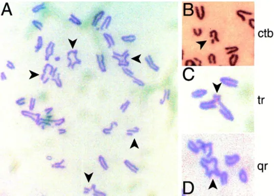
inaccurate chromosome segregation81, 129and genome instability which are hallmarks of tumorigenesis70. The structural aberrations typically include breaks affecting a single sister chromatid, as well as tri-radial and quatri-radial chromosomes (Fig. 15). These abnormalities denote defects in HR and can be explained by error-prone
36 mechanisms like NHEJ or SSA82, 130, 131 that take over the DNA lesions in the absence of
BRCA2. These pathways promiscuously re-ligate broken DNA ends, particularly across short microhomologies. Consistent with this notion, human cancers harbouring homozygous mutations in BRCA2 display not only extensive structural rearrangements in chromosomes but also many short deletions (≤50 bp) with overlapping microhomology at breakpoint junctions132.
The abnormal chromosome number may be explained by other functions of BRCA2 in mitosis such its role in cytokinesis83, 98, 104 and in the alignment of chromosomes that we have recently reported84.
As mention above, BRCA2 mutation carriers typically inherit a single mutated copy in their germline. Following the Knudson 2-hits theory for tumor suppressors320, the wild-type copy is lost in a process called loss-of-heterozygosity (LOH)134, 135, 136. However, recent genomic studies have shown that a fraction of these tumor retain BRCA2 wild-type allele, suggesting that heterozygous mutations affecting BRCA2 might suffice for carcinogenesis137. If so, haploinsufficiency may be the driver condition for tumorigenesis. Along these lines, a recent study proposes that exposure to naturally occurring
Figure 15. Structural chromosomal aberrations in metaphase spreads from murine embryonic fi-broblasts homozygous for a targeted truncation in Brca2. (A) Typical metaphase spread, with arrows
marking abnormal chromosomes. Chromatid-type aberrations enlarged in (B-D) show breaks affecting a single sister chromatid, tri-radial and quatri-radial chromosomes, respectively. (From Patel et al. 199881)