RESEARCH OUTPUTS / RÉSULTATS DE RECHERCHE
Author(s) - Auteur(s) :
Publication date - Date de publication :
Permanent link - Permalien :
Rights / License - Licence de droit d’auteur :
Institutional Repository - Research Portal
Dépôt Institutionnel - Portail de la Recherche
researchportal.unamur.be
University of Namur
Studies of cell signaling in a reconstructed human epidermis exposed to sensitizers
Frankart, Aurélie; Coquette, Alain; Schroeder, Klaus-Rudolf; Poumay, Yves
Published in:
Archives of Dermatological Research
DOI:
10.1007/s00403-012-1209-5
Publication date:
2012
Document Version
Early version, also known as pre-print
Link to publication
Citation for pulished version (HARVARD):
Frankart, A, Coquette, A, Schroeder, K-R & Poumay, Y 2012, 'Studies of cell signaling in a reconstructed human epidermis exposed to sensitizers: IL-8 synthesis and release depend on EGFR activation', Archives of
Dermatological Research, vol. 304, no. 4, pp. 289-303. https://doi.org/10.1007/s00403-012-1209-5
General rights
Copyright and moral rights for the publications made accessible in the public portal are retained by the authors and/or other copyright owners and it is a condition of accessing publications that users recognise and abide by the legal requirements associated with these rights. • Users may download and print one copy of any publication from the public portal for the purpose of private study or research. • You may not further distribute the material or use it for any profit-making activity or commercial gain
• You may freely distribute the URL identifying the publication in the public portal ? Take down policy
If you believe that this document breaches copyright please contact us providing details, and we will remove access to the work immediately and investigate your claim.
Studies of cell signalling in a reconstructed human
epidermis exposed to sensitizers: IL-8 synthesis and
release depend on EGFR activation
Aurélie Frankart1, Alain Coquette2, Klaus-Rudolf Schroeder3 and Yves Poumay1
1Cell and Tissue Laboratory, URPHYM (Research Unit for Molecular Physiology), NARILIS, University of Namur (FUNDP), Namur, Belgium; 2Department of Biology, SGS Life Science, Wavre, Belgium and 3Henkel AG & Co., Düsseldorf, Germany
Corresponding author: Professor Yves Poumay,
Cell and Tissue Laboratory, URPHYM-NARILIS, University of Namur (FUNDP),
61 Rue de Bruxelles, B-5000 Namur, Belgium. E-mail: yves.poumay@fundp.ac.be
Tel: +32-81-724257 Fax: +32-81-724261
ABSTRACT
Models of reconstructed human epidermis (RHE) holding proliferating and fully differentiated cultured keratinocytes allow in vitro investigation of early molecular and cellular epidermal events during the complex response of keratinocytes at the onset of allergic contact dermatitis (ACD) or sensitization. In this study, data collected on RHE exposed to well-characterized sensitizing chemicals, such as dinitrofluorobenzene, oxazolone, cinnamaldehyde and isoeugenol, revealed a transient expression of 8 mRNA in association with abundant IL-8 cell release. Investigations of keratinocyte signalling illustrate transient activation by tissue exposure to sensitizing chemicals of the epidermal growth factor receptor (EGFR). This activation of EGFR tyrosine kinase is involved in the expression and release of IL-8. The IL-8 release appears also to be partially dependent on p38 and ERK 1/2 MAPK activation. Moreover, data suggest that heparin-binding EGF-like growth factor (HB-EGF) expression and release induced after exposure of RHE to sensitizing chemicals is also under the control of EGFR tyrosine kinase activity, independently of the IL-8 expression and release. Mechanistic approach of keratinocyte responses in the context of RHE underlying regulation of expression and release of epidermal cytokines and growth factors after topical application of sensitizing chemicals is proposed to identify biomarkers which could then be analysed for in vitro toxicological screening of new or undefined compounds.
Keywords: keratinocytes, RHE (reconstructed human epidermis), sensitizers, IL-8, EGFR signalling
Abbreviations used: AD: atopic dermatitis, ACD: allergic contact dermatitis, BC: benzalkonium chloride, Cin:
cinnamaldehyde, DC: dendritic cell, DNFB: 2,4-dinitrofluorobenzene, GPCR: G protein-coupled receptor, ICD: irritant contact dermatitis, IL interleukin, Iso: isoeugenol, KGF: keratinocyte growth factor, MAPK: mitogen- activated protein kinase, MCD: methyl--cyclodextrin, Oxa: oxazolone, RHE: reconstructed human epidermis.
INTRODUCTION
The skin is the largest organ of the body and covers its surface. Among other functions, a superficial barrier is provided by the epidermis in order to protect the body against potential harmful environmental aggressions, including by chemicals. The epidermis is mainly composed of proliferating and terminally differentiating keratinocytes which create and maintain the barrier. A frequent evidence of skin toxicity in modern life is allergic contact dermatitis (ACD) following exposure to sensitizing chemicals. ACD is characterized by skin inflammatory response through a delayed hypersensitivity reaction that is mediated by the immune system [16]. However, the involvement of keratinocytes is highly suspected in the initiation of the tissue response [42,41]. Indeed, due to their localization under the cornified barrier and their ability to secrete cytokines and growth factors, living keratinocytes play a crucial role as initiators of events underlying skin inflammation and immunologic reaction during ACD [12,2,16,17,3]. Fully differentiated keratinocytes embedded in reconstructed human epidermis (RHE) where they produce a cornified barrier represent a simplified but physiologically relevant environment for investigating the keratinocyte response during exposure to sensitizing chemicals through the barrier.
To date, many cellular events, including signalling, that happen in keratinocytes exposed to harmful chemicals as sensitizers are not yet fully understood or even identified [54,13,23]. Notably, release of interleukin-8 (IL-8) from keratinocytes embedded in RHE has been reported following exposure to sensitizing chemicals [8,7]. This is similar to the response of immune cells [53,40,36,35], but no investigation of the mechanisms involved in this keratinocyte response was still reported. Here, using RHE grown in our lab [47] in order to topically expose keratinocytes to sensitizers, we focused our analysis on keratinocyte responses and we investigated whether an induction of cell signalling could bring explanation for the expression and release of IL-8. We demonstrate that signalling through the EGF receptor (EGFR), a major pathway for inflammatory/immune reaction of the skin, is largely induced by exposure to sensitizing chemicals and is involved in the production and release of IL-8 by keratinocytes. Concomitantly with EGFR activation, we report phosphorylation of p38 and ERK 1/2 MAPK’s partially responsible for the IL-8 release observed after topical application of sensitizers. EGFR kinase activity, observed only after sensitizing application, regulates transient expression of the heparin-binding EGF-like growth factor (HB-EGF). This mechanistic approach of keratinocyte responses after exposure to sensitizers can bring some explanation to the physiopathology of ACD and helps to define biomarkers which can be studied in toxicological sensitizing investigation.
MATERIAL AND METHODS
Antibodies and chemicals
Antibodies were purchased from Cell Signalling/Bioké (Leiden, the Netherlands) except for the rabbit human phospho-EGFR (Tyr 1173) (Invitrogen, Merelbeke, Belgium) for human HB-EGF and for anti-human IL-8 (R&D Systems, Abingdon, U.K.). PD 153035 was purchased from A.G. Scientific (Marcq, Belgium). AG 1478, PD 169316 and PD 98059 were obtained from Merck-Calbiochem (Overijse, Belgium). Neutralizing antibody (clone LA1) against EGFR was obtained from Millipore (Brussels, Belgium). Sensitizing chemicals dinitrofluorobenzene (DNFB), oxazolone (Oxa), cinnamaldehyde (Cin), isoeugenol (Iso), irritating chemical benzalkonium chloride (BC) and CRM 197 were purchased from Sigma-Aldrich (Bornem, Belgium).
Reconstructed epidermal model
The RHE was prepared according to our previously described method [47]. Briefly, we used third-passage proliferating keratinocytes originally isolated from adult skin samples obtained from plastic surgery (Dr B. Bienfait, Clinique St. Luc, Namur-Bouge, Belgium). The keratinocytes were plated at high density (2.5 X 10⁵
cells/cm2) in polycarbonate culture inserts with 0.4 µm diameter pore size (Millipore, Brussels, Belgium) in
keratinocyte complete culture medium. This medium consists of EpiLife medium containing HKGS (Invitrogen-Cascade Biologics, Merelbeke, Belgium) and CaCl2 in order to reach a total Ca2+ concentration of 1.5 mM. After
24h, the cells were exposed to the air-liquid interface by careful aspiration of the culture medium above keratinocytes. After this step, Epilife medium containing HKGS, 1.5 mM Ca2+, 50µg/ml vitamin C and 10ng/ml
keratinocyte growth factor (KGF; R&D systems, Abingdon, U.K.) was used to feed the cells from the bottom of the polycarbonate insert filter. The medium was changed every two days. For all experiments, the RHE were used 11 days after seeding.
Topical application of compounds on the RHE
For application on the surface of RHE, compounds of interest were first dissolved in vehicle. In order to obtain adequate concentrations, subsequent series of dilutions were prepared with culture medium. In practice, 140µl of the diluted chemical were applied directly onto the cornified layer on top of the RHE. With this large volume, the whole surface of the tissue was evenly in contact with each chemical at right concentration. However, 120 µl of the initial 140 µl volume applied on the tissue were immediately removed in order to finally keep 20 µl of each chemical at defined concentrations for the duration of the treatment. In control conditions, the RHE was treated with the same volume of vehicle (DMSO or culture medium) in order to assess the possible influence of this vehicle. DNFB, Oxa, Iso, Cin, DNCB or DCNB were dissolved in DMSO. BC was dissolved in culture medium.
IL-1 and IL-8 measurements
The extracellular release of IL-1 or IL-8 was determined in the culture medium using quantitative sandwich immunoassay technique (Duoset, R&D systems, Abigdon, U.K.) according to the manufacturer recommendations. Briefly, Maxisorp Nunc immunoplates (Thermo Scientific) were coated overnight at room temperature. Subsequently, plates were blocked for 1h. After several washes, 100 µl of test sample or standard were pipetted into the appropriate wells and incubated for 2h at room temperature. After washings, 100 µl of
biotinylated anti-human antibody was added for 2h. The incubation mixture was removed and after washings, diluted streptavidin conjugated to horseradish-peroxidase was added for 20 min. The enzyme reaction was initiated by tetramethylbenzidine/H2O solution (R&D systems, Abingdon, U.K.) and stopped by addition of
H2SO4. Using a microplate reader (Molecular Devices, Sunnyvale, CA, USA), the plates were read at
wavelength set to 450 nm and 540 nm. The later one is being used as a correction wavelength. The amount of interleukins present in the culture supernatants was calculated on the basis of a standard curve with the SoftMax Pro 5.2 program.
Measurement of cell survival: MTT assay
Thiazolyl blue tetra-zolium bromide (MTT) (Sigma-Aldrich, Bornem, Belgium) was used at a concentration of 0.5 mg/ml. The optical density of MTT extraction solution was determined using a microplate reader (550 nm) (Molecular Devices, Sunnyvale, CA). For the presentation of results, the viability was expressed as the percentage of surviving cells compared with 100% survival in untreated control tissues.
Protein extraction and Western blotting
RHE were harvested in twice-concentrated Laemmli sample buffer (62.5 mM tris-HCl, 2% SDS, 8.7 % glycerol, 0.2 % dithiothreitol). The proteins extracted were separated by 10% SDS-PAGE and transferred onto polyvinylidene difluoride (PVDF) membranes (GE Healthcare Bio-Sciences, Diegem, Belgium). Membranes were then incubated with specific antibodies diluted in blocking buffer (PBS containing 1% Tween20 and 5% nonfat powdered milk). POD chemiluminescence substrate (Roche Diagnostics, Vilvoorde, Belgium) was used in order to visualize the recognized protein bands in an ImageQuant 350 device (GE Healhcare Bio-Sciences,
Diegem, Belgium).
Total RNA isolation and real-time polymerase chain reaction
Total RNA was extracted from the RHE by the RNeasy kit (Qiagen, KJ Venlo, The Netherlands). The collected RNA was reverse transcribed into cDNA using the Super Script II RNase H-Reverse transcriptase kit (Invitrogen, Merelbeke, Belgium). Power SYBR Green PCR Master Mix (Applied Biosystems, Lennik, Belgium) was used for the real-time polymerase chain reaction (PCR). The oligonucleotide primer sequences (300 nmol/l; Sigma-Aldrich, Bornem, Belgium) used were the following: RPLP0
(F-5’-ATCAACGGGTACAAACGAGTC-3’, R-5’-CAGATGGATCAGCCAAGAAGG -3’), IL-1 (F-5’-AACCAGTGCTGCTGAAGGAG-3’, R-5’-TGGTCTCACTACCTGTGATG-3’), IL-8 (F-5’-GCAGAGGGTTGTGGAGAAGTTT-3’, R-5’-TTGGATACCACAGAGAATGAATTTTT-3’). We analysed the stability of gene expression of several candidate housekeeping genes (HKG). RPLP0, encoding a ribosomal protein, was chosen after testing the stability of this gene expression in each experimental condition as described [38].
Immunohistochemical labeling of RHE
After the respective treatments, RHE were embedded in paraffin and sections were made with a microtome. Sections were first deparaffined (toluol) and deshydrated (methanol). In order to inhibit endogenous peroxidases, every tissue section was incubated in 3% H2O2 for 10 min, then blocked and permeabilized in PBS containing
0.2% bovine serum albumin (BSA) and 0.02% triton X-100. After the blocking step, all sections were incubated overnight at 4°C with the primary antibody anti- phospho EGFR (Tyr 1173) diluted in PBS/0.2% BSA/0.02% TritonX-100. All the slides were then washed with PBS before incubation with HRP-secondary antibody (Vectastain ABC kit, Vector Laboratories, Burlingame, USA). Detection of HRP was performed with AEC substrate chromogen and counterstaining with heamalun was carried out for 5 min. All slides were mounted in Glycergel for observation in an Olympus AX70 microscope.
RESULTS
Well-known sensitizing chemicals induce transient expression of IL-8
The profile of released amounts of IL-1 and IL-8 by keratinocytes exhibiting a progressively reduced viability (around 50% after 22h of topical exposure) is typical for contact of cultured RHE with sensitizing chemicals as we previously published [47,7,46] and was currently confirmed with four compounds using RHE grown in our lab (Online Resource 1). Indeed, such compounds induce important IL-8 release in the medium underneath the tissue, while very low levels of extracellular IL-1 can be measured. Using quantitative RT-PCR, we hereby demonstrate that the release of IL-8 follows an early transient induction of IL-8 mRNA expression, as illustrated during exposure to four typical sensitizers (Figure 1). Dinitrofluorobenzene (DNFB) and oxazolone (Oxa) were selected as strong sensitizers, whereas cinnamaldehyde (Cin) and isoeugenol (Iso) were taken as moderate. Using these four compounds at respective concentrations that produce losses of viability under 50% after 22h exposures of the RHE (Online Resource 1), the relative mRNA expression for both IL-1 and IL-8 was determined after 0, 1, 4, 8, 20 and 24 hours of exposure. This kinetic approach revealed that, after exposure with each chemical, a transient mRNA expression of IL-8 was induced around 4h later, whereas there was nearly no simultaneous induction of IL-1 mRNA expression was observed (Figure 1). This transient induction of IL-8 expression seems characteristic of sensitizers, although the fold–change varies between the different compounds. Indeed, exposure to the typical irritant benzalkonium chloride (BC) conversely induces a release of IL-8 from keratinocytes which happens together with some release of IL-1, maybe as a result of the late (20-24h) induction in these conditions of both IL-8 and IL-1 mRNA expression (Online Resource 2 a and b).
Exposure of RHE to sensitizing chemicals induces early transient phosphorylation of EGF receptor
EGFR is the founding member of the type I family of receptors exhibiting tyrosine kinase activity, is crucial for keratinocyte survival and growth, and controls tissue homeostasis by regulating epidermal proliferation and differentiation [43,22]. Recent data have suggested the crucial involvement of EGFR signalling during inflammatory skin disorders such as psoriasis, atopic dermatitis, ACD and irritant contact dermatitis (ICD) [21,48,43,6,54]. Moreover, induced phosphorylation of EGFR was reported in human keratinocytes exposed to various stresses such as oxidative stress (H2O2), scratch wound, or disruption of plasma membrane lipid rafts
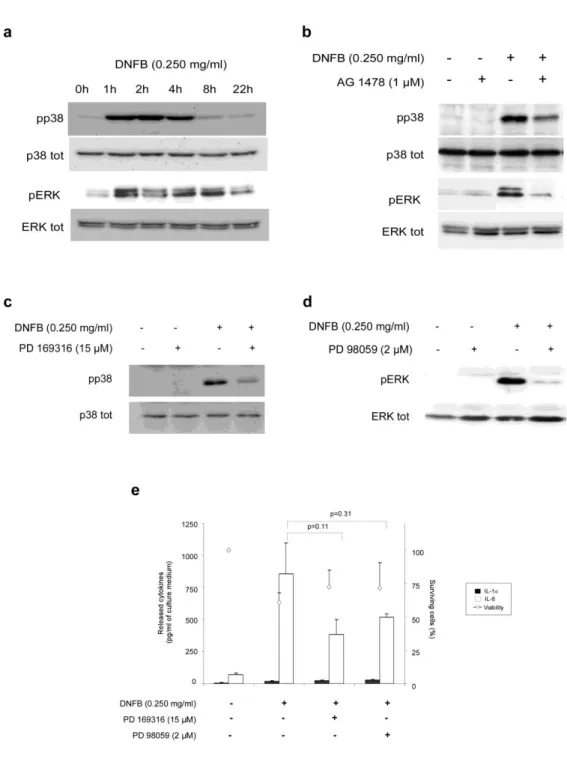
[33,25,15]. In consequence, we have investigated whether EGFR signalling could be part of the keratinocyte response following exposure of the RHE to sensitizing chemicals : RHE were exposed for 1 to 22h to 0.250 mg/ml DNFB (Figure 2a), 5 mg/ml Oxa (Figure 2b), 2.5 mg/ml Cin (Figure 2c) or 3 mg/ml Iso (Figure 2d), then Western blot analysis of tissue extracts was performed in order to assess EGFR phosphorylation on tyrosine 1173, one of the main phosphorylated tyrosine residue implicated in downstream signalling. The detection of total form of EGFR was also performed as a loading control. All sensitizing chemicals were able to induce EGFR phosphorylation in keratinocytes, mainly between 1 to 4 hours of exposure of the RHE (Figure 2). Thus, EGFR signalling precedes the transiently induced IL-8 mRNA expression reported on Figure 1. Interestingly, on the other hand, such an EGFR phosphorylation was not detected after exposure of RHE to the typical irritant BC (Online Resource 2c).
The inhibition of EGFR tyrosine kinase activity reduces the phosphorylation of the EGFR and the release of IL-8 from RHE exposed to sensitizing chemicals
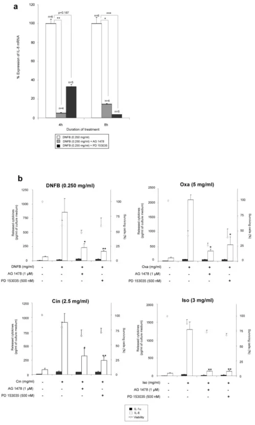
Because of the potential relationship between EGFR phosphorylation (Figure 2) and upregulation of IL-8 mRNA expression (Figure 1) in RHE exposed to sensitizers, we investigated whether EGFR tyrosine kinase activity could be involved in regulation of IL-8 expression and release. For this purpose, RHE were pre-treated or not with AG 1478 and PD 153035, specific inhibitors of the EGFR tyrosine kinase activity, before exposure to sensitizing chemicals. Firstly, using DNFB as a prototype sensitizing chemical, we demonstrated, using Western blot analysis, the effect of both inhibitors on the EGFR tyrosine 1173 phosphorylation (Figure 3a). Moreover, inhibition of tyrosine kinase activity using AG 1478 was also underlined by the decrease in the phosphorylated form of EGFR on tyrosine 1173 which is mainly detected in the basal layer of RHE (Figure 3b). The inhibition properties of AG 1478 and PD 153035 were also proved at the transcriptional level of IL-8 gene in RHE exposed to DNFB for 4 or 8 h (Figure 4a). Finally, pre-treatments of RHE with either AG 1478 or PD 153035 were found to significantly inhibit IL-8 release from RHE exposed to DNFB, Oxa, Cin or Iso (Figure 4b), strongly arguing for a control of IL-8 expression and release through EGFR signalling in these situations. Again, IL-1release from RHE exposed to sensitizing chemicals remained very low in all circumstances, as well as insensitive to EGFR signalling (Figure 4b).
Phosphorylation of p38 and ERK 1/2 MAPK, induced in response to EGFR activation, is partially involved in IL-8 release observed after topical application of DNFB
Phosphorylation of mitogen-activated protein kinase (MAPK) upon activation of EGFR has already been reported in dendritic cells and keratinocytes treated with sensitizing chemicals [53,40,23,1]. Since MAPK p38 plays major roles in cellular responses to environmental and physiological stresses [32,20,15,14] and MAPK ERK 1/2 promotes cellular proliferation needed for tissue reconstruction [24], phosphorylation of both MAPK’s wasinvestigated in our model. RHE were exposed for 1, 2, 4, 8 or 22h to DNFB (0.250 mg/ml) as a prototype sensitizing chemical, then tissue extracts were analyzed by Western blot in order to detect phosphorylated forms of p38 and ERK 1/2 MAPK’s (Figure 5a). Phosphorylation of both MAPK’s started after 1h of exposure, concomitantly with EGFR phosphorylation as reported on Figure 2. Phosphorylation of p38 ended after 4 hours of sensitizing topical application, while ERK 1/2 phosphorylation was maintained for 8 hours. Same MAPK activations were systematically observed after exposure to the other sensitizers (Oxa, Iso and Cin ; data not shown). Moreover, using one inhibitor of EGFR tyrosine kinase activity (AG 1478) we confirmed in our experimental conditions that phosphorylation of both p38 and ERK 1/2 MAPK’s is under the control of EGFR activation. However, AG 1478 only partially reduces the phosphorylation of p38 and ERK 1/2 observed after topical application of DNFB (Figure 5b). These results suggest that p38 and ERK 1/2 MAPK’s activation observed after phosphorylation of EGFR may be part of the keratinocyte responses that precede IL-8 mRNA upregulation. In order to control this hypothesis, we used specific inhibitors targeting p38 (PD 169316) and MEK 1/2, an upstream activator of ERK 1/2. Firstly, we show an inhibitory effect of PD 169316 and PD 98059 respectively on p38 and ERK 1/2 phosphorylation observed after DNFB (0.250 mg/ml) application (Figure 5c and 5d). The release of IL-8 from RHE exposed for 22h to DNFB was also measured after pre-treatment with both inhibitors (Figure 5e). The IL-8 release observed after DNFB application decreased partially after a tissue pre-treatment with PD 169316 and PD 98059. However, large standard deviations do not allow concluding in a
statistically significant effect of PD 169316 or PD 98059. However, low levels of statistical “p value” calculated using Student’s t-test suggest some partial involvement of both p38 and ERK 1/2 MAPK’s activation on IL-8 release after DNFB exposure. The IL-8 release measurement after topical application of Oxa, Cin and Iso on RHE pre-treated with PD 169316 confirms the partial involvement of p38 MAPK on IL-8 release after sensitizing application (Online Resource 3). However, the IL-8 release observed after Oxa and Iso exposure was affected to a lesser extent by PD 98059, suggesting that the signalling through ERK1/2 MAPK observed after sensitizing treatments is not systematically involved in IL-8 release.
The keratinocyte response to sensitizing chemicals depends on ligand-dependent and ligand-independent activation of EGFR
We have highlighted above that phosphorylation and activation of EGFR regulates expression and release of IL-8, after exposure of RHE to sensitizing chemicals. Epidermal keratinocytes can produce EGFR ligands, including transforming growth factor-alpha (TGF-), amphiregulin (AR) or HB-EGF. These ligands are able to bind and activate EGFR. EGFR can also be activated through ligand-independent mechanisms since treatment of keratinocytes with UV or hydrogen peroxide results in activation of EGFR [5,55]. Similarly, lipid rafts disruption in keratinocytes also induces EGFR phosphorylation in absence of ligand [27,25,20]. In order to investigate whether EGFR activation and IL-8 release induced after exposure of RHE to sensitizing chemicals were due to ligand-dependent or -independent mechanisms, we used the EGFR-specific neutralizing antibody LA1. LA1 is able to impede access to the ligand-binding pocket of EGFR. RHE pre-treated with LA1 were exposed to DNFB and Western blot analysis revealed that the phosphorylation of EGFR was partially reduced by this neutralizing antibody (Figure 6a), suggesting that phosphorylation of EGFR is partly due to ligand-binding. We also demonstrated that the IL-8 release after exposure to the sensitizing chemicals for 22h was also sensitive to LA1 pre-treatment (Figure 6b). One can observe that RHE pre-treated with LA1 exhibit reproducible although incomplete statistically significant decrease in IL-8 release induced by each chemical. These results suggest that ligand-dependent and independent activation of EGFR are involved in RHE exposed to sensitizers.
HB-EGF is not the ligand involved in EGFR activation responsible for IL-8 release
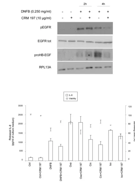
Previous studies have shown the large involvement of HB-EGF in inflammatory skin disorders (Piepkorn et al., 2003; Mathay et al., 2011). In addition, it was demonstrated in keratinocytes stressed by lipid raft disruption that HB-EGF must be considered as a key factor during epidermal stress conditions linked to EGFR activation [32,15]. Thus, expression of HB-EGF precursor form (ProHB-EGF) was investigated in RHE exposed for 1, 4, 8, 20 or 24h to DNFB, Oxa, Cin and Iso at the respective concentrations of 0.250 mg/ml, 5 mg/ml, 2.5 mg/ml and 3 mg/ml. Results in Figure 7 suggest that the rather late expression of proHB-EGF, observed between 4h and 20h after exposure to sensitizers, is probably not linked to the earlier phosphorylation of EGFR and the downstream IL-8 expression and release. In order to confirm that HB-EGF was unable to activate EGFR, RHE were incubated in the presence of CRM 197, a non-toxic mutant of diphtheria toxin, highly specific for HB-EGF, which impairs the binding between EGFR and HB-EGF. CRM 197 is able to bind proHB-EGF as well as the mature form of HB-EGF [39]. The effect of CRM 197 on EGFR phosphorylation after 2h and 4h of topical application of DNFB was investigated and indicated no modification of the phosphorylation of EGFR after 2h of treatment with DNFB. However, the activation of this receptor observed after 4h of treatment slightly decreases
in the presence of CRM 197 as a HB-EGF neutralizing agent. These results reveal that HB-EGF is not involved in the early phosphorylation of EGFR observed after the topical application of DNFB, but this growth factor seems implicated in the longer maintenance of EGFR activation signal (Figure 8a). Moreover, the IL-8 accumulation in the extracellular medium after 22h of exposure to chemicals (DNFB, Oxa, Cin and Iso) was not significantly modified by the use of CRM 197 (Figure 8b), suggesting that HB-EGF, despite his involvement in late EGFR phosphorylation, is not involved in IL-8 release.
The release of IL-8 is not responsible for HB-EGF expression and release.
As reported in Figure 3, impairment of EGFR signalling drastically reduces the proHB-EGF expression observed after 4h of exposure to DNFB (Figure 9a). Since we report here that inhibition of EGFR tyrosine kinase activity reduces the IL-8 release observed after exposure of RHE to DNFB, and since it was reported that IL-8 induction in epithelial cells contributes to activate some release of EGFR ligands into the extracellular environment, notably through activation of a specific G protein-coupled receptor (GPCR) like the 8 receptor [19,34], an IL-8 neutralizing antibody was used in order to investigate the involvement of IL-IL-8 in induction of HB-EGF expression. The neutralization of released IL-8 did not modify the phosphorylation of EGFR and the proHB-EGF expression (Figure 9b), suggesting that 8 is not able to control these phenomena. Using recombinant IL-8 protein, we further confirmed that IL-IL-8 added in extracellular medium is not involved in activation of EGFR signalling and HB-EGF expression in RHE (data not shown).
DISCUSSION
The effects of sensitizing chemicals entering in contact with the skin target dendritic cells of the immune system, but critical interactions with keratinocytes are believed to be implicated in the initial epidermal response that results in the onset of skin inflammation. Therefore, RHE exposed to chemicals constitute relevant 3D epidermal models for in vitro investigation of keratinocyte responses following an exposure to sensitizing chemicals. Such models provide more valuable information than 2D monolayer-based cultures of keratinocytes. Indeed, in RHE, non-differentiating, differentiating and fully differentiated keratinocytes stratify to create an epithelium where the keratinization process leads to the formation of a superficial cornified barrier. As it happens in vivo, chemicals can be topically applied onto the keratinized layer of the model in order to investigate early molecular and cellular events which illustrate the keratinocyte response to different classes of molecules as sensitizers [47]. IL-1 and IL-8 profiles, obtained after measurements of interleukins release into extracellular medium from keratinocytes of RHE exposed to chemicals, have been previously reported as characteristic of sensitizing compounds (Online Resource 1) [7]. Decoding the cellular responses given by keratinocytes towards the advert effects of chemicals and understanding the mechanisms of cell signalling underlying this particular regulation of interleukins expression and release should permit refinements of assays developed to characterize sensitizing properties of chemicals.
In this study, we focused on four sensitizers (DNFB, Oxa, Iso, Cin) and we identified rapid transient upregulations of IL-8 expression in RHE after 4h of exposure to these chemicals. A good candidate for cell signalling linked to early IL-8 expression and release on different stress condition was EGFR [49,43,41,30]. Accordingly, our results on RHE exposed to sensitizers clearly illustrate an important transient activation of EGFR in keratinocytes, in correlation with IL-8 mRNA expression and release by this cell type. The topical application of one irritant as BC on RHE induced rather late IL-1 and IL-8 expression and release without any phosphorylation of EGFR, suggesting that other cellular mechanisms are involved on RHE after topical application of certain irritating compounds such as BC. Contrarily to irritants, sensitizing chemicals are electrophilic compounds presenting highly reactive properties which could potentially justify the rapid cellular responses and signalling pathway activations, as notably EGFR phosphorylation, observed only after topical application of sensitizers on RHE [11,10]. Moreover, we also found that transient and sustained p38 phosphorylation observed on RHE treated with sensitizers is partially required for the extracellular release of IL-8. However, experimental results obtained on RHE pre-treated with PD 98059 advise that the signalling through ERK1/2 MAPK observed after sensitizing treatments (Figure 5) was not systematically involved on IL-8 release. These results suggest that complex cell signalling pathways downstream of EGFR activation lead to rapid IL-8 expression and release after application of sensitizing chemicals. The phosphorylation of EGFR also regulates the transient membrane expression of ProHB-EGF after topical application of sensitizers. However, despite the release of this ligand, HB-EGF is not responsible for this phosphorylation of EGFR. So, the expression of IL-8 and HB-EGF are under the control of EGFR phosphorylation and ligands involved in the rapid EGFR ligand-dependent phosphorylation are not yet identified. TGF-is largely expressed in skin and this growth factor is involved in skin inflammatory disorders such as ACD and psoriasis. Indeed, it is reported that TGF-, as a ligand of EGFR, regulates the expression and release of several chemokines, as IL-8, implicated in cell migration of components of the immune system in skin [37,30]. Possibly, a similar mechanism might regulate sensitizer-induced EGFR phosphorylation and IL-8 release. Indeed, the quantification of TGF- in the extracellular
medium of RHE treated with sensitizing chemicals as well as the neutralization of this EGFR-ligand using a
specific neutralising antibody against TGF-should be deserve in further study.
TGF-, firstly expressed as a transmembrane precursor, is enzymatically-cleaved as a soluble growth factor in the extracellular medium [28]. TNF- converting enzyme (TACE) is a member of a disintegrin and metalloprotease family, a group of zinc-dependent transmembrane metalloproteinases [29]. TACE cleaves ectodomains of various transmembrane proteins including TNF- and EGFR ligands as TGF-. TACE-mediated ectodomain shedding appears to be a central component in many important physiological and pathological activities as this enzyme is expressed in most tissues and meanwhile its expression is enhanced during the inflammation process [4,18]. A specific TACE inhibition using TAPi-1 should be performed in order to evaluate
the involvement of TGF- on cellular responses induced on RHE after sensitizing exposure [52].
Sensitized mice, treated with a selective inhibitor of EGFR kinase activity before antigen challenging, exhibited enhanced contact hypersensitivity in response to DNFB induced by the down regulation of CXCL8/IL-8 [31,44]. In psoriasis, enhanced IL-8 expression can lead to epidermal hyperplasia. Thus IL-8 is sometimes considered as an autocrine growth factor involved in epidermal proliferation and repair [9,57,48]. Previous studies have also shown some involvement of HB-EGF in inflammatory skin disorders [45,33] where this growth factor is responsible for epidermal hyperplasia. Indeed, after topical application of sensitizing chemicals on RHE, our report illustrated expression and release of IL-8, followed by expression and release of proHB-EGF into the extracellular epidermal environment. Both IL-8 and HB-EGF, involved in cellular proliferation and skin repair, are likely involved in the maintenance of tissue homeostasis in the epidermis after stress such as sensitization. Ligand-independent activation of EGFR also appears to play a role in the initiation of cell responses after topical application of sensitizing chemicals. However, mechanistic explanation for the ligand-independent activation of EGFR in such conditions remains puzzling. Such activation could result from inhibition of particular tyrosine phosphatases by oxidative stress, after exposure to H2O2 or UV [5,55]. It has been already reported that reactive
oxygen species mediate sensitization effects in dendritic cell in vitro [35]. Experiments are currently undertaken to investigate the induction of such oxidative stress and the potential involvement of reactive oxygen species in EGFR phosphorylation in RHE after topical application of sensitizers on the tissue (Frankart and Poumay, unpublished results). Moreover, the most likely reactivity of electrophilic molecules such as sensitizers happens with amino acid side chains exhibiting nucleophilic properties like cysteine sulfhydryl group (-SH) for instance [11]. The catalytic domain of protein tyrosine phosphatases is rich in cysteine-residues and becomes rapidly impaired when interactions occur with electrophilic molecules such as sensitizers [50,5]. Thus, since both electrophilic and oxidative stresses inactivate protein tyrosine phosphatases, one may postulate that investigating the phosphatase activity could bring information about an involvement of the enzyme in the sensitizer-induced phosphorylation of EGFR. In other words, analysis of EGFR phosphorylation induced by tyrosine phosphatase inhibition after electrophilic stress on RHE could potentially be used as a particular cell biomarker for electrophilic stress induced by topical application of sensitizers.
Similarly to protein tyrosine phosphatises, the catalytic domain of TACE is sensitive to redox state due to the presence of a cysteine-zinc bond. Disruption of this bond, following the induction of intracellular oxidative stress, results in a conformational change and activation of the enzyme. In this way, the production of ROS could lead to TACE activation through cysteine-zinc bound disruption, thereby contributing to increased release of EGFR ligands and subsequent receptor activation [56].
In addition, it could be of interest to mention that EGFR is mainly localized within cholesterol-enriched membrane microdomains named lipid rafts [51]. The functional significance of lipid rafts in keratinocytes has been considerably studied [32,25,33,20] and our lab has shown EGFR activation and internalization in keratinocytes after lipid raft disruption, but also after treatment of keratinocyte monolayers with DNFB [26,25]. Moreover, significant transient expression and release of IL-8 and HB-EGF were also observed [33]. Since transcriptional profiling of lipid raft-disrupted keratinocytes revealed regulations found in atopic dermatitis (AD), altogether those results suggest that lipid raft organization and related cell signalling are likely perturbed in skin disease [33]. Those similarities with our present data obtained with keratinocytes embedded in RHE and exposed to sensitizing chemicals lead us to hypothesize that, during sensitization, interactions of keratinocytes with sensitizing molecules may also involve membrane microdomains. More direct analysis of keratinocyte membrane domains are now required in order to test this hypothesis [27,25,20].
To conclude, studies performed on complex models composed of keratinocytes embedded in RHE allow better identification of early keratinocyte response to harmful chemicals. Particularly, investigation of keratinocyte signalling that controls expression and release of cytokines appears of great interest in highlighting potential biomarkers for their use in discrimination of sensitizing versus non-sensitizing chemicals. Transient and sustained phosphorylation of EGFR, due to tyrosine kinase activation that also regulates IL-8 and HB-EGF expression and release, seems to specifically occur in RHE treated with sensitizers. However, more known sensitizing chemicals should ideally be applied on RHE in order to sustain our current data. How chemicals interact with keratinocytes, especially with their membranes, in order to activate EGFR signalling is still hypothetical, but represents an interesting topic to further explore physiological keratinocyte response to chemicals and to bring new ideas in order to develop more relevant and accurate alternative methods useful for cutaneous toxicology.
ACKNOWLEDGEMENTS
Technical help from D. Van Vlaender, F. Herphelin and V. De Glas is gratefully acknowledged.
CONFLICT OF INTEREST
The authors have no conflict of interest to declare.
REFERENCES
1. Aiba S, Manome H, Nakagawa S, Mollah ZU, Mizuashi M, Ohtani T, Yoshino Y, Tagami
H (2003) p38 Mitogen-activated protein kinase and extracellular signal-regulated
kinases play distinct roles in the activation of dendritic cells by two representative
haptens, NiCl2 and 2,4-dinitrochlorobenzene. J Invest Dermatol 120 (3):390-399.
doi:12065 [pii]
10.1046/j.1523-1747.2003.12065.x
2. Albanesi C, De Pita O, Girolomoni G (2007) Resident skin cells in psoriasis: a special look
at the pathogenetic functions of keratinocytes. Clin Dermatol 25 (6):581-588.
doi:S0738-081X(07)00157-5 [pii]
10.1016/j.clindermatol.2007.08.013
3. Albanesi C, Scarponi C, Giustizieri ML, Girolomoni G (2005) Keratinocytes in
inflammatory skin diseases. Curr Drug Targets Inflamm Allergy 4 (3):329-334
4. Blaydon DC, Biancheri P, Di WL, Plagnol V, Cabral RM, Brooke MA, van Heel DA,
Ruschendorf F, Toynbee M, Walne A, O'Toole EA, Martin JE, Lindley K, Vulliamy
T, Abrams DJ, MacDonald TT, Harper JI, Kelsell DP (2011) Inflammatory skin and
bowel disease linked to ADAM17 deletion. N Engl J Med 365 (16):1502-1508.
doi:10.1056/NEJMoa1100721
5. Chiarugi P, Cirri P (2003) Redox regulation of protein tyrosine phosphatases during
receptor tyrosine kinase signal transduction. Trends Biochem Sci 28 (9):509-514.
doi:S0968000403001749 [pii]
6. Cook PW, Piepkorn M, Clegg CH, Plowman GD, DeMay JM, Brown JR, Pittelkow MR
(1997) Transgenic expression of the human amphiregulin gene induces a psoriasis-like
phenotype. J Clin Invest 100 (9):2286-2294. doi:10.1172/JCI119766
7. Coquette A, Berna N, Vandenbosch A, Rosdy M, De Wever B, Poumay Y (2003) Analysis
of interleukin-1alpha (IL-1alpha) and interleukin-8 (IL-8) expression and release in in
vitro reconstructed human epidermis for the prediction of in vivo skin irritation and/or
sensitization. Toxicol In Vitro 17 (3):311-321. doi:S0887233303000195 [pii]
8. Coquette A, Berna N, Vandenbosch A, Rosdy M, Poumay Y (1999) Differential expression
and release of cytokines by an in vitro reconstructed human epidermis following
exposure to skin irritant and sensitizing chemicals. Toxicol In Vitro 13 (6):867-877.
doi:S0887-2333(99)00076-4 [pii]
9. De Luca A, Carotenuto A, Rachiglio A, Gallo M, Maiello MR, Aldinucci D, Pinto A,
Normanno N (2008) The role of the EGFR signaling in tumor microenvironment. J
Cell Physiol 214 (3):559-567. doi:10.1002/jcp.21260
10. Dinkova-Kostova AT, Holtzclaw WD, Cole RN, Itoh K, Wakabayashi N, Katoh Y,
Yamamoto M, Talalay P (2002) Direct evidence that sulfhydryl groups of Keap1 are
the sensors regulating induction of phase 2 enzymes that protect against carcinogens
and
oxidants.
Proc
Natl
Acad
Sci
U
S
A
99
(18):11908-11913.
doi:10.1073/pnas.172398899
172398899 [pii]
11. Divkovic M, Pease CK, Gerberick GF, Basketter DA (2005) Hapten-protein binding: from
theory to practical application in the in vitro prediction of skin sensitization. Contact
Dermatitis 53 (4):189-200. doi:COD683 [pii]
10.1111/j.0105-1873.2005.00683.x
12. Esche C, de Benedetto A, Beck LA (2004) Keratinocytes in atopic dermatitis:
inflammatory signals. Curr Allergy Asthma Rep 4 (4):276-284
13. Funding AT, Johansen C, Gaestel M, Bibby BM, Lilleholt LL, Kragballe K, Iversen L
(2009) Reduced oxazolone-induced skin inflammation in MAPKAP kinase 2 knockout
mice. J Invest Dermatol 129 (4):891-898. doi:jid2008322 [pii]
10.1038/jid.2008.322
14. Giltaire S, Herphelin F, Frankart A, Herin M, Stoppie P, Poumay Y (2009) The CYP26
inhibitor R115866 potentiates the effects of all-trans retinoic acid on cultured human
epidermal keratinocytes. Br J Dermatol 160 (3):505-513. doi:BJD8960 [pii]
10.1111/j.1365-2133.2008.08960.x
15. Giltaire S, Lambert S, Poumay Y (2011) HB-EGF synthesis and release induced by
cholesterol depletion of human epidermal keratinocytes is controlled by extracellular
ATP and involves both p38 and ERK1/2 signaling pathways. J Cell Physiol.
doi:10.1002/jcp.22496
16. Gober MD, Gaspari AA (2008) Allergic contact dermatitis. Curr Dir Autoimmun 10:1-26.
doi:131410 [pii]
10.1159/000131410
17. Grone A (2002) Keratinocytes and cytokines. Vet Immunol Immunopathol 88 (1-2):1-12.
doi:S0165242702001368 [pii]
18. Guinea-Viniegra J, Zenz R, Scheuch H, Hnisz D, Holcmann M, Bakiri L, Schonthaler HB,
Sibilia M, Wagner EF (2009) TNFalpha shedding and epidermal inflammation are
controlled by Jun proteins. Genes Dev 23 (22):2663-2674. doi:10.1101/gad.543109
19. Itoh Y, Joh T, Tanida S, Sasaki M, Kataoka H, Itoh K, Oshima T, Ogasawara N, Togawa
S, Wada T, Kubota H, Mori Y, Ohara H, Nomura T, Higashiyama S, Itoh M (2005)
IL-8 promotes cell proliferation and migration through metalloproteinase-cleavage
proHB-EGF in human colon carcinoma cells. Cytokine 29 (6):275-282.
doi:S1043-4666(05)00014-1 [pii]
10.1016/j.cyto.2004.11.005
20. Jans R, Atanasova G, Jadot M, Poumay Y (2004) Cholesterol depletion upregulates
involucrin expression in epidermal keratinocytes through activation of p38. J Invest
Dermatol 123 (3):564-573. doi:10.1111/j.0022-202X.2004.23221.x
JID23221 [pii]
21. Johnston A, Gudjonsson JE, Aphale A, Guzman AM, Stoll SW, Elder JT (2011) EGFR
and IL-1 Signaling Synergistically Promote Keratinocyte Antimicrobial Defenses in a
Differentiation-Dependent
Manner.
J
Invest
Dermatol
131
(2):329-337.
doi:jid2010313 [pii]
10.1038/jid.2010.313
22. Jost M, Kari C, Rodeck U (2000) The EGF receptor - an essential regulator of multiple
epidermal functions. Eur J Dermatol 10 (7):505-510
23. Koeper LM, Schulz A, Ahr HJ, Vohr HW (2007) In vitro differentiation of skin sensitizers
by
cell
signaling
pathways.
Toxicology
242
(1-3):144-152.
doi:S0300-483X(07)00653-1 [pii]
10.1016/j.tox.2007.09.019
24. Kyriakis JM, Avruch J (2001) Mammalian mitogen-activated protein kinase signal
transduction pathways activated by stress and inflammation. Physiol Rev 81
(2):807-869
25. Lambert S, Ameels H, Gniadecki R, Herin M, Poumay Y (2008) Internalization of EGF
receptor following lipid rafts disruption in keratinocytes is delayed and dependent on
p38 MAPK activation. J Cell Physiol 217 (3):834-845. doi:10.1002/jcp.21563
26. Lambert S, Frankart A, Poumay Y (2009) p38 MAPK-regulated EGFR internalization
takes place in keratinocyte monolayer during stress conditions. Arch Dermatol Res
302 (3):229-233. doi:10.1007/s00403-009-1020-0
27. Lambert S, Vind-Kezunovic D, Karvinen S, Gniadecki R (2006) Ligand-independent
activation of the EGFR by lipid raft disruption. J Invest Dermatol 126 (5):954-962.
doi:5700168 [pii]
10.1038/sj.jid.5700168
28. Lee DC, Sunnarborg SW, Hinkle CL, Myers TJ, Stevenson MY, Russell WE, Castner BJ,
Gerhart MJ, Paxton RJ, Black RA, Chang A, Jackson LF (2003) TACE/ADAM17
processing of EGFR ligands indicates a role as a physiological convertase. Ann N Y
Acad Sci 995:22-38
29. Li Y, Yan J, Xu W, Wang H, Xia Y (2011) Lentivirus-Mediated ADAM17 RNA
Interference Inhibited Interleukin-8 Expression via EGFR Signaling in Lung Epithelial
Cells. Inflammation. doi:10.1007/s10753-011-9386-5
30. Mascia F, Cataisson C, Lee TC, Threadgill D, Mariani V, Amerio P, Chandrasekhara C,
Souto Adeva G, Girolomoni G, Yuspa SH, Pastore S (2010) EGFR regulates the
expression of keratinocyte-derived granulocyte/macrophage colony-stimulating factor
in vitro and in vivo. J Invest Dermatol 130 (3):682-693. doi:jid2009336 [pii]
10.1038/jid.2009.336
31. Mascia F, Mariani V, Girolomoni G, Pastore S (2003) Blockade of the EGF receptor
induces a deranged chemokine expression in keratinocytes leading to enhanced skin
inflammation. Am J Pathol 163 (1):303-312
32. Mathay C, Giltaire S, Minner F, Bera E, Herin M, Poumay Y (2008) Heparin-binding
EGF-like growth factor is induced by disruption of lipid rafts and oxidative stress in
keratinocytes and participates in the epidermal response to cutaneous wounds. J Invest
Dermatol 128 (3):717-727. doi:5701069 [pii]
10.1038/sj.jid.5701069
33. Mathay C, Pierre M, Pittelkow MR, Depiereux E, Nikkels AF, Colige A, Poumay Y
(2011) Transcriptional profiling after lipid raft disruption in keratinocytes identifies
critical mediators of atopic dermatitis pathways. J Invest Dermatol 131 (1):46-58.
doi:jid2010272 [pii]
10.1038/jid.2010.272
34. McGovern T, Risse PA, Tsuchiya K, Hassan M, Frigola G, Martin JG (2010) LTD
induces HB-EGF-dependent CXCL8 release through EGFR activation in human
bronchial epithelial cells. Am J Physiol Lung Cell Mol Physiol 299 (6):L808-815.
doi:ajplung.00438.2009 [pii]
10.1152/ajplung.00438.2009
35. Migdal C, Foggia L, Tailhardat M, Courtellemont P, Haftek M, Serres M (2010)
Sensitization effect of thimerosal is mediated in vitro via reactive oxygen species and
calcium signaling. Toxicology 274 (1-3):1-9. doi:S0300-483X(10)00204-0 [pii]
10.1016/j.tox.2010.04.016
36. Migdal C, Tailhardat M, Courtellemont P, Haftek M, Serres M (2010) Responsiveness of
human monocyte-derived dendritic cells to thimerosal and mercury derivatives.
Toxicol Appl Pharmacol 246 (1-2):66-73. doi:S0041-008X(10)00134-1 [pii]
10.1016/j.taap.2010.04.007
37. Miller LS, Sorensen OE, Liu PT, Jalian HR, Eshtiaghpour D, Behmanesh BE, Chung W,
Starner TD, Kim J, Sieling PA, Ganz T, Modlin RL (2005) TGF-alpha regulates TLR
expression and function on epidermal keratinocytes. J Immunol 174 (10):6137-6143.
doi:174/10/6137 [pii]
38. Minner F, Poumay Y (2009) Candidate housekeeping genes require evaluation before
their selection for studies of human epidermal keratinocytes. J Invest Dermatol 129
(3):770-773. doi:jid2008247 [pii]
39. Mitamura T, Higashiyama S, Taniguchi N, Klagsbrun M, Mekada E (1995) Diphtheria
toxin binds to the epidermal growth factor (EGF)-like domain of human
heparin-binding EGF-like growth factor/diphtheria toxin receptor and inhibits specifically its
mitogenic activity. J Biol Chem 270 (3):1015-1019
40. Mitjans M, Galbiati V, Lucchi L, Viviani B, Marinovich M, Galli CL, Corsini E (2010)
Use of IL-8 release and p38 MAPK activation in THP-1 cells to identify allergens and
to assess their potency in vitro. Toxicol In Vitro 24 (6):1803-1809.
doi:S0887-2333(10)00143-8 [pii]
10.1016/j.tiv.2010.06.001
41. Pastore S, Mascia F, Girolomoni G (2006) The contribution of keratinocytes to the
pathogenesis of atopic dermatitis. Eur J Dermatol 16 (2):125-131
42. Pastore S, Mascia F, Mariani V, Girolomoni G (2006) Keratinocytes in skin inflammation.
Expert Rev Dermatol 1 (2):279-291
43. Pastore S, Mascia F, Mariani V, Girolomoni G (2008) The epidermal growth factor
receptor system in skin repair and inflammation. J Invest Dermatol 128 (6):1365-1374.
doi:5701184 [pii]
10.1038/sj.jid.5701184
44. Pastore S, Mascia F, Mariotti F, Dattilo C, Mariani V, Girolomoni G (2005) ERK1/2
regulates epidermal chemokine expression and skin inflammation. J Immunol 174
(8):5047-5056. doi:174/8/5047 [pii]
45. Piepkorn M, Predd H, Underwood R, Cook P (2003) Proliferation-differentiation
relationships in the expression of heparin-binding epidermal growth factor-related
factors and erbB receptors by normal and psoriatic human keratinocytes. Arch
Dermatol Res 295 (3):93-101. doi:10.1007/s00403-003-0391-x
46. Poumay Y, Coquette A (2007) Modelling the human epidermis in vitro: tools for basic
and applied research. Arch Dermatol Res 298 (8):361-369.
doi:10.1007/s00403-006-0709-6
47. Poumay Y, Dupont F, Marcoux S, Leclercq-Smekens M, Herin M, Coquette A (2004) A
simple reconstructed human epidermis: preparation of the culture model and
utilization
in
in
vitro
studies.
Arch
Dermatol
Res
296
(5):203-211.
doi:10.1007/s00403-004-0507-y
48. Reich K, Garbe C, Blaschke V, Maurer C, Middel P, Westphal G, Lippert U, Neumann C
(2001) Response of psoriasis to interleukin-10 is associated with suppression of
cutaneous type 1 inflammation, downregulation of the epidermal
interleukin-8/CXCR2 pathway and normalization of keratinocyte maturation. J Invest Dermatol
116 (2):319-329. doi:jid1248 [pii]
10.1046/j.1523-1747.2001.01248.x
49. Roupe KM, Nybo M, Sjobring U, Alberius P, Schmidtchen A, Sorensen OE (2010) Injury
is a major inducer of epidermal innate immune responses during wound healing. J
Invest Dermatol 130 (4):1167-1177. doi:jid2009284 [pii]
10.1038/jid.2009.284
50. Samet JM, Tal TL (2010) Toxicological disruption of signaling homeostasis: tyrosine
phosphatases
as
targets.
Annu
Rev
Pharmacol
Toxicol
50:215-235.
doi:10.1146/annurev.pharmtox.010909.105841
51. Simons K, Ikonen E (1997) Functional rafts in cell membranes. Nature 387
(6633):569-572. doi:10.1038/42408
52. Terao M, Murota H, Kitaba S, Katayama I (2010) Tumor necrosis factor-alpha processing
inhibitor-1 inhibits skin fibrosis in a bleomycin-induced murine model of scleroderma.
Exp Dermatol 19 (1):38-43. doi:10.1111/j.1600-0625.2009.00973.x
53. Trompezinski S, Migdal C, Tailhardat M, Le Varlet B, Courtellemont P, Haftek M, Serres
M (2008) Characterization of early events involved in human dendritic cell maturation
induced by sensitizers: cross talk between MAPK signalling pathways. Toxicol Appl
Pharmacol 230 (3):397-406. doi:S0041-008X(08)00143-9 [pii]
10.1016/j.taap.2008.03.012
54. White KJ, Maffei VJ, Newton-West M, Swerlick RA (2011) Irritant Activation of
Epithelial Cells Is Mediated via Protease-Dependent EGFR Activation. J Invest
Dermatol. doi:jid2010308 [pii]
10.1038/jid.2010.308
55. Xu Y, Voorhees JJ, Fisher GJ (2006) Epidermal growth factor receptor is a critical
mediator of ultraviolet B irradiation-induced signal transduction in immortalized
human keratinocyte HaCaT cells. Am J Pathol 169 (3):823-830. doi:169/3/823 [pii]
56. Zhang Z, Oliver P, Lancaster JR, Jr., Schwarzenberger PO, Joshi MS, Cork J, Kolls JK
(2001) Reactive oxygen species mediate tumor necrosis factor alpha-converting,
enzyme-dependent ectodomain shedding induced by phorbol myristate acetate.
FASEB J 15 (2):303-305. doi:10.1096/fj.00-0371fje
57. Zhu YM, Woll PJ (2005) Mitogenic effects of interleukin-8/CXCL8 on cancer cells.
Future Oncol 1 (5):699-704. doi:10.2217/14796694.1.5.699
FIGURE LEGENDS
Figure 1. Well-known sensitizing chemicals induce transient expression of IL-8. RHE were treated with
DNFB (a), Oxa (b), Cin (c) or Iso (d) for 1, 4, 8, 20 or 24 hours. Total RNA extracts prepared from three independent cultures were analysed by real-time PCR to determine relative IL-1 and IL-8 mRNA expression. Data show the mean +/- SEM of the relative mRNA expression.
Figure 2. Exposure of RHE to sensitizing chemicals induces early transient phosphorylation of EGF receptor. Proteins extracted from RHE exposed for 0, 1, 2, 4, 8 or 22 hours to DNFB (a), Oxa (b), Cin (c) or Iso
phosphorylated form of EGFR (tyrosine 1173) (pEGFR). Data are representative of at least three independent experiments.
Figure 3. The inhibition of EGFR tyrosine kinase activity reduce the phosphorylation of the EGFR from RHE exposed to sensitizing chemicals. RHE were pre-treated or not with 1µM AG 1478 (a) (b) or 500nM PD
153035 (a) specific inhibitors of EGFR tyrosine kinase activity. RHE were then topically treated for 2h with 0.250 mg/ml DNFB. (a) RHE were lysed and subjected to Western blotting analysis using antibodies against total (EGFR tot) and phosphorylated forms of EGFR (tyrosine 1173) (pEGFR). (b) Paraffin sections of RHE were labelled against phosphorylated forms of EGFR on tyrosine 1173. Section of negative control do not receive primary antibody.
Figure 4. The inhibition of EGFR tyrosine kinase activity reduce the expression and release of IL-8 from RHE exposed to sensitizing chemicals RHE were pre-treated or not with specific inhibitors of EGFR tyrosine
in the presence or absence of AG 1478 or PD 153035. Total RNA extracts were analysed by real-time PCR in order to determine relative IL-8 mRNA expression. For each time point of the experiment, IL-8 expression was set up at 100% in RHE exposed to DNFB only. Statistical analysis was performed by ANOVA 1 after testing the homogeneity of the variance. Post hoc comparisons were performed by Tukey test (* p<0.05, **p<0.01, ***p<0.001). Data are shown as means +/- SEM. (b) RHE were topically exposed to DNFB, Oxa, Cin or Iso for 22h. After this incubation, cell survival in RHE was measured by MTT assay, and determination of IL-1 and IL-8in culture medium performed by ELISA assay. Statistical analysis of released cytokines was performed using ANOVA 1. Post hoc comparisons were performed by Dunnet’s tests (* p<0.05, **p<0.01, ***p<0.001). Data show the means +/- SEM of values obtained in three independent experiments.
Figure 5. Phosphorylation of p38 and ERK 1/2 MAPK’s, linked to EGFR activation, are partially involved on IL-8 release observed after topical application of DNFB. (a) Proteins extracted from RHE exposed for 0,
then topically treated for 2h with 0.250 mg/ml DNFB. RHE were pre-treated or not with 15µM of PD 169316 (c) or with 2µM of PD 98059 (d), RHE were then topically treated for 2h with 0.250 mg/ml DNFB. (a) (b) (c) (d) RHE were subjected to SDS-PAGE and Western blot analysis using antibodies against total and phosphorylated forms of p38 and ERK 1/2 MAPK’s. Data are representative of at least three independent experiments. (e) RHE were pre-treated or not with 15µM of PD 169316 or with 2µM of PD 98059. RHE were then topically treated for 22h with 0.250 mg/ml DNFB. After this incubation, cell survival in RHE was measured by MTT assay, and determination of IL-1 and IL-8in culture medium performed by ELISA assay. Statistical analysis of released cytokines was performed using ANOVA 1. Statistical “p values” are calculated using Student’s t-tests as Post
Figure 6. The keratinocyte response to sensitizing chemicals depends on dependent and ligand-independent activation of EGFR. RHE were incubated for 2h with neutralizing LA1 antibody (10µg/ml). (a)
Western blotting using specific antibodies for total (EGFR tot) and phosphorylated form of EGFR (pEGFR) (Tyr 1173). (b) RHE were exposed for 22h to DNFB (0.250 mg/ml), Oxa (5 mg/ml), Cin (2.5 mg/ml) or Iso (3 mg/ml)
before cell survival assays. IL-1 and IL-8 were measured in collected culture medium. Data show the mean +/- SEM of values obtained in triplicates. Statistical analysis of released cytokines was performed using ANOVA 1.
Post hoc comparisons were performed by Student’s t-tests (* p<0.05, **p<0.01, ***p<0.001). Data show the
Figure 7. Expression of ProHB-EGF is induced by sensitizers. RHE were exposed for 0, 1, 4, 8, 20 or 24h to
prepared and analysed by Western blot analysis for proHB-EGF and RPL13A (60S ribosomal protein L13a), as a loading control.
Figure 8. HB-EGF is not the ligand involved on EGFR activation responsible for IL-8 release observed after topical application of sensitizers. (a) RHE were incubated for 2h with CRM 197 (10 µg/ml), then
exposed or not for 2h or 4h to DNFB (0.250 mg/ml). Proteins were then extracted and analyzed by Western blotting using specific antibodies for total (EGFR tot), as a loading control, or phosphorylated form of EGFR (pEGFR). ProHB-EGF and RPL13A (60S ribosomal protein L13a), as a loading control, was also investigated. (b) RHE were incubated for 2h with CRM 197 (10 µg/ml), then exposed for 22h to DNFB (0.250 mg/ml), Oxa (5 mg/ml), Cin (2.5 mg/ml) or Iso (3 mg/ml) before cell survival assays. IL-8 were measured in collected culture medium. Data show the mean +/- SEM of values obtained in triplicates.
Figure 9. The release of IL-8 is not responsible for HB-EGF expression and release. RHE were pre-treated
or not with (a) 1µM AG 1478 as a specific inhibitor of EGFR tyrosine kinase activity or with (b) 0.5 µg/ml of IL-8 neutralizing antibody. (a) (b) RHE were then topically treated for 2h with 0.250 mg/ml DNFB, lysed and
subjected to Western blotting analysis using antibodies against total (EGFR tot) and phosphorylated forms of EGFR (pEGFR). ProHB-EGF and RPL13A (60S ribosomal protein L13a), as a loading control, were also investigated.
ONLINE RESOURCE LEGENDS
Online Resource 1. Topical application of sensitizers on RHE induces similar profile of IL-1 and IL-8.
RHE were treated for 22h with increasing concentrations of DNFB (a), Oxa (b), Cin (c) or Iso (d). After the treatments, the culture medium under the tissues was collected and analysed using ELISA assays in order to quantify the amount of released IL-1 and IL-8. Tissues were analyzed by the MTT assay in order to evaluate the cell survival after the treatment. This was expressed as percentage of remaining viability compared with the viability measured in control conditions which was fixed at 100%. Statistical analysis of extracellular released cytokines was performed by ANOVA 1 after testing the homogeneity of the variance. Post hoc comparisons were performed by Dunnet’s test, comparing each value to the extracellular release of cytokine from control RHE (* p<0.05, **p<0.01, ***p<0.001). Data show the mean +/- SEM of at least three independent experiments (n=3), each experiment performed with at least duplicate samples. In order to obtain homoscedasticity, values for released cytokines (x) were replaced by their logarithmic values [log (x)].
Online Resource 2. Benzalkonium chloride (BC) induces different keratinocyte response in RHE exposed to this typical irritant. (a) RHE were exposed for 22h to increasing concentrations of BC. After incubation, the
of IL-1 and IL-8 released. Tissues were analyzed by the MTT assay in order to evaluate the cell survival after exposure. This was expressed as percentage of remaining viability compared with the viability measured in control conditions which was fixed at 100%. (b) RHE were exposed to BC for 1, 4, 8, 20 or 24 hours. Total RNA extracts were prepared from three independent cultures and then analysed by real-time PCR in order to determine relative IL-1and IL-8 mRNA expression. Data show means +/- SEM of relative mRNA expression. (c) RHE were exposed to 0.250 mg/ml BC for 0, 1, 2, 4, 8 or 22 hours, then analyzed by Western blotting using antibodies against total (EGFR tot) and phosphorylated form (pEGFR) of EGFR. These data are representative of three independent experiments.
Online Resource 3. Involvement of p38 and ERK 1/2 MAPK’s phosphorylation, linked to EGFR activation, on IL-8 release observed after topical application of Oxa, Cin and Iso. (a) (b) (c) RHE were
22h with 5 mg/ml Oxa (a), 2.5 mg/ml Cin (b) or 3 mg/ml Iso (C). After this incubation, cell survival in RHE was
measured by MTT assay, and determination of IL-1 and IL-8in culture medium performed by ELISA assay. Statistical analysis of released cytokines was performed using ANOVA 1. Statistical “p values” are calculated using Student’s t-tests as Post hoc comparisons. Data show the means +/- SEM of values obtained in at least three independent experiments.