HAL Id: tel-03008570
https://tel.archives-ouvertes.fr/tel-03008570
Submitted on 16 Nov 2020HAL is a multi-disciplinary open access
archive for the deposit and dissemination of sci-entific research documents, whether they are pub-lished or not. The documents may come from teaching and research institutions in France or abroad, or from public or private research centers.
L’archive ouverte pluridisciplinaire HAL, est destinée au dépôt et à la diffusion de documents scientifiques de niveau recherche, publiés ou non, émanant des établissements d’enseignement et de recherche français ou étrangers, des laboratoires publics ou privés.
Improving visual function through concomitant use of
perceptual learning and brain stimulation : the case of
macular degeneration
Giulio Contemori
To cite this version:
Giulio Contemori. Improving visual function through concomitant use of perceptual learning and brain stimulation : the case of macular degeneration. Neurons and Cognition [q-bio.NC]. Université Paul Sabatier - Toulouse III; Università degli studi (Padoue, Italie), 2020. English. �NNT : 2020TOU30054�. �tel-03008570�
Ph.D. THESIS
Head Office: Università degli Studi di Padova Department of General Psychology
Etablissement: Université Toulouse III - Paul Sabatier
Unité de recherche: CERCO Centre de Recherche Cerveau et Cognition
___________________________________________________________________
Ph.D. COURSE IN: PSYCHOLOGICAL SCIENCES SERIES XXXII°
ECOLE DOCTORALE: CLESCO - Comportement, Langage, Education, Socialisation, Cognition DOCTORAT: NEUROPSYCHOLOGIE - 1ere inscription en thèse: 1 octobre 2016
IMPROVING VISUAL FUNCTION THROUGH CONCOMITANT USE OF PERCEPTUAL LEARNING AND BRAIN STIMULATION: THE CASE OF MACULAR DEGENERATION
Thesis written with the financial contribution of Fondazione Cariparo and Bando Vinci (Cap II) of Università Italo-Francese
Coordinator: Prof. Giovanni Galfano Supervisor: Prof. Clara Casco Co-Supervisore: Prof. Yves Trotter
Ph.D. student: Giulio Contemori
Abstract
Macular degeneration (MD) is a common visual disorder in the aging population characterized by a loss of central vision, reduced visual acuity contrast sensitivity, and increased crowding. This impairment strongly affects the quality of life and personal autonomy. There is currently no cure for AMD, available treatment options are only able to slow down the disease, and even palliative treatments are rare. After the emergence of the central scotoma, patients with MD develop one or more eccentric fixation areas - preferred retinal loci (PRLs) - that are used for fixation, reading, tracking, and other visual tasks that require finer ocular abilities. The final goal of the project was to investigate and to improve the residual visual abilities in the PRL. Four studies were conducted in total. Study 1 was conducted in MD patients to investigate whether after the emergence of the scotoma, the PRL acquire enhanced abilities in the processing of the visual information through spontaneous or use-dependent adaptive plasticity. Study 2 aimed to assess the effects of a single administration of transcranial random noise electrical stimulation (tRNS), a subtype of non-invasive transcranial electrical
stimulation, on the spatial integration in the healthy visual cortex. Study 3 aimed to assess the between session effect of daily repeated tRNS coupled with perceptual training. The objective of study 4 was to translate the previous findings into a clinically applicable treatment approach by combining tRNS and perceptual training in adult patients with MD.
Contrary to previous results, we found neither a phenomenon of spontaneous nor use-dependent cortical plasticity undergoing in the PRL before the training. We also found that the tRNS was able to modulate the visuospatial integration in the early visual processing, promoting plastic changes in the stimulated network. Its effects were not limited to the short-term modulation but also produced a boosting of the learning in a crowding task. The final experiment showed that a combination of tRNS and perceptual training could result in greater improvements and larger transfer to untrained visual tasks in adults with MD than training alone. Overall, our results indicate that tRNS of the visual cortex has potential application as an additional therapy to improve vision in adults with bilateral central blindness.
Résumé
La dégénérescence maculaire (DM) est une pathologie visuelle fréquente dans la population vieillissante, qui se caractérise par une perte de la vision centrale, une
diminution de la sensibilité au contraste et de l'acuité visuelle. Cette déficience affecte fortement la qualité de vie et l'autonomie . Il n'existe actuellement aucun traitement curatif de la DMLA, les options thérapeutiques disponibles ne permettant que de ralentir l’évolution de la maladie, avec de rares traitements palliatifs. Après l'apparition du scotome central, les patients atteints de DM développent une ou plusieurs zones de fixations excentrées - les lieux rétiniens préférentiels (PRLs) - qui sont utilisées pour la fixation, la lecture, et d'autres tâches visuelles qui nécessitent des capacités oculaires plus fines. L'objectif principal du projet était d'étudier et d'améliorer les capacités visuelles résiduelles dans les PRL. En tout quatre études ont été menées L'étude 1 a été menée chez des patients atteints de DMLA afin de déterminer si, après l'apparition du scotome, le PRL acquiert des capacités accrues dans le traitement de l'information visuelle grâce à une plasticité adaptative spontanée ou dépendante de l'utilisation. L'étude 2 visait à évaluer les effets d'une seule stimulation électrique transcrânienne à bruit aléatoire (tRNS), une variante de stimulation électrique non-invasive, sur l'intégration spatiale dans le cortex visuel sain. L'étude 3 visait à évaluer chez des sujets sains l'effet d'une répétition quotidienne du tRNS associée à un entraînement perceptif entre les séances.
L'objectif de l'étude 4 était de traduire ces résultats en une approche clinique en combinant la tRNS et l'entraînement perceptif chez des patients adultes atteints de DMLA. Nous n'avons trouvé aucun phénomène de plasticité corticale spontanée ou dépendante de l'utilisation dans la PRL avant l'entraînement contrairement à ce qui avait été montré dans d’autres études. Nous avons cependant constaté que le tRNS était capable de moduler l'intégration visuospatiale dans le traitement visuel précoce, en favorisant les changements plastiques dans le réseau stimulé. Les effets de la tRNS ne se sont pas limités à la modulation à court terme, mais ont également produit un
renforcement de l'apprentissage dans une tâche d'encombrement spatial. L'expérience finale a montré chez les adultes atteints de DMLA qu'une combinaison de la tRNS et de l’apprentissage perceptif pouvait induire des améliorations plus importantes et un transfert accentué vers des tâches visuelles non entrainées que le seul apprentissage perceptif. Dans l'ensemble, nos résultats indiquent que la tRNS du cortex visuel peut être utilisée comme thérapie supplémentaire pour améliorer la vision chez les adultes atteints de cécité centrale bilatérale.
Riassunto
La degenerazione maculare (MD) è una patologia visiva che si verifica per lo più nell’età adulta ed è caratterizzata da una perdita della visione centrale, una riduzione della sensibilità al contrasto, una riduzione dell’acuità visiva, e un aumento dell’affollamento visivo. Questo deterioramento compromette in maniera significativa la qualità della vita e l’autonomia dell’individuo. Attualmente non esistono cure per l’MD ed anche le opzioni di trattamento palliativo sono scarse, tuttavia esistono alcuni trattamenti che permettono di rallentare il decorso della malattia. Con l’insorgenza di uno scotoma centrale, i pazienti con degenerazione maculare sviluppano una o più aree eccentriche di fissazione – loci retinici preferenziali (PRLs)– usati per compiti che richiedono fini abilità oculari e stabilità di fissazione quali ad esempio la lettura, la ricerca visiva, ed il riconoscimento di volti o oggetti. Gli obiettivi principali di questo progetto erano due: investigare le abilità visive residue nel PRL e verificare l’efficacia di un nuovo protocollo di riabilitazione visiva nei pazienti affetti da MD. Il progetto si è sviluppato attraverso quattro differenti studi. Il primo è stato condotto in pazienti con MD per analizzare se dopo lo sviluppo dello scotoma, il PRL acquisisca rafforzate abilità nel processamento dell’informazione visiva attraverso processi di plasticità corticale spontanea o uso-dipendente. Il secondo studio era atto a determinare gli effetti sull’integrazione spaziale nella corteccia visiva, di una tecnica di promozione della plasticità neurale basata sulla stimolazione elettrica transcranica non-invasiva a frequenza casuale (tRNS). Con il terzo studio, abbiamo
testato l’efficacia della tRNS nell’accelerare l’apprendimento percettivo ed il
trasferimento dell’apprendimento nella periferia del campo visivo. Infine, l’obiettivo del quarto ed ultimo studio è stato quello di testare la fattibilità e l’efficacia di un protocollo di riabilitazione visiva per i pazienti affetti da MD che prevedesse l’uso combinato della tRNS e del training percettivo. Contrariamente a studi precedenti, nessun fenomeno di plasticità corticale spontaneo o uso-dipendente è stato trovato nei PRL prima del training. Inoltre, la tRNS si è dimostrata capace di modulare l’integrazione visuo-spaziale nel processamento visivo precoce, promuovendo cambiamenti nella plasticità della popolazione neurale stimolate. Gli effetti della stimolazione non si sono limitati alla modulazione a breve termine, ma hanno prodotto anche un aumento dell’apprendimento in compiti di affollamento visivo. L’ultimo studio ha mostrato come la combinazione tra tRNS e training percettivo nei pazienti affetti da MD produca un incremento
dell’apprendimento e – parzialmente – del trasferimento verso compiti visivi non allenati. In generale, i nostri risultati indicano che la tRNS sulla corteccia visiva potrebbe avere possibili applicazioni come terapia aggiuntiva al training neuro-comportamentale nel trattamento della cecità centrale bilaterale. Questi risultati hanno inoltre potenziali ricadute anche per altre popolazioni cliniche che soffrono di bassa visione.
Acknowledgments
Firstly, I would like to express my gratitude to my supervisor, Professor Clara Casco, for her priceless continuous guidance.
I would also like to sincerely thank my co-supervisor, Professor Yves Trotter, for his immense help.
Sincere thanks to all members of the NeuroVis.US Laboratory, with particular gratitude to Dr. Luca Battaglini and the former member Dr. Marcello Maniglia, who have been great friends and colleagues.
Sincere thanks to Tania, Elena, Evelin, Max, and all the people at the DPG for sharing with me this valuable experience.
Sincere thanks to the CerCo members for sharing with me a healthy, pleasant, and stimulating working environment.
Sincere thanks to all my participants, the one with low vision, they are the real protagonists of this work.
Sincere thanks to my friends for forcing me to have a normal social life even in the darkest time.
Sincere thanks to my family, no expression of thanks can be sufficient for your love, care, patience, wisdom, and support you have given me during my whole life.
Lastly, but most importantly, I would like to thank my other half for the patience she always demonstrated during these three years.
Foreword
This project stems from the experience gained during my master's internship and the post-graduate Erasmus internship. Without these previous experiences, there would not have been the network of international collaborations that allowed the realization of the project. This thesis conveys my interest in translational research and goes through a multidisciplinary research field that ranges from optometry, and neuropsychology, to psychophysics. Many are the knowledge learned, and the people met along this path. I thank you all who will read this work of mine, science is a silly game if there is no dissemination.
Table of content Abstract ... iii Résumé ...v Riassunto ... vii Acknowledgments... ix Foreword ...x Table of content ... xi
List of Tables ... xiv
List of Figures ...xv
Glossary of Abbreviations ... xvii
Chapter I. Introduction ...1
Chapter II. Macular Degeneration: Clinical Presentation ...7
The PRL ...10
Plasticity in the Adult Visual System ...13
Is there a remapping process for the neurons inside the lesion projection zone (LPZ)? ...17
Structural Changes ...18
Functional Changes ...22
Psychophysical Evidence of Cortical Plasticity in MD ...30
Macular Degeneration: Final Consideration ...34
Chapter III. Perceptual Learning as a Tool to Improve Visual Functions ...36
Specificity vs. Generalization ...40
The Lateral Masking Paradigm ...44
Visual Crowding ...47
From Basic Experiments to Clinics ...49
Chapter IV. Non-invasive Brain Stimulation Techniques: The tRNS ...53
Safety considerations ...59
Chapter V. Contextual Influences on the Peripheral Retina of Patients with Macular Degeneration ...63
Introduction ...64
Materials and Methods ...68
Results ...77
Discussion ...84
Chapter VI. tRNS Modulates Excitatory and Inhibitory Lateral Interactions in Contrast Detection ...88
Introduction ...89
Materials and Methods ...94
Results ...100
Discussion ...103
Chapter VII. tRNS Boosts Perceptual Learning in Peripheral Vision ...115
Materials and Methods ...120
Results ...129
Discussion ...134
Chapter VIII. tRNS Boosts Perceptual Learning in MD Patients ...141
Introduction ...142
Materials and Methods ...150
Results ...162
Discussion ...172
Conclusion ...178
Chapter IX. Contextual Influences on the Peripheral Retina of Patients with Macular Degeneration: further considerations. ...179
Results ...182
Conclusion ...184
Chapter X. Overview and Future Directions ...185
Current perspective on Macular Degeneration Treatment ...189
Can tRNS be used in visual rehabilitation? ...192
Limitation of the thesis ...194
Future direction and general conclusion ...196
Supplementary material ...200
List of Tables
Table 1. Details of participants. ... 69 Table 2. Contrast range for the target at each target-to-flankers distance () ... 96 Table 3. Results one-tail t-tests based on the null hypothesis of 0 sensitivity change. .. 101 Table 4. Results table form statistical data analysis ... 130 Table 5. Details of the experimental sample. ... 152 Table 6. Summary of the results relative to the transfer tasks. ... 174
List of Figures
Figure 1. Illustrative example of stimuli placement. ... 71
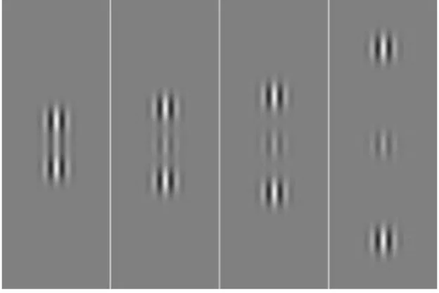
Figure 2. Stimuli used for the lateral masking paradigm. ... 74
Figure 3. Contrast threshold and TE values for PRL and controls ... 78
Figure 4. Contrast threshold and TE values for PRL and non-PRL ... 80
Figure 5. Visual acuity non-PRL vs. Visual acuity PRL. ... 81
Figure 6. Critical space PRL vs. Critical space non-PRL. ... 82
Figure 7. TEs as a function of flankers/orthogonal contrast ratio. ... 86
Figure 8. The stimulus configuration used in the experiments. ... 96
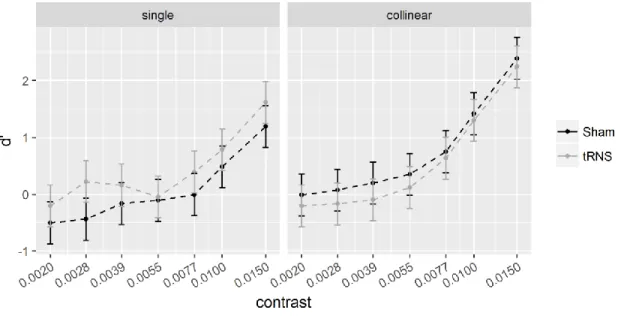
Figure 9. Sensitivity (d’) for the single (left) and collinear target at 6. ... 104
Figure 10. Sensitivity changes (SC) plotted as a function of target contrast with flankers at a distance of 6. ... 105
Figure 11. Sensitivity (d’) for the single (left) and collinear target at 2. ... 106
Figure 12. Sensitivity changes (SC) plotted as a function of target contrast with flankers at a distance of 2. ... 106
Figure 13. The figure shows the way the Criterion varies as a function of contrast in the two main experiments (Experiment 1, left panel; Experiment 2. right panel). ... 108
Figure 14. Configurations used in the crowding experiment. ... 124
Figure 15. Electrode positioning and modeled electrical field strength (normE). ... 126
Figure 16. Between days crowding thresholds. ... 130
Figure 18. The range of lateral interactions in the paracentral vision. ... 146
Figure 19. PRL position e scotoma size... 152
Figure 20. Training configuration with the stimuli and the control of fixation. ... 159
Figure 21. Contrast threshold in the trained task as a function of group, λ, and session. 163 Figure 22. Threshold elevation (TE) in the trained task as a function of group, λ, and session. ... 164
Figure 23. Improvement in contrast sensitivity. ... 166
Figure 24. Results from the Sloan visual acuity test. ... 168
Figure 25. Results from the tachistoscopic visual acuity test. ... 170
Figure 26. Results from the tachistoscopic crowding test. ... 171
Figure 27. Threshold Elevation (TE) as a function of log(flanker/target) contrast ratio. 182 Figure 28. Threshold Elevation (TE) as a function of log(flanker/target) contrast ratio and session. ... 183
Figure 29. Training and follow-up results for the Sham (n = 5) and tRNS (n = 5) subgroup. ... 200
Glossary of Abbreviations
2AFC – Two-alternative forced choice
2IFC – Two-interval forced choice
AMD – Age-Related Macular Degeneration
AR – Augmented reality
CPD – Cycles per Degree
CS – Contrast sensitivity
CSF – Contrast sensitivity function
CRF – Cell’s receptive field
dMRI – diffusion-weighted imaging
EEG – Electroencephalography
ETDRS – Early Treatment Diabetic Retinopathy Study logMAR chart
fMRI – Functional Magnetic Resonance Imaging
HF-tRNS – High-frequency transcranial Random Noise Stimulation
LF-tRNS – Low-frequency transcranial Random Noise Stimulation
LTP – Long Term Potentiation
LTD – Long Term Depression
MD – Macular Degeneration
MT – Medio-temporal area
NMDA – N-methyl –D- Aspartate
NIBS – Non-invasive Brain Stimulation
PET – Positron Emission Tomography
PL – Perceptual Learning
PRL – Preferred Retinal Locus
rTMS – Repetitive Transcranial Magnetic Stimulation
RF – Receptive Field
SNR – Signal to Noise Ratio
tACS – Transcranial Alternating Current Stimulation
tDCS – Transcranial Direct Current Stimulation
TES – Transcranial Electrical Stimulation
TMS – Transcranial Magnetic Stimulation
tRNS – Transcranial Random Noise Stimulation
TVC – Tachistoscopic Visual Crowding
V1 – Primary Visual Cortex
VA – Visual Acuity
VAS – Visual Acuity with Sloan letters
VR – Virtual Reality
Chapter I. Introduction
The continued increase in average age in developed western countries has transformed macular degeneration (MD) into the main cause of visual impairment in the modern age. This condition involves the loss of central vision, including loss of contrast sensitivity and visual acuity, mainly caused by a foveal scotoma. The current treatment options available are limited and mostly aim only to slow progression rather than restore vision. People with MD to cope with the loss of the sharp central vision, begin to retrain their visual habits so that they can fixate with a peripheral preferred retinal locus (PRL) instead of the impaired macula. PRL is usually found in a region near the scotomatous retina (Klaver, 1998; Klein, Klein, & Linton, 1992). So far, current efforts to monitor and treat the macular disease have focused on the retina, and no therapy has been able to restore patients' vision as it was before the onset of the disease. Pharmacological
treatments can slow down or even stabilize the formation of the visual defect; once it has manifested, treatment and rehabilitation must face its irreversibility. In the absence of a restorative treatment, the use of optical aids and compensative strategies might help to reduce the impact of the visual impairment (Calabrèse et al., 2018; N. X. Nguyen, Weismann, & Trauzettel-Klosinski, 2009; Rohrschneider, 2013; Romayananda, Wong, Elzeneiny, & Chan, 1982), on the other hand it is also possible to focus on enhancing the preserved visual functions (Maniglia, Cottereau, Soler, & Trotter, 2016; Pijnacker,
Verstraten, Van Damme, Vandermeulen, & Steenbergen, 2011). The main categories of vision-enhancing training are eccentric viewing training, eye movement training, and Perceptual Learning [PL] training (M. Li, Zhu, & Sun, 2015). PL is an improvement in a perceptual task resulting from the fine-tuning of sensory neurons through experience-dependent plasticity (Gilbert, Sigman, & Crist, 2001). PL has been proven effective in improving a wide series of visual dysfunctions, ranging from cortical blindness to mild refractive defects (Campana & Maniglia, 2015a; Das & Huxlin, 2010; M. Li et al., 2015). However, PL requires many sessions to be effective, and this represents a practical difficulty for patients that are not autonomous. In order to achieve a faster and larger improvement, recent studies combined PL with other techniques that have the ability to increase brain plasticity like brain stimulation (Camilleri, Pavan, Ghin, Battaglini, & Campana, 2014; Campana, Camilleri, Pavan, Veronese, & Giudice, 2014; Gall et al., 2015; Rufener, Ruhnau, Heinze, & Zaehle, 2017) and drug administration (Grieb, Jünemann, Rekas, & Rejdak, 2016; Kang, Huppé-Gourgues, Vaucher, & Kang, 2014; Rokem & Silver, 2013). Recently, non-invasive brain stimulation has been used, alone or coupled with PL, to enhance visual abilities (Camilleri, Pavan, & Campana, 2016;
Camilleri, Pavan, Ghin, Battaglini, et al., 2014; Campana et al., 2014; A. Fertonani, Pirulli, & Miniussi, 2011; Pirulli, Fertonani, & Miniussi, 2013; Thompson, Mansouri, Koski, & Hess, 2008). In particular, transcranial random noise electrical stimulation (tRNS), in which a weak current is delivered through the scalp on a cortical region at random frequencies, has shown promising results in boosting PL and reducing the
number of sessions needed to observe significant improvements (Camilleri, Pavan, Ghin, Battaglini, et al., 2014; A. Fertonani et al., 2011). Specifically, Camilleri et al. (2014) and
Campana et al. (2014) showed that when tRNS is repeatedly applied during a contrast-detection training it induced greater transfer (the post-training improvement observed in an untrained task) to visual acuity (VA) with respect to PL alone in both amblyopic and myopic patients (Camilleri, Pavan, Ghin, Battaglini, et al., 2014; Campana et al., 2014). In general, tRNS appears to boost both the early (within-session (A. Fertonani et al., 2011) and late (between sessions/days (Camilleri, Pavan, Ghin, Battaglini, et al., 2014) components of PL. The research presented in this thesis aimed to evaluate the therapeutic potential of tRNS in the treatment of human adults with bilateral central vision loss. The results presented in the thesis also contributed to increase our knowledge of the effects of tRNS on the visual cortex.
Conceptually this thesis has been divided into two parts. The first part is a review on the MD whose purpose is to critically review the evidence of adaptive neural plasticity in central vision loss. Based on the most recent literature, I will propose new
interpretations of old results in this field. The second part will focus on my experimental activity.
The first part of the literature review (CHAPTER II) introduces the MD and its clinical features. Particular relevance has been given to the structural and functional alterations that accompany the ocular disease at the level of the central nervous system. I have also discussed how some of the results that were previously interpreted as evidence of adaptive reorganization can be reinterpreted in an alternative more conservative way. I have concluded exposing the importance of developing an effective rehabilitative
protocol for MD patients. In CHAPTER III, PL is presented as a tool to improve visual functions. The concepts of learning, transfer, and specificity are discussed. CHAPTER IV
provides an overview of the action mechanisms proposed for the tRNS, the main results of previous studies, and some safety considerations. Finally, the possibility of extending the use of tRNS as a therapeutic intervention for other visual disorders is discussed. This thesis presents the data of four experiments, two of these were carried out on patients affected by bilateral maculopathy while the other two involved normal-viewing subjects. Each experiment was a necessary step to understand the interaction mechanism of tRNS and perceptual learning on the visual cortex of adults. Each subsequent study represented a further advancement from the basic scientific results towards the clinical application of the tRNS in MD. My hypotheses were:
• The spontaneous and use-dependent cortical adaptations in the region around the scotoma produces as perceptual consequences some alterations in the early spatial integration that would be measurable through psychophysical methods
(CHAPTER V).
• tRNS modulates early visual-spatial integration by altering the balance between excitatory and inhibitory lateral interactions. (CHAPTER VI)
• tRNS boosts between session perceptual learning and transfer (CHAPTER VII) • tRNS enhances and accelerate the outcomes of perceptual training in adults with
bilateral macular degeneration (CHAPTER VIII).
CHAPTER IX combines datasets from different studies to further test the hypotheses presented in CHAPTER V.
The first study is presented in CHAPTER V. This experiment aimed to evaluate the presence of spontaneous or use-dependent plasticity in the patients’ PRL. In the previous literature about plasticity in MD (D. D. Dilks, Baker, Peli, & Kanwisher, 2009;
Maniglia, Soler, Cottereau, & Trotter, 2018; Plank et al., 2017), there is debate whether the spared periphery undergoes a general process of adaptation triggered by the presence of the scotoma (“use-dependent reorganization” hypothesis) or whether the preferential use of the PRL for active high demanding tasks triggers a more specific use-dependent adaptation (D. D. Dilks et al., 2009; Maniglia et al., 2018; Plank et al., 2017). Knowing the type of adaptation process to which the PRL is subject is important to avoid damaging any compensatory mechanisms in place and instead to try to maximize them with the training. In this study, we tried to investigate this issue psychophysically by comparing spatial integration in the PRL, in a symmetrical retinal position (non-PRL) and a region with matched eccentricity in a control group. To do this, we probed the contextual influences by measuring the contrast gain for a vertical Gabor target, flanked by two high-contrast collinear masks compared to the orthogonal baseline condition. In line with previous literature (Maniglia et al., 2018), our prediction was to find evidence for
plasticity, in the form of reduced collinear inhibition. Moreover, we expected the reduction of inhibition to be stronger in the PRL.
CHAPTER VI includes the results of a study that aimed at investigating whether the contextual influences are modulated by tRNS applied to the occipital cortex of human observers during task performance. Given that the tRNS main effect is to increase cortical excitability, it could expand the sensitivity of the neurons to weak stimuli and thus
lowering the contrast threshold for the target. At the same time, this increased excitability could also modulate how the target is integrated with its context by altering the relative strength of excitatory (E) or inhibitory (I) influences from the flanking elements,
depending on which is weaker. In this case, we might expect an effect of tRNS even if tRNS shows no effect at all on target perceived contrast.
The study presented in CHAPTER VII investigated whether tRNS can effectively boost PL on a peripheral visual task over a small number of daily training sessions. Additionally, we tested whether learning transferred to untrained spatial location and task variation. We expected the tRNS to be able to increase both the learning rate and the transfer of learning.
The final study presented in CHAPTER VIII and CHAPTER IX brought together the findings of the experiments presented in this dissertation, investigating the combined effect of tRNS and perceptual learning in a visual rehabilitation protocol in MD patients.
This thesis explores a new path for visual improvement in MD and opens new possibilities for other clinical population.
Chapter II.
Macular Degeneration: Clinical Presentation
Macular degeneration [MD] can lead to severe visual impairment and blindness. Throughout the disease, the central retina responsible for our sharp vision undergoes a series of irreversible changes that endangers visual acuity.
The consequences of a bilateral central scotoma are severe visual impairments, especially in reading, face recognition, and visual search. Patients with central scotoma develop one or more eccentric fixation areas - preferred retinal loci (PRLs) - that are used for fixation, reading, tracking, and other visual tasks that require fine ocular abilities. This area, the preferred retinal locus (PRL), is a useful adaptation to central visual loss, but its function is weaker than that of the fovea because of the relatively poor visual resolution and the fixation instability.
There are different types of MD corresponding to different aetiology and progressions. Juvenile macular degeneration (JMD) is a quite rare type and includes several inherited eye diseases like Stargardt's disease, Best disease, and juvenile retinoschisis. In these cases, loss of central vision may begin in childhood or young adulthood. Unfortunately, there is no treatment available for these diseases, which are caused by gene mutations passed down in families. Early age-related macular
degeneration (AMD) consists of retinal or subretinal drusen, yellow or white accumulations of extracellular material, and retinal pigment abnormalities. It is not known whether drusen promote AMD or if they are common results of an underlying process. Late AMD is divided into neovascular AMD and geographic atrophy (GA).
that is a result of neovascularisation within the retina with leakage of fluid in the macula. There are different outcomes for the three type of AMD: patients with early AMD can retain some visual functionality, on the contrary, GA patients have no residual visual function in the affected area due to permanent loss of photoreceptors. In wet AMD, retinal scarring and deterioration can lead to blindness. No curative treatments are available until now for any of the diseases, but there are therapies able to slow down or freeze the progression. For wet AMD, treatment consists mainly of inhibitors of vascular endothelial growth factor (VEGF) repeatedly injected into the eye. AMD has been
identified as the third leading cause of blindness in the world, and the first considering only the developed part (Wong et al., 2014). Current efforts for tracking and treating macular disease have focused on the retina, for instance, quantification of drusen
distributions, photodynamic therapy (Wormald, Evans, Smeeth, & Henshaw, 2007), and even retinal prostheses for degenerations of the entire retina (Lohmann et al., 2019; Roux et al., 2016; Weiland & Humayun, 2013).
Moreover, the impact of degeneration is not limited within the scotoma, but it may also compromise the peripheral retinal region generally considered as "spared". Some studies have reported a higher frequency of peripheral lesions such as drusen, atrophy, and changes to the retinal pigment epithelium, in the retina outside the scotoma when compared to control eyes. (Domalpally et al., 2017; Johansen Forshaw, Minör, Subhi, & Sørensen, 2019; Laíns et al., 2018; Lengyel et al., 2015). This is in line with psychophysical studies reporting reduced visual acuity and contrast sensitivity in MDs in respect to age-matched controls at the same eccentricity (Fletcher & Schuchard, 2006; Maniglia, Pavan, et al., 2016). The indication that patients might show some impairments
even outside the scotoma has also to be considered when discussing results from experimental protocols that match normal viewers and MD patients.
So far, no therapies able to restore patients’ vision as it was before the onset of the disease are available. Drug treatments can slow down or even stabilize the formation of the visual defect, but once formed, treatment and rehabilitation must face against its irreversibility. In the case of visual impairment, in the absence of Restorative approaches, it is possible to adopt other types of rehabilitations based on Compensatory and
Substitutive techniques (Bouwmeester, Heutink, & Lucas, 2007; Lane, Smith, & Schenk, 2008). In particular, to ensure a better outcome in the daily life activities, the treatment of MD often require combined approaches that reckon on optical aids (N. X. Nguyen et al., 2009; Rohrschneider, 2013) and multiple kinds of training. Some possible training options are reading training (Seiple, Grant, & Szlyk, 2011), scotoma awareness training (Verghese & Janssen, 2015), oculomotor training (Rosengarth et al., 2013) and
Microperimetric biofeedback training (Vingolo, Cavarretta, Domanico, Parisi, &
Malagola, 2007; Vingolo, Salvatore, & Limoli, 2013). What I will focus on in this thesis is a training based on a perceptual learning paradigm whose purpose is to reshape the way in which visual information relating to multiple concomitant elements is integrated to form a single percept (Uri Polat, 2009). We will see this technique in more detail in CHAPTER III.
The PRL
It is still unclear how the PRL develops and if it acquires over time, particular processing ability with respect to the other quadrants of the functional periphery. Since it may be found in every direction around the scotoma, it is difficult to predict where the PRL might develop. Possibly would be the region with the best-spared sensitivity outside the scotoma (Timberlake, Peli, Essock, & Augliere, 1987) but this idea has been
challenged recently since in average the PRL doesn’t seem the absolute best retinal spot for visual acuity (Bernard & Chung, 2018; Shima, Markowitz, & Reyes, 2010) or contrast sensitivity and crowding (Contemori, Battaglini, & Casco, 2019). Some other factors known to play a role are the form and density of the central scotoma, the distance from the dysfunctional macula, and also the distance from the scotoma border (Crossland, Culham, Kabanarou, & Rubin, 2005; Erbezci & Ozturk, 2018; S N Markowitz &
Aleykina, 2010; Riss-Jayle, Giorgi, & Barthes, 2008a, 2008b). Sometimes, multiple PRLs may co-exist, and they could be interchanged depending on the viewing distance, the task, or the luminance (Crossland et al., 2005; Déruaz, Whatham, Mermoud, & Safran, 2002; González, Tarita-Nistor, Mandelcorn, Mandelcorn, & Steinbach, 2018; Lei & Schuchard, 1997). Probably it is worthy of thinking of the PRL as the best region in terms of functional efficiency instead of best sensitivity. Shima and colleagues (2010) have proposed that the oculomotor efficiency is likely the driving force for the functional adaptive changes in MD. They suggest that the spot with the best sensitivity and the one with the best fixational abilities are not necessarily the same but that they are often nearby each other and functionally linked (Samuel N. Markowitz & Daibert-Nido, 2019;
Shima et al., 2010). Moreover, Kabanarou et al. (2006) showed that there is a shift in fixation in one or both eyes when comparing monocular versus binocular viewing conditions. Since the two monocular PRLs often fall on noncorresponding areas (Kisilevsky et al., 2016) one or both might switch to another position, to facilitate conjugation in binocular tasks (Kabanarou et al., 2006; Verezen, Hoyng, Meulendijks, Keunen, & Klevering, 2011). In general, visual acuity, eye movement, and fixation stability are driven by the better eye (Kabanarou et al., 2006; Schneider et al., 2013; Tarita-Nistor, Brent, Steinbach, & González, 2011) but the overall binocular oculomotor efficiency is still disrupted in patients who have a low correspondence between the two PRLs. As we might expect, the lower the difference in quality of vision between the two eyes end the better the oculomotor efficiency. In fact, the best predictors for the
oculomotor efficiency are the contrast sensitivity ratio between the two eyes and the amount of stereoacuity – if measurable – not the absolute visual acuity (Shanidze, Heinen, & Verghese, 2017). The same concept holds also for the reading speed, in this case binocular integration and acuity gain have a stronger impact than absolute visual acuity. Indeed patients with interocular inhibition are reading significantly slower than those with no inhibition or summation (Tarita-Nistor, Brent, Markowitz, Steinbach, & González, 2013). It has been reported that some patients also show characteristics of binocular inhibition at low and medium spatial frequencies (Valberg & Fosse, 2002) but surprisingly in average binocular acuity gain is not different from that of age-matched control participants (Tarita-Nistor, González, Markowitz, & Steinbach, 2006). A better understanding of how the PRLs develop and how people with bilateral MD conjugate the two eyes is fundamental to design accurately any visual rehabilitation protocols that aims
at improving the residual visual ability in MD, but this requires ad hoc tools with
different characteristics from those generally used in clinical evaluation and rehabilitation (Tarita-Nistor et al., 2015). As we will see later in CHAPTER III, the rehabilitative protocol discussed in this thesis partially overcomes this limitation. In fact, even if carried out monocularly, PL obtained with the lateral masking paradigm is able to transfer to the untrained eye with consequent benefit in binocular vision (Dorais & Sagi, 1997; C. Yu, Klein, & Levi, 2004).
As we will see in CHAPTER VII and VIII, one of the thesis aims is to improve PL outcome by maximizing the transfer through the concomitant use of the tRNS.
Plasticity in the Adult Visual System
Macular degeneration is the most frequent, but not the only cause of central scotoma. Regardless of aetiology, the onset of the central blindness generally occurs in adulthood. (Ferris, 1983).
To date it has been established, that once the critical period for the development of the visual system is over, there is still a certain level of residual plasticity that allows for adaptative changes of the system even in advanced age (C Darian-Smith & Gilbert, 1995; Kaas et al., 1990; Knudsen, 2004). It is also known, however, that this ability is slowly declining with time and that it is subject to the limits given by the functional equilibrium of the entire system (Haak, Morland, & Engel, 2015; Knudsen, 2004; Morishita, Miwa, Heintz, & Hensch, 2010). Any change in the network must be
compatible with the subsequent, and previous stages of processing, otherwise perceptive distortions due to maladaptive plasticity may arise (Rosa, Silva, Ferreira, Murta, & Castelo-Branco, 2013). In some cases, visual hallucinations are a necessary step during visual recovery and tend to disappear when the plastic adaptive process matures, but if their presence persists, they could be functionally invalidating (Burke, 2002; C. S. H. Tan, Sabel, & Goh, 2006). When visual cortical neurons in adult mammals are deprived of their normal afferent input from retinae, they can acquire new receptive fields by modifying the effectiveness of existing intrinsic connections, a basis for topographic map reorganization. If the retinal lesions are relatively small (< 5 degrees), after long periods, the lesion projection zone (LPZ) shows partial recovery and exhibit normal receptive
and the contrast sensitivity is notably reduced (Y. M. Chino, 1995; Avinoam B. Safran & Landis, 1996; Sur, Nagakura, Chen, & Sugihara, 2013). Some other
experience-dependent mechanisms may overlap on top of this spontaneous cortical plasticity, for example by prolonged practice it is possible to trigger a perceptual learning mechanism (PL) that benefits from reinforcement-dependent plasticity (Manfred. Fahle & Poggio, 2002; Karmarkar & Dan, 2006). Recent studies have shown how it is possible to take advantage of this residual plasticity to obtain significant improvements during
rehabilitation protocols although with great limitations (M. Li et al., 2015; Maniglia, Cottereau, et al., 2016). Wandell & Smirnakis (2009) in their fundamental review state: “...it can be no serious debate as to whether the brain is plastic or not: it is both. A better question is to investigate distinct systems and understand the conditions under which each system is plastic or stable”. More recently Haak and colleagues by reviewing contrasting results about cortical remapping in MD suggest that differently from animal studies, evidence for extensive reorganization in the adult human primary visual cortex is just limited. According to their model, this could be due to the costs associated with making changes at the very root of the visual processing hierarchy (Haak et al., 2015). A retinal lesion during (or before) the sensitive period can modify the architecture of the visual system radically, but after the development is completed, the patterns of
connectivity become highly stable. At this point, the higher visual areas for their correct functioning rely on the retinotopic and functional organizations of the lower-level visual areas. Huge alterations of the low-level structures could have negative consequences in the readout of the upper areas. After the end of the sensitive period, the residual plasticity could be able to alter the connectivity patterns exclusively if the change respects the
architectural constraints established during development. In case of retinal damage, it would make more sense for the visual system to make slow structural adjustments at later stages of visual processing than a fast and extensive remapping at a lower-level, because the later stages have fewer dependencies that may be adversely affected (Merav Ahissar & Hochstein, 2004; Haak et al., 2015; Hochstein & Ahissar, 2002; Lillard & Erisir, 2011). It is important to note that this constraint would be less strict in the case of a simpler brain with less processing nodes such as mice or cats that are the main non-primate models for the study of retinal lesions.
According to the previous literature, a necessary factor for an extensive cortical reorganization might be the presence of a dense bilateral scotoma rather than a monocular scotoma or a bilateral scotoma with some spared islands of vision (Y. M. Chino, Kaas, Smith, Langston, & Cheng, 1992; Daniel D. Dilks, Julian, Peli, & Kanwisher, 2014; Schumacher et al., 2008). Also, the presence of a stable eccentric PRL seems to be relevant (Daniel D. Dilks et al., 2014; Plank et al., 2017; Schumacher et al., 2008). Undoubtedly if the central vision in one of the two eyes is preserved, there is no need for a re-referencing of the oculomotor activity, much less a cortical remapping, with the result that the mechanism of suppression of the best eye with respect to the defective one could be activated (Wiecek, Lashkari, Dakin, & Bex, 2015). This suppressive mechanism could be based on the consolidation of pre-existing inhibitory networks that are also transiently activated in normal viewers, for example during binocular rivalry (Holopigian, 1989; V. A. Nguyen, Freeman, & Wenderoth, 2001). Possibly it could easily be reversed with perceptual learning or dichoptic training, in the same way as of clinical suppression
(Barollo, Contemori, Battaglini, Pavan, & Casco, 2017a; R. F. Hess, Mansouri, & Thompson, 2010; Robert F. Hess, Mansouri, & Thompson, 2010).
Also, the re-activation at the level of the LPZ that is found in patients with bilateral partially overlapping scotoma could indicate a strengthening of a pre-existing feedback network already active in normal vision rather than an extensive cortical reorganization. This interpretation of the BOLD activity in the LPZ was originally suggested by Masuda, but to date it has not been confirmed (Masuda, Dumoulin, Nakadomari, & Wandell, 2008). We will discuss this possibility extensively in the ‘Functional changes’ section of this review.
The idea that the reorganization relies more on slow structural adjustments at later stages rather than extensive remapping at lower-levels undoubtedly re-launches the role of visual PL – and the active training in general – in the rehabilitation of visual deficits. Later, we will see how the major evidence of cortical reorganization in MD can be explained as the product of short-term non-specific spontaneous plasticity mechanisms. This form of plasticity contrasts with the one promoted by the perceptual training whose changes are slow but stable over time. (Maniglia, Cottereau, et al., 2016; Sabel, Henrich-Noack, Fedorov, & Gall, 2011; Trauzettel-Klosinski, 2011).
Is there a remapping process for the neurons inside the lesion projection zone (LPZ)? Retrograde (presynaptic) degeneration of retinal ganglion cells and retinal nerve fibers following damage to the occipital lobe has been documented extensively (Beatty, Sadun, Smith, Vonsattel, & Richardson, 1982; Cowey & Stoerig, 1989; Dinkin, 2017). On the other hand, evidence for anterograde (postsynaptic) degeneration are not as prevalent, but the phenomenon is nonetheless accredited (You, Gupta, Graham, & Klistorner, 2012) as well as the fact that retinal degeneration could lead to secondary brain damage to the visual pathways through a trans-synaptic degeneration mechanism (Ito, Shimazawa, & Hara, 2010).
On the other hand, we also know that the adult visual system has residual
plasticity that can counterbalance this phenomenon through the functional reorganization of neurons whose afferences have been suppressed (Gilbert & Li, 2012; Karmarkar & Dan, 2006). In this regard, the long-term visual deprivation might work as a trigger for the replacement (loss and formation) of spines in the deafferented cortex favouring the formation of new networks (Keck et al., 2008). One might wonder what the changes at a structural and functional level after a long period of adaptation to the loss of central vision are and whether these changes lead to compensation for the damage. Over the last two decades, a growing body of research has used magnetic resonance imaging (MRI) to answer these questions (Prins, Hanekamp, & Cornelissen, 2016). We will try to
Structural Changes
In MRI research that studies alterations following the loss of central vision, it is a common practice to compare patients with a corresponding age-matched control group.
The body of evidence that has gradually accumulated over time shows a reduction in the grey matter of the lateral geniculate nuclei, and of the visual cortex as well as in the white matter of the optic radiations (Boucard et al., 2009; Hernowo et al., 2014; Plank et al., 2011; Prins, Hanekamp, et al., 2016; Yoshimine et al., 2018). Grey matter loss in the visual cortex appears to overlap with the retinotopic region corresponding to the retinal lesion (Boucard et al., 2009) and to be correlated with scotoma size (Plank et al., 2011). Also, differences between JMD and AMD have been evidenced, with JMD showing more marked signs of grey matter loss (Prins, Plank, et al., 2016) and white matter loss in the optic nerves and the chiasm (Hernowo et al., 2014). If the long-term visual deprivation would be the mechanism behind those structural changes, we should see alterations in higher-level visual areas as a consequence of specific functional deprivation in the
absence of damage in the early visual cortex. This is precisely what has been found in the case of late monocular blind patients where a volumetric decrease in the superior lateral occipital cortices is present in the absence of a decrease in the early visual cortex (Prins, Jansonius, & Cornelissen, 2017). On the contrary, in MD, observed data are consistent with a transsynaptic degeneration propagating from the degenerated retinal axons to the central visual system (Malania, Konra, Jägle, Werner, & Greenlee, 2017; Ogawa et al., 2014; Yoshimine et al., 2018).
Some very recent studies based on tractography and diffusion-weighted imaging (dMRI) have provided strong support to the existence of transsynaptic anterograde
degeneration in case of damage to the peripheral visual nervous system (Balk et al., 2015, 2014; Malania et al., 2017; Ogawa et al., 2014; Tur et al., 2016; Yoshimine et al., 2018). In 2014 Ogawa and colleagues showed a reduction in fractional anisotropy – a measure of fiber integrity – in the optic tract and the optic radiation of patients with central vision loss. Other two studies have later corroborated their main finding. Malania et al., (2017) found a correlation between photoreceptors loss, atrophy of the ganglion cell axons, and diffusivity in the optic tract fibers. Moreover, they found that retinal nerve fiber layer thinning and the lower fractional anisotropy in the optic radiation was not limited to the foveal afferent fibers but also extended to the peripheral fibers (Malania et al., 2017). This is in agreement with the psychophysical and physiological finding showing that the “spared” periphery outside the scotoma maybe not so spared (Domalpally et al., 2017; Fletcher & Schuchard, 2006; Johansen Forshaw et al., 2019; Laíns et al., 2018; Lengyel et al., 2015; Maniglia, Pavan, et al., 2016). Despite this, it is clear that the main driver of the degeneration is the loss of central vision. Yoshimine and colleagues (2018) also found a reduction in fractional anisotropy along the optic radiation, but it affected fascicles primarily projecting to the central visual field in respect to the one projecting to mid or far-periphery. Contrary to the previous study, they did not find an alteration in the optic tract, but they found a positive correlation between the integrity of the optic radiation and the visual acuity (Yoshimine et al., 2018). Although the results of these studies have some minor inconsistencies, they provide convergent evidence that cell death within the retina causes significant alterations along the visual pathways to the cortex. This finding
challenges the understanding that macular degeneration is confined within the retinal tissue and questions the effectiveness of many restorative treatments (Lemos, Pereira, & Castelo-Branco, 2016a). The structural changes listed above are not, however, the only ones that have been found. Alongside with anterograde degeneration, another more general degenerative mechanism could be related to the etiopathology of AMD. For instance, Hernowo and colleagues (2014) have also found that AMDs but not JMDs show a reduction of white matter volume in frontal areas that are not strictly linked with visual processing. This could suggest a link with other forms of neural degeneration such as mild cognitive impairment and Alzheimer disease (AD). Indeed this hypothesis is corroborated by some studies that evidenced some common histopathologic features found in AD and AMD that might be led back to a common pathogenic mechanism (Lynn et al., 2017; Ohno-Matsui, 2011; Ong et al., 2019; Peiretti et al., 2014).
Nevertheless, AD and AMD don’t have a strong association with each other (Keenan, Goldacre, Goldacre, & Hyman, 2014; Michael A. Williams, Silvestri, Craig, Passmore, & Silvestri, 2014). In fact, the hazard ratio for developing probable or possible AD for people with established diagnoses of AMD (more than 5 years) is quite low,
approximately 1.50 (C. S. Lee et al., 2019). A ratio of 1.50 does not surprise if we consider that there might be some common risk factors linked to lifestyle habits (Klaver et al., 1999).
Not all the structural alterations found in AMD have a negative connotation. Some studies have also reported changes that can be traced to a positive adaptation to the presence of the scotoma. For example, Plank et al. (2011) found increased grey matter volume in a region slightly anterior to the frontal eye fields, and this increase was
correlated positively with fixation stability. They proposed that this could be a consequence of oculomotor learning. Other evidence for structural changes linked to functional adaptive plasticity comes from the studies of Sabbah et al., (2017) and Sanda et al., (2018). In this second study, they provided evidence of structural modification linked to enhanced peripheral visual field processing in associative visual areas that compensate for the central visual field loss. In fact, despite a thinning of the visual cortex, they found an increase in synaptic complexity in some regions of the lateral occipital cortex (hOc4la) and the fusiform gyrus (FG1) that are involved in shape, place, object, and face processing (Lorenz et al., 2017; Malikovic et al., 2016). This is in agreement with the previous study of Sabbah (2017) that found increased resting-state functional connectivity between the peripheral early visual cortex and the fusiform gyrus in patients with central visual field loss. If we consider the results presented so far, we can imagine two mechanisms that act oppositely: an anterograde degeneration that spreads from the retinal lesion to the visual cortex and a compensatory mechanism that enhances the connections between the preserved peripheral visual field and the visual associative areas. In this context, the use of PL as a way to bolster activity-dependent plasticity during visual rehabilitation could have the dual purpose of reducing the effects of anterograde degeneration and guiding the strengthening of connections between the cortical
projection of the PRL and the higher-level visual areas (Crair & Mason, 2016; Ganguly & Poo, 2013). PL might also work in combination with genetic or pharmacological strategies that reintroduce conditions for reorganization in the adult brain by promoting homeostatic rescaling and synaptic remodelling (Crair & Mason, 2016).
Functional Changes
The use of neuroimaging techniques in the study of central vision loss was not limited to structural investigations, and there are functional anomalies that have also been reported (for review on the topic see (Haak et al., 2015; Lemos et al., 2016a; Wandell & Smirnakis, 2009a). Given the retinotopic structure of the visual system, one of the most interesting questions is what happens to the deafferented part of the visual cortex. One hypothesis is that the neurons coding for the diseased part of the retina that is still functional could be recruited in the processing of stimuli outside the scotoma (Daniel D. Dilks et al., 2014; Masuda et al., 2008; Morland, Baseler, Hoffmann, Sharpe, & Wandell, 2001). By responding to stimuli originally outside the retinotopic area of their
competence, these neurons would, therefore, undergo a functional remapping. This type of extensive reorganization is documented in animal models (Corinna Darian-Smith & Gilbert, 1994; U T Eysel & Schweigart, 1999; Ulf Th Eysel, Gonzalez-Aguilar, & Mayer, 1980; Keck et al., 2008) as well as in other domains such as the somatosensory cortex (Flor, 2003; Gaetz et al., 2018; Rigato, Begum Ali, Van Velzen, & Bremner, 2014; Winship & Murphy, 2009). In the human vision, this has been found in early partial blindness (Baseler et al., 2002; L. Muckli, Naumer, & Singer, 2009) and recently also in adults suffering from degeneration of the peripheral retina (Ferreira et al., 2017). In case of central vision loss, some authors observed a stimulus-related activation in the lesion projection zone (LPZ) (Chris I. Baker, Dilks, Peli, & Kanwisher, 2008; Masuda et al., 2008; Schmid, Panagiotaropoulos, Augath, Logothetis, & Smirnakis, 2009). There are two competing explanations for the activation in the LPZ: it could indicate structural
reorganization of the visual cortex or unmasking of previously silent cortico-cortical connections (Masuda et al., 2008). Although the first hypothesis finds support in animals and juvenile lesions, there are valid reasons to think that this activation does not represent an index of cortical reorganization in adults with MD. First of all the reactivation of the LPZ seems to be present only under some conditions (Masuda et al., 2008, 2010) and only for some patients (Chris I. Baker et al., 2008) and it has not been reported in all the studies (T. Liu et al., 2010; Sunness, Liu, & Yantis, 2004). Moreover, such an extensive reorganization could cause dysfunction at higher visual areas that rely on that input. The input could be misinterpreted as producing distortion or illusions (D. D. Dilks, Serences, Rosenau, Yantis, & McCloskey, 2007). In the case of extensive central vision loss, a huge rearrangement could prevent the long-term stabilization of the initial short-term plasticity (Haak et al., 2015). Based on the recent literature, here we propose a more conservative version of the second hypothesis based on the concept of reinforcement of already existing (and active) cortico-cortical connection instead of the unmasking of silent connection. Only about 20% of V1 activity comes from excitatory projections from the lateral geniculate nucleus (LGN) (Carandini, 2005), while most activity in V1
receives major contributions from top-down, feedback and lateral input (Budd, 1998). In particular, if it is true that collicular input drives the earliest spiking activity that is linked with local processing it is also true that there is a prolonged later response that is more influenced by contextual feature of the stimuli (T. S. Lee, Mumford, Romero, & Lamme, 1998). Recent research shows that feedback from higher-level areas plays more than a modulatory function in V1 but can drive a task-related activity that can spread to non-stimulated regions (Lars Muckli & Petro, 2013). For example activation in V1 carries
information of task-dependent and position-independent representations of imagined (Thirion et al., 2006), remembered (Serences, 2016), or compared stimuli (Mark A. Williams et al., 2008). Baseler and colleagues (2011) in analysing the population receptive fields centre location in MD and controls with artificial scotoma, found the presence of ectopic receptive fields that were not restricted to the LPZ and were equally present in both the patient and control group. They also hypothesized that feedback or lateral connections could be the source of this ectopic signal and that it could be more active in the absence of feedforward signals as in the patient group. The fact that the visual processing in V1 might not be strictly retinotopic plays against the interpretation of this “re-activation” of the LPZ as a sign of extensive cortical reorganization. Moreover, reactivation in the LPZ has been proven to be absent during passive viewing but present during an active task like a discrimination task (Masuda et al., 2008, 2010). This type of task is indeed difficult for the patient given the central scotoma, the instability of fixation and the narrowness visual field stimulated in the scanner. However, why the reactivation of the LPZ can be measured only with difficult active task and not during the classic retinotopic mapping? A possible explanation comes from the paper of Williams et al. (2008), in fact they have found a foveal activation during a peripheral matching task in normal viewers. This activation was related to task difficulty and performance and was stronger after practice. A series of psychophysical (Fan, Wang, Shao, Kersten, & He, 2015; Weldon, Rich, Woolgar, & Williams, 2016; Q. Yu & Shim, 2016) studies have later demonstrated that delayed foveal feedback is critical for peripheral perception in fine details processing. Moreover, this feedback has a precise time course, altering the processing of the feedback utilizing a foveal mask or a TMS pulse at around 350ms that
produces a decrease in the peripheral task performance (Chambers, Allen, Maizey, & Williams, 2013). This feedback seems to be activated “on-demand” only for active peripheral task that requires processing of the details of the image. It has been suggested that since the foveal region has higher spatial resolution and small receptive fields, it might act as a sort of “enhancing software” that reconstruct and solve the details of the blurry peripheral image (Shim, Jiang, & Kanwisher, 2013; Mark A. Williams et al., 2008). Is seems very plausible that the AMD take advantage of this foveal feedback even more than normal viewers. We might also expect a strengthening of this feedback
connection, but this is far away from the concept of cortical remapping as found in the sensorimotor system or case of early visual impairments. However, where this feedback comes from? There is consistent literature about a “frontoparietal control system” active during a wide range of cognitive tasks including perceptual discrimination (Cole & Schneider, 2007; Duncan & Owen, 2000) that code for a range of different types of task-relevant information and that is sensitive to changing task demands (Duncan, 2010). Woolgar, Williams, & Rich (2015) showed that during a peripheral task, representation of visual stimuli in the frontoparietal cortex was stronger when stimuli were hard to perceive, and coding in early visual cortex was weak. On the contrary, coding in higher visual areas was sensitive to the allocation of attention but robust to changes in perceptual difficulty. This result is consistent with a feedback process that reinforces the visual percept at an early stage of visual processing. For the patients with central vision loss the possibility of reinforcing such feedback loop seems more probable than the possibility of an extensive rearrangement occurring at lower-order visual areas (Haak et al., 2015). Increased prefrontal and parietal activation in AMD patients compared to controls might
be a sign of increased top-down involvement in basic visual processing to compensate for the sensory loss (Szlyk & Little, 2009). However, some visual and non-visual high-level areas could be affected by the trans-synaptic anterograde degeneration which in turn could weaken the feedback to the lower-level areas. Ramanoël et al., (2018) testing AMD patients with low-pass, high-pass and nonfiltered scenes, found that in respect of controls patients had a much worse performance with high-pass scenes than low-pass. In patients, this was associated with reduced BOLD activity in the cortex corresponding to both the central and the peripheral visual fields. Moreover, they found reduced activation in the parahippocampal place area, a cerebral region specialized for scene perception.
Increasing the contrast of the scene produced a benefit in the processing of high-pass images but surprisingly only the activation in the occipital cortex was spurred by the increase in contrast, while the BOLD signal in the parahippocampal place area was not significatively increased (Ramanoël et al., 2018). This could mean that the prolonged decreased sensitivity for high spatial frequencies, and therefore for details, has produced a long-term impoverishment in the area specialized in perception which becomes less responsive.
In contrast, motion sensitivity seems to be improved in MD. It is relevant to mention the case of a monkey who developed juvenile MD. Interestingly the animal showed limited reorganization and no remapping in V1 and V2, but area V5/MT showed increased spread of activation compared to controls (Shao et al., 2013). This finding has been further supported by another animal study that found enhanced peripheral vision as a result of the sensitization for motion processing relying on feedback from V5/PMLS and area 7 to area 17 (Burnat, Hu, Kossut, Eysel, & Arckens, 2017). The idea of
improved motion sensitivity in MD is partially corroborated by a human behavioural study which shows increased effect of large moving visual fields display in the vection of MD patients than controls (Tarita-Nistor, González, Markowitz, Lillakas, & Steinbach, 2008). While in the central visual field the geniculate inputs to the striate cortex are predominantly parvocellular the Parvo to Magno ratio progressively decreases as a function of eccentricity (Azzopardi, Jones, & Cowey, 1999). Moreover, the receptive field size of magnocellular neuron is, on average, much larger compared to the
parvocellular one (Croner & Kaplan, 1995). If we consider these assumptions together, we might expect increased sensitivity to motion stimuli due to a compensatory
mechanism that in the absence of the foveal input rely mostly upon the spared peripheral abilities. Future studies should better address the modification in motion sensitivity in patients. Moreover, the study of a possible dissociation in the performance of MD patients when tested with stimuli that are selective for the Magnocellular pathway (low spatial frequency and fast motion) or the Parvocellular pathway (high spatial frequency and slow motion) could have potentials implication for rehabilitation.
Receptive Field Expansion
In animal research it is well established that neurons in the border of and inside the LPZ after a given period of recovery display an enlarged receptive that might also be shifted outside the scotoma (Y. M. Chino et al., 1992; Y. Chino et al., 2001; U T Eysel et al., 1999; Gilbert & Wiesel, 1992; Heinen & Skavenski, 1991; Kaas et al., 1990). In adult mammals, this reorganization occurs within hours of the lesion, but only if associated with the absence of input from the fellow eye (Y. M. Chino et al., 1992). The receptive field enlargement can be measured non-invasively by means of a functional MRI method that estimate the neuronal population receptive field size and location (pRF) (Dumoulin & Wandell, 2008; Harvey & Dumoulin, 2011). Results of this measurement in normal human subjects have been found to be in agreement with electrophysiological
measurements in the corresponding areas in monkeys (Dumoulin & Wandell, 2008). Shao et colleagues (2013) in a monkey affected by MD found an average increase in the pRF size of non-deafferented V1 voxels of about 20%. Similarly to the MD monkey, even MD patients show increased pRF sizes when compared to healthy controls, but unexpectedly the same effect was also present when healthy controls were tested with an artificial central scotoma (Baseler et al., 2011a). Recent studies with the artificial
scotoma suggest that the receptive field enlargement is part of a transient mechanism of homeostatic disinhibition that leads to increased cortical response in the cortical zone deprived of the visual stimulation (Haak, Cornelissen, & Morland, 2012; Papanikolaou, Keliris, Lee, Logothetis, & Smirnakis, 2015; Parks & Corballis, 2012). The fact that this expansion is very rapid and present also in case of artificial scotoma lets us think of a
reversible, spontaneous and automatic mechanism which has nothing to do with the retinal damage in the strict sense, but which instead comes into play whenever there is a significant decrease in the levels of visual activity in a given part of the visual field, and that is partly modulated by feedback signals from extrastriate visual areas (Haak et al., 2012).
This short-term plasticity comes at the expense of the neural response tuning as it causes a reduction in the inhibitory shaping of selectivity (Gannon, Long, & Parks, 2017). Even if it cannot be considered a reorganization process in itself, the increase in receptive field size together with the transient increase in cortical response might be the starter for a long-term topographical reorganization (Gannon et al., 2017).
At the same time, it could produce behavioural effects that in some cases might be detrimental. In AMD patients, some studies have shown a specific impairment for the elaboration of precise details transmitted from high spatial frequencies in the scenes, while the perception of global forms transmitted by low spatial frequencies remains relatively well preserved. (Musel et al., 2011; Peyrin, Ramanoël, Roux-Sibilon, Chokron, & Hera, 2017). This could be a consequence of the widening of the receptive fields that leads to a greater spatial summation, but also to a loss of neural tuning. Improving the sensitivity specifically to the medium and high spatial frequencies is one of the objectives of the neural-based perceptual learning that will be discussed in CHAPTER VIII of this thesis.
In the next section, I will discuss how, through the use of psychophysical methods it is possible to investigate the mechanisms of adaptation to the presence of the central scotoma.
Psychophysical Evidence of Cortical Plasticity in MD
We have previously seen what are the main visual deficits that emerge following the presence of bilateral central scotomas, we then saw what are the structural and functional alterations that ensue at the level of the central nervous system. Now we will focus on the psychophysical studies that have investigated the presence of cortical reorganization. Patients with retinal scotomas experience perceptual filling-in for scotomas as large as 6 degrees (Gerrits & Timmerman, 1969; Zur & Ullman, 2003). According to the classification in Weil & Rees (2011), this completion process falls under Stimulus-independent and instantaneous type. The exact mechanism through which this process takes place is still uncertain. There can be three possible mechanisms that can act independently or combined. First, there might be active lateral propagation of the visual information from the border to interior of the scotoma (Weerd, Gattass, Desimone, & Ungerleider, 1995); alternatively there might a be a remapping of receptive fields (Y. Chino et al., 2001). Lastly this completion might be driven by the activity of some location-independent large receptive fields like the one found in the lateral occipital and inferior temporal cortex that are activated even in case of partial visual stimulation as in the presence of an occluder (Hegdé, Fang, Murray, & Kersten, 2008; Weigelt, Singer, & Muckli, 2007). How much of this process can be attributed to a permanent and slow adaptive reorganization (long-range rewire) and how much is part of transient plastic mechanisms (receptive field enlargement and shift) is not given. Crossland and Bex (2009) in a Vernier task found an advantage when stimuli were presented across the physiological blind spot than over equally eccentric temporal retina, but the same