HAL Id: tel-01835003
https://tel.archives-ouvertes.fr/tel-01835003
Submitted on 11 Jul 2018HAL is a multi-disciplinary open access archive for the deposit and dissemination of sci-entific research documents, whether they are pub-lished or not. The documents may come from teaching and research institutions in France or abroad, or from public or private research centers.
L’archive ouverte pluridisciplinaire HAL, est destinée au dépôt et à la diffusion de documents scientifiques de niveau recherche, publiés ou non, émanant des établissements d’enseignement et de recherche français ou étrangers, des laboratoires publics ou privés.
Development of S-nitrosoglutathione loaded particles
adapted to oral administration for preventing
Inflammatory Bowel Disease relapses
Hui Ming
To cite this version:
Hui Ming. Development of S-nitrosoglutathione loaded particles adapted to oral administration for preventing Inflammatory Bowel Disease relapses. Human health and pathology. Université de Lor-raine, 2017. English. �NNT : 2017LORR0325�. �tel-01835003�
AVERTISSEMENT
Ce document est le fruit d'un long travail approuvé par le jury de
soutenance et mis à disposition de l'ensemble de la
communauté universitaire élargie.
Il est soumis à la propriété intellectuelle de l'auteur. Ceci
implique une obligation de citation et de référencement lors de
l’utilisation de ce document.
D'autre part, toute contrefaçon, plagiat, reproduction illicite
encourt une poursuite pénale.
Contact : [email protected]
LIENS
Code de la Propriété Intellectuelle. articles L 122. 4
Code de la Propriété Intellectuelle. articles L 335.2- L 335.10
http://www.cfcopies.com/V2/leg/leg_droi.php
Ecole Doctorale BioSE (Biologie-Santé-Environnement)
Thèse
Présentée et soutenue publiquement pour l’obtention du titre de
DOCTEUR DE l’UNIVERSITE DE LORRAINE
Mention : « Sciences de la Vie et de la Santé » par
Hui MING
Développement de formulations polymériques de
S-nitrosoglutathion comme traitement per os pour
prévenir les maladies inflammatoires chroniques
de l’intestin
07 Décembre 2017
Membres du jury :
Rapporteurs : Gilles Ponchel
Aurélie Malzert-Fréon Examinateurs : Anne Sapin-Minet
Franck Hansmannel Membre invité : David Moulin
PR, UMR 8612, CNRS, Université Paris-Sud, Paris PR, EA 4258, CNRS, Université de Caen Normandie, Caen
HDR, EA 3452, Université de lorraine, Nancy directeur de thèse
MCU, UMR 954, INSERM, Université de lorraine, Nancy co-directeur de thèse
DR, UMR 7365, CNRS, Université de lorraine, Nancy
--- --- EA 3452 CITHEFOR : Cibles Thérapeutiques, Formulation et Expertise Préclinique du Médicament
ACKNOWLEDGEMENTS
I would like to express my gratitude to all those who helped me to survive from these 3 years of PhD study here.
I am sincerely grateful to Pr. Ponchel (Université Paris-Sud), Pr. Malzert-Fréon (Université de Caen Normandie) and Dr. Moulin (Université de lorraine): Pr. Ponchel and Pr. Malzert-Fréon for accepting to be my reviewers of this work; Pr. Ponchel and Dr. Moulin for their participation in my thesis committee and helpful suggestions. I gratefully acknowledge Pr. Hu (my supervisor during master study in Wuhan University) and Pr. Maincent (Université de lorraine) for giving me the opportunity to start my PhD study here. Pr. Hu provided the research platform for me during the two years of master study in the lab, which helped me improve the experimental skills and inspired my research enthusiasm. Pr. Maincent gave me some really useful suggestions of the formulation part. The conversation with him was always relaxed and lively. His smile comforted me a lot when I did the presentation during the formulation meeting. He looks always optimistic. His friendly encouragement made me stronger when I lost my confidence.
I feel grateful to Pr. Leroy (the director of EA3452 CITHEFOR) for his critical opinions. His strong sense of responsibility, serious and detailed attitude of work influenced me a lot.
My deepest gratitude goes to my supervisors, Anne and Franck, for their instructive and valuable advices on my thesis. Anne helped me a lot not only in the guidance of thesis but also in the manipulation of experiment part. She is always nice and patient in analyzing and solving experimental problems with me. Besides the life in the lab, Anne also helped me to make my casual life easier in France. I learned a lot from Franck’s profound knowledge in physiology part and analysis of data. He was very patient in explaining basic knowledge to me, such as western blot, statistic study, intestine physiology and pathology. Without their patient instruction, insightful criticism and expert guidance, the completion of this thesis would not have been possible.
I would like to express my gratitude to Caroline G. and Isabelle F. for their useful suggestion and helpful manipulation in Caco-2 experiment part. Caroline’s smart mind and efficient work capacity impressed me a lot.
Great thanks shall be transported to Philippe G. for his work on GSNO preparation, apparatus setting and validation. His easygoing character and warm introduction helped me get used to this lab more easily and quickly.
My grateful words should go to Marianne as well for her sharing of interesting and useful literatures. She is very efficient in literature searching, analyzing and summarizing, which inspired me a lot.
I also own a truthful gratitude to Romain whose thesis is crossed with mine under the same supervisor of Anne. We shared a lot of happy moments together. He helped me a lot also in computer technical part. He is very good at communication which I can learn a lot from him.
Heartful gratitude is sent to Pascale, Nathalie and Ghizlane for their friendly smile every time and help in administrative things. Even though the language barrier exists, we still make us understandable to each other.
Special gratitude should go to my dear friends Haiyan and Yi, who accompanied me, comforted me and encouraged me a lot.
I also own my sincere gratitude to all my colleges for their help at any time for any cases.
At last, my thanks would go to my beloved family for their loving consideration and great support in me for all these years.
Hui MING In Nancy
TABLE OF CONTENTS
SCIENTIFIC WORKS ... I LIST OF TABLES ... III LIST OF FIGURES ... IV LIST OF ABBRIEVIATIONS ... VII
Résumé francophone du manuscrit ... i
Chapter 1. ... 1
Introduction ... 1
1.1 Inflammation bowel disease ... 2
1.1.1 IBD epidemiology ... 2
1.1.2 IBD physiopathology ... 3
1.2 Intestine physiology ... 9
1.2.1 Intestinal barrier components ... 11
1.2.2 Epithelial passage routes ... 27
1.2.3 Intestinal permeability measurement ... 31
1.3 IBD therapy ... 35 1.3.1 Aminosalicylates ... 35 1.3.2 Corticosteroids ... 36 1.3.3 Immunosuppressive drugs ... 37 1.3.4 Anti-TNF agents ... 38 1.3.5 Innovative therapy ... 38 1.4 Nitric oxide ... 39
1.4.2 NO donors ... 43
1.4.3 S-Nitrosoglutathione: a potent S-nitrosothiol ... 44
1.5 S-nitrosoglutathione related delivery system ... 52
1.5.1 S-nitrosoglutathione conjugated delivery system ... 57
1.5.2 S-nitrosation of glutathione related delivery system ... 57
1.5.3 Direct S-nitrosoglutathione encapsulation ... 58
1.6 Polymer nanocomposites for drug oral delivery: development strategies and potentialities ... 60
Chapter 2. ... 84
Luminal GSNO effects on the intestinal barrier integrity ... 84
2.1 Introduction ... 85
2.2 Study of the impact of the GSNO on the intestinal barrier permeability ... 87
Chapter 3. ... 108
Formulations of GSNO nanocomposites adapted to oral delivery ... 108
3.1 Introduction ... 109
3.2 Alginate nanocomposite particles optimization ... 113
3.2.1 Pre-optimization ... 113
3.2.2 S-nitrosoglutathione loaded alginate nanocomposite particles development ………118
3.2.3 Supplementary study: GSNO alginate nanocomposite particles in vitro release in acidic pH ... 144
3.3 Alginate/Eudragit®E 100 nanocomposite particles development ... 148
3.3.1 Pre-experiment ... 148
3.3.3 Result and discussion: ... 152
3.3.4 Conclusion ... 154
3.4 Conclusion and perspectives: ... 155
Chapter 4. ... 157
General discussion and perspectives ... 157
4.1 Discussion ... 158
4.1.1 Impact of luminal GSNO on the integrity of the intestinal mucosa ... 158
4.1.2 Oral administration of GSNO ... 159
4.2 Conclusion and Perspectives... 162
Reference ... 170
Attachments ... 203
Attachment 1: Article-Polymer nanocomposites enhance S-nitrosoglutathione intestinal absorption and promote the formation of releasable nitric oxide stores in rat aorta .. 204
I
SCIENTIFIC WORKS
Publications:
Polymer nanocomposites enhance S-nitrosoglutathione intestinal absorption and promote the formation of releasable nitric oxide stores in rat aorta. Wen Wu, Caroline Perrin-Sarrado, Hui Ming, Isabelle Lartaud, Philippe Maincent, Xian-Ming Hu, Anne Sapin-Minet, Caroline Gaucher. Nanomedicine: Nanotechnology, Biology and
Medicine (2016), volume 12, issue 7, page 1795-1803.
Single step alginate nanocomposite formulation for GSNO intestine delivery: an in vitro and in cellulo characterization. Hui Ming, Franck Hansmannel, Wen Wu, Romain Schmitt, Caroline Gaucher, Philippe Maincent, Xian-Ming Hu, Pierre Leroy, Anne Sapin-Minet. Journal of microencapsulation (submitted)
Luminal S-nitrosoglutathione impacts intestinal barrier permeability by dose-dependent way in an ex vivo model of Ussing Chamber. Romain Schmitt, Hui Ming (same contribution), Franck Hansmannel, Caroline Gaucher, Pierre Leroy, Isabelle Lartaud, Anne Sapin-minet. Journal of Gastroenterology. (in progress)
Booker chapter: Polymer nanocomposites for drug oral delivery: development strategies and potentialities. Hui Ming, Wen Wu, Philippe Maincent, Marianne Parent, Caroline Gaucher, Anne Sapin-Minet. Volume 9: Organic particles, Composites in Biomedical Engineering: Particles (Series C) – ELSEVIER, Edited by Alexandru Mihai GRUMEZESCU (in progress)
Oral communications:
NutriOx 2015, November 19th - 20th 2015 in Luxembourg: Formulations of Nitric Oxide
Donors Adapted to Oral Delivery: Perspectives for Preventing Inflammatory Bowel Disease Relapses. Hui Ming, Wen Wu, Franck Hansmannel, Caroline Gaucher, Philippe Maincent, Xian-Ming Hu, Pierre Leroy, Anne Sapin-Minet.
Journée scientifique de l'école doctorale BioSE, December 11th 2015 in Nancy (France):
Formulation of S-nitrosoglutathione(GSNO) Adapted to Oral Delivery: Perspectives for Inflammatory Bowel Disease Relapses Prevention. Hui Ming, Franck Hansmannel, Wen Wu, Caroline Gaucher, Philippe Maincent, Xian-Ming Hu, Jean-Louis Guéant, Laurent Peyrin-Biroulet, Pierre Leroy, Anne Sapin-Minet.
The 4th edition of the conference DocSciLor, June 15th, 2017 in Nancy (France):
S-nitrosoglutathione and intestine barrier integrity on an ex vivo rat model of LPS-induced inflammation. Romain Schmitt, Hui Ming, Franck Hansmannel, Caroline Gaucher, Isabelle Lartaud, Pierre Leroy, Anne Sapin-Minet.
II
Poster communications:
BioBarriers 2016, March 07th – 09th, 2016 in Saarbruecken (Germany): Formulations
of nitric oxide donors adapted to oral delivery: perspectives for intestinal bowel disease relapses prevention. Hui Ming, Wen Wu, Franck Hansmannel, Caroline Gaucher, Philippe Maincent, Xian-Ming Hu, Pierre Leroy, Anne Sapin-Minet.
DocLor – Doctorial de Lorraine 2016, April 17th – 22th, 2016 in La Bolle Saint-Dié
(France): Development of S-Nitrosoglutathione Formulation for the Prevention of Inflammatory Bowel Disease Relapses. Hui Ming.
OCC World Congress 2017 and Annual SFRR-E Conference, June 21th-23th, 2017 in
Berlin (German): Intestine permeability of S-nitrosoglutathione as a potential nitric oxide donor via oral administration. Justinea Bonetti, Haiyan Yu, Hui Ming, Anne Sapin-Minet, Isabelle Fries, Igor Clarot, Patrick Chaimbault, Pierre Leroy, Caroline Gaucher.
III
LIST OF TABLES
N◦ Captions Page
Résumé francophone
Tableau 1 Exemples d’actions protectrices des donneurs de NO
observée au niveau intestinal. iii
Tableau 2 Charge et libération du GSNO dans les particules
composites formulées par complexation de
polyélectrolytes et par gélification ionotropique
x
Table 1 The differences between the innate and adaptive immunity. 5
Table 2 GSNO related delivery system. 54
Table 3 Optimization of alginate nanocomposite. 117
Table 4 Experiment of interaction between TPP and Eudragit®E
100. 150
Table 5 Characterization of GSNO-aeNCP. 152
Article 1
Table 1 Simplified summary of drugs, nanoplateforms, polymer
matrix and their efficiencies. 68
Table 2 Simple summary of proceeding methods to prepare
polymer nanocomposites. 72
Table 3 Elimination of burst effect by polymer nanocomposites. 76
Article 2
Table 1 Evolution of the apparent permeability coefficient (Papp)
of sodium fluorescein (NaFlu, 1 mg/mL). 99
Article 3
IV
LIST OF FIGURES
N◦ Captions Page
Résumé francophone
Figure 1 Rôles protecteurs de l’oxyde nitrique dans les conditions
physiologiques : le maintien de l’intégrité de la barrière intestinale.
iv
Figure 2 Description d’un système particulaire composite délivrant un
RSNO. viii
Figure 3 Schéma expérimental des expériences qui pourront être
menées in vivo. xi
Figure 1 IBD and immune dysfunction. 6
Figure 2 Anatomy of intestine. 10
Figure 3 Elements comprising the intestinal mucosal barrier. 11
Figure 4 Spatial and longitudinal variations in microbial numbers and
composition across the length of the gastrointestinal tract. 12
Figure 5 The junctional complexes (a)The junctional complexes,
comprised of tight junctions, adherens junctions, gap junctions, and desmosomes (b) Electron micrograph of apical junctional complex between two intestinal epithelial cells of human ileal mucosa.
18
Figure 6 Molecular structures of TJ proteins and their interaction. 19
Figure 7 The multiple domains of ZO proteins. 22
Figure 8 AJ belt-like connection structure formed by cadherin-catenin
complex. 24
Figure 9 Molecular components and model of desmosomal protein
organization. 25
Figure 10 Key feature of intestinal immune system. 26
Figure 11 Epithelial pathway route: transcellular pathway and
paracellular pathway. 27
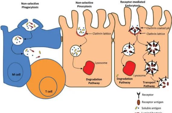
Figure 12 Transcellular pathway: selective phagocytosis,
non-selective pinocytosis and receptor-mediated endocytosis. 28
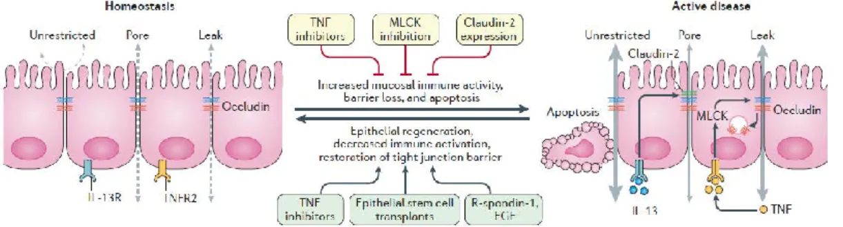
Figure 13 Three distinct paracellular epithelial permeability pathways in
homeostasis and active disease state. 30
Figure 14 Ussing Chamber system. 33
Figure 15 Transwell system: cell culture monolayer. 34
Figure 16 Endogenous synthesis of nitric oxide using L-arginine,
oxygen and cofactors as the substrates. 39
Figure 17 Concentration dependency of NO functions. . 40
Figure 18 Major beneficial actions of NO in intestine. 41
Figure 19 Chemical structure of S-nitrosoglutathione. 45
Figure 20 In vivo metabolism of GSNO by enzymes: 1. GSNO reductase
(GSNOR) and carbonyl reductase 1 (CR1); 2. thioredoxin system (Trx); 3. protein disulfide isomerase (PDI); 4, γ-glutamyltranspeptidase (GGT).
46
Figure 21 A model for cellular GSNO uptake 1: GSNO was metabolised
by GGT forming S-nitrosocysteinylglycine, which further deliver NO spontaneously and then diffused into cell, or
V
transfer NO to L-cysteine. 2: PDI catalyzes transnitrosation and denitrosation of GSNO to release NO. 3: GSNO transfers NO to L-cysteine forming CysNO, which is transported into the cell through the L-amino acid transporter system (L-AT).
Figure 22 Function of GSH as an antioxidant. 52
Figure 23 Strategies of polymer matrix. 112
Figure 24 Drug (GSNO and degradation product nitrite ions) release
from GSNO-NCP (free GSNO, ionotropic gelation, polyelectrolyte complexation) in acidic buffer (pH 1.2, 37 °C).
146
Figure 25 Advantages and drawbacks of two different alginate-NCP. 147
Figure 26 Possible mechanism of GSNO release in different
alginate-NCP in in vitro environment. 148
Figure 27 Perspectives of in cellulo study to explore the mechanism of
NO localization in intestine. 149
Figure 28 Mechanism of Interaction between TPP and chitosan. 149
Figure 29 Possible mechanism of Interaction between TPP and
Eudragit®E 100. 150
Figure 30 SEM images of GSNO-aeNCPs: a) surface morphology and
b) zoomed in nanoscale. 154
Figure 31 In vitro cytotoxicity of GSNO-aeNCP on 2 cells.
Caco-2 cells were treated with indicated polymer concentrations. 154
Figure 32 A crucial goal for the patients: Inflammation prevention. 164
Figure 33 Perspectives of NO action with intestine. 167
Figure 34 In vivo evaluation of GSNO loaded formulations in
inflammation model. 169
Article 1
Figure 1 Representative scheme of polymer nanocomposites: drug,
nanoplatforms and polymer matrix. They may present as A) core-shell structures, B) particles and C) bulk/film.
65
Article 2
Figure 1 Criteria of NaFlu permeability and TEER result during 2 h in
Ussing chamber. 97
Figure 2 Correlation between NaFlu permeability and TEER result
during 2 h in Ussing chamber. 97
Figure 3 NaFlu flux: Evolution of sodium fluorescein (NaFlu, 1
mg/mL) flux; TEER: Evolution of trans-epithelial electrical resistance
99
Figure 4 Expression of cell junction proteins by western blotting after
different concentration of GSNO exposure in Ussing Chamber for 2 h.
100
Figure 5 Possible mechanism of intestinal barrier integrity modulation
by NO. 103
Article 3
Figure 1 Scanning Electronic Microscopy images of GSNO-NCPs: A,
GSNO-NCP prepared by ionotropic gelation method; B, GSNO-NCP prepared by polyelectrolyte complexation.
129
Figure 2 Drug (GSNO and degradation product nitrite ions) release
from GSNO-NCPs. 130
VI
complexation) swell in PBS (0.148 M, pH 7.4, 37 °C).
Figure 4 In vitro cytotoxicity of GSNO-NCPs (A: ionotropic gelation;
B: polyelectrolyte complexation) on Caco-2 cells. Caco-2 cells were treated with indicated concentrations of GSNO-NCP for 24 h at 37 °C.
131
Figure 5 GSNO permeability evaluation of free GSNO and
GSNO-NCP (ionotropic gelation, polyelectrolyte complexation) in Caco-2 cells monolayers.
VII
LIST OF ABBRIEVIATIONS
Abbreviation Full name
6MP 6-mercaptopurine
aeNCP alginate/Eudragit®E 100 nanocomposite particles
AJ adherens junction
AMP antimicrobial proteins
aNCP alginate nanocomposite particles
Arm armadillo
ASA Aminosalicylates
AZA azathioprine
BP blood pressure
BUC(NO)2 S,S’-dinitrosobucillamine
CAR coxsackie virus and adenovirus receptor
CD Crohn’s disease
Cgmp cyclic guanosine monophosphate
CR1 carbonyl reductase 1
CysGlyNO S-nitrosocysteinylglycine
DCM dichloromethane
DCs dendritic cells
EDRF endothelial derived relaxing factor
EE encapsulation efficiency
EGCs enteric glia cells
EGFR epithelial growth factor receptor
ELISA enzyme-linked immunosorbent assay
eNOS endothelial nitric oxide synthases
FAE follicle-associated epithelium
GGT γ-glutamyltranspeptidase
GI gastrointestinal
GSH glutathione
GSNO S-nitrosoglutathion
GSNOR S-nitrosoglutathion reductase
GSSG oxidized glutathione
GTN nitroglycerin
GTP guanosine-5’-triphosphate
HPLC-MS high-performance liquid chromatography coupled with
mass spectrometry
HRP horseradish peroxidase
IBD inflammatory bowel diseases
IBS irritable bowel syndrome
IC50 half maximal inhibitory concentration
IECs intestinal epithelial cells
VIII
Ig immunoglobulin
IL interleukin
ILFs isolated lymphoid follicles
iNOS inducible nitric oxide synthases
ISDN isosorbide dinitrate
ISMN isosorbide mononitrate
JAM junctional adhesion molecule
L-AT L-amino acid transporter system
L-CysNO S-nitroso-L-cysteine
L-NAME L-NG-nitroarginine methyl ester
LP lamina propria
LPS lipopolysaccharide
M Cells microfolds cells
MAMPs microbial-associated molecular patterns
MDCK Madin-Darby canine kidney
MLC myosin light chain
MLCK myosin light chain kinase
mLNs mesenteric lymph nodes
MTX methotrexate
NACNO S-nitroso-N-acetylcysteine
NADPH nicotine adenine disphosphonucleotide
NCP nanocomposite particles
NF nuclear factor
NH2OH hydroxylamine
NK natural killer
NLRs nucleotide-binding oligomerization domain-like
receptors
nNOS neuronal nitric oxide synthases
NO nitric oxide
NOD nucleotide-binding oligomerization domain
NP nanoparticles
Papp apparent permeability coefficient
PDI protein disulfide isomerase
PETN pentaerythrityl nitrate
PKC protein kinase C
PKCs protein kinase Cs
P-MLC phosphorylation of myosin light chain
PPs Peyer’s patches
PRR pattern recognition receptor
RELMβ resistin-like molecule-β
RNOS reactive nitrogen oxide species
RSNO S-nitrosothiols
IX
Ser serine
sGC soluble guanylate cyclase
SNAP S-nitroso-N-acetylpenicillamine
SNOPCs S-nitrosophytochelatins
SNP Sodium nitroprusside
SPIONs superparamagnetic iron oxide nanoparticles
sprr2A small proline-rich protein 2A
TEER transepithelial electrical resistance
TFF3 trefoil factor 3
Thr threonine
TJ tight junction
TLRs toll-like receptors
TNF tumor necrosis factor
TPP sodium tripolyphosphate
TrxR thioredoxin reductase
UC ulcerative colitis
i
Résumé francophone du manuscrit
Les maladies inflammatoires chroniques de l’intestin (MICI) sont un groupe d’affections idiopathiques incluant la rectocolite hémorragique (RCH) et la maladie de Crohn (MC) qui affectent 200.000 personnes en France et 2,2 millions de personnes en Europe. La prévalence des MICI a augmenté principalement dans les pays occidentaux dans les 50 dernières années jusqu'à 200 / 100.000 pour les RCH et 200 / 100.000 pour la MC (Cosnes et al., 2011). Elles apparaissent chez des sujets jeunes (15‐35 ans) et sont incurables actuellement.
Elles représentent un véritable problème de santé publique compte tenu de leurs fréquences, de leurs pronostics à court et à long termes, du coût de leurs prises en charge ainsi que de leurs répercussions sur la qualité de vie des malades. Elles se caractérisent par des épisodes inflammatoires (poussées) répétés et invalidants pour le patient au cours desquels il ressent toutes les manifestations cliniques de la maladie. Actuellement la prise en charge des patients MICI se limite à la prise en charge de ces poussées inflammatoires. A plus ou moins long terme, les MICI conduisent fréquemment à des chirurgies visant à éliminer la zone enflammée (Cosnes et al., 2011). De plus, l’inflammation chronique de la muqueuse du colon (colite) du patient MICI peut conduire à la mise en place d’un cancer colorectal (CCR) (Jess et al., 2012).
Leurs étiologies sont inconnues, mais la pathogénie des MICI reposerait sur une activation inappropriée du système immunitaire intestinal, dirigé contre la flore intestinale de l’hôte chez des patients prédisposés (génétiquement ou épigénétiquement), dans un contexte de dysbiose. Cette dérégulation dans les interactions hôte-microbiote conduit à l’inflammation intestinale.
Des approches chirurgicales lourdes permettent de réaliser l’ablation de la zone lésée chez les malades lorsque le traitement médical n’est plus efficace, ou en cas de complications graves liées à la sévérité de la lésion. Ces interventions ne peuvent être envisagées qu’en seconde instance car elles sont d’une part très traumatisantes et d’autre part elles n’ont pas d’effet curatif réel puisque les récidives sont nombreuses après la chirurgie. Le développement de nouveaux axes de recherche et de nouveaux traitements chroniques permettant de prévenir les récidives inflammatoires améliorerait la qualité de vie des patients atteints de ces maladies et de diminuer le risque de
ii
développement de cancer. Les traitements conventionnels des MICI visent à limiter l’inflammation de la muqueuse des patients et incluent les aminosalicylates, les corticostéroïdes, les antibiotiques et les agents immunosuppresseurs, qui sont nécessairement administrés pendant de longues périodes et par conséquent, entraînent des effets secondaires (Meissner and Lamprecht, 2008). De plus, les dernières innovations thérapeutiques proposées comme l’utilisation d’agent anti-Tumor Necrosis Factor (anti-TNF) ont de puissants effets anti-inflammatoires mais une perte de réponse est observée lors d’un traitement au long cours et seulement un tiers des patients présente une rémission clinique totale après un an. L’identification de nouvelles cibles, de nouveaux agents, produits naturellement par l’organisme sain, pourrait ouvrir de nouvelles perspectives thérapeutiques pour ces pathologies.
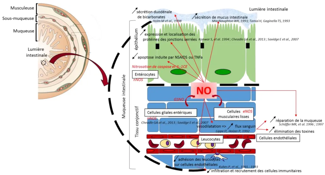
Lors d’une inflammation, l’oxyde nitrique (NO) joue un rôle pivot et paradoxal, discuté de façon récurrente dans de nombreuses études. Ainsi, le NO intervient à la fois dans des processus physiologiques se déroulant dans des tissus sains et dans des processus physiopathologiques. C’est particulièrement le cas au niveau du tractus gastro-intestinal, où le NO intervient à la fois comme médiateur important dans la préservation et la réparation de la muqueuse (Figure 1) mais contribue également directement aux dommages constatés dans les pathologies digestives, par une action pro-inflammatoire. Compte tenu de cette dualité, le développement de principes actifs pouvant moduler la synthèse ou l’action du NO constitue évidemment un véritable challenge et le rôle pivot du NO justifie les recherches de nouvelles stratégies thérapeutiques autour de ce composé. Le rôle bénéfique du NO dans ce contexte pathologique implique une action préventive en délivrant de faibles concentrations en NO (nM à µM) dans un environnement peu engagé dans l’inflammation (la balance redox doit être peu perturbée) afin qu’il exerce son action anti-inflammatoires.
L’apport de NO à l’état gazeux est particulièrement délicat pour remplir ces conditions, en raison d’une demi-vie très courte (de l’ordre de la seconde) et de sa très grande réactivité qui l’amène à interagir très rapidement avec les éléments de son environnement, de façon non contrôlable. Toutefois, différentes molécules libérant le NO ou des espèces apparentées, appelées « donneurs de NO », sont décrites voire même utilisées en thérapeutique (dans un contexte cardiovasculaire notamment) (Parent et al., 2013c). Certains donneurs de NO ont ainsi été étudiés dès les années 90, dans le contexte des MICI pour promouvoir les actions bénéfiques du NO au niveau intestinal
iii
(tableau 1).
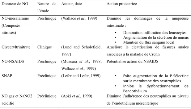
Tableau 1 Exemples d’actions protectrices des donneurs de NO observée au niveau intestinal
Parmi les donneurs de NO évoqués, les S-nitrosothiols (RSNO) représentent une classe de composés particulièrement intéressante pour un traitement de différentes pathologies inflammatoires chroniques (cérébrovasculaires, pulmonaires et intestinales) en jouant sur l’intégrité des barrières physiologiques.
Donneur de NO Nature de l’étude
Auteur, date Action protectrice
NO-mesalamine (Composés nitrosés)
Préclinique (Wallace et al., 1999) Diminue les dommages de la muqueuse intestinale :
• Diminution infiltration des leucocytes • Augmentation de la sécrétion de mucus • Maintien du flux sanguin local Glyceryltrinitrate Clinique (Lund and Scholefield,
1997)
Améliore la cicatrisation de fissures anales associées à la maladie de Crohn
NO-NSAIDS Préclinique (Muscará et al., 1998, Wallace et al., 1999)
Potentialise action du NSAIDS
SNAP Préclinique (Lefer and Lefer, 1999) • Evite augmentation de la P-Sélectine sur la membrane des neutrophiles • Inhibe le dysfonctionnement de
l’endothélium NO gaz et NaNO2
acidifié
Préclinique (Aoki et al., 1990) Diminue l’adhérence des neutrophiles au niveau de l’endothélium mésentérique
iv
Figure 1 Rôles protecteurs de l’oxyde nitrique dans les conditions physiologiques : le maintien de l’intégrité de la barrière intestinale (Wallace and Miller, 2000, Heanue and Pachnis, 2007)
v
En effet, quand le NO est combiné avec un donneur (R) au niveau d’un groupement thiol pour former un RSNO, la demi-vie du NO se trouve augmentée (plusieurs heures) et le NO est alors délivré aux cellules et aux tissus de façon plus contrôlée que sous la forme d’anion peroxynitrite. Ceci est d’autant plus intéressant dans un contexte inflammatoire lorsque le donneur (R) est une molécule antioxydante naturellement produite par l’organisme, par exemple le glutathion formant ainsi le S-nitrosoglutathion (GSNO). Il a d’ailleurs été démontré que le GSNO sécrété par les cellules gliales entériques à la suite d’une stimulation du nerf vague permet de prévenir un épisode inflammatoire au niveau intestinal et de préserver l’intégrité de la barrière (Savidge et
al., 2007, Cheadle et al., 2013). La mise en place d’un traitement chronique
préventif, basé sur l’administration orale de S-nitrosoglutathion (GSNO) représente une nouvelle stratégie pour limiter les récidives inflammatoires et les dommages au niveau de la barrière intestinale chez des patients atteints de MICI. Cependant, le développement d’un traitement chronique préventif des MICI, par voie orale, à base de GSNO représente un véritable challenge. Il s’agit de proposer une forme pharmaceutique stable adaptée à la voie orale, administrée à distance d’un épisode inflammatoire aigu, contrôlant la concentration de NO délivré au niveau intestinal pour assurer une action locale bénéfique.
Ce projet de thèse répond à une réelle attente des patients pour améliorer leur confort mais répond aussi à un réel besoin économique, proposant une solution pour diminuer le taux de récidives des MICI après un épisode aigu et pour réduire la période et le coût des traitements chroniques actuels. Le traitement préventif des MICI tel qu’il a été envisagé ci-dessus requiert donc la vérification de plusieurs points autour desquels s’articule cette thèse.
La première étape de cette thèse consiste à vérifier que le GSNO apporté dans la lumière intestinale (donc administré par voie orale) maintient l’intégrité de la barrière intestinale au même titre que le GSNO produit de manière endogène par les cellules gliales
vi
entériques présentes dans la muqueuse intestinale (Savidge et al., 2007, Cheadle et al., 2013). Pour ce faire, une méthode expérimentale ex vivo a été utilisée pour suivre la capacité du GSNO à préserver l’intégrité de la barrière intestinale. Nous avons utilisé la chambre de Ussing qui est un dispositif permettant l’étude de la diffusion, du transport de molécules à travers une membrane. Dans ce dispositif, du tissu intestinal peut-être positionné de façon à séparer deux compartiments : un compartiment donneur (mimant la lumière intestinale) et un compartiment accepteur (mimant le tissu conjonctif sous-jacent voire le compartiment sanguin). Cette étude a été réalisée sur des tissus intestinaux prélevés chez des rats Wistar sains. Pour ce faire, différentes concentrations en GSNO ont été déposées (de 0,1 µM à 100 µM) dans le compartiment donneur de la chambre de Ussing et nous avons évalué plusieurs critères :
- la perméabilité d’une molécule fluorescente « de référence » (la fluorescéine sodique) au cours du temps,
- la mesure de la résistance transépithéliale (TEER),
- le niveau d’expression des protéines de jonctions serrées et de cytokines pro-inflammatoires (par Western Blot).
Nos études, conduites pendant 2 h, ont montré pour la première fois, que la perméabilité de la fluorescéine sodique (marqueur du passage entre les cellules intestinales, au travers des jonctions cellulaires) est significativement diminuée, en présence d’une faible concentration en GSNO (0,1 µM), par rapport aux plus fortes concentrations testées (10 et 100 µM). En revanche, aucune modification
significative de la résistance transépithéliale (TEER) et de la quantité des protéines de jonction étudiées n’a été observée. Seule une diminution de 50 % de la quantité d’occludine (protéine constitutive des jonctions serrées) a été constatée en présence de 100 µM de GSNO. Ainsi, nos résultats indiquent qu’une faible concentration en GSNO (0,1 µM) retarderait la perte d’intégrité de la muqueuse intestinale dans ce système ex
vivo, alors que les fortes concentrations en GSNO (100 µM) accélèreraient cette perte
vii
Une étude plus précise de l’expression des protéines de jonction (par la technique de Western Blot sur extraits membranaires uniquement, et non sur lysats tissulaires totaux ou par ELISA) pourrait affiner ces résultats. Pour compléter ces travaux, l’internalisation des protéines des jonctions cellulaires pourrait également être étudiée par immunofluorescence en microscopie confocale. Le TEER et l’expression des protéines de jonction à la membrane sont finalement des marqueurs tardifs des effets précoces du GSNO. Dans un tissu sain présentant une balance RedOx contrôlée, la capacité de la cellule à lutter contre le stress pourrait masquer ces effets précoces du NO. A l’inverse, dans un tissu inflammatoire, cette capacité de la cellule est altérée et l’effet du NO pourrait être observé plus aisément car ils pourraient résulter d’une nitrosation modérée de protéines impliquées dans l’intégrité de la barrière. Ce résultat constitue la première « preuve de concept », la première étude de faisabilité quant à l’utilisation de GSNO comme traitement préventif des MICI.
L’administration orale requiert une mise en forme adaptée du GSNO et constitue une seconde partie majeure de ce manuscrit. Si le NO voit sa demi-vie augmentée sous la forme GSNO (de quelques minutes à plusieurs heures), ces donneurs de NO n’en demeurent pas moins fragiles et très réactifs vis-à-vis de leur environnement. Sensible à différents facteurs environnementaux comme par exemple la lumière, les variations de température ou la présence d’ions métalliques divalents, la liaison S-NO est maintenue dans le temps très difficilement (Gaucher et al., 2013). L’établissement d’un traitement chronique avec ce type de molécules nécessite une mise en forme galénique adaptée afin de protéger le NO jusqu’à son lieu d’action. De plus, cette forme galénique doit permettre un contrôle des doses de NO libéré. Les systèmes particulaires à libération contrôlée, tel que des nano- ou microparticules polymériques, peuvent tout à fait répondre à ces contraintes puisqu’elles sont basées sur l’utilisation de polymères biocompatibles, approuvés par les autorités de santé, et que les particules développées offrent une surface d’échange avec leur environnement particulièrement adaptée à la muqueuse intestinale.
viii
L’encapsulation directe de GSNO est un véritable challenge, liée notamment à la difficulté de préserver la liaison S-NO durant le procédé de formulation des systèmes particulaires. Cette stratégie offre la possibilité d’obtenir des charges en NO très intéressantes pour minimiser les quantités de particules à administrer, pour protéger le GSNO véhiculé et pour proposer des libérations et des actions plus prolongées dans le temps. Pour une application orale et un effet au contact de l’intestin, une libération sur 24 h serait plus adaptée. Ce travail de thèse fait suite à différents travaux entrepris
au sein de l’équipe CITHEFOR ayant démontré la faisabilité de cette encapsulation
directe du GSNO par différentes techniques de formulation (Parent et al., 2013c, Wu et
al., 2015a, Diab et al., 2016) par exemple démontré une encapsulation efficace du
GSNO (54% d’encapsulation de GSNO, soit 40 mmol de GSNO/L de suspension) dans
des nanoparticules d’Eudragit® RL (un polymère polycationique de type méthacrylique
utilisé couramment dans de très nombreuses formes orales sous forme de film de pelliculage et actuellement sur le marché), libérant un GSNO actif de façon plus prolongée dans le temps et limitant sa dégradation en ions nitrite et nitrate (Wu et al., 2015a). Afin d’augmenter la stabilité et de prolonger la libération du GSNO au cours du temps, l’incorporation des nanoparticules dans une matrice polymérique pour former les systèmes particulaires composites (Wu et al., 2015b) a été proposée (Figure 2).
ix
Ce type de système est recommandé dans la littérature pour réduire la libération initiale massive de principe actif (Hassan et al., 2009). Des systèmes particulaires pouvant mesurer jusqu’à plusieurs dizaines de micromètre ont été préparés par un procédé de gélification ionique, menant à des particules de chitosane ou d’alginate de calcium (Nasti et al., 2009, Koukaras et al., 2012). Ces systèmes particulaires composites ont été caractérisés en termes de taille, de taux d’encapsulation du GSNO, de cinétique de libération à pH 7,4 dans un milieu tamponné mais aussi de toxicité cellulaire par établissement de tests MTT sur une lignée d’adénocarcinome colique humaine (Caco-2).
Au cours de cette thèse, de nouveaux systèmes composites ont été formulés et caractérisés pour renforcer la protection du GSNO tout au long du tractus gastro-intestinal et contrôler davantage la libération de ce dernier : les nanoparticules
d’Eudragit® RL préalablement développées au sein de l’équipe ont été elles-mêmes
encapsulées dans une matrice polymérique à base d’alginate, polymère adapté à la voie orale et décrivant des propriétés favorisant la mucopénétration au niveau intestinal (Laffleur and Bernkop-Schnürch, 2013, Kumar et al., 2014).
Pour ce faire, deux procédés de formulation de la matrice d’alginate ont été développés et comparés (article soumis dans Journal of Microencapsulation). Un procédé simple
de complexation de polyélectrolytes (entre les nanoparticules d’Eudragit® RL chargées
positivement et l’alginate de sodium chargé négativement) a été comparé à un procédé plus classique de gélification ionotropique, utilisant le chlorure de calcium comme agent structurant des chaines d’alginate (« cross-linker »). Le procédé simple de complexation de polyélectrolytes a conduit à l’obtention de particules composites avec une charge en GSNO plus importante mais une libération in vitro plus rapide que celles formulées par gélification ionotropique (tableau 2).
x
Tableau 2 : Charge et libération du GSNO dans les particules composites formulées par complexation de polyélectrolytes et par gélification ionotropique.
Procédés de formulation Charge en GSNO (mg/g de polymère) Libération du GSNO à 3h (%) Complexation de polyélectrolytes 4,4 ± 0,4 27,9 ± 4,7
Gélification ionotropique 2,7 ± 0,2 97,8 ± 7,9
Cette différence de libération du GSNO observée in vitro, est minimisée au contact des
cellules (cellules Caco-2 cultivées en système de Transwell®) : la perméabilité du
GSNO et de ses produits de dégradation (ions nitrite et nitrate) est retardée de la même façon quelle que soit la formulation choisie. Le procédé de complexation de
polyelectrolytes, plus simple et plus facilement industrialisable que le procédé de gélification ionotropique, est adapté à la libération du GSNO. Ces deux procédés répondent donc au cahier des charges mentionné plus haut, en termes d’encapsulation, de libération et de délivrance locale au niveau intestinal. Il sera
néanmoins nécessaire de confirmer ces caractéristiques par des essais chez l’animal, sur un tissu intestinal.
Un autre type de formulation a été proposé en parallèle de cette étude. Il s’agissait de proposer une nouvelle association de polymères, non décrite dans la littérature et répondant au même cahier des charges, pour constituer la matrice des particules
composites par un procédé de gélification ionotropique (Wu et al., 2016). Le chitosan
a été remplacé par un autre polymère polycationique (l’Eudragit®E) pour constituer la
matrice en association avec l’alginate. Contrairement au chitosan qui est connu pour ouvrir les jonctions serrées, donc inadapté à notre problématique, (Thanou et al., 2001,
Wu et al., 2016), l’Eudragit® E pourrait limiter la libération du GSNO au niveau
intestinal. L’encapsulation de GSNO dans ces systèmes particulaires est efficace (2,5±0,6 mg de GSNO/g de polymère) et équivalente à celles des particules composites à base d’alginate seul ou associé avec le chitosan. La caractérisation physico-chimique de ces particules composites doit être complétée par des études in vitro et in cellulo de
xi
libération du GSNO.
En conclusion, ce travail s’attache donc au développement et à la caractérisation, limitée à la détermination des paramètres physico-chimiques des particules, à l’évaluation de leur cytotoxicité et à l’étude de la perméabilité du GSNO délivré sur un modèle de barrière intestinale en culture. A l’issue de ces travaux de thèse, trois formulations adaptées à la délivrance orale de GSNO sont proposées. La
démarche entreprise devra être poursuivie par :
- des études de stabilité des formulations proposées
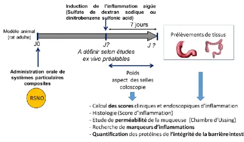
- des études in vivo (modèles animaux de MICI) évaluant la capacité des systèmes particulaires composites de GSNO les plus efficaces à prévenir le développement et à limiter l’apparition d’épisodes inflammatoires aigus. Il s’agira d’administrer oralement (par gavage) les formulations (sous la forme de suspensions) de manière préalable à l’induction de l’inflammation induite par des composés chimiques (sulfate de dextran sodique par exemple) (Tran CD et al., 2012) (Figure 3).
Figure 3 : Schéma expérimental des expériences qui pourront être menées in vivo.
Pour présenter ces travaux de thèse, le manuscrit s’articule de la manière suivante :
xii
et les traitements innovants sont présentés. Parmi ces derniers, le choix du GSNO et de l’oxyde nitrique est argumenté en décrivant les actions bénéfiques de ce dernier au niveau intestinal. La mise en forme du GSNO pour proposer cette molécule comme candidat à un traitement oral chronique est ensuite évoquée. L’intérêt des formulations appelées « nanoparticules composites » pour la délivrance de principe actif par voie orale est alors documentée au travers d’une revue bibliographique (Polymer
nanocomposites for drug oral delivery: development strategies and potentialities. Hui Ming, Wen Wu, Philippe Maincent, Marianne Parent, Caroline Gaucher, Anne Sapin-Minet. Volume 9: Organic particles, Composites in Biomedical Engineering: Particles (Series C) – ELSEVIER, Edited by Alexandru Mihai GRUMEZESCU (in progress)).
La seconde partie rapporte l’étude du maintien, du renforcement de la barrière
intestinale par le GSNO (non formulé) apporté dans la lumière intestinale : cette étude originale constitue la preuve de concept, l’étude de faisabilité de ce projet. Elle s’articule autour d’un article devant être prochainement soumis dans Journal of
gastroenterology.
La formulation de GSNO constitue la troisième partie du manuscrit. Un article soumis dans Journal of Microencapsulation décrit et compare deux systèmes particulaires composites. Un troisième système est ensuite décrit en données complémentaires.
A ce stade du travail, nous avons proposé, caractérisé et évalué plusieurs méthodologies de formulation dont les avantages et les limites sont discutées dans une dernière partie du manuscrit. L’étude de ces formulations peut maintenant être poursuivie in vivo par l’évaluation de leur capacité à prévenir les récidives inflammatoires au niveau intestinal.
1
Chapter 1.
Introduction
2
1.1 Inflammation bowel disease
Inflammatory bowel diseases (IBD) are chronic and relapsing disorders affecting gastrointestinal (GI) tract and associated with intestinal mucosa lesions and inflammation. The most known subtypes are Crohn’s disease (CD) and ulcerative colitis (UC). While both subtypes can lead to symptoms such as abdominal pain, cramping, diarrhea or weight loss, there are differences between them. It is important to note that CD can occur all over the digestion tract, from the mouth to the anus, but most often affects the large and small intestines, whereas UC primarily affects only the colon and rectum. As chronic disease, IBD are disabling, lead to significant health care costs and cause patients to suffer a lot in life quality and work capacity (Bannaga and Selinger, 2015).
1.1.1 IBD epidemiology
IBD has become a worldwide disease. Incidence and prevalence of IBD are higher in developed countries than in developing countries, and in urban areas than in rural areas (Molodecky et al., 2012). Currently, the highest annual incidence of CD is in Canada (20.2 per 100 000), New Zealand (16.5 per 100 000), northern Europe (10.6 per 100 000), and Australia (29.3 per 100 000) (Torres et al., 2016). Meanwhile the highest incidences of UC have been reported in northern Europe (24.3 per 100 000), Canada (19.2 per 100 000), and Australia (17.4 per 100 000) (Ungaro et al., 2016b). The prevalence of both UC and CD are highest in Europe (505 and 322, per 100,000 respectively), Canada (319 and 248 per 100 000 respectively), and the USA (214 per 100 000 for both) (Molodecky et al., 2012, Ponder and Long, 2013, Torres et al., 2016, Ungaro et al., 2016b). A significant increasing trend of IBD incidence was observed with developing countries, such as Asian, Middle East, South America and Africa while a plateaued of incidence was achieved for the western areas (Ng et al., 2013, Ng et al., 2016, Torres et al., 2016, Ungaro et al., 2016b). IBD has become more common in Asia over the past few decades. The rate of increase in prevalence of the disease varies
3
greatly in Asia, with several countries in East Asia experiencing a more than doubled increase in IBD prevalence over the past decade (Ng et al., 2016).
1.1.2 IBD physiopathology
Till now, the exact physiopathology remains unknown. Genetic susceptibility, environment factors, intestinal microbial flora dysbiosis and immune dysfunction are all correlated with the pathogenesis.
1.1.2.1 Genetic susceptibility
About 12% of patients have a family history of CD while the ratio for UC is 8–14% of patients (Moller et al., 2015, Torres et al., 2016, Ungaro et al., 2016b). Ashkenazi Jews have higher rates of IBD than in non-Jewish populations, and African-American and Asian ancestries are associated with the lowest risk (Ananthakrishnan, 2015, Huang et
al., 2015). Genome-wide association studies have identified more than 200 alleles
associated with IBD, of which most genes contributing to both ulcerative colitis and Crohn’s disease phenotypes (Jostins et al., 2012, Liu et al., 2015b). The discovery of genes associated with bacterial sensing, innate immunity, Th17-cell function (NOD2, ATG16L1, LRRK2, IRGM, Il23R, HLA, STAT3, JAK2, and Th17 pathways) and an altered mucus layer (MUC2), human leukocyte antigen, barrier function, such as HNF4A and CDH1 brought major insights into disease pathogenesis (Barrett et al., 2009, Jostins et al., 2012, McGovern et al., 2015). However, only 13.1% of CD and 7.5% of UC heritability is explained by genetic variation (Torres et al., 2016, Ungaro
et al., 2016b). The importance of epigenetic and other nongenetic environmental factors
are highlighted (Huang et al., 2015). Therefore, genetics alone has little predictive capacity for phenotype, and currently are of limited clinical use.
1.1.2.2 Environment factors
The increasing incidence of IBD worldwide suggests the importance of environmental factors in its development. Smoking is one of the well-studied factor and strongest
4
environmental risk for CD and UC. The protective effect of current smoking on the development of UC was confirmed. The active smokers are less likely to develop UC compared with former and non-smokers and have a milder disease course (Birrenbach and Böcker, 2004, Cosnes, 2004). Contrary to its effect on UC, smoking is associated an increasing risk of developing CD by two-fold (Mahid et al., 2006). However, the mechanism how smoking impacts IBD remains unclear as does the reason for its protective effect in UC but deleterious impact on CD (Cosnes et al., 2011). The medications such as aspirin, non-steroidal anti-inflammatory drugs, oral contraceptives and antibiotics are potentially associated with increased risk (Ananthakrishnan, 2013), whereas statins have been related with a decreased risk, especially in older people (Ungaro et al., 2016a). Stress is another environment factor that plays a role in the pathogenesis of CD (Mawdsley and Rampton, 2007). Mood components of perceived stress including depression and anxiety exacerbated the disease related to stress (Cámara et al., 2011) while individuals with lower levels of stress and better coping mechanisms had a reduced risk of disease flare (Bitton et al., 2008). Micronutrients (zinc and iron) and vitamin D has also been proposed for a role of environmental role (Ananthakrishnan, 2015). Diet has been implicated with an important factor of IBD pathology. An increasing risk of UC and CD was observed with reduction in dietary fiber and increase in saturated fat intake (Ananthakrishnan et al., 2014). Many environmental factors need to be proven in the further study.
1.1.2.3 Intestinal microbial flora dysbiosis
The involvement of the intestinal microbial flora alteration in the pathophysiology of IBD has recently been highlighted (Matsuoka and Kanai, 2015). The altered balance of the gut microbiota is referred to dysbiosis. Dysbiosis of IBD patient includes reduced diversity of the gut microbiota (Tong et al., 2013), a decrease in Bacteroides and Firmicutes bacteria and an increase in Gammaproteobacteria, Actinobacteria, Enterobacteriaceae and Proteobacteria (Matsuoka and Kanai, 2015, Torres et al., 2016,
5
Ungaro et al., 2016b). The reduced diversity of the gut microbiota observed in IBD patients is largely owing to a reduction in the diversity of Firmicutes. An increase abundance of Escherichia coli which is mucosa associated adherent-invasive bacteria were observed in about one third CD patient (Darfeuille-Michaud et al., 2004, Lapaquette et al., 2010, Torres et al., 2016). These strains trigger high amount of tumor necrosis factor-α (TNF-α) secretion. The mechanism is that Escherichia coli goes through the intestinal mucosal barrier, adhere to and invade intestinal epithelial cells, then survive and replicate within macrophages (Torres et al., 2016). Although the alternation of intestinal microbial flora is a highly active research area, it has not yet applied to practices, because of the difficulties in microbiota manipulation (Torres et
al., 2016).
1.1.2.4 Immune dysfunction
Immunity is one of physiological functions in humans, which can be divided into innate and adaptive immunity (Huang and Chen, 2016). Two major processes of the innate immunity are usage of a large set of different pattern recognition receptors (PRRs) and a system for random and nonselective generation of antigen specific receptors. The adaptive immunity has been considered as a complement to the innate immunity and a definitive solution to pathogen recognition (Huang and Chen, 2016). The differences between innate immunity and adaptive immunity are presented in Table 1.
Table 1 The differences between the innate and adaptive immunity (Huang and Chen, 2016)
Item Innate immunity Adaptive immunity
Acquired form Inherent (or congenital) Acquired
Do not need to contact the antigens Need to contact with the antigens Time to play roles Early, rapid (minutes-4 days) 4-5 days
Immune recognition
receptors Pattern recognition receptor
Specific antigen recognition receptors
Immune memory Nothing Generation of memory cells
Examples Antibacterial substances, bactericidal substances, inflammatory cytokines, phagocytic cells, NK cells, NK T cells
T cells (cell immunity) B cells (humoral immunity)
6
Inflammation happens when the immune system dysfunction occurs. The Figure 1 showed the link of IBD and immune dysfunction. The innate immune abnormalities of inflammatory bowel disease result in adaptive immune disorders (Th1/Th2 regulation imbalance and Th17/Treg transformation imbalance). The inflammatory cytokines in turn increase the innate immune damages (apoptosis of intestinal epithelial cells, reduction of connection protein expression, decrease of antibacterial peptides secreted by Paneth cells, etc.), abate the intestinal barrier function, and aggravate inflammation.
Figure 1 IBD and immune dysfunction (Huang and Chen, 2016). 1.1.2.4.1 Innate immunity defects in IBD
The innate intestinal immune system is constituted of intestinal mucosal barrier (see paragraph 1.2.1), innate immune cells, innate immune molecules… Innate immunity defects in IBD are associated with intestinal barrier by apoptosis of intestinal epithelial cells and increasing intestinal permeability.
7
epithelial cell apoptosis related proteins, such as Caspase-1 by producing inflammation factors, such as tumor necrosis factor-α (TNF-α) and interferons-γ (IFN-γ), which, and inhibit expression of anti-apoptotic proteins, such as Bcl-2, which lead to induced epithelial cell apoptosis, weakened function of epithelial cells resisting pathogens and increased permeability of intestinal mucosa (Bouma and Strober, 2003). During an active stage of IBD, junction protein and its corresponding mRNA in intestinal tissue decrease significantly, resulting in the disruption of intestinal barrier integrity (Gassler
et al., 2001). Innate immune cells include Macrophages, Dendritic cells (DCs) and
Natural killer (NK) cells. Macrophages can decompose engulfed pathogens into specific antigenic determinants, such as peptides and lipopolysaccharide, using proteases and oxygen free radicals (Huang and Chen, 2016). During the acute phase of IBD, quantity of macrophages in intestinal mucosa dramatically increases, leading to secretion of macrophage pro-inflammatory factors (Baj-Krzyworzeka et al., 2006). Mature DCs are antigens presenting cells. In colonic mucosa of IBD patients, interactions between DCs and T cells increase the secretion of inflammatory cytokines and cause inflammation (Drakes et al., 2005). NK cells can control viral and bacterial infections, produce cytokines, kill tumor cells, and build a relationship between innate immunity and acquired immunity (Colucci et al., 2003). NK cells that express interleukin 22 (IL-22) have a protective effect on initiation of IBD (Zenewicz et al., 2008).
Innate immune molecules contain defensins and PRRs. Defensins, which contain two types: α and β, have lots of functions. They play an important role in cell immunity by serving as a bridge between innate immunity and adaptive immunity. Thus, a decline of defensins not only weakens innate immunity, but also influences initiation and regulation of adaptive immunity (Huang and Chen, 2016). More bacteria invasion of intestinal mucosa was observed with a loss of mucosal defensins, which resulted in inflammation (Wehkamp et al., 2005). PRRs contain Toll-like receptors (TLRs) and nucleotide-binding oligomerization domain like receptors (NLRs) (Huang and Chen,
8
2016). PRRs also act as a bridge between innate immunity and adaptive immunity. They can activate innate immunity by secreting cytokines to fight against pathogens(Huang and Chen, 2016).
1.1.2.4.2 Adapted immune response in IBD
Adapted immunity works as a complementary immune response of innate immunity against the invasion of pathogenic microorganisms. After antigens stimulating, the original T cells amplify and are differentiated into different subsets such as Th1, Th2, Th17 and Treg cells (Huang and Chen, 2016). Th1 cells eliminate pathogens inside cells; Th2 cells protect human body from harmful parasites and adjust allergic reactions; Th17 cells remove extracellular bacteria and fungi; Treg cells help tissue repairs(Huang and Chen, 2016). However, disorders of T cell responses and imbalance of Th1/Th2 cells and Th17/Treg cells promote excessive secretion of cytokines and chemokines, resulting inflammation (Huang and Chen, 2016). Thl and Th2 cells are in dynamic equilibrium under normal conditions, and are regulated and inhibited by cytokines produced by each other (Huang and Chen, 2016). Imbalance of Th1/Th2 cells is associated with pathogenesis of IBD (Kanai et al., 2006). Th1 cells release pro-inflammatory cytokines, such as IL-1, IL-2, IL-6 and IL-8 which involved in cellular immune response, while Th2 cells secret anti-inflammatory cytokines, such as 4, IL-10 and IL-13 which is correlated with humoral immune responses (Huang and Chen, 2016). Thus, balance of Thl and Th2 cells decides balance of pro-inflammatory and anti-inflammatory cytokines, which then leads to whether and which kinds of immune reactions occur (Huang and Chen, 2016). Under normal condition, balance of Th17 cells and Treg cells exist in dynamic way. If the balance was broken, intestinal mucosa damages occur. In IBD patients, the balance is disrupted with over-increases of Th17 cells, over-enhancements of immunogenicity and decreases or abnormal functions of Treg cells (Huang and Chen, 2016).
The disruption of intestinal barrier has been implicated in the pathology of IBD as well. The intestinal barrier plays a crucial role in observed in the preventing microbiota
9
invasion. This barrier defect was observed in IBD patients (Söderholm et al., 1999), which caused by decreased expression and redistribution of tight junction proteins, including occludin, claudin-3, -5, -8, and JAM-1, and increased expression of pore-forming claudin-2, and cytoskeletal dysregulation caused by alternation of MLCK expression and MLC phosphorylation (Zeissig et al., 2007, Vetrano et al., 2008, Blair et al., 2006). Therefore, the improvement of intestinal mucosal barrier represents a potential therapeutic target of IBD.
1.2 Intestine physiology
The intestine is a remarkable organ. It is filled with 1013–1014 bacteria and also with
digestive enzymes, which has two important function for human health: digestion and protection from external environment. For these functions, intestine covers a surface of
about 400 m2 and requires approximately 40% of the body’s energy expenditure
(Bischoff et al., 2014). The body has two types of intestine: small intestine which begins at the pyloric sphincter of the stomach and extends to the cecum with a length of approximately 6.7 meters; large intestine which is also called colon with a length of 1.8 meters. The small intestine is divided in three major segments: the duodenum, the jejunum, and the ileum. The large intestine is composed of four parts: the ascending colon, the transverse colon, the descending colon and the sigmoid colon.
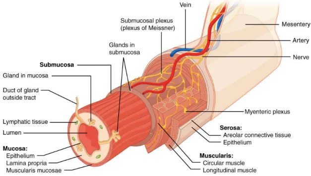
The intestine tissue is composed of four concentric layers: the serosa, muscularis, submucosa, and mucosa (Figure 2) (Rubin and Langer, 2009). The serosa (the outmost layer) is a smooth membrane that is also a thin layer of connective tissue. It is constituted of a thin layer of cells (epithelium) that produce serous fluid. Serous fluid is a lubricating fluid which decrease friction from the muscularis movement. The
muscularis is in charge of the movement or peristalsis of the intestine. It normally has
two obvious layers of smooth muscle: circular and longitudinal. The submucosa is the dense layer of irregular connective tissue that supports the mucosa, as well as joins the mucosa to the bulk of underlying smooth muscle. The mucosa is a mucous membrane,
10
which is the innermost tissue layer of the small intestine It includes digestive enzymes and hormones. The intestinal villi are part of the mucosa, which has bigger surface that can enhance the nutrients absorption. The mucosal layer of the gut consists of epithelium overlying the lamina propria, and resting on a narrow layer of smooth muscle, the muscularis mucosae. It represents the obvious physical boundary between the external and internal environment to constitute an important defense barrier of human body.
Figure 2 Anatomy of intestine
In direct contact with external environment, intestinal mucosa plays a critical role in the maintenance of the intestinal homeostasis. It has two critical functions. Firstly, it helps the absorption of nutrients, electrolytes and water into vascular, followed by circulation in the whole body. Secondly, the intestinal mucosa plays a crucial role of barrier as it prevents the uncontrolled translocation of luminal contents such as microorganisms or toxins into the intestinal tissue circulatory system and in the end in the whole organism (Groschwitz and Hogan, 2009). This barrier function is particularly important in the defense against the invasion by microorganisms of the gut microbiota and to avoid septic shock. The intestinal barrier is a complex biological system including several components with two main functions: the prevention of the mucosa
11
invasion and the elimination of the microorganism who succeeded in the invasion.
1.2.1 Intestinal barrier components
Intestinal barrier is a complex multilayer system, consisting in an external "physical" barrier and an inner "functional" immunological barrier. The tight interaction of these two barriers maintains an equilibrated permeability to nutrients, electrolytes and avoids the entry of pathogens, toxins, antigens (Bischoff et al., 2014). To understand this complex barrier, the functions of its components is very crucial to be described. Several components are assembled to form a functional intestinal barrier (Figure 3).
Figure 3 Elements comprising the intestinal mucosal barrier(Salim and Soderholm, 2011).
First, the endothelial cell layer is covered with two mucus layers, a thick, unstirred and relatively sterile mucus layer and an outer layer in direct contact with the cell which forms a protective area. The single layer of epithelial cells interspersed with mucus producing goblet cells, anti-microbial peptide producing Paneth cells, and specialized luminal sampling enterocytes M-cells forms the main barrier between the outside world and the internal body proper. In the lamina propria, innate and adaptive immune cells such as T cells, B cells, eosinophils, mast cells, dendritic cells, and macrophages, comprise the mucosal immune system that responds with “active” eradication or “toleragenic-reaction” towards foreign antigens. In addition, the degradative properties of gastric acids, pancreatic and biliary juices, and the intestinal propulsive motility have
12
also been recognized to be important factors in intestinal barrier function. It is imperative to keep in mind that although these components might be presented individually, they are inextricably linked and can affect the functional responses of each other (Salim and Soderholm, 2011).
1.2.1.1 The role of the commensal microbiota
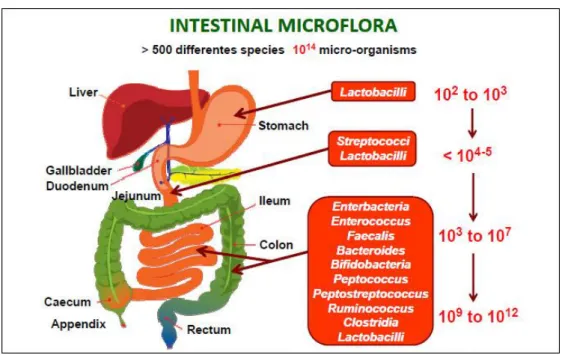
More than 500 different microbial species constituting up to 100 trillion (1014)
microorganisms per human colonize the intestinal tract (Figure 4) (Konturek et al., 2015). The gut microbiota maintains a symbiotic relationship with the gut mucosa and imparts substantial metabolic, immunological and gut protective functions in the healthy individual (Jandhyala et al., 2015). The normal human gut microbiota comprises of two major phyla, namely Bacteroidetes and Firmicutes (Jandhyala et al., 2015).
Figure 4 Spatial and longitudinal variations in microbial numbers and composition across the length of the gastrointestinal tract (Konturek et al., 2015). The number of bacterial cells present in the mammalian gut shows a continuum that goes from 102 to 103 bacteria per gram of contents in the stomach and duodenum, progressing to 104 to 107 bacteria per gram in the jejunum and ileum and culminating in 109 to 1012 cells per gram in the colon.
The composition of the gut microbiota remains relatively unchanged from late childhood to old age, the changes occur when aging process starts (Petschow et al.,