RESEARCH OUTPUTS / RÉSULTATS DE RECHERCHE
Author(s) - Auteur(s) :
Publication date - Date de publication :
Permanent link - Permalien :
Rights / License - Licence de droit d’auteur :
Bibliothèque Universitaire Moretus Plantin
Institutional Repository - Research Portal
Dépôt Institutionnel - Portail de la Recherche
researchportal.unamur.be
University of Namur
Inflammation and vasomotry during reperfusion
Gourdin, Maximilien; Dubois, Philippe
Published in:
Artery Bypass
DOI:
10.5772/54508
Publication date:
2014
Document Version
Publisher's PDF, also known as Version of record
Link to publication
Citation for pulished version (HARVARD):
Gourdin, M & Dubois, P 2014, Inflammation and vasomotry during reperfusion: Chapter 2. in Artery Bypass.
InTech, Croatia, pp. 19-35. https://doi.org/10.5772/54508
General rights
Copyright and moral rights for the publications made accessible in the public portal are retained by the authors and/or other copyright owners and it is a condition of accessing publications that users recognise and abide by the legal requirements associated with these rights. • Users may download and print one copy of any publication from the public portal for the purpose of private study or research. • You may not further distribute the material or use it for any profit-making activity or commercial gain
• You may freely distribute the URL identifying the publication in the public portal ?
Take down policy
If you believe that this document breaches copyright please contact us providing details, and we will remove access to the work immediately and investigate your claim.
Inflammation and Vasomotricity During Reperfusion
Maximilien Gourdin and Philippe Dubois
Additional information is available at the end of the chapter http://dx.doi.org/10.5772/54508
1. Introduction
Restoration of perfusion and reoxygenation of ischemic tissues restores aerobic metabo‐ lism and supports postischemic functional recovery but also generates significant dam‐ age related to the ischemia/reperfusion (I/R) phenomenon. At the level of a blood vessel, lesions of I/R are mainly characterized by the perturbation of vasomotion and endothe‐ lial dysfunction. Moreover, despite the fact that ischemia occurs in a sterile environment, reperfusion induces a significant activation of innate and adaptive immune responses: massive reactive oxygen species (ROS) production; activation of pattern-recognition re‐ ceptors or toll-like receptors (TLRs); activation of complement, coagulation, cytokine and chemokine production; and inflammatory cell trafficking into the diseased organ.1 I/R ac‐
tivates different programs of cell death (necrosis, apoptosis or autophagy-associated cell death) and generates a systemic inflammatory response that lasts several days and that can lead, in some cases, to multi-organ failure and death. [2-4]
2. Posthypoxic blood vessel motricity and posthypoxic endothelial
dysfunction
Blood vessels, and especially endothelium located at the blood-organ interface, are partic‐ ularly susceptible to ischemia-reperfusion injuries. Endothelial stunning or the loss of en‐ dothelial functions during reperfusion contributes to IR injuries and compromises the postischemic recovery. [5-7]
The basal vascular tone is a continual balance between vasoconstrictors and vasodilators acting on the blood vessel. Vascular smooth muscle cells (VSMCs) and endothelium play pivotal roles in this control.
© 2013 Gourdin and Dubois; licensee InTech. This is an open access article distributed under the terms of the Creative Commons Attribution License (http://creativecommons.org/licenses/by/3.0), which permits unrestricted use, distribution, and reproduction in any medium, provided the original work is properly cited.
Posthypoxic vasoconstriction, in response to vasoconstrictors, and endothelium-independ‐ ent vasodilation, induced by direct vasodilators (direct action on VSMCs), are slightly af‐ fected by I/R, demonstrating the relative resistance of VSMCs. [8]-[10] In contrast, endothelium-dependent dilatation is deeply affected. Despite the fact that endothelial cells seem relatively more resistant than other cells types (cardiomyocytes, neurons, renal tubular cell), I/R modifies their phenotype: diminution of their anticoagulant properties, increased vascular permeability, increased leukoadhesivity and establishment of a proin‐ flammatory state in the endovascular milieu.
The production of some bioactive agents decreases (e.g., prostacyclin, nitric oxide), while that of others increases during I/R (e.g., endothelin, thromboxane A2). [1],[11]-[16] These endothe‐ lial modifications are called endothelial dysfunction and are widely described in human and animals studies.[15],[17]-[21] IR-related endothelial dysfunction is mainly characterized by the loss of NO availability and seems to be related to the reperfusion more than to ischemia. [10] In normal situations, NO acts in numerous pathways: direct vasodilation, indirect vasodilation by inhibiting the influences of vasoconstrictors (e.g., inhibiting angiotensin II and sympathetic vasoconstriction), inhibiting platelet adhesion to the vascular endothelium (anti-thrombotic effect), inhibiting leukocyte adhesion to vascular endothelium (anti-inflammatory effect), and inhibiting smooth muscle hyperplasia by scavenging superoxide anion (anti-proliferative effect). The diminution of NO concentration jeopardizes these functions.
Multiple hypotheses have been proposed to explain postischemic endothelial dysfunction: massive ROS production by mitochondria, activation of immune cells, activation of xanthine oxidase and NADPH2 oxidase by the ceramide/sphingosine kinase pathway, the depletion of
dihydrobiopterin (an essential cofactor of nitric oxide synthase), increased arginine consump‐ tion in other intracellular pathways, the production of chemokines and cytokines (tumor necrosis factor-alpha (TNF-α), interleukin-1, -6, and -8) or the activation of the complement system (C3a fraction, C5b-9 fraction). [21]-[31]
In normoxic conditions, the endothelium permits only restricted diffusion. During hypoxia, the modifications of the cytoskeleton of endothelial cells, induced by hypoxia and low intracellular cyclic adenosine monophosphate phosphate (cAMP) concentration, increase vascular permea‐ bility, leading to capillary leakage and perivascular interstitial edema.[1] Complement system activation, leukocyte endothelial adhesion and platelet-leukocyte aggregation increase after re‐ perfusion.[1],[32] A clinical example is the acute respiratory failure with hypoxia and pulmona‐ ry edema observed in several surgeries. Acute respiratory distress syndrome is caused by heart failure but also by a disruption of the alveolar-capillary barrier.[33]-[36]
3. The inflammatory response
Ischemia-reperfusion induces a vigorous inflammatory reaction including activation of the complement system; activation of the innate and adaptive immune systems; increased ROS, cytokine, chemokine and other proinflammatory metabolite production; and activation of programmed cell death. If inflammation concerns mainly ischemic organs, its effects will
extend to the whole body and, particularly, the organs with a high capillary density, such as lung, brain and kidney. [1],[12],[37],[38]
3.1. Activation of the complement system
Reperfusion injury is characterized by autoimmune responses, including natural antibodies recognizing neoantigens and subsequent activation of the complement system (auto-im‐ munity). 1 Locally produced and activated, the complement system amplifies inflammation
during ischemia and reperfusion through complement-mediated recognition of damaged cells and anaphylatoxin release. The anaphylatoxins C3a, C4a and C5a lead to the recruit‐ ment and stimulation of immune cells, which promotes cell-cell interactions by increasing the expression of adhesion molecules (vascular cell adhesion molecule-1, ICAM-1, E-selectin and P-selectin) on the surface of the endothelial cells and neutrophils. [12],[39] Moreover, C5a is a chemotactic factor that directly stimulates leukocytes to synthesize and secrete cyto‐ kines such as interleukin (IL)-1, IL-6, monocyte chemoattractant protein-1 (MCP-1) and TNF-α. iC3b is implicated in neutrophil-endothelium interactions. C5b-9, known as the final cytolytic membrane attack complex complement, is a powerful chemotactic agent that caus‐ es direct lesions to the endothelial cells, stimulates the endothelial production of IL-8, MCP-1, and ROS and inhibits endothelium-dependent vasodilatation. [12],[39]
3.2. Cell-cell interactions during reperfusion
3.2.1. Neutrophil–endothelium interaction
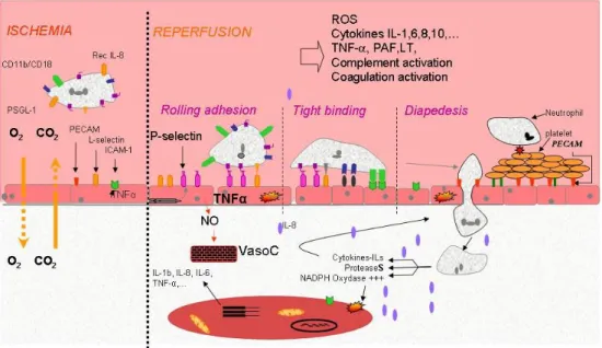
During reperfusion, neutrophils play a central part in the inflammatory response and in the genesis of the I/R injuries. Activated neutrophils produce high amounts of cytokines, che‐ mokines, and ROS in the vascular lumen but also in the parenchyma that directly contacts cells. These neutrophils and endothelial cells activated by cytokines (e.g., IL-6, TNF-α, IL-8, IL-1β) and other proinflammatory mediators (e.g., platelet-activating factor, ROS) promote a close interaction between these cell types that will result in a significant concentration of ac‐ tivated neutrophils in the interstitium. [1],[13],[15],[17],[32],[40]-[43] This complex process can be summarized in four steps: chemoattraction, weak neutrophil adhesion to the endo‐ thelium, followed by a stronger adhesion and, finally, neutrophil migration (Figure 1). Three families of sarcoplasmic adhesion molecules are implicated in the neutrophil-endothelium interaction: selectins, β2-integrins and immunoglobulins.
• Chemoattraction:
Upon reperfusion, the endothelium, parenchyma and resident immune cells (mainly macro‐ phages and neutrophils) release cytokines such as IL-1, TNF-α and chemokines, inducing the production of selectins by endothelial and immune cells. Circulating leukocytes are concen‐ trated towards the site of injury by the concentration gradient of chemokines.
• Rolling adhesion
Endothelial L-selectin interacts with the P-selectin and the E-selectin-specific ligand-1 (ESL-1) expressed by neutrophils. [44],[45] The activation of TLR-2, ROS production, the complement
system and thrombin and a high intracellular calcium concentration promotes the expression of endothelial P-selectin from the Weibel–Palade bodies. Its peak of expression occurs 10–20 min after the beginning of reperfusion.[40],[46] P-selectin interacts with P-selectin glycoprotein ligand-1 (PSGL-1) expressed by neutrophils. These interactions are weak and reversible, providing transitory neutrophil adherence, slowing down leukocytes and allowing them to “roll” along the endothelial surface. During this rolling motion, transitory bonds are formed and broken between selectins and their ligands. This phase prepares the neutrophils and the endothelium for the following stage.
• Tight adhesion
At the same time, chemokines released by endothelial and immune cells activate the rolling neutrophils. Stimulated by ROS, platelet-activating factor (PAF), IL-1, TNF-α and leukotriene B4 (LTB4), neutrophils present CD11a/CD18, CD11b/CD18 and CD11c/CD18 from intracellu‐ lar granules. These sarcoplasmic proteins interact with the iC3a fraction of the complement system and ICAM-1, an endothelial protein whose expression is reinforced by TNF-α and IL-1. [47],[48] This interaction switches from a low-affinity link to a high-affinity state and firmly attaches the neutrophil to the surface of the endothelial cell, despite the shear forces of the blood flow.
Figure 1. Ischemia–reperfusion-induced neutrophils accumulation in the interstitium is a mechanism described in
three phases implicating specific complementary proteins. CD11b/CD18, sarcoplasmic neutrophil integrin; CO2, car‐
bon dioxide; ESL-1, E-selectin-specific ligand-1; I/R, ischemia– reperfusion; O2, oxygen; PECAM, platelet–endothelial
cell adhesion molecule-1; PSGL-1, P-selectin glycoprotein ligand-1; Rec IL-8, neutrophil IL-8 receptor; ROS, reactive oxygen species; TNF-α, tumour necrosis factor-a; WPB, Weibel–Palade body.
• Migration into the interstitium or diapedesis
Intercellular adhesion molecule-1 (ICAM-1) and platelet-endothelium adhesion molecule-1 (PECAM-1) are sarcoplasmic adhesion molecules belonging to the superfamily of the immu‐ noglobulins. They are implicated in the transfer of neutrophils towards the interstitium, termed diapedesis. Leukocytes extravasation comprises many stages, which are not fully understood. Nevertheless, it seems that PECAM-1, found on neutrophil and endothelial cell membranes, is necessary for diapedesis. [1],[49] It interacts with several sarcoplasmic pro‐ teins of neutrophils. The cytoskeleton of the neutrophil is reorganized to allow the projec‐ tion of pseudopodia between endothelial cells. This transfer is facilitated by inflammatory mediators, the CD11/CD18–ICAM-1 interaction and ROS, which combine to decrease the ex‐ pression of cadherin and induce the phosphorylation vascular endothelial-cadherin and cat‐ enin, components of the intercellular junctions. [50]-[53] There is controversy concerning the mechanisms underlying this transfer through the basal membrane of the endothelium. Once into the interstitium, the neutrophil migrates along a chemotactic gradient towards the site of injury, where it causes considerable damage.
The neutrophil-related injuries in the interstitium are mainly related to the massive ROS production, proteases from the intracellular neutrophilic granules and the metabolites of arachidonic acid (PAF and LTB4). PAF and LTB4 are powerful chemoattractants that stimu‐ late neutrophil degranulation. The neutrophil granules contain proteases, collagenases, ela‐ stases, lipoxygenases, phospholipases and myeloperoxidases that digest the protein network of the extracellular matrix. For example, elastase digests substrates such as collagen types III and IV, immunoglobulins, fibronectin and proteoglycans. Several cells, such as cardiomyo‐ cytes, stimulated by IL-6, express ICAM-1. The neutrophil binds to its receptor and empties its granules directly near the cell. [54],[55]
3.2.2. Neutrophil-platelet interaction
The role of platelets in ischemia-reperfusion injuries is unclear. However, it seems that they participate directly and indirectly in posthypoxic endothelial injury. [32],[56] Platelets affect neutrophil activation by releasing thromboxane A2, platelet-derived growth factor, seroto‐ nin, lipoxygenase products, proteases and adenosine. During reperfusion, approximately 25% of the fixed platelets are directly bound to the endothelium and the remaining 75% to neutrophils linked to the endothelium. [32],[57] This platelet-neutrophil interaction potenti‐ ates the neutrophils’ capacity to produce superoxide and platelet-activating factor. [58],[59] Moreover, the neutrophil-platelet aggregates contribute to the no-reflow phenomenon and jeopardize the quality of the microcirculation. 60
3.3. Reactive oxygen species or oxygen free radicals
Reactive oxygen species, such as superoxide anion (O2−•), hydrogen peroxide (H2O2) and
ing proteins, DNA, phospholipids and other biological structures. During reperfusion, PAF, TNF-α, IL-6, IL-1β, granulocyte-macrophage colony-stimulating factor, complement fraction C5a and the ROS themselves stimulate endothelial and neutrophil ROS production. [49], [61],[62] On the other hand, ROS activate nuclear factor-κB, promote cytokine production (e.g., TNF-α, IL-6, PAF), and induce the synthesis and expression of endothelial and leuko‐ cyte adhesion molecules. [15],[41],[63]
In the reperfused tissue, the principal sources of ROS are neutrophil NADPH-oxidase, xan‐ thine oxidase, mitochondria and the arachidonic acid pathways. [64]-[66] The massive ROS production quickly exceeds the capacity of cellular defense systems (catalase, superoxide dismutase, glutathione peroxidase and vitamins C and E). ROS directly cause much struc‐ tural damage, increase the susceptibility to the opening of the mitochondrial permeability transition pore, activate immune and endothelial cells and induce apoptosis. [67]
ROS can also be produced by monoamine oxidase (MAO) of the outer mitochondrial mem‐ brane. MAO transfers electrons from amine compounds with oxygen to produce hydrogen peroxide. [68] p66Shc, a cytosolic adaptor protein for tyrosine kinase receptors that has been implicated in signal transduction, translocates to the mitochondrial matrix during reperfu‐ sion and oxidizes the reduced cytochrome c, which generates oxygen peroxide. [67],[69]
3.4. Ischemia-reperfusion-induced apoptosis
Reperfusion is vital for the functional recovery of an ischemic organ but also initiates the apoptosis pathways. [70],[71] Apoptosis is an active mechanism of cellular death, is geneti‐ cally programmed, consumes energy, requires the expression or activation of specific en‐ zymes, and can be induced by the oxidative stress of reperfusion. Reperfusion-induced apoptosis occurs in many organs, including heart, brain, kidney and liver. The reperfusion of an organ can induce apoptosis in other, distant organs. For example, reperfusion of a low‐ er limb or the small bowel can induce apoptosis of cardiomyocytes or lung cells, respective‐ ly. [72],[73] The TNF-α production by the reperfused organ seems to play a crucial part in the induction of apoptosis. [70],[74]-[76] TNF-α initiates a receptor-dependent death path‐ way by activating downstream caspases. [70],[76],[77] Other causes of reperfusion-induced apoptosis are also important: mitochondrial depolarization, high intracellular calcium, mPTP opening and the release of some mitochondrial proteins into the cytoplasm, such as cytochrome c. When this protein is released from mitochondria into the cytoplasm, it inter‐ acts with apoptotic protease activating factor-1 (Apaf-1) and ATP to form the apoptosome, a large oligomeric protein complex that can activate caspase 9, which activates the caspase-de‐ pendent apoptosis pathway.
Endothelial cell apoptosis precedes and influences the apoptosis of the subjacent parenchymal cells. For example, a reduction in endothelial apoptosis decreases the apoptosis of subjacent cardiomyocytes. This suggests that signals emanating from the endothelium during apoptosis can induce or reinforce that of the cardiomyocytes.
4. Integration of different aspects of ischemia-reperfusion
4.1. Blood vessel
According to the level of the vascular system considered (small arteries, capillaries and post‐ capillary veins), the repercussions of I/R are identical, but the clinical pictures differ.
4.1.1. At the arteriolar level
The principal manifestation of I/R in arterioles is a loss of the vasodilatation-dependent en‐ dothelium and the appearance of spasms. [78] Widespread endothelial lesions decrease the production of nitric oxide and do not counterbalance the arterioles’ tendency toward vaso‐ constriction. This tendency is highlighted in several tissues, such as skeletal muscle, heart, lung and brain. [79]-[82] The combined effects of IR and inflammation on arteriolar vasomo‐ tricity are well documented. The increase in the contractile response of the pulmonary and mesenteric microcirculation after cardiac surgery predisposes the patient to the develop‐ ment of pulmonary shunt or mesenteric ischemia, particularly during the administration of vasopressive drugs in the postextracorporeal circulation. [83 ],[84]
4.1.2. At the capillary level
The posthypoxic recovery of an organ depends on the quality of its microcirculation and the re‐ sultant nutrient delivery and gaseous exchange. However, the microcirculation is the site of a paradoxical phenomenon called “no reflow”, characterized by a major reduction in the capilla‐ ry density. Despite the reestablishment of complete blood flow, an incomplete and heterogene‐ ous perfusion of microcirculation persists. [85],[86] The capillaries are blocked by the parenchymatous and endothelial edema and the adhesion of the neutrophils and platelets to the surface of the endothelium, aided by the reduction in the production of nitric oxide. [15],[81], [85]-[87] Increased ROS and the depletion of ATP modify the cytoskeleton and the intercellular junctions, contributing to the loss of liquid from the vascular bed towards the interstitium. [88], [89] The phenomenon of no reflow persists several weeks after reperfusion. [85]
4.1.3. At the postcapillary vein level
The postcapillary veins are the sites of the inflammatory reaction. The margination and ex‐ travasation of the leukocytes are facilitated by the slower blood flow. Venous blood, arriving from the reperfused zones, is rich in proinflammatory mediators and activated neutrophils. These cause lesions both directly and indirectly through their interactions with platelets. [15],[90] Endothelial lesions prevent the intravascular oncotic pressure from recovering the excess liquid from the interstitium, thereby increasing the edema and contributing to the phenomenon of “no reflow”.
4.2. Organs
In pulmonary transplantation surgery, I/R-induced lung injury is characterized by non‐ specific alveolar damage, lung edema and hypoxemia. The most severe form may lead to
primary graft failure and remains a significant cause of morbidity and mortality after lung transplantation.[91] Pulmonary microvascular permeability appears to have a bimo‐ dal pattern, peaking at 30 min and 4 h after reperfusion. [92] Mechanical ventilation, car‐ diopulmonary bypass during cardiac surgery and lung resection can also induce apoptosis and I/R-induced lung injury. [93]-[96]
Perioperative acute renal failure is associated with a high incidence of morbidity and mortality. According to the type of surgery, IR injuries in the kidney are direct or indi‐ rect. [97] For example, acute renal failure is the most important complication of remote tissue damage following abdominal aortic surgery. [98] I/R induces renal tubular injuries and contributes to the decrease of glomerular filtration. Recent data suggest that 13% of patients with acute kidney injury (AKI) evolve to end-stage renal disease within 3 years. In the case of patients with preexisting renal disease, the progression to end-stage renal disease rises to 28% within the same period. [98] These results suggest that AKI predis‐ poses to chronic renal complication. I/R reduces blood vessel density and promotes renal fibrosis. The mechanisms mediating vascular loss are not clear but may be related to the lack of effective vascular repair responses. [99]
In cardiac surgery and in myocardial ischemia, cell death following I/R has features of apoptosis and necrosis. The loss of cardiomyocytes, which can hibernate in “no reflow” zones, and stunning, led by free radicals and calcium overload, explain the contractile posthypoxic dysfunction. The stunned cardiomyocytes can take several hours and days to recover. Intracellular ionic perturbation favors ventricular arrhythmias, such as ventricular fibrillation, ventricular tachycardia or ventricular extrasystole. [10 ]0 During ischemia, cardiomyocytes
express ICAM-1. Neutrophils bind to this receptor and empty the contents of their granules onto the cells. [54],[55]
The mechanisms of I/R-induced brain injury have many similar aspects compared with those of I/R-induced myocardial injury. Many mediators and cytokines upregulated by I/R, such as bradykinin, purine nucleotides, nitric oxide and ROS, increase blood–brain barrier permea‐ bility and induce cerebral edema. [10 ]1 Although leukocyte infiltration into the ischemic brain
increases cerebral damage, leukocyte accumulation in the microcirculation reduces reperfu‐ sion and increases the “no reflow” phenomenon.
The indirect repercussions of I/R on organs remote from the reperfused site are much more insidious. Neutrophils, complement activation, and massive production of cytokines and chemokines install a proinflammatory state that affects the functioning of other organs. During abdominal aortic surgery, I/R injuries are not only limited to the lower extremities but also cause damage to remote organs such as the lungs, kidneys, heart and bowel. [36],[97],[102-[104] Lung injuries following abdominal aortic aneurysm surgery are characterized by progressive hypoxemia, pulmonary hypertension, decreased lung compliance and nonhydro‐ static pulmonary edema, consistent with adult respiratory distress syndrome. [36],[103] In comparison with surgery, endovascular abdominal aortic aneurysm repair decreases I/R and I/R-induced-intestinal mucosal, renal and pulmonary dysfunction. [104]
Author details
Maximilien Gourdin and Philippe Dubois*
Department of Anaesthesiology, Université Catholique de Louvain, University Hospital of Mont Godinne, Belgium
References
[1] Eltzschig HK, Eckle T: Ischemia and reperfusion--from mechanism to translation. Nat Med 2011 17: 1391-401
[2] Berthonneche C, Sulpice T, Boucher F, Gouraud L, de Leiris J, O'Connor SE, Herbert JM, Janiak P: New insights into the pathological role of TNF-alpha in early cardiac dysfunction and subsequent heart failure after infarction in rats. Am J Physiol Heart Circ Physiol 2004; 287: H340-50
[3] Hotchkiss RS, Strasser A, McDunn JE, Swanson PE: Cell death. N Engl J Med 2009; 361: 1570-83
[4] Moro C, Jouan MG, Rakotovao A, Toufektsian MC, Ormezzano O, Nagy N, Tosaki A, de Leiris J, Boucher F: Delayed expression of cytokines after reperfused myocar‐ dial infarction: possible trigger for cardiac dysfunction and ventricular remodeling. Am J Physiol Heart Circ Physiol 2007; 293: H3014-9
[5] Garcia SC, Pomblum V, Gams E, Langenbach MR, Schipke JD: Independency of my‐ ocardial stunning of endothelial stunning? Basic Res Cardiol 2007; 102: 359-67 [6] Lefer AM, Tsao PS, Lefer DJ, Ma XL: Role of endothelial dysfunction in the patho‐
genesis of reperfusion injury after myocardial ischemia. Faseb J 1991; 5: 2029-34 [7] Qi XL, Nguyen TL, Andries L, Sys SU, Rouleau JL: Vascular endothelial dysfunction
contributes to myocardial depression in ischemia-reperfusion in the rat. Can J Physi‐ ol Pharmacol 1998; 76: 35-45
[8] Besse S, Tanguy S, Boucher F, Bulteau AL, Riou B, de Leiris J, Swynghedauw B: Aort‐ ic vasoreactivity during prolonged hypoxia and hypoxia-reoxygenation in senescent rats. Mech Ageing Dev 2002; 123: 275-85
[9] Piana RN, Wang SY, Friedman M, Sellke FW: Angiotensin-converting enzyme inhibi‐ tion preserves endothelium-dependent coronary microvascular responses during short-term ischemia-reperfusion. Circulation 1996; 93: 544-51
[10] Quillen JE, Sellke FW, Brooks LA, Harrison DG: Ischemia-reperfusion impairs endo‐ thelium-dependent relaxation of coronary microvessels but does not affect large ar‐ teries. Circulation 1990; 82: 586-94
[11] Deanfield JE, Halcox JP, Rabelink TJ: Endothelial function and dysfunction: testing and clinical relevance. Circulation 2007; 115: 1285-95
[12] Eltzschig HK, Collard CD: Vascular ischaemia and reperfusion injury. Br Med Bull 2004; 70: 71-86
[13] Maxwell SR, Lip GY: Reperfusion injury: a review of the pathophysiology, clinical manifestations and therapeutic options. Int J Cardiol 1997; 58: 95-117
[14] Pinsky DJ, Yan SF, Lawson C, Naka Y, Chen JX, Connolly ES, Jr., Stern DM: Hypoxia and modification of the endothelium: implications for regulation of vascular homeo‐ static properties. Semin Cell Biol 1995; 6: 283-94
[15] Seal JB, Gewertz BL: Vascular dysfunction in ischemia-reperfusion injury. Ann Vasc Surg 2005; 19: 572-84
[16] Zhang Y, Oliver JR, Horowitz JD: Endothelin B receptor-mediated vasoconstriction induced by endothelin A receptor antagonist. Cardiovasc Res 1998; 39: 665-73 [17] Engelman DT, Watanabe M, Engelman RM, Rousou JA, Flack JE, 3rd, Deaton DW,
Das DK: Constitutive nitric oxide release is impaired after ischemia and reperfusion. J Thorac Cardiovasc Surg 1995; 110: 1047-53
[18] Loukogeorgakis SP, Panagiotidou AT, Yellon DM, Deanfield JE, MacAllister RJ: Post‐ conditioning protects against endothelial ischemia-reperfusion injury in the human forearm. Circulation 2006; 113: 1015-9
[19] Loukogeorgakis SP, Williams R, Panagiotidou AT, Kolvekar SK, Donald A, Cole TJ, Yellon DM, Deanfield JE, MacAllister RJ: Transient limb ischemia induces remote preconditioning and remote postconditioning in humans by a K(ATP)-channel de‐ pendent mechanism. Circulation 2007; 116: 1386-95
[20] Pernow J, Bohm F, Beltran E, Gonon A: L-arginine protects from ischemia-reperfu‐ sion-induced endothelial dysfunction in humans in vivo. J Appl Physiol 2003; 95: 2218-22
[21] Weyrich AS, Ma XL, Lefer AM: The role of L-arginine in ameliorating reperfusion in‐ jury after myocardial ischemia in the cat. Circulation 1992; 86: 279-88
[22] Corda S, Laplace C, Vicaut E, Duranteau J: Rapid reactive oxygen species production by mitochondria in endothelial cells exposed to tumor necrosis factor-alpha is medi‐ ated by ceramide. Am J Respir Cell Mol Biol 2001; 24: 762-8
[23] Endres M, Laufs U: Effects of statins on endothelium and signaling mechanisms. Stroke 2004; 35: 2708-11
[24] Lefer AM, Ma XL: Cytokines and growth factors in endothelial dysfunction. Crit Care Med 1993; 21: S9-14
[25] Moens AL, Kietadisorn R, Lin JY, Kass D: Targeting endothelial and myocardial dys‐ function with tetrahydrobiopterin. J Mol Cell Cardiol; 51: 559-63
[26] Salvemini D, Cuzzocrea S: Superoxide, superoxide dismutase and ischemic injury. Curr Opin Investig Drugs 2002; 3: 886-95
[27] Tiefenbacher CP, Chilian WM, Mitchell M, DeFily DV: Restoration of endothelium-dependent vasodilation after reperfusion injury by tetrahydrobiopterin. Circulation 1996; 94: 1423-9
[28] Werner ER, Blau N, Thony B: Tetrahydrobiopterin: biochemistry and pathophysiolo‐ gy. Biochem J; 438: 397-414
[29] Wu G, Meininger CJ: Impaired arginine metabolism and NO synthesis in coronary endothelial cells of the spontaneously diabetic BB rat. Am J Physiol 1995; 269: H1312-8
[30] Zhang C, Hein TW, Wang W, Ren Y, Shipley RD, Kuo L: Activation of JNK and xan‐ thine oxidase by TNF-alpha impairs nitric oxide-mediated dilation of coronary arte‐ rioles. J Mol Cell Cardiol 2006; 40: 247-57
[31] Zhang C, Xu X, Potter BJ, Wang W, Kuo L, Michael L, Bagby GJ, Chilian WM: TNF-alpha contributes to endothelial dysfunction in ischemia/reperfusion injury. Arterios‐ cler Thromb Vasc Biol 2006; 26: 475-80
[32] Rodrigues SF, Granger DN: Role of blood cells in ischaemia-reperfusion induced en‐ dothelial barrier failure. Cardiovasc Res 2010; 87: 291-9
[33] Klausner JM, Paterson IS, Mannick JA, Valeri R, Shepro D, Hechtman HB: Reperfu‐ sion pulmonary edema. Jama 1989; 261: 1030-5
[34] Ogawa S, Gerlach H, Esposito C, Pasagian-Macaulay A, Brett J, Stern D: Hypoxia modulates the barrier and coagulant function of cultured bovine endothelium. In‐ creased monolayer permeability and induction of procoagulant properties. J Clin In‐ vest 1990; 85: 1090-8
[35] Ogawa S, Koga S, Kuwabara K, Brett J, Morrow B, Morris SA, Bilezikian JP, Silver‐ stein SC, Stern D: Hypoxia-induced increased permeability of endothelial monolay‐ ers occurs through lowering of cellular cAMP levels. Am J Physiol 1992; 262: C546-54 [36] Paterson IS, Klausner JM, Pugatch R, Allen P, Mannick JA, Shepro D, Hechtman HB:
Noncardiogenic pulmonary edema after abdominal aortic aneurysm surgery. Ann Surg 1989; 209: 231-6
[37] Laipanov Kh I, Petrosyan EA, Sergienko VI: Morphological changes in the lungs dur‐ ing experimental acute ischemia and reperfusion of the limb. Bull Exp Biol Med 2006; 142: 105-7
[38] Yassin MM, Harkin DW, Barros D'Sa AA, Halliday MI, Rowlands BJ: Lower limb is‐ chemia-reperfusion injury triggers a systemic inflammatory response and multiple organ dysfunction. World J Surg 2002; 26: 115-21
[39] Arumugam T, Magnus T, Woodruff T, Proctor L, Shiels I, Taylor S: Complement me‐ diators in ischemia-reperfusion injury. Clin Chim Acta. 2006; 374:: 33-45
[40] Sluiter W, Pietersma A, Lamers JM, Koster JF: Leukocyte adhesion molecules on the vascular endothelium: their role in the pathogenesis of cardiovascular disease and the mechanisms underlying their expression. J Cardiovasc Pharmacol 1993; 22 Suppl 4: S37-44
[41] Vinten-Johansen J: Involvement of neutrophils in the pathogenesis of lethal myocar‐ dial reperfusion injury. Cardiovasc Res 2004; 61: 481-97
[42] Wung BS, Ni CW, Wang DL: ICAM-1 induction by TNFalpha and IL-6 is mediated by distinct pathways via Rac in endothelial cells. J Biomed Sci 2005; 12: 91-101 [43] Yadav SS, Howell DN, Gao W, Steeber DA, Harland RC, Clavien PA: L-selectin and
ICAM-1 mediate reperfusion injury and neutrophil adhesion in the warm ischemic mouse liver. Am J Physiol 1998; 275: G1341-52
[44] Hidalgo A, Peired AJ, Wild MK, Vestweber D, Frenette PS: Complete identification of E-selectin ligands on neutrophils reveals distinct functions of PSGL-1, ESL-1, and CD44. Immunity 2007; 26: 477-89
[45] Steegmaier M, Levinovitz A, Isenmann S, Borges E, Lenter M, Kocher HP, Kleuser B, Vestweber D: The E-selectin-ligand ESL-1 is a variant of a receptor for fibroblast growth factor. Nature 1995; 373: 615-20
[46] Kuo MC, Patschan D, Patschan S, Cohen-Gould L, Park HC, Ni J, Addabbo F, Goli‐ gorsky MS: Ischemia-induced exocytosis of Weibel-Palade bodies mobilizes stem cells. J Am Soc Nephrol 2008; 19: 2321-30
[47] Gu Q, Yang XP, Bonde P, DiPaula A, Fox-Talbot K, Becker LC: Inhibition of TNF-al‐ pha reduces myocardial injury and proinflammatory pathways following ischemia-reperfusion in the dog. J Cardiovasc Pharmacol 2006; 48: 320-8
[48] Ikeda U, Ikeda M, Kano S, Shimada K: Neutrophil adherence to rat cardiac myocyte by proinflammatory cytokines. J Cardiovasc Pharmacol 1994; 23: 647-52
[49] Jordan JE, Zhao ZQ, Vinten-Johansen J: The role of neutrophils in myocardial ische‐ mia-reperfusion injury. Cardiovasc Res 1999; 43: 860-78
[50] Alexander JS, Alexander BC, Eppihimer LA, Goodyear N, Haque R, Davis CP, Kalo‐ geris TJ, Carden DL, Zhu YN, Kevil CG: Inflammatory mediators induce sequestra‐ tion of VE-cadherin in cultured human endothelial cells. Inflammation 2000; 24: 99-113
[51] Allingham MJ, van Buul JD, Burridge K: ICAM-1-mediated, Src- and Pyk2-depend‐ ent vascular endothelial cadherin tyrosine phosphorylation is required for leukocyte transendothelial migration. J Immunol 2007; 179: 4053-64
[52] Kevil CG, Ohno N, Gute DC, Okayama N, Robinson SA, Chaney E, Alexander JS: Role of cadherin internalization in hydrogen peroxide-mediated endothelial permea‐ bility. Free Radic Biol Med 1998; 24: 1015-22
[53] Wang Y, Jin G, Miao H, Li JY, Usami S, Chien S: Integrins regulate VE-cadherin and catenins: dependence of this regulation on Src, but not on Ras. Proc Natl Acad Sci U S A 2006; 103: 1774-9
[54] Davani EY, Dorscheid DR, Lee CH, van Breemen C, Walley KR: Novel regulatory mechanism of cardiomyocyte contractility involving ICAM-1 and the cytoskeleton. Am J Physiol Heart Circ Physiol 2004; 287: H1013-22
[55] Niessen HW, Lagrand WK, Visser CA, Meijer CJ, Hack CE: Upregulation of ICAM-1 on cardiomyocytes in jeopardized human myocardium during infarction. Cardiovasc Res 1999; 41: 603-10
[56] Tailor A, Cooper D, Granger DN: Platelet-vessel wall interactions in the microcircula‐ tion. Microcirculation 2005; 12: 275-85
[57] Granger DN: Role of xanthine oxidase and granulocytes in ischemia-reperfusion in‐ jury. Am J Physiol 1988; 255: H1269-75
[58] Herd CM, Page CP: Pulmonary immune cells in health and disease: platelets. Eur Re‐ spir J 1994; 7: 1145-60
[59] Suzuki K, Sugimura K, Hasegawa K, Yoshida K, Suzuki A, Ishizuka K, Ohtsuka K, Honma T, Narisawa R, Asakura H: Activated platelets in ulcerative colitis enhance the production of reactive oxygen species by polymorphonuclear leukocytes. Scand J Gastroenterol 2001; 36: 1301-6
[60] Botto N, Sbrana S, Trianni G, Andreassi MG, Ravani M, Rizza A, Al-Jabri A, Palmieri C, Berti S: An increased platelet-leukocytes interaction at the culprit site of coronary artery occlusion in acute myocardial infarction: a pathogenic role for "no-reflow" phenomenon? Int J Cardiol 2007; 117: 123-30
[61] Takahashi T, Hato F, Yamane T, Fukumasu H, Suzuki K, Ogita S, Nishizawa Y, Kita‐ gawa S: Activation of human neutrophil by cytokine-activated endothelial cells. Circ Res 2001; 88: 422-9
[62] Takahashi T, Nishizawa Y, Hato F, Shintaku H, Maeda N, Fujiwara N, Inaba M, Ko‐ bayashi K, Kitagawa S: Neutrophil-activating activity and platelet-activating factor synthesis in cytokine-stimulated endothelial cells: reduced activity in growth-arrest‐ ed cells. Microvasc Res 2007; 73: 29-34
[63] Gourdin MJ, Bree B, De Kock M: The impact of ischaemia-reperfusion on the blood vessel. Eur J Anaesthesiol 2009; 26: 537-47
[64] Abramov AY, Scorziello A, Duchen MR: Three distinct mechanisms generate oxygen free radicals in neurons and contribute to cell death during anoxia and reoxygena‐ tion. J Neurosci 2007; 27: 1129-38
[65] Kang SM, Lim S, Song H, Chang W, Lee S, Bae SM, Chung JH, Lee H, Kim HG, Yoon DH, Kim TW, Jang Y, Sung JM, Chung NS, Hwang KC: Allopurinol modulates reac‐ tive oxygen species generation and Ca2+ overload in ischemia-reperfused heart and hypoxia-reoxygenated cardiomyocytes. Eur J Pharmacol 2006; 535: 212-9
[66] Szocs K: Endothelial dysfunction and reactive oxygen species production in ische‐ mia/reperfusion and nitrate tolerance. Gen Physiol Biophys 2004; 23: 265-95
[67] Giorgio M, Migliaccio E, Orsini F, Paolucci D, Moroni M, Contursi C, Pelliccia G, Lu‐ zi L, Minucci S, Marcaccio M, Pinton P, Rizzuto R, Bernardi P, Paolucci F, Pelicci PG: Electron transfer between cytochrome c and p66Shc generates reactive oxygen spe‐ cies that trigger mitochondrial apoptosis. Cell 2005; 122: 221-33
[68] Droge W: Free radicals in the physiological control of cell function. Physiol Rev 2002; 82: 47-95
[69] Pinton P, Rimessi A, Marchi S, Orsini F, Migliaccio E, Giorgio M, Contursi C, Minuc‐ ci S, Mantovani F, Wieckowski MR, Del Sal G, Pelicci PG, Rizzuto R: Protein kinase C beta and prolyl isomerase 1 regulate mitochondrial effects of the life-span determi‐ nant p66Shc. Science 2007; 315: 659-63
[70] Bajaj G, Sharma RK: TNF-alpha-mediated cardiomyocyte apoptosis involves cas‐ pase-12 and calpain. Biochem Biophys Res Commun 2006; 345: 1558-64
[71] Wu D, Chen X, Ding R, Qiao X, Shi S, Xie Y, Hong Q, Feng Z: Ischemia/reperfusion induce renal tubule apoptosis by inositol 1,4,5-trisphosphate receptor and L-type Ca2+ channel opening. Am J Nephrol 2008; 28: 487-99
[72] An S, Hishikawa Y, Liu J, Koji T: Lung injury after ischemia-reperfusion of small in‐ testine in rats involves apoptosis of type II alveolar epithelial cells mediated by TNF-alpha and activation of Bid pathway. Apoptosis 2007; 12: 1989-2001
[73] Lu X, Hamilton JA, Shen J, Pang T, Jones DL, Potter RF, Arnold JM, Feng Q: Role of tumor necrosis factor-alpha in myocardial dysfunction and apoptosis during hin‐ dlimb ischemia and reperfusion. Crit Care Med 2006; 34: 484-91
[74] Misseri R, Meldrum DR, Dinarello CA, Dagher P, Hile KL, Rink RC, Meldrum KK: TNF-alpha mediates obstruction-induced renal tubular cell apoptosis and proapop‐ totic signaling. Am J Physiol Renal Physiol 2005; 288: F406-11
[75] Sugano M, Hata T, Tsuchida K, Suematsu N, Oyama J, Satoh S, Makino N: Local de‐ livery of soluble TNF-alpha receptor 1 gene reduces infarct size following ischemia/ reperfusion injury in rats. Mol Cell Biochem 2004; 266: 127-32
[76] Sun HY, Wang NP, Halkos M, Kerendi F, Kin H, Guyton RA, Vinten-Johansen J, Zhao ZQ: Postconditioning attenuates cardiomyocyte apoptosis via inhibition of JNK and p38 mitogen-activated protein kinase signaling pathways. Apoptosis 2006; 11: 1583-93
[77] Pru JK, Lynch MP, Davis JS, Rueda BR: Signaling mechanisms in tumor necrosis fac‐ tor alpha-induced death of microvascular endothelial cells of the corpus luteum. Re‐ prod Biol Endocrinol 2003; 1: 17
[78] Ruel M, Khan TA, Voisine P, Bianchi C, Sellke FW: Vasomotor dysfunction after car‐ diac surgery. Eur J Cardiothorac Surg 2004; 26: 1002-14
[79] Davenpeck KL, Guo JP, Lefer AM: Pulmonary artery endothelial dysfunction follow‐ ing ischemia and reperfusion of the rabbit lung. J Vasc Res 1993; 30: 145-53
[80] Meredith IT, Currie KE, Anderson TJ, Roddy MA, Ganz P, Creager MA: Postische‐ mic vasodilation in human forearm is dependent on endothelium-derived nitric ox‐ ide. Am J Physiol 1996; 270: H1435-40
[81] Nanobashvili J, Neumayer C, Fuegl A, Blumer R, Prager M, Sporn E, Polterauer P, Malinski T, Huk I: Development of 'no-reflow' phenomenon in ischemia/reperfusion injury: failure of active vasomotility and not simply passive vasoconstriction. Eur Surg Res 2003; 35: 417-24
[82] Stauton M, Drexler C, Dulitz MG, Ekbom DC, Schmeling WT, Farber NE: Effects of hypoxia-reoxygenation on microvascular endothelial function in the rat hippocampal slice. Anesthesiology 1999; 91: 1462-9
[83] Friedman M, Wang SY, Stahl GL, Johnson RG, Sellke FW: Altered beta-adrenergic and cholinergic pulmonary vascular responses after total cardiopulmonary bypass. J Appl Physiol 1995; 79: 1998-2006
[84] Sato K, Li J, Metais C, Bianchi C, Sellke F: Increased pulmonary vascular contraction to serotonin after cardiopulmonary bypass: role of cyclooxygenase. J Surg Res 2000; 90: 138-43
[85] Reffelmann T, Hale SL, Dow JS, Kloner RA: No-reflow phenomenon persists long-term after ischemia/reperfusion in the rat and predicts infarct expansion. Circulation 2003; 108: 2911-7
[86] Reffelmann T, Kloner RA: The no-reflow phenomenon: A basic mechanism of myo‐ cardial ischemia and reperfusion. Basic Res Cardiol 2006; 101: 359-72
[87] Tauber S, Menger MD, Lehr HA: Microvascular in vivo assessment of reperfusion in‐ jury: significance of prostaglandin E(1) and I(2) in postischemic "no-reflow" and "re‐ flow-paradox". J Surg Res 2004; 120: 1-11
[88] Hinshaw DB, Burger JM, Miller MT, Adams JA, Beals TF, Omann GM: ATP deple‐ tion induces an increase in the assembly of a labile pool of polymerized actin in en‐ dothelial cells. Am J Physiol 1993; 264: C1171-9
[89] Hinshaw DB, Sklar LA, Bohl B, Schraufstatter IU, Hyslop PA, Rossi MW, Spragg RG, Cochrane CG: Cytoskeletal and morphologic impact of cellular oxidant injury. Am J Pathol 1986; 123: 454-64
[90] Han JY, Horie Y, Li D, Akiba Y, Nagata H, Miura S, Oda M, Ishii H, Hibi T: Attenuat‐ ing effect of Myakuryu on mesenteric microcirculatory disorders induced by ische‐ mia and reperfusion. Clin Hemorheol Microcirc 2006; 34: 145-50
[91] de Perrot M, Liu M, Waddell TK, Keshavjee S: Ischemia-reperfusion-induced lung in‐ jury. Am J Respir Crit Care Med 2003; 167: 490-511
[92] Eppinger MJ, Deeb GM, Bolling SF, Ward PA: Mediators of ischemia-reperfusion in‐ jury of rat lung. Am J Pathol 1997; 150: 1773-84
[93] Chang WC, Murota SI, Nakao J, Orimo H: Age-related decrease in prostacyclin bio‐ synthetic activity in rat aortic smooth muscle cells. Biochim Biophys Acta 1980; 620: 159-66
[94] Klass O, Fischer UM, Antonyan A, Bosse M, Fischer JH, Bloch W, Mehlhorn U: Pneu‐ mocyte apoptosis induction during cardiopulmonary bypass: effective prevention by radical scavenging using N-acetylcysteine. J Invest Surg 2007; 20: 349-56
[95] Massoudy P, Zahler S, Becker BF, Braun SL, Barankay A, Meisner H: Evidence for in‐ flammatory responses of the lungs during coronary artery bypass grafting with car‐ diopulmonary bypass. Chest 2001; 119: 31-6
[96] Ng CS, Wan S, Yim AP, Arifi AA: Pulmonary dysfunction after cardiac surgery. Chest 2002; 121: 1269-77
[97] O'Donnell D, Clarke G, Hurst P: Acute renal failure following surgery for abdominal aortic aneurysm. Aust N Z J Surg 1989; 59: 405-8
[98] Basile DP: The endothelial cell in ischemic acute kidney injury: implications for acute and chronic function. Kidney Int 2007; 72: 151-6
[99] Basile DP, Fredrich K, Chelladurai B, Leonard EC, Parrish AR: Renal ischemia reper‐ fusion inhibits VEGF expression and induces ADAMTS-1, a novel VEGF inhibitor. Am J Physiol Renal Physiol 2008; 294: F928-36
[100] Ferdinandy P, Schulz R, Baxter GF: Interaction of cardiovascular risk factors with myocardial ischemia/reperfusion injury, preconditioning, and postconditioning. Pharmacol Rev 2007; 59: 418-58
[101] Abbott NJ: Inflammatory mediators and modulation of blood-brain barrier permea‐ bility. Cell Mol Neurobiol 2000; 20: 131-47
[102] Adembri C, Kastamoniti E, Bertolozzi I, Vanni S, Dorigo W, Coppo M, Pratesi C, De Gaudio AR, Gensini GF, Modesti PA: Pulmonary injury follows systemic inflamma‐ tory reaction in infrarenal aortic surgery. Crit Care Med 2004; 32: 1170-7
[103] Fantini GA, Conte MS: Pulmonary failure following lower torso ischemia: clinical evidence for a remote effect of reperfusion injury. Am Surg 1995; 61: 316-9
[104] Junnarkar S, Lau LL, Edrees WK, Underwood D, Smye MG, Lee B, Hannon RJ, Soong CV: Cytokine activation and intestinal mucosal and renal dysfunction are re‐ duced in endovascular AAA repair compared to surgery. J Endovasc Ther 2003; 10: 195-202