HAL Id: hal-03121681
https://hal.archives-ouvertes.fr/hal-03121681
Submitted on 26 Jan 2021HAL is a multi-disciplinary open access archive for the deposit and dissemination of sci-entific research documents, whether they are pub-lished or not. The documents may come from teaching and research institutions in France or abroad, or from public or private research centers.
L’archive ouverte pluridisciplinaire HAL, est destinée au dépôt et à la diffusion de documents scientifiques de niveau recherche, publiés ou non, émanant des établissements d’enseignement et de recherche français ou étrangers, des laboratoires publics ou privés.
Targeting 5-HT 2B Receptor Signaling Prevents Border
Zone Expansion and Improves Microstructural
Remodeling after Myocardial Infarction
J Snider, Lance Riley, Noah Mallory, Matthew Bersi, Prachi Umbarkar,
Rekha Gautam, Qinkun Zhang, Anita Mahadevan-Jansen, Antonis
Hatzopoulos, Luc Maroteaux, et al.
To cite this version:
J Snider, Lance Riley, Noah Mallory, Matthew Bersi, Prachi Umbarkar, et al.. Targeting 5-HT 2B Receptor Signaling Prevents Border Zone Expansion and Improves Microstructural Remodeling after Myocardial Infarction. Circulation, American Heart Association, 2021, �10.1161/CIRCULATION-AHA.120.051517�. �hal-03121681�
Title: Targeting 5-HT2B Receptor Signaling Prevents Border Zone Expansion and 1
Improves Microstructural Remodeling after Myocardial Infarction.
2
Authors: J. Caleb Snider1, Lance A. Riley1, Noah T. Mallory1, Matthew R. Bersi1, Prachi
3
Umbarkar2, Rekha Gautam1, Qinkun Zhang2, Anita Mahadevan-Jansen1, Antonis K.
4
Hatzopoulos3, Luc Maroteaux4, Hind Lal2, and W. David Merryman1*
5
Affiliations:
6
1
Department of Biomedical Engineering, Vanderbilt University, Nashville, TN 37232
7
2
Division of Cardiovascular Disease, The University of Alabama at Birmingham, Birmingham,
8
AL 35294
9
3
Division of Cardiovascular Medicine, Department of Medicine and Department of Cell and
10
Developmental Biology, Vanderbilt University Medical Center, Nashville, TN 37232
11
4
INSERM UMR-S 1270, 75005 Paris, France; Sorbonne Universités, 75005 Paris, France;
12
Institut du Fer à Moulin, 75005 Paris, France
13
* Address for Correspondence:
14 W. David Merryman 15 Room 9445, MRB4 16 2213 Garland Ave 17 Nashville, TN 37232 18 615.322.7219 19 david.merryman@vanderbilt.edu 20 21
Short Title: 5-HT2B in Myocardial Infarction
22
Total Word Count: 9605 (excluding supplement), Word Count of Body: 4933
1
Abstract
1
Background: Myocardial infarction (MI) induces an intense injury response which ultimately
2
generates a collagen-dominated scar. While required to prevent ventricular rupture, the fibrotic
3
process is often sustained in a manner detrimental to optimal recovery. Cardiac myofibroblasts
4
are the cells tasked with depositing and remodeling collagen and are a prime target to limit the
5
fibrotic process post-MI. Serotonin 2B receptor (5-HT2B) signaling has been shown to be harmful
6
in a variety of cardiopulmonary pathologies and could play an important role in mediating scar
7
formation after MI.
8
Methods: We employed two pharmacologic antagonists to explore the effect of 5-HT2B ablation
9
on outcomes post-MI and characterized the histological and microstructural changes involved in
10
tissue remodeling. Inducible, 5-HT2B ablation driven by Tcf21MCM and PostnMCM were used to
11
evaluate resident cardiac fibroblast- and myofibroblast-specific contributions of 5-HT2B,
12
respectively. RNA sequencing was used to motivate subsequent in vitro analyses to explore
13
cardiac fibroblast phenotype.
14
Results: 5-HT2B antagonism preserved cardiac structure and function by facilitating a less
15
fibrotic scar, indicated by decreased scar thickness and decreased border zone area. 5-HT2B
16
antagonism resulted in collagen fiber redistribution to thinner collagen fibers which were more
17
anisotropic, enhancing left ventricular contractility, while fibrotic tissue stiffness was decreased,
18
limiting the hypertrophic response of uninjured cardiomyocytes. Using a tamoxifen-inducible
19
Cre, we ablated 5-HT2B from Tcf21-lineage resident cardiac fibroblasts and saw similar
20
improvements to the pharmacologic approach. Tamoxifen-inducible Cre-mediated ablation of
5-21
HT2B after onset of injury in Postn-lineage myofibroblasts also improved cardiac outcomes.
22
RNA sequencing and subsequent in vitro analyses corroborate a decrease in fibroblast
2
proliferation, migration, and remodeling capabilities through alterations in Dnajb4 expression
1
and Src phosphorylation.
2
Conclusions: Together, our findings illustrate that 5-HT2B expression in either cardiac
3
fibroblasts or activated myofibroblasts directly contributes to excessive scar formation, resulting
4
in adverse remodeling and impaired cardiac function after MI.
5 6
Keywords: Myocardial infarction, Serotonin 2B receptor, Cardiac fibrosis, Collagen
7
remodeling
8 9
3
Clinical Perspective
1
What is new?
2
Antagonism of the serotonin 2B receptor (5-HT2B) improves outcomes after myocardial
3
infarction through limiting the fibrotic process of scar formation.
4
Biomechanical characterization of the scar and adjacent border zone provides useful
5
insight into cardiac fibroblast-mediated fibrosis which results in the associated
6
echocardiographic metrics of tissue structure and function.
7
Using pharmacological and genetic approaches, this study pinpoints 5-HT2B expression in
8
myofibroblasts as a regulator of cell proliferation and invasion after myocardial
9
infarction.
10
What are the clinical implications?
11
This study suggests that early inhibition of 5-HT2B signaling after myocardial infarction
12
is sufficient to optimize scar formation, resulting in a functional scar which is less likely
13
to expand beyond the initial infarct and cause long-term remodeling.
14
Prolonged presence of the antagonist was not required to maintain the benefits observed
15
in the early stages after injury, indicating that acute treatment can alter chronic
16
remodeling.
17
5-HT2B blockade does not negatively affect any of the vast array of contributors to the
18
healing response after MI provided by non-cardiac fibroblast cell types.
19 20
4
Introduction
1 2
Cardiac fibroblasts (CFs) comprise approximately 11% of cells in the healthy adult
3
heart.1 Quiescent CFs reside in healthy ventricles and maintain homeostasis through low-level
4
extracellular matrix (ECM) turnover and organization.2,3 Injurious environmental stimuli, such as
5
hypoxia, altered tissue mechanics, and cytokines like TGF-β, signal the phenotypic switch of
6
CFs to a more proliferative, hypercontractile, and hypersecretory state.4 In the case of myocardial
7
infarction (MI), Tcf21 lineage-traced resident CFs transdifferentiate into myofibroblasts, which
8
can then be traced through their expression of the marker periostin – a secreted matricellular
9
protein produced in adults exclusively following injury.5–7 Myofibroblasts localize to both
10
infarcted tissue and the surrounding border zone (BZ) to potentiate the reparative fibrotic injury
11
response due to the lack of intrinsic regenerative capacity of the myocardium.8–10
12
Following MI, myofibroblasts play the indispensable role of stabilizing and reinforcing
13
the left ventricle (LV) via formation of a collagen-dominated scar.4 Insufficient ECM deposition
14
can lead to LV rupture or aneurysm, while excessive ECM deposition leads to tissue stiffening,
15
scar expansion, and arrhythmias.11 Furthermore, replacement of contractile myocardium with a
16
collagenous scar creates a local increase in mechanical strain at the BZ of surviving myocardium
17
and scar tissue. To compensate for biomechanical alterations, connective tissue often expands
18
beyond the original injury, creating a subsequent decline in tissue compliance and cardiac
19
output.12,13 Most chronic myocardial conditions are associated with excessive deposition of
20
fibrotic tissue, making the myofibroblast a desirable therapeutic target to limit fibrotic
21
overactivity.14,15
22
Serotonergic dysfunction has long been understood to contribute to a myriad of
23
cardiopulmonary pathologies. Specifically, signaling through the serotonin 2B receptor (5-HT2B)
5
is particularly involved in valvular heart disease and pulmonary hypertension.16,17 In response to
1
TGF-β, fibroblast-like valve interstitial cells undergo myofibroblast transformation, which can be
2
prevented with 5-HT2B antagonism.18 In a murine model of experimental pulmonary
3
hypertension, it has been shown that 5-HT2B in the myeloid compartment is necessary to develop
4
disease and instigate the stiffening of arterioles.19,20 5-HT2B has also been shown to play an
5
integral role in mediating isoproterenol-induced cardiac hypertrophy through modulating the
6
inflammatory milieu in a CF-dependent manner.21 There are multiple downstream 5-HT2B
7
signaling pathways, but the mitogen-activated protein kinase effector p38 and the tyrosine kinase
8
Src are two of the most commonly observed. Both of which are known regulators of cell
9
contractility, ECM deposition, and ECM stiffness.22,23
10
Here, we hypothesized that 5-HT2B antagonism could hinder excessive fibrotic
11
remodeling after MI and create a post-MI scar that preserved cardiac function. We successfully
12
limited adverse remodeling following MI using two independent 5-HT2B antagonists. Using
13
newly developed, tamoxifen-inducible Cre recombinase mouse models for either resident
14
fibroblast- or myofibroblast-targeted ablation of 5-HT2B, we illustrate that CFs have a 5-HT2B-
15
mediated deleterious effect on cardiac structure and function after infarct. We show that targeted
16
ablation of 5-HT2B only from myofibroblasts after injury is sufficient to improve cardiac
17
outcomes. These results are attributed to decreased scar thickness, limited BZ expansion into
18
healthy myocardium, and reduced stiffness of scar tissue. RNA sequencing pointed to a limited
19
proliferative and remodeling capacity in myofibroblasts lacking 5-HT2B driven by an increased
20
expression of Dnajb4, which encodes a heat shock protein with known anti-proliferative and
21
anti-invasive properties, and decreased activation of the tyrosine kinase Src.
22 23
6
Methods
1 2
The data that support the reported findings and code used for analyses in this study are
3
available from the corresponding author upon reasonable request. All mouse experiments were
4
approved by the Vanderbilt Institutional Animal Care and Use Committee before their
5
commencement. MI was induced via permanent coronary artery occlusion as previously
6
described with consistent infarct size confirmed via TTC staining (Supplemental Figure 1A)24–26.
7
Serial echocardiography (example image in Supplemental Figure 1B) was used to assess cardiac
8
structure and function on mice receiving a 5-HT2B antagonist or mice with cell-specific 5-HT2B
9
ablation. Htr2bfl/flTcf21MCM/+ and Htr2bfl/flPostnMCM/+ mice (inducible resident fibroblast- and 10
myofibroblast-specific 5-HT2B knockouts, respectively) were generated to compare with Htr2bfl/fl
11
littermate controls; all mice were crossed with Rosa26-stop-tdTomato reporter mice to visually
12
verify Cre activation. Tissue was collected for analysis to determine properties of the scar, BZ,
13
and uninjured myocardium. Picrosirius red staining was used to identify ECM deposition and
14
associated fibrotic measurements (scar thickness, BZ area, and interstitial fibrosis). Polarized
15
light imaging was used to determine collagen fiber thickness and second-harmonic generation
16
imaging to quantify fiber orientation. Atomic force microscopy (AFM) was used to acquire
17
mechanical properties of the tissue and wheat germ agglutinin to identify cardiomyocyte borders
18
for cross-sectional area measurements. CFs (PDGFRα+) were isolated one week after MI from
19
Htr2bfl/fl and Htr2bfl/flPostnMCM/+ animals to perform bulk RNA sequencing. CFs were isolated
20
from adult WT and 5-HT2B knockout mice for in vitro studies. An expanded methods section is
21
available in the Online Data Supplement.
22 23
7
Results
1 2
5-HT2B antagonism preserves cardiac structure and function following MI 3
4
To test our hypothesis that 5-HT2B regulates scar formation following MI, we first
5
confirmed there was a marked increase in Htr2b expression three days after injury which was
6
sustained into the fibrotic healing phase seven days after infarction (Supplemental Figure 2).
7
Htr2a, which encodes the only other member of the 5-HT2 receptor subfamily expressed in the
8
cardiovascular system,27 is not upregulated until seven days post-MI, and it is still only induced
9
to a fraction of Htr2b levels. These findings motivated our exploration into 5-HT2B signaling. To
10
determine if 5-HT2B signaling influences ventricular remodeling after MI, wild-type (WT) mice
11
underwent permanent coronary artery ligation and were administered either vehicle control
12
(dimethyl sulfoxide, DMSO) or the 5-HT2B antagonist SB204741 (SB) at the time of injury
13
(Figure 1A). Interestingly, female mice showed no response to SB treatment (Supplemental
14
Figure 3A-C) which may be due to sex-specific differences of 5-HT2B governed cardiac activity,
15
so subsequent antagonist studies were conducted in male mice.
16
We observed a preservation of cardiac function indicated by decreased reduction in
17
ejection fraction (EF) and fractional shortening (FS) one week after injury in mice treated with
5-18
HT2B antagonist, highlighting a slowed LV impairment following MI. This effect was maintained
19
for the six-week period after injury, even with the removal of the antagonist three weeks post-MI
20
(Figure 1B-C). The observed effects were due to the improved systolic inner dimension and
21
volume of the LV (Figure 1D-G). We employed a speckle-tracking algorithm to obtain the global
22
longitudinal strain (GLS) of the LV as another sensitive measure of LV function. While GLS
23
was identically reduced in both groups the first week after injury, cardiac contractility of
8
treated animals stabilized, while the contractility of DMSO-treated control animals continued to
1
deteriorate over the duration of the experiment (Figure 1H). In order to confirm these results, the
2
experiment was repeated using another selective, high affinity 5-HT2B antagonist RS127445 (RS)
3
which provided a comparable effect (Supplemental figure 4A-H). Heart rate was not affected by
4
inhibition of 5-HT2B signaling (Supplemental Table 1). These data suggest increased 5-HT2B
5
signaling following ischemic injury plays a detrimental role in tissue healing.
6 7
Fibrotic injury response is altered by 5-HT2B antagonism 8
9
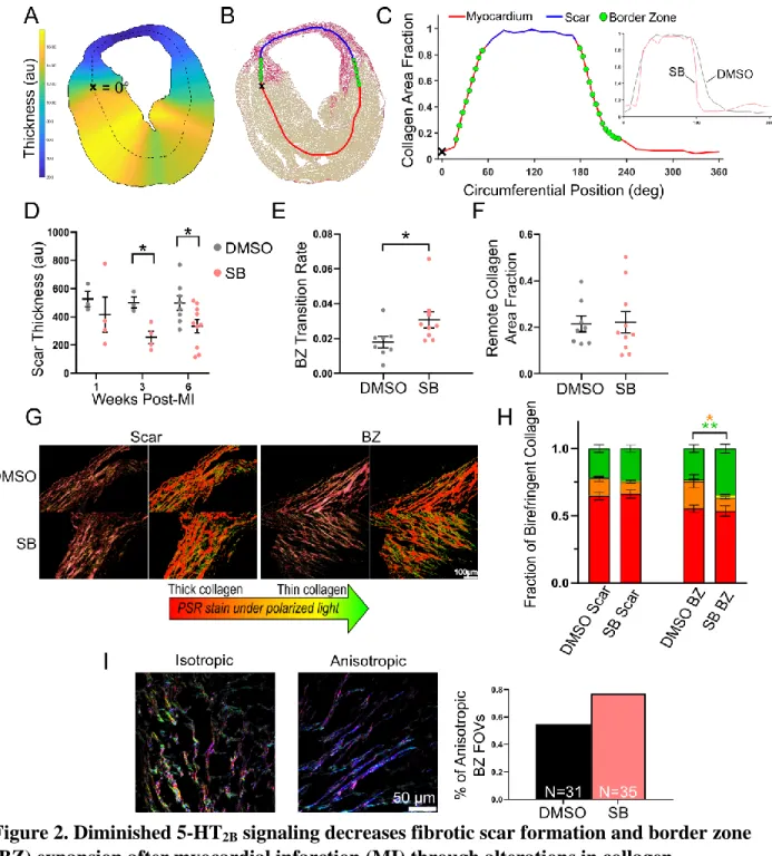
Using a custom-built image processing pipeline,4 short-axis tissue sections were analyzed
10
six weeks following MI (unless otherwise noted) to identify the role of 5-HT2B signaling in the
11
healing process post-MI. Tissue thickness was calculated and picrosirius red staining demarcated
12
the viable myocardium (yellow) from collagenous scar tissue (red; Figure 2A-B). The BZ (green
13
dots; Figure 2B) was defined as the transition region between collagen-dominated scar tissue
14
(stained >85% red) and myocardium-dominated tissue (stained >85% yellow) (Figure 2C). In
15
SB-treated mice, the deposition of fibrotic tissue was diminished as indicated by the formation of
16
a thinner scar than control mice (Figure 2D). Scars in the SB-treated group appear to form
17
thinner in the first week after injury and retain this thinness from week one to week six. This
18
composition did not compromise scar integrity as there was no difference in mortality between
19
the two groups (Supplemental Figure 5).
20
We then calculated the BZ transition rate (indicated by the slope of the green dot region
21
plotted in Figure 2C) to quantify the area of material mismatch between the contractile
22
myocardium and stiff, collagenous scar. We observed an increased rate of change from collagen
9
to myocardium (i.e. a shorter BZ region) with 5-HT2B antagonism revealing decreased BZ
1
infiltration and scar disruption of viable myocardium (Figure 2E). There were no differences in
2
the area fraction of collagen in the remote uninjured myocardium to indicate tissue-wide fibrosis
3
(Figure 2F).
4
Since we observed differences in fibrotic response in both the scar and BZ of SB-treated
5
animals, picrosirius red staining was imaged under polarized light to observe collagen fiber
6
thickness. While there was no shift in the distribution of collagen fiber thickness in the scar, a
7
higher percentage of thin collagen fibers was observed in response to 5-HT2B antagonism within
8
the BZ (Figure 2G-H). We also used second-harmonic generation (SHG) imaging to analyze
9
collagen fiber orientation (Supplemental Figure 6). SB-treated mice had a higher fraction of
10
fields of view (FOV) dominated by strongly aligned, anisotropic collagen fibers in the BZ, which
11
would impart an improved contractility (Figure 2I). Mice treated with RS also demonstrated
12
similar alterations in the scar formation process (Supplemental Figure 7A-C). Histological
13
analyses indicate that treatment with a 5-HT2B antagonist is capable of controlling scar formation
14
after MI and limiting BZ expansion through alterations in collagen fiber formation and
15
organization.
16 17
Microstructural changes in response to impaired 5-HT2B signaling 18
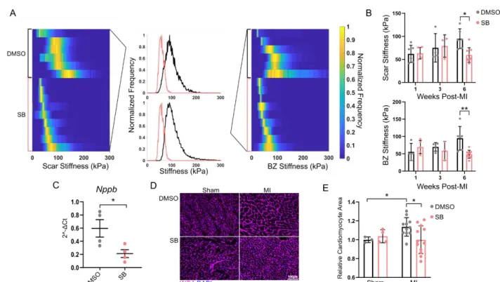
19
We next explored how alterations in the fibrotic healing process affects tissue stiffness.
20
AFM was used to investigate mechanical changes of the fibrotic tissue area to determine if tissue
21
compliance could play a role in preserving cardiac function in response to SB treatment. While
22
there were no differences in tissue stiffness in the early stages of scar formation,
10
treated groups showed a decreased tissue stiffness in both the scar and BZ at six weeks following
1
injury (Figure 3A-B, Supplemental Figure 7D). There were no changes in the myocardial
2
stiffness in uninjured myocardium (Supplemental Figure 8).
3
Since increased tissue stiffening can lead to a hypertrophic response in cardiomyocytes,28
4
we quantified the expression of the gene encoding the cardiomyocyte injury marker natriuretic
5
peptide B (Nppb). Six weeks after MI, there was a significant reduction in Nppb in the SB group
6
(Figure 3C). To further characterize the cardiomyocyte response to diminished fibrotic
7
remodeling, short-axis cross-sectional area of cardiomyocytes distant to the infarcted tissue was
8
quantified. We observed a significant increase in cardiomyocyte area six weeks after MI in
9
vehicle treated animals compared to SB-treated animals (Figure 3D-E); these results again held
10
with RS treatment (Supplemental Figure 7E). There was also a significant increase in
11
cardiomyocyte area of vehicle-treated animals over their sham counterparts which was prevented
12
with SB treatment (Figure 3E). These results indicate a biophysical alteration in cardiac
13
composition downstream of the scar formation process influences remote cardiomyocyte
14
hypertrophy after MI.
15 16
Deletion of 5-HT2B in resident fibroblasts improves cardiac response to MI 17
18
We next examined potential cell populations responsible for the effects seen following
19
systemic administration of a 5-HT2B antagonist. While it is known 5-HT2B influences
20
cardiomyocyte development and mitogenesis,29,30 we did not observe alterations in cardiac
21
structure or function in sham-operated animals given antagonist (Supplemental Figure 9A-C).
22
Therefore, we ruled out that 5-HT2B antagonism in the absence of injury affects the resident cell
11
populations (primarily cardiomyocytes) with regards to LV dimensions or output. In the context
1
of MI, we investigated Htr2b expression via quantitative polymerase chain reaction (qPCR) in
2
three cell populations. First, adult cardiomyocytes were isolated from infarcted LVs seven days
3
after injury. Compared to sham operation, there was approximately a 5-fold increase in Htr2b
4
expression (Supplemental Figure 10A). However, since we observed an 17-fold increase in bulk
5
tissue at this time point (Supplemental Figure 2), we sought other contributing cell types. The
6
next cell population investigated was endothelial cells. WT mice were subjected to MI, and
7
CD31-expressing cells were isolated via fluorescence-activated cell sorting (FACS). qPCR
8
revealed that CD31+ cells had a negligible contribution to the 17-fold induction of Htr2b
9
following MI (Supplemental Figure 10B-C). Furthermore, there was not an alteration in the
10
expression for the gene encoding E-selectin (Sele) in scar tissue of mice treated with a 5-HT2B
11
antagonist, indicating blocking 5-HT2B signaling does not mediate endothelial cell activation
12
(Supplemental Figure 10E). While there was no increase in Htr2b expression in CD45+ cells
13
seven days after MI compared to sham operation (Supplemental Figure 10B,D), there was the
14
possibility that the immune cells, which infiltrate early after injury, had already been cleared
15
from the injury. To test this hypothesis, we used a bone marrow transplant model based on
16
previous findings showing that mice lacking 5-HT2B in the bone marrow compartment exhibit
17
decreased tissue remodeling and arteriole stiffness in an experimental model of pulmonary
18
hypertension.19,20 Age- and sex-matched donors (WT or Htr2b-/-) were transplanted into WT
19
mice with engraftment success of approximately 95% (Supplemental Figure 11A-B). We
20
observed no differences in cardiac structure or function between the transplant groups after MI,
21
indicating a cell type of non-hematopoietic origin mediated the results observed in the 5-HT2B
22
antagonist studies (Supplemental Figure 12A-D).
12
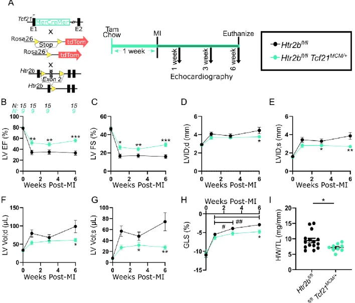
To determine if 5-HT2B signaling in the remaining major cardiac cell population, resident
1
CFs, impacts healing after MI, we deleted the gene encoding 5-HT2B in resident CFs using a
2
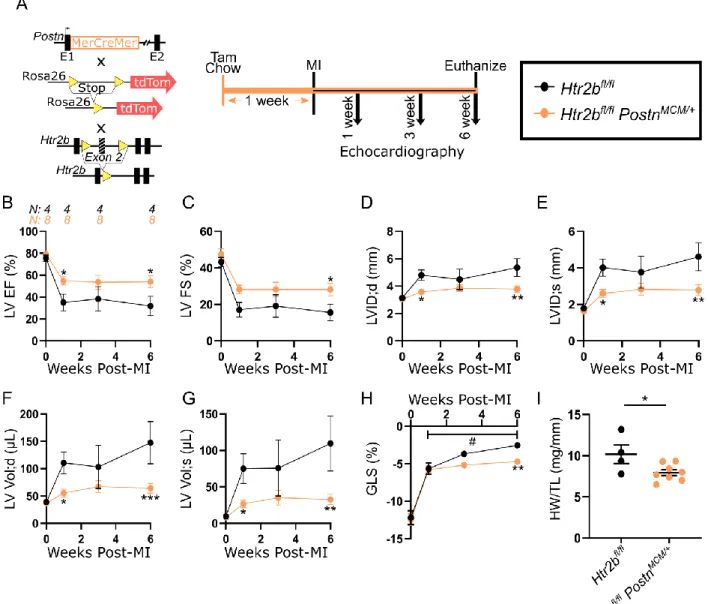
tamoxifen-inducible, Tcf21MerCreMer (Tcf21MCM) transgene7,31 (Figure 4A). The tdTomato
3
fluorescent reporter was observed in all Htr2bfl/flTcf21MCM/+ mice indicating successful
4
recombination in the CFs without affecting survival (Supplemental Figure 13A-B, Supplemental
5
Figure 14A). Following MI, Htr2bfl/flTcf21MCM/+ mice exhibited significantly improved EF and 6
FS (Figure 4B-C). These mice also had preserved LV inner dimension and volume in both
7
diastole and systole six weeks post-MI (Figure 4D-G). GLS steadily decreased over time in
8
Htr2bfl/fl mice lacking the Tcf21MCM transgene and was significantly lower than the stabilized 9
contractility of mice harboring the Tcf21MCM transgene (Figure 4H). Six weeks after MI, 10
morphometric analysis revealed increased heart weight to tibia length in Htr2bfl/fl mice indicative 11
of cardiac hypertrophy (Figure 4I). These results show that ablation of the 5-HT2B receptor from
12
resident CFs is effective in improving cardiac outcomes after MI in a manner similar to that
13
achieved with 5-HT2B antagonism.
14 15
Myofibroblast-specific deletion of 5-HT2B improves cardiac response to MI 16
17
Tcf21MCM driven ablation of 5-HT2B targets residential CFs present even in the absence of
18
injury. We wanted to assess the functionality of ablating 5-HT2B after the induction of an MI. We
19
utilized a mouse model which expressed the MCM cDNA driven by the periostin promoter
20
(PostnMCM) (Figure 5A). This mouse model has been well-characterized to demonstrate the
21
marking of nearly all newly activated fibroblasts (myofibroblasts) following an injury, without
22
induction prior to injury.6,32 Following MI, all Htr2bfl/flPostnMCM/+ hearts exhibited signal from
13
the tdTomato reporter, indicating successful recombination without affecting survival
1
(Supplemental Figure 14B, Supplemental Figure 15). Htr2bfl/flPostnMCM/+ mice demonstrated a 2
significant improvement in the functional metrics of EF and FS one week after MI compared to
3
Htr2bfl/fl animals which were maintained six weeks following the injury (Figure 5B-C). 4
Myofibroblast-specific 5-HT2B ablation resulted in improved LV inner dimension and volume in
5
both diastole and systole six weeks after MI, indicating a preserved cardiac structure (Figure
5D-6
G). Ventricular deformation measured by GLS was preserved in the Htr2bfl/flPostnMCM/+ group
7
while the control group continued to deteriorate over the six-week experiment (Figure 5H).
8
Finally, morphometric analyses revealed a decreased heart weight with 5-HT2B ablation (Figure
9
5I). As it has been shown that tissue-resident fibroblasts of the Tcf21 lineage are the primary
10
source of subsequent injury-activated, periostin-expressing myofibroblasts 6, these results
11
confirm 5-HT2B expression in the CF population worsens the injury response to MI and that
12
blocking 5-HT2B signaling after injury is sufficient to improve cardiac outcomes.
13 14
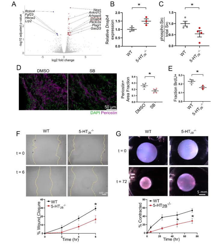
5-HT2B knockout decreases CF proliferative and remodeling capacity 15
16
In order to investigate how periostin-driven ablation of 5-HT2B imparts beneficial
17
outcomes following MI, we performed RNA sequencing on PDGFRα+ cells isolated via FACS
18
from the scar tissue of infarcted hearts seven days after MI (Supplemental Figure 16A-B).
19
Despite only about 20% of PDGFRα+ cells being tdTomato+ in the Htr2bfl/fl
PostnMCM/+ group 20
(indicating 5-HT2B knockout), we observed differential expression of 63 genes which, following
21
Gene Ontology (GO) analysis, were overwhelmingly associated with biological processes linked
22
to the control of cell cycle and mitosis as well as individual genes (i.e. Nexn, Robo4, Fgf23, and
14
Arhgap5) governing cell migration and remodeling (Figure 6A, Supplemental Table 2). One such 1
gene of interest that was upregulated in the Htr2bfl/flPostnMCM/+ group was the tumor suppressor 2
Dnajb4 which reduces the invasive and metastatic behavior cellular phenotype in lung 3
cancer.33,34 In isolated 5-HT2B-/- CFs, it was observed that loss of 5-HT2B resulted in an increased
4
expression of Dnajb4 (Figure 6B). As previous reports have shown Dnajb4 controls the
5
activation of Src33, we probed Src phosphorylation in CFs and observed a decreased
6
phosphorylation of Src in 5-HT2B-/- CFs (Figure 6C, Supplemental Figure 17). The decrease in
7
these molecular mediators of cell proliferation and invasion led us to investigate the capabilities
8
of CFs to populate the wounded tissue after MI. We observed a decreased area fraction of
9
periostin staining in the infarct of SB-treated mice one week after injury, indicative of decreased
10
myofibroblast proliferation and infiltration (Figure 6D). Similarly, in vitro analysis of 5-HT2B
-/-11
CFs revealed a decreased proliferative capacity as seen by fewer cells incorporating BrdU
12
(Figure 6E). CFs lacking 5-HT2B demonstrated an approximate 50% decrease in migratory
13
capacity in an in vitro wound healing assay (Figure 6F). Finally, CFs were seeded into
free-14
floating collagen gels in order to test their ability to remodel a collagen matrix. In gels seeded
15
with 5-HT2B-/- CFs, gel contraction after 72 hours was significantly hindered (Figure 6G). These
16
results point to a mechanism of 5-HT2B-mediated myofibroblast proliferation and matrix
17
remodeling leading to adverse scar formation following MI.
18 19
Discussion
20 21
Experimental MI induced by permanent coronary artery occlusion triggers the expansion
22
and activation of resident CFs from their quiescent, homeostatic state. CFs transdifferentiate into
23
highly active myofibroblasts following injury, migrating to and proliferating at the site of tissue
15
damage to secrete ECM and contract scar tissue.35,36 The quality of initial scar formation has
1
lasting effects on cardiac outcomes. A structurally sufficient scar is necessary to reinforce the LV
2
wall, but persistent fibrotic activity leads to chronic cardiac deterioration.11,37 Therefore, it is
3
desirable to properly tune the fibrotic response following MI such that a functional scar is able to
4
form but is dampened before the activity of myofibroblasts becomes deleterious.
5
Prior studies have shown anti-fibrotic effects of 5-HT2B disruption in various
6
cardiopulmonary pathologies.19–21,38,39 To our knowledge, this study is the first investigation to
7
report the direct contribution of 5-HT2B signaling in wound healing after MI. Through the
8
implementation of two pharmacological inhibitors and two models of genetically targeted
9
ablation, we have shown that the 5-HT2B receptor is an effective target to limit fibrosis following
10
MI injury in mice. Due to advances in identification of genetic markers for CFs, we were able to
11
isolate myofibroblasts as the cell population responsible for the improved recovery after MI seen
12
in animals treated with a 5-HT2B antagonist.
13
In the present study, echocardiographic analysis revealed global 5-HT2B antagonism
14
improves cardiac structure and function one week after MI versus vehicle treatment. While there
15
were not significant changes in these metrics between one and six weeks after injury, GLS
16
deteriorated from week one to six in the control groups whereas 5-HT2B inhibition stabilized this
17
measure of cardiac contractility. GLS has been shown to be an independent predictor of adverse
18
remodeling after ST-elevated MI in humans and can be a more sensitive functional output than
19
the traditionally used EF.40
20
We further explored changes to tissue architecture which led to an improvement in
21
cardiac function and alterations in scar formation. A heavier collagen burden increases passive
22
tissue stiffness, heightening afterload and hindering systolic function.41 5-HT2B blockade
16
successfully decreased collagen burden, indicated by decreased scar thickness, without
1
negatively affecting survival rate. The BZ is a vulnerable region of tissue which experiences
2
heightened wall stress and where scar expansion occurs, further damaging cardiomyocytes not
3
directly affected by the initial ischemic event.13,42 We found that targeting 5-HT2B resulted in an
4
increased transition rate from scar tissue to surviving myocardium (i.e. less BZ region), revealing
5
decreased intrusion of collagen fibers from the scar into uninjured myocardium. The prevention
6
of scar expansion can minimize the disruption of the cardiac syncytium, providing a more
7
coordinated systolic cycle.43 Similar to reports linking 5-HT2B to collagen content and
8
composition through activation of lung and valve fibroblasts, 38,44 our results show a
9
redistribution of collagen fiber thickness in the BZ, favoring less mature, more compliant
10
collagen fibers. This difference was not seen in the scar, further suggesting the formation of a
11
mechanically sound scar with limited capacity to expand beyond the BZ. The collagen fibers in
12
BZs of SB-treated animals exhibited more frequent regions of anisotropic collagen fiber
13
distributions which are able to undergo elastic deformation along with cardiomyocyte
14
contraction and increase LV contraction.45 This effect is enhanced by the softer scar and BZ.
15
Since these effects prevented early hypertrophic signs observed in control animals, 5-HT2B
16
antagonism reveals a desirable mechanical and biophysical outcome after MI achieved by a
17
muted initial fibrotic response that provides adequate scar formation without hindering the
18
systolic capabilities of the heart.
19
It is clear that the translational approach of pharmacological inhibition of 5-HT2B
20
signaling is effective to establish a therapeutic effect on scar formation and gives insight into the
21
mechanistic alterations of collagen deposition and remodeling, but the lack of specificity renders
22
it insufficient to identify the cell population which mediates the observed effect. Therefore, we
17
implemented several models to manipulate 5-HT2B in different cell populations. Cardiomyocytes
1
express 5-HT2B, and blockade of this receptor can partially mitigate noradrenaline
overload-2
induced hypertrophy.46 Cardiomyocytes isolated seven days after infarct do show an increase in
3
Htr2b expression, but only to a fraction of the degree to which expression is increased in bulk 4
tissue. CD31+ cells neither exhibited increased Htr2b gene expression after infarct nor altered
5
gene expression for the marker of endothelial activation, E-selectin. Similarly, Htr2b expression
6
in CD45+ cells did not change after MI, and bone-marrow transplants from 5-HT2B-knockout
7
mice did not recapitulate the results from antagonist studies, pointing to a mechanism different
8
from previous reports exploring fibrotic remodeling in pulmonary disease.19,20 Therefore, we
9
developed a novel model of CF-specific 5-HT2B ablation using Tcf21-driven Cre expression.
10
This approach circumvented developmental defects seen with gestational ablation of 5-HT2B;30
11
however, it could also have imparted benefits prior to MI. Regardless, eliminating 5-HT2B from
12
resident, Tcf21-expressing CFs resulted in a vastly improved cardiac phenotype compared to
13
control animals. Similar improvements as the antagonist studies were achieved with even more
14
pronounced structural benefits observed in the improvement in both LV systolic and diastolic
15
metrics, as well as a decreased heart weight. The enhanced effects of the genetic approach over
16
the pharmacological approach are most likely due to a sufficient recombination of the Htr2b
17
gene locus to convey a stronger effect than what is achieved by transient binding of the
18
antagonist which is subject to metabolic processing and removal from the system.
19
To implement an approach which more closely replicates the pharmacological strategy,
20
we utilized a Postn-driven model of 5-HT2B ablation. Myofibroblasts, which are overwhelmingly
21
derived from a Tcf21 lineage,6 are absent in healthy tissue and populate the infarct zone 2-4 days
22
after injury.14 Targeting this specialized cell type achieved nearly identical results as the
18
driven model and further hones in on the population affected by 5-HT2B antagonism. Knowing
1
that myofibroblasts are derived from resident fibroblasts, the overlapping results importantly
2
demonstrate that targeting injury-responsive myofibroblasts without affecting their progenitor
3
cells is sufficient to affect scar formation after MI. The mechanistic implication of this result is
4
that 5-HT2B-dependent infiltration and scar remodeling occurs after the transdifferentiation of
5
resident CFs. Furthermore, it demonstrates the redundancy in the two genetic models where
6
targeting either resident CFs in the absence of injury or myofibroblasts derived from resident
7
CFs achieves the same result.
8
While only the contribution of bone marrow-derived populations was explicitly
9
eliminated, initial evidence reveals that the contribution of cardiomyocyte or endothelial
10
expression of 5-HT2B plays a negligible role in improving scar formation after MI. We reported a
11
dramatic increase in Htr2b expression after MI which cannot be accounted for by
12
cardiomyocytes, CD31+, and/or CD45+ cells. Considering the utility of the CF-specific genetic
13
ablation of 5-HT2B, we are confident that this increase is due to CFs. Pericytes cannot be
14
excluded as potential contributors as these cells express Tcf21 and function in a multiplicity of
15
roles involved in MI healing.7,9,47 The periostin-driven model of 5-HT2B ablation in
16
myofibroblasts should circumvent pericyte contribution, and so we are confident that cells which
17
produce and organize collagen are the primary effector cells. While cardiomyocyte expression of
18
Htr2b is increased after MI, it accounts for a small fraction of the total increase. In healthy 19
hearts, cardiomyocytes are bountiful with CFs making up a small portion of cells.1 While these
20
populations shift after injury, it is still conceivable that the contribution of cardiomyocyte 5-HT2B
21
is minimal since a more abundant cell population is a minor contributor with mesenchymal cells
22
accounting for the vast majority of Htr2b expression. In addition, reports show blocking 5-HT2B
19
signaling after injury may reduce cardiomyocyte hypertrophy,46,48 providing additional
1
protection in the antagonist studies. Regardless, cardiomyocyte expression of 5-HT2B is intact
2
with the Tcf21- and Postn-driven 5-HT2B ablation models, further supporting that the alteration
3
in scar formation and cardiac outcomes is mediated by a fibroblast cell population. Because of
4
this evidence, utilization of 5-HT2B antagonists provides an approach to further investigate the
5
mechanism behind improved cardiac outcomes. Since myofibroblasts are not immediately
6
present after MI, it may not be necessary to begin treatment instantly after injury. Furthermore,
7
since echocardiographic readouts were improved as early as seven days after injury, it is possible
8
that 5-HT2B inhibition during the acute healing phase of initial scar formation is sufficient to
9
preserve cardiac performance.
10
It is interesting that 5-HT2B antagonism was ineffective in females. It has been shown that
11
global knockout of 5-HT2B results in more severe histopathological lesions and a stronger
12
systolic dysfunction in adult male mice compared to females as a result of impaired cardiac
13
development.30 Furthermore, sex-specific differences in hepatocellular carcinoma has been
14
attributed to increased serotonin production in males compared to females, resulting in a 5-HT2B
-15
mediated increase of fibrogenic hepatic stellate cells density.49 It must be noted that both males
16
and females were included in both genetic models, and both sexes responded similarly to 5-HT2B
17
ablation. While genetic manipulation is sufficient to improve outcomes in females, these
18
observations indicate that there is a biological difference in the way that female mice respond to
19
5-HT2B antagonism, potentially through metabolic processing of the molecule or ligand/receptor
20
interactions.
21
RNA sequencing revealed 5-HT2B as a regulator of proliferation and matrix remodeling in
22
CFs. We employed a surrogate CF marker PDGFRα50 to select for CFs, and despite only 20% of
20
these cells displaying the tdTomato fluorescent reporter in the Htr2bfl/flPostnMCM/+ group, we still
1
observed differential regulation of multiple genes in myofibroblasts with 5-HT2B ablation.
2
Supported by differences in associated GO terms, we primarily investigated CF proliferation as
3
well as ECM remodeling. We saw upregulation of genes such as Plagl1 which inhibits cell
4
growth and proliferation, potentially through PPARγ,51
and Nexn whose loss is associated with
5
dilated cardiomyopathy.52 Several interesting transcripts were also downregulated: Lrp2 which
6
increases proliferation of epicardial cells (CF precursors),53 Fgf23 which demonstrates broad
7
mitogenic and cell survival actions specifically in the heart,54 and Robo4 which is associated
8
with increased matrix metalloproteinase expression and predisposition for aortic valve disease.55
9
Of note, we have shown that in CFs lacking 5-HT2B, Dnajb4 is upregulated. It is known that the
10
heat shock protein encoded by this gene is highly expressed in human CFs.56 There is a direct
11
link to increased expression of this protein and the inhibition of Src,33 and consistent with this
12
prior report, we found decreased phosphorylation of Src in 5-HT2B-/- CFs. Dnajb4 is known to
13
control cell proliferation and migration, supporting our observations in which myofibroblasts
14
were sparser in damaged tissue of SB-treated animals and 5-HT2B knockout decreasing
15
proliferation, migration, and collagen matrix remodeling. The known link between Dnajb4, Src
16
phosphorylation, and cell proliferation and remodeling capacity provides a mechanistic insight
17
into the improved phenotype observed with 5-HT2B inhibition. Blocking 5-HT2B signal
18
transduction in the beginning stages of scar formation hinders the proliferation and migration of
19
myofibroblasts into the infarct zone through increasing Dnajb4 expression, which, in turn,
20
inhibits endogenous activation of Src. This approach allowed myofibroblasts to form a functional
21
scar while avoiding a mechanical environment that predisposes the tissue to scar expansion and
22
hypertrophy.
21
Due to the complexities associated with the intricate coordination involved in the
1
inflammatory response post-MI, we set out to target the effector cells of fibrosis (i.e. CFs) to
2
control the initial reparative response and limit adverse fibrotic remodeling. Targeting 5-HT2B on
3
CFs can control scar mechanics and limit fibrosis without affecting scar stability.57 Further,
4
inhibiting the activity of 5-HT2B has an acute benefit that is sustained well beyond the initial
5
healing phase and demonstrates that cardiac hypertrophy subsequent to an ischemic event can be
6
curtailed. Taken together, this work has identified 5-HT2B as a potential therapeutic target for
7
muting the over activity of myofibroblasts following MI to preserve cardiac phenotype and
8
prevent the initiation and progression of cardiac fibrosis and heart failure.
9 10
Sources of Funding
11 12
This work was supported by the National Institutes of Health: R35-HL135790 (WDM),
13
R01-HL115103 (WDM), R01-HL133290 (HL), R01-HL143074 (HL), R01-HL138519 (AKH), K99-14
HL146951 (MRB), F32- HL154596 (LAR), T32-HL007411 (MRB), T32-EB021937 (LAR); American
15
Heart Association: 18PRE34060078 (JCS); and a grant from the Fondation Leducq.
16 17
Acknowledgements
18 19
We thank Dr. Lin Zhong in the Cardiovascular Physiology Core for performing all of the
20
echocardiography and Vanderbilt Technologies for Advanced Genomics (VANTAGE) for
21
performing RNA sequencing. We thank Dr. Michelle Tallquist and Dr. Jeffery Molkentin for
22
providing the Tcf21MCM and PostnMCM animals, respectively. 23
22 Disclosures 1 2 None. 3 4 References 5 6
1. Pinto AR, Ilinykh A, Ivey MJ, Kuwabara JT, D’Antoni ML, Debuque R, et al. Revisiting
7
Cardiac Cellular Composition. Circ Res 2016;118:400–409. doi:
8
10.1161/CIRCRESAHA.115.307778.
9
2. Valiente-Alandi I, Potter SJ, Salvador AM, Schafer AE, Schips T, Carrillo-Salinas F, et al.
10
Inhibiting Fibronectin Attenuates Fibrosis and Improves Cardiac Function in a Model of
11
Heart Failure. Circulation 2018;138:1236–1252. doi:
12
10.1161/CIRCULATIONAHA.118.034609.
13
3. Nagaraju CK, Dries E, Popovic N, Singh AA, Haemers P, Roderick HL, et al. Global
14
fibroblast activation throughout the left ventricle but localized fibrosis after myocardial
15
infarction. Sci Reports 2017 71 2017;7:10801. doi: 10.1038/s41598-017-09790-1.
16
4. Schroer AK, Bersi MR, Clark CR, Zhang Q, Sanders LH, Hatzopoulos AK, et al.
17
Cadherin-11 blockade reduces inflammation-driven fibrotic remodeling and improves
18
outcomes after myocardial infarction. JCI Insight 2019;4. doi: 10.1172/jci.insight.131545.
19
5. Snider P, Standley KN, Wang J, Azhar M, Doetschman T, Conway SJ. Origin of cardiac
20
fibroblasts and the role of periostin. Circ Res 2009;105:934–947. doi:
21
10.1161/CIRCRESAHA.109.201400.
22
6. Kanisicak O, Khalil H, Ivey MJ, Karch J, Maliken BD, Correll RN, et al. Genetic lineage
23
tracing defines myofibroblast origin and function in the injured heart. Nat Commun
23
2016;7:12260. doi: 10.1038/ncomms12260.
1
7. Acharya A, Baek ST, Huang G, Eskiocak B, Goetsch S, Sung CY, et al. The bHLH
2
transcription factor Tcf21 is required for lineage-specific EMT of cardiac fibroblast
3
progenitors. Development 2012;139:2139–2149. doi: 10.1242/dev.079970.
4
8. Chistiakov DA, Orekhov AN, Bobryshev Y V. The role of cardiac fibroblasts in
post-5
myocardial heart tissue repair. Exp Mol Pathol 2016;101:231–240. doi:
6
10.1016/J.YEXMP.2016.09.002.
7
9. Prabhu SD, Frangogiannis NG. The Biological Basis for Cardiac Repair After Myocardial
8
Infarction: From Inflammation to Fibrosis. Circ Res 2016;119:91–112. doi:
9
10.1161/CIRCRESAHA.116.303577.
10
10. Paik DT, Rai M, Ryzhov S, Sanders LN, Aisagbonhi O, Funke MJ, et al. Wnt10b
gain-of-11
function improves cardiac repair by arteriole formation and attenuation of fibrosis. Circ
12
Res 2015;117:804–816. doi: 10.1161/CIRCRESAHA.115.306886. 13
11. Daseke MJ, Tenkorang MAA, Chalise U, Konfrst SR, Lindsey ML. Cardiac fibroblast
14
activation during myocardial infarction wound healing. Matrix Biol 2020;91–92:109–116.
15
doi: 10.1016/j.matbio.2020.03.010.
16
12. Talman V, Ruskoaho H. Cardiac fibrosis in myocardial infarction—from repair and
17
remodeling to regeneration. Cell Tissue Res 2016;365:563–581. doi:
10.1007/s00441-016-18
2431-9.
19
13. Jackson BM, Gorman JH, Salgo IS, Moainie SL, Plappert T, St. John-Sutton M, et al.
20
Border zone geometry increases wall stress after myocardial infarction: contrast
21
echocardiographic assessment. Am J Physiol Circ Physiol 2003;284:H475–H479. doi:
22
10.1152/ajpheart.00360.2002.
24
14. Humeres C, Frangogiannis NG. Fibroblasts in the Infarcted, Remodeling, and Failing
1
Heart. JACC Basic to Transl Sci 2019;4:449–467. doi: 10.1016/j.jacbts.2019.02.006.
2
15. van Putten S, Shafieyan Y, Hinz B. Mechanical control of cardiac myofibroblasts. J Mol
3
Cell Cardiol 2016;93:133–142. doi: 10.1016/j.yjmcc.2015.11.025. 4
16. Rothman RB, Baumann MH, Savage JE, Rauser L, McBride A, Hufeisen SJ, et al.
5
Evidence for Possible Involvement of 5-HT 2B Receptors in the Cardiac Valvulopathy
6
Associated With Fenfluramine and Other Serotonergic Medications. Circulation
7
2000;102:2836–2841. doi: 10.1161/01.CIR.102.23.2836.
8
17. Launay J-M, Hervé P, Peoc’h K, Tournois C, Callebert J, Nebigil CG, et al. Function of
9
the serotonin 5-hydroxytryptamine 2B receptor in pulmonary hypertension. Nat Med
10
2002;8:1129–1135. doi: 10.1038/nm764.
11
18. Hutcheson JD, Ryzhova LM, Setola V, Merryman WD. 5-HT(2B) antagonism arrests
12
non-canonical TGF-β1-induced valvular myofibroblast differentiation. J Mol Cell Cardiol
13
2012;53:707–714. doi: 10.1016/j.yjmcc.2012.08.012.
14
19. Launay J-M, Hervé P, Callebert J, Mallat Z, Collet C, Doly S, et al. Serotonin 5-HT2B
15
receptors are required for bone-marrow contribution to pulmonary arterial hypertension.
16
Blood 2012;119. 17
20. Bloodworth NC, Clark CR, West JD, Snider JC, Gaskill C, Shay S, et al. Bone Marrow–
18
Derived Proangiogenic Cells Mediate Pulmonary Arteriole Stiffening via Serotonin 2B
19
Receptor Dependent Mechanism. Circ Res 2018;123:e51–e64. doi:
20
10.1161/CIRCRESAHA.118.313397.
21
21. Jaffré F, Bonnin P, Callebert J, Debbabi H, Setola V, Doly S, et al. Serotonin and
22
angiotensin receptors in cardiac fibroblasts coregulate adrenergic-dependent cardiac
25
hypertrophy. Circ Res 2009;104:113–123. doi: 10.1161/CIRCRESAHA.108.180976.
1
22. He L, Huang X, Kanisicak O, Li Y, Wang Y, Li Y, et al. Preexisting endothelial cells
2
mediate cardiac neovascularization after injury. J Clin Invest 2017;127:2968–2981. doi:
3
10.1172/JCI93868.
4
23. West JD, Carrier EJ, Bloodworth NC, Schroer AK, Chen P, Ryzhova LM, et al. Serotonin
5
2B Receptor Antagonism Prevents Heritable Pulmonary Arterial Hypertension. PLoS One
6
2016;11:e0148657. doi: 10.1371/journal.pone.0148657.
7
24. Gao E, Lei YH, Shang X, Huang ZM, Zuo L, Boucher M, et al. A Novel and Efficient
8
Model of Coronary Artery Ligation and Myocardial Infarction in the Mouse. Circ Res
9
2010;107:1445–1453. doi: 10.1161/CIRCRESAHA.110.223925.
10
25. Wu L, Dalal R, Cao CD, Postoak JL, Yang G, Zhang Q, et al. IL-10–producing B cells are
11
enriched in murine pericardial adipose tissues and ameliorate the outcome of acute
12
myocardial infarction. Proc Natl Acad Sci U S A 2019;116:21673–21684. doi:
13
10.1073/pnas.1911464116.
14
26. Lal H, Ahmad F, Zhou J, Yu JE, Vagnozzi RJ, Guo Y, et al. Cardiac fibroblast glycogen
15
synthase kinase-3β regulates ventricular remodeling and dysfunction in ischemic heart.
16
Circulation 2014;130:419–430. doi: 10.1161/CIRCULATIONAHA.113.008364. 17
27. Maroteaux L, Ayme-Dietrich E, Aubertin-Kirch G, Banas S, Quentin E, Lawson R, et al.
18
New therapeutic opportunities for 5-HT2 receptor ligands. Pharmacol Ther 2017;170:14–
19
36. doi: 10.1016/j.pharmthera.2016.10.008.
20
28. Schelbert EB, Butler J, Diez J. Why Clinicians Should Care About the Cardiac
21
Interstitium. JACC Cardiovasc Imaging 2019;12:2305–2318. doi:
22
10.1016/j.jcmg.2019.04.025.
26
29. Nebigil CG, Choi DS, Dierich A, Hickel P, Le Meur M, Messaddeq N, et al. Serotonin 2B
1
receptor is required for heart development. Proc Natl Acad Sci U S A 2000;97:9508–9513.
2
doi: 10.1073/pnas.97.17.9508.
3
30. Nebigil CG, Hickel P, Messaddeq N, Vonesch JL, Douchet MP, Monassier L, et al.
4
Ablation of serotonin 5-HT(2B) receptors in mice leads to abnormal cardiac structure and
5
function. Circulation 2001;103:2973–2979. doi: 10.1161/01.cir.103.24.2973.
6
31. Acharya A, Baek ST, Banfi S, Eskiocak B, Tallquist MD. Efficient inducible
Cre-7
mediated recombination in Tcf21cell lineages in the heart and kidney. Genesis
8
2011;49:870–877. doi: 10.1002/dvg.20750.
9
32. Molkentin JD, Bugg D, Ghearing N, Dorn LE, Kim P, Sargent MA, et al.
Fibroblast-10
Specific Genetic Manipulation of p38 Mitogen-Activated Protein Kinase In Vivo Reveals
11
Its Central Regulatory Role in Fibrosis. Circulation 2017;136:549–561. doi:
12
10.1161/CIRCULATIONAHA.116.026238.
13
33. Chen CH, Chang WH, Su KY, Ku WH, Chang GC, Hong QS, et al. HLJ1 is an
14
endogenous Src inhibitor suppressing cancer progression through dual mechanisms.
15
Oncogene 2016;35:5674–5685. doi: 10.1038/onc.2016.106. 16
34. Wang CC, Tsai MF, Dai TH, Hong TM, Chan WK, Chen JJW, et al. Synergistic
17
activation of the tumor suppressor, HLJ1, by the transcription factors YY1 and activator
18
protein 1. Cancer Res 2007;67:4816–4826. doi: 10.1158/0008-5472.CAN-07-0504.
19
35. Driesen RB, Nagaraju CK, Abi-Char J, Coenen T, Lijnen PJ, Fagard RH, et al. Reversible
20
and irreversible differentiation of cardiac fibroblasts. Cardiovasc Res 2014;101:411–422.
21
doi: 10.1093/cvr/cvt338.
22
36. Herum KM, Choppe J, Kumar A, Engler AJ, McCulloch AD. Mechanical regulation of
27
cardiac fibroblast profibrotic phenotypes. Mol Biol Cell 2017;28:1871–1882. doi:
1
10.1091/mbc.e17-01-0014.
2
37. Li L, Zhao Q, Kong W. Extracellular matrix remodeling and cardiac fibrosis. Matrix Biol
3
2018;68–69:490–506. doi: 10.1016/J.MATBIO.2018.01.013.
4
38. Driesbaugh KH, Branchetti E, Grau JB, Keeney SJ, Glass K, Oyama MA, et al. Serotonin
5
receptor 2B signaling with interstitial cell activation and leaflet remodeling in
6
degenerative mitral regurgitation. J Mol Cell Cardiol 2018;115:94–103. doi:
7
10.1016/j.yjmcc.2017.12.014.
8
39. Janssen W, Schymura Y, Novoyatleva T, Kojonazarov B, Boehm M, Wietelmann A, et al.
9
5-HT2B Receptor Antagonists Inhibit Fibrosis and Protect from RV Heart Failure. Biomed
10
Res Int 2015;2015:1–8. doi: 10.1155/2015/438403. 11
40. Reindl M, Tiller C, Holzknecht M, Lechner I, Eisner D, Riepl L, et al. Global longitudinal
12
strain by feature tracking for optimized prediction of adverse remodeling after
ST-13
elevation myocardial infarction. Clin Res Cardiol 2020:1–11. doi:
10.1007/s00392-020-14
01649-2.
15
41. Gonzalez A, Lopez B, Ravassa S, San Jose G, Diez J. The complex dynamics of
16
myocardial interstitial fibrosis in heart failure. Focus on collagen cross-linking. BBA - Mol
17
Cell Res 2019;1866:1421–1432. doi: https://doi.org/10.1016/j.bbamcr.2019.06.001. 18
42. Dick SA, Macklin JA, Nejat S, Momen A, Clemente-Casares X, Althagafi MG, et al.
Self-19
renewing resident cardiac macrophages limit adverse remodeling following myocardial
20
infarction. Nat Immunol 2019;20:29–39. doi: 10.1038/s41590-018-0272-2.
21
43. Weber KT, Sun Y, Bhattacharya SK, Ahokas RA, Gerling IC. Myofibroblast-mediated
22
mechanisms of pathological remodelling of the heart. Nat Rev Cardiol 2013;10:15–26.
28
doi: 10.1038/nrcardio.2012.158.
1
44. Fabre A, Marchal-Sommé J, Marchand-Adam S, Quesnel C, Borie R, Dehoux M, et al.
2
Modulation of bleomycin-induced lung fibrosis by serotonin receptor antagonists in mice.
3
Eur Respir J 2008;32:426–436. doi: 10.1183/09031936.00126907. 4
45. Holmes JW, Laksman Z, Gepstein L, Holmes J. Making Better Scar: Emerging
5
Approaches for Modifying Mechanical and Electrical Properties Following Infarction and
6
Ablation HHS Public Access. Prog Biophys Mol Biol Prog Biophys Mol Biol
7
2016;120:134–148. doi: 10.1016/j.pbiomolbio.2015.11.002.
8
46. Bai C-F, Liu J-C, Zhao R, Cao W, Liu S-B, Zhang X-N, et al. Role of 5-HT2B receptors
9
in cardiomyocyte apoptosis in noradrenaline-induced cardiomyopathy in rats. Clin Exp
10
Pharmacol Physiol 2010;37:e145–e151. doi: 10.1111/j.1440-1681.2010.05388.x. 11
47. Lee LL, Chintalgattu V. Pericytes in the heart. Adv. Exp. Med. Biol., vol. 1122, Springer,
12
Cham; 2019, p. 187–210. doi: 10.1007/978-3-030-11093-2_11.
13
48. Jaffré F, Callebert J, Sarre A, Etienne N, Nebigil CG, Launay J-M, et al. Involvement of
14
the serotonin 5-HT2B receptor in cardiac hypertrophy linked to sympathetic stimulation:
15
control of interleukin-6, interleukin-1beta, and tumor necrosis factor-alpha cytokine
16
production by ventricular fibroblasts. Circulation 2004;110:969–974. doi:
17
10.1161/01.CIR.0000139856.20505.57.
18
49. Yang Q, Yan C, Yin C, Gong Z. Serotonin Activated Hepatic Stellate Cells Contribute to
19
Sex Disparity in Hepatocellular Carcinoma. CMGH 2017;3:484–499. doi:
20
10.1016/j.jcmgh.2017.01.002.
21
50. Swonger JM, Liu JS, Ivey MJ, Tallquist MD. Genetic tools for identifying and
22
manipulating fibroblasts in the mouse. Differentiation 2016;92:66–83. doi:
29
10.1016/j.diff.2016.05.009.
1
51. Vega-Benedetti AF, Saucedo CN, Zavattari P, Vanni R, Royo F, Llavero F, et al.
2
PLAGL1 gene function during hepatoma cells proliferation. Oncotarget 2018;9:32775–
3
32794. doi: 10.18632/oncotarget.25996.
4
52. Hassel D, Dahme T, Erdmann J, Meder B, Huge A, Stoll M, et al. Nexilin mutations
5
destabilize cardiac Z-disks and lead to dilated cardiomyopathy. Nat Med 2009;15:1281–
6
1288. doi: 10.1038/nm.2037.
7
53. Baardman ME, Zwier M V., Wisse LJ, Gittenberger-De Groot AC, Kerstjens-Frederikse
8
WS, Hofstra RMW, et al. Common arterial trunk and ventricular non-compaction in Lrp2
9
knockout mice indicate a crucial role of LRP2 in cardiac development. DMM Dis Model
10
Mech 2016;9:413–425. doi: 10.1242/dmm.022053. 11
54. Faul C. Cardiac actions of fibroblast growth factor 23. Bone 2017;100:69–79. doi:
12
10.1016/j.bone.2016.10.001.
13
55. Gould RA, Aziz H, Woods CE, Seman-Senderos MA, Sparks E, Preuss C, et al. ROBO4
14
variants predispose individuals to bicuspid aortic valve and thoracic aortic aneurysm. Nat
15
Genet 2019;51:42–50. doi: 10.1038/s41588-018-0265-y. 16
56. Uhlen M, Fagerberg L, Hallstrom BM, Lindskog C, Oksvold P, Mardinoglu A, et al.
17
Tissue-based map of the human proteome. Science (80- ) 2015;347:1260419–1260419.
18
doi: 10.1126/science.1260419.
19
57. Kaur H, Takefuji M, Ngai C, Carvalho J, Bayer J, Wietelmann A, et al. Targeted Ablation
20
of Periostin-Expressing Activated Fibroblasts Prevents Adverse Cardiac Remodeling in
21
Mice. Circ Res 2016;118:1906–1917. doi: 10.1161/CIRCRESAHA.116.308643.
22
58. Belmer A, Quentin E, Diaz SL, Guiard BP, Fernandez SP, Doly S, et al. Positive
30
regulation of raphe serotonin neurons by serotonin 2B receptors.
1
Neuropsychopharmacology 2018;43:1623–1632. doi: 10.1038/s41386-018-0013-0. 2
59. Bonhaus DW, Flippin LA, Greenhouse RJ, Jaime S, Rocha C, Dawson M, et al.
RS-3
127445: a selective, high affinity, orally bioavailable 5-HT 2B receptor antagonist. Br J
4
Pharmacol 1999;127:1075–1082. doi: 10.1038/sj.bjp.0702632. 5
60. Yezzi AJ, Prince JL. An Eulerian PDE Approach for Computing Tissue Thickness. IEEE
6
Trans Med Imaging 2003;22:1332–1339. doi: 10.1109/TMI.2003.817775. 7
61. Bersi MR, Khosravi R, Wujciak AJ, Harrison DG, Humphrey JD. Differential cell-matrix
8
mechanoadaptations and inflammation drive regional propensities to aortic fibrosis,
9
aneurysm or dissection in hypertension. J R Soc Interface 2017;14:20170327. doi:
10
10.1098/rsif.2017.0327.
11
62. Cox G, Kable E, Jones A, Fraser I, Manconi F, Gorrell MD. 3-Dimensional imaging of
12
collagen using second harmonic generation. J Struct Biol 2003;141:53–62. doi:
13
10.1016/S1047-8477(02)00576-2.
14
63. Watson SR, Liu P, Peña EA, Sutton MA, Eberth JF, Lessner SM. Comparison of Aortic
15
Collagen Fiber Angle Distribution in Mouse Models of Atherosclerosis Using
Second-16
Harmonic Generation (SHG) Microscopy. Microsc Microanal 2016;22:55–62. doi:
17
10.1017/S1431927615015585.
18
64. Adams W, Mehl B, Leiser E, Wang M, Patton S, Throckmorton G, et al. Multimodal
19
Nonlinear Optical and Thermal Imaging Platform for Label-Free Characterization of
20
Biological Tissue. BioRxiv 2020:Preprint posted online August 28, 2020. doi:
21
10.1101/2020.04.06.023820.
22
65. Chen X, Nadiarynkh O, Plotnikov S, Campagnola PJ. Second harmonic generation
31
microscopy for quantitative analysis of collagen fibrillar structure. Nat Protoc
1
2012;7:654–669. doi: 10.1038/nprot.2012.009.
2
66. Schriefl AJ, Wolinski H, Regitnig P, Kohlwein SD, Holzapfel GA. An automated
3
approach for three-dimensional quantification of fibrillar structures in optically cleared
4
soft biological tissues. J R Soc Interface 2012;10:20120760–20120760. doi:
5
10.1098/rsif.2012.0760.
6
67. Lal H, Zhou J, Ahmad F, Zaka R, Vagnozzi RJ, Decaul M, et al. Glycogen synthase
7
kinase-3α limits ischemic injury, cardiac rupture, post-myocardial infarction remodeling
8
and death. Circulation 2012;125:65–75. doi: 10.1161/CIRCULATIONAHA.111.050666.
9
68. Jones TR, Kang IH, Wheeler DB, Lindquist RA, Papallo A, Sabatini DM, et al.
10
CellProfiler Analyst: Data exploration and analysis software for complex image-based
11
screens. BMC Bioinformatics 2008;9:482. doi: 10.1186/1471-2105-9-482.
12
69. Joll JE, Clark CR, Peters CS, Raddatz MA, Bersi MR, Merryman WD. Genetic ablation of
13
serotonin receptor 2B improves aortic valve hemodynamics of Notch1 heterozygous mice
14
in a high-cholesterol diet model. PLoS One 2020;15:e0238407. doi:
15
10.1371/journal.pone.0238407.
16 17 18
32 1
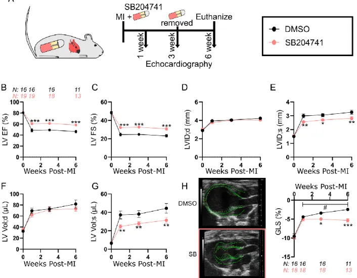
Figure 1. Antagonism of 5-HT2B preserves cardiac structure and function following 2
myocardial infarction (MI) and prevents deterioration of myocardial contractile capability.
3
A, Experimental approach. 12-week-old mice were subjected to MI surgery and coincidentally
4
treated with dimethyl sulfoxide (DMSO) control or the 5-HT2B antagonist, SB204741 (SB).
5
Treatment was ceased three weeks following injury, and serial echocardiography was performed
6
at times shown. B, Left ventricular ejection fraction (LV EF). C, Left ventricular fractional
7
shortening (LV FS). D,E, Left ventricular internal dimension at diastole (LVID;d) and
end-8
systole (LVID;s). F,G, Left ventricular volume at end-diastole (LV Vol;d) and end-systole (LV
9
Vol;s). H, Vector diagram showing magnitude and direction of myocardial deformation in
10
systole and quantified global longitudinal strain (GLS). B-H, Mean ± SEM, *P<0.05, **P<0.01,
11
***P<0.001 between DMSO and SB treatments, #P<0.05 between timepoints within treatment
12
group following 2-way ANOVA and Holm-Sidak post hoc test. Number of mice denoted in B
13
applies to subsequent groups except H where explicitly labeled.
14 15
33 1
Figure 2. Diminished 5-HT2B signaling decreases fibrotic scar formation and border zone 2
(BZ) expansion after myocardial infarction (MI) through alterations in collagen
3
composition. A-C, Analytical approach of calculating tissue thickness (A), demarcation of scar
4
vs. healthy myocardium (B), and (C) mathematical definition of BZ as the transition region
5
between scar dominated (>85% collagen stained red with picrosirius red; PSR) and myocardium
6
dominated (>85% myocardium stained yellow) with inset illustrating a representative curve from
7
each treatment. D, Thickness of the formed scar is decreased with SB204741 (SB) treatment but
8
does not thin over time (N=3-10). E, Decreased BZ infiltration with 5-HT2B antagonism as
9
indicated by the rapid transition from scar to myocardium (N=8-10). F, No difference in
10
interstitial fibrosis was observed (N=8-10). G-H, PSR stain imaged under polarized light in the
34
BZ and scar revealed an increased proportion of thinner, less mature collagen fibers in the BZ of
1
SB-treated mice (N=9-10). I, Analysis of collagen fiber orientation in the BZs of DMSO- (31
2
FOVs across 4 mice) and SB- (35 FOVs across 5 mice) treated animals to quantify the
3
distribution of orientations to classify as isotropic or anisotropic. All data 6 weeks post-MI
4
except where noted in D. D-F, H, Mean ± SEM, *P<0.05, **P<0.01 (color denotes difference
5
between corresponding color proportion in H) (D,H) 2-way ANOVA and Holm-Sidak post hoc
6
test or (E,F) 2-tailed Student t test.
7 8