HAL Id: tel-02918664
https://tel.archives-ouvertes.fr/tel-02918664
Submitted on 21 Aug 2020HAL is a multi-disciplinary open access archive for the deposit and dissemination of sci-entific research documents, whether they are pub-lished or not. The documents may come from teaching and research institutions in France or abroad, or from public or private research centers.
L’archive ouverte pluridisciplinaire HAL, est destinée au dépôt et à la diffusion de documents scientifiques de niveau recherche, publiés ou non, émanant des établissements d’enseignement et de recherche français ou étrangers, des laboratoires publics ou privés.
Pro-apoptotic and radiosensitizing potential of four
candidate microtubule regulators in breast cancer cells
Elsie Nolte
To cite this version:
Elsie Nolte. Pro-apoptotic and radiosensitizing potential of four candidate microtubule regulators in breast cancer cells. Cellular Biology. Université Grenoble Alpes; University of Pretoria, 2019. English. �NNT : 2019GREAV002�. �tel-02918664�
THÈSE
Pour obtenir le grade de
DOCTEUR DE LA COMMUNAUTE UNIVERSITE
GRENOBLE ALPES
préparée dans le cadre d’une cotutelle entre la
Communauté Université Grenoble Alpes et
l'Université de Pretoria (Afrique du Sud)
Spécialité : Biologie Cellulaire
Arrêté ministériel : le 6 janvier 2005 – 25 mai 2016 Présentée par
Elsie NOLTE »
Thèse dirigée par Anne MERCIER et Laurence LAFANECHERE
préparée au sein de l'Équipe Régulation et Pharmacologie du Cytosquelette de L'Institut pour l'Avancée de Biosciences,
Grenoble, France and the Department of Physiology, Cancer Cell Physiology Laboratory, University of Pretoria, Pretoria, South Africa
dans les Écoles Doctorales de Chimie et Sciences du Vivant et de School of Medicine, Department of Physiology,
University of Pretoria
Étude du potentiel pro-apoptotique et
radiosensibilisateur de quatre
candidats-médicaments régulateurs des microtubules,
sur les cellules de cancer du sein
Thèse soutenue publiquement le 20 Février 2019 devant le jury composé de :
Monsieur Jean-Luc COLL
Directeur de Recherche, Institut pour l'Avancée des Biosciences, Président
Monsieur Siamak HAGHDOOST
Professeur, Université de Stockholm, Suède, Rapporteur
Madame Megan Jean BESTER
Professeur, Université de Pretoria, Afrique du Sud, Rapporteur
Madame Anne Mercier
MCU-PH, Université de Pretoria, Afrique du Sud, Directrice de Thèse
Madame Laurence LAFANECHERE
Directeur de Recherche, Institut pour l'Avancée des Biosciences, Directrice de thèse
i
Thesis submitted in fulfilment of the requirements for the
degree
Philosophiæ Doctor (Ph.D) in Human Physiology
Title:
Pro-apoptotic and radiosensitizing potential of four candidate
microtubule regulators in breast cancer cells
by
Elsie Magdalena Nolte
(UP:10005022; UGA:61600881)
Department of Physiology
School of Medicine, Faculty of Health Sciences
University of Pretoria
ii
Project supervisors
Supervisor:
Dr. AE Mercier
Department of Physiology, University of Pretoria,
Pretoria, South Africa
Prof. L Lafanechère
Institute for Advanced Biosciences, INSERM U1209/
UMR
CNRS 5309, Université Grenoble Alpes, Grenoble,
France
Co-supervisor: Prof. AM Joubert
Department of Physiology
University of Pretoria, Pretoria, South Africa
Prof. R Lakier
Department of Radiation Oncology
Steve Biko Academic Hospital
iii
“I am just a child who has never grown up. I still keep asking these
'how' and 'why' questions. Occasionally, I find an answer.”
- Stephen Hawking -
“Tog, ondanks ons stukkende vlerke, leer ons vlieg. Hoër en verder as
wat ons ooit kon droom”
iv
"En as ek die dag my hand in joune sit, sal ek my padkaarte bêre met die wete:
my reise opsoek na jou is nou verby!" - Antjie Krog.
Zander Coetzee, dankie dat jy jou lewe (en Whispers) met my deel!
v
Summary
Microtubule targeting agents are effective anti-cancer drugs. Their use as part of a combined treatment modality with ionising radiation is also a promising strategy. However, the emergence of chemical and radiation resistance requires searching for alternative treatments. Our laboratories have recently described several drugs that directly or indirectly target the cellular microtubules. 2-Ethyl-3-O-sulphamoyl-estra-1,3,5(10)16-tetraene (ESE-16), an analogue of 2-methoxyestradiol, is a microtubule targeting agent that binds to microtubules causing the formation of abnormal mitotic spindles. 9-Benzoyloxy-5,11-dimethyl-2H,6H-pyrido[4,3-b]carbazol-1-one (LimPyr1) is a novel inhibitor of LIM kinases that indirectly induces microtubule stabilization. As microtubule-targeting drugs, both agents, ESE-16 and LimPyr1, induce mitotic defects. We thus hypothesized that they could sensitize cells to radiation as the G2/M phase is the most radiosensitive phase of the cell
cycle. The aim of this PhD project was to test that hypothesis and, more specifically, to investigate whether low-doses of ESE-16 and LimPyr1 could increase apoptosis and delay nuclear repair induced by radiation in breast cancer cells in vitro.
Breast cancer cell lines namely MCF-7-, MDA-MB-231- and BT-20 cells, were exposed to ESE-16 and LimPyr1 for 24-hours prior to 8 Gy radiation. The effects of these combination therapies were compared to those obtained from cells exposed to the compounds alone or only to radiation. The activation of the survival and intrinsic apoptotic pathways were investigated. Results revealed altered survival and death signaling in cells exposed to the individual treatments. The combination treatments decreased cell survival while apoptotic signaling was increased, resulting in increased cell death. Furthermore, the combination treatments significantly increased the presence of micronuclei in BT-20 cells, indicating an increase in deoxyribonucleic acid (DNA) damage. MCF-7- and MDA-MB-231 cells displayed similar micronuclei formation when exposed to the combination treatments or radiation only. Phosphorylation of H2AX (γH2AX) (normally increased upon DNA damage) and Ku70 expression (required for DNA repair) were decreased in pre-treated breast cancer cells 2
vi
hours after irradiation compared to cells exposed to radiation only. The expression of γH2AX and Ku70, however, were significantly increased 24 hours after irradiation in the pre-treated cells. LimPyr1 decreased radiation resistance development from dose fractionation by increasing the permeability of the mitochondrial membrane. An increase in the generation of reactive oxygen species (ROS) was not observed in ESE-16 pre-treated cells exposed to fractionated radiotherapy. We also observed pro-apoptotic signaling between cells exposed to radiation and non-exposed cells via the radiation-induced bystander effect.
In conclusion, the anti-mitotic effect of ESE-16 and LimPyr1 renders the chromosomes more exposed to radiation damage as assessed by the increased occurrence of micronuclei. Moreover, both compounds decreased the signaling and trafficking of DNA damage and repair proteins. Additionally, LimPyr1 prevented the development of radiation resistance in cells exposed to fractionated radiation doses. Future studies will aim to elucidate the molecular mechanisms responsible for ESE-16 and LimPyr1 radiosensitization, as well as validate the safety and efficacy of this approach in vivo.
Key words: Cancer, radiation therapy, 2-methoxyestradiol, LIM kinase, ESE-16, LimPyr1, radiosensitization, Akt, FoxO1/3a, reactive oxygen species, Bax, mitochondria, caspase, DNA damage and repair, γH2AX, Ku70, apoptosis, radiation-induced bystander effect, radiation resistance
vii
Research outputs
1. Internationally peer‐reviewed accredited scientific publications
1. Nolte EM, Joubert AM, Lakier R, Van Rensburg A, Mercier AE. Exposure of breast and lung cancer cells to a novel estrone analog prior to radiation enhances Bcl-2-mediated cell death. Int J Mol Sci. 2018;19(10):2887.
2. Sadoul K, Joubert C, Michallet S, Nolte E, Peronne L, Ramirez-Rios S, Ribba AS, Lafanechère L. Déchiffrage du code tubuline-Le voile se lève sur le rôle de l’acétylation et de la détyrosination. Med Sci. 2018;34(12):1047-1055.
3. Helena JM, Joubert AM, Grobbelaar S, Nolte EM, Nel M, Pepper MS, Coetzee M, Mercier AE. Deoxyribonucleic acid damage and repair: Capitalizing on our understanding of the mechanisms of maintaining genomic integrity for therapeutic purposes. Int J Mol Sci. 2018;19(4):1148.
4. Verwey M, Nolte EM, Joubert AM, Theron AE. Autophagy induced by a sulphamoylated estrone analogue contributes to its cytotoxic effect on breast cancer cells. Cancer Cell Int. 2016;16(1):91.
5. Klionsky DJ, et al. Guidelines for the use and interpretation of assays for monitoring autophagy. Autophagy. 2016;12(1):1-222.
6. Theron A, Nolte E, Lafanechere L, Joubert A. Molecular crosstalk between apoptosis and autophagy induced by a novel 2-methoxyestradiol analogue in cervical adenocarcinoma cells. Cancer Cell Int. 2013;13(1):87.
7. AE Theron, R Prudent, E Nolte, I van den Bout, R Punchoo, S Marais, P Du Toit, Y Hlope, D van Papendorp, L Lafanechère and A Joubert. Novel in silico-designed estradiol analogues are cytotoxic to a multidrug-resistant cell line at nanomolar concentrations. Cancer Chemother. Pharmacol. 2015;75(2):431-437.
viii
2. Published conference proceedings and conference presentations
1. Nolte EM, Joubert AM, Lakier R, Sebopa L, Prudent R, Lafanechère L, Mercier AE. Intra- and intercellular signaling pathways driving the radiosensitizating effect of a Lim kinase inhibitor in vitro. Oral presentation at the 56th annual South African Association of Physicists in Medicine and Biology congress (SAAPMB) (22-26 October 2018).
2. Helena JM, Joubert AM, Mabeta P, Coetzee M, Lakier R, Nolte EM, Nel M, Sebopa L, Rathidili M, Mercier AE. Radiosensitization of in vitro metastasis models by a spindle disruptor potentially involves increased DNA damage, proapoptotic signaling and inhibiting neovascularization. Oral presentation at the 56th annual South African Association of Physicists in Medicine and Biology congress (SAAPMB) (22-26 October 2018).
3. Nolte EM, Joubert AM, Lakier R, Etsebeth M, Sebopa L, Prudent R, Lafanechère, Mercier AE. Disruption of both actin- and microtubule dynamics sensitizes triple negative breast cancer cells to radiation. Oral presentation at the 45th Conference of the Physiology Society of Southern Africa (27-31 August 2017).
4. Nolte EM, Joubert AM, Lakier R, Etsebeth M, Sebopa L, Lafanechère L & Mercier AE. A novel estradiol analogue delays deoxyribonucleic acid repair in triple negative breast cancer cells exposed to radiation in vitro. Physica Medica. 2017;41:S11.
5. Nolte EM, Joubert AM, Lakier R, Etsebeth M, Sebopa L, Prudent R, Lafanechère L & Mercier AE. Targeting actin dynamics with a novel LIM kinase inhibitor results in radiosensitization of triple negative breast cancer cells and radiation-induced pro-apoptotic bystander effects. Physica Medica. 2017;41:S14-15.
6. Helena JM, Joubert AM, Mabeta P, Coetzee M, Lakier R, Nolte E, Verwey M, Etsebeth M, Moosa S, Sebopa L, Mercier AE. Radiosensitization capacities of a novel microtubule disruptor assessed on a deconstructed bone metastasis model. Physica Medica. 2017;41:S6.
7. Nolte EM, Joubert AM, Hargrave SD, Helena JM, Lakier R, Etsebeth M, Verwey M, Theron AE. O41. Novel microtubule disruptors radiosensitize cancer cells in vitro. Physica Medica. 2016;32:154.
ix
8. Verwey M, Joubert AM, Meijer W, Nolte EM, Lakier R, Etsebeth M, Helena J, Theron AE. P10. Radiosensitization of breast adenocarcinoma cells by a novel estrone analogue is dependent on reactive oxygen species signaling. Physica Medica. 2016;32:164.
9. Nolte EM, Lakier R, Etsebeth M, Helena JM, Verwey M, Joubert A.M, Lafanechère L & Theron AE. Cellular mechanisms involved in the radiosensitization of breast- and lung cancer cells by novel agents targeting microtubule dynamics. Oral presentation at the 18th International Conference on Integrative Biology and Physiology (3-4 November 2016).
10. Nolte EM, Lakier R, Van Rensburg A, Helena JM, Verwey M, Matlala P, Punchoo R, du Toit P, Joubert AM and Theron AE. Potential radiosensitization of breast cancer cells by a novel anti-mitotic estrone analogue. Poster at the 17th National Congress of South African Society of Medical Oncology (SASMO) and South African Society of Clinical & Radiation Oncology (SASCRO) (7-9 Augusts 2015)
11. EM Nolte, R. Lakier, A Van Rensburg, JM Helena, M Verwey, AM. Joubert, and AE Theron. P.11-Potential radiosensitisation of breast-and lung cancer cells by a novel anti-mitotic oestrone analogue. Physica Medica. 2015;31: S17-S18.
12. Helena JM, Lakier R, van Rensburg A, Nolte EM, Verwey M, Matlala P, Mlambo R, Joubert AM and AE Theron. Radiosensitization of cancer cells with a novel estradiol analogue affects nuclear repair mechanisms. Poster at the Microscopy Association of Southern Africa Annual Congress (29 November - 4 December 2015).
13. Nolte, E.M., Joubert, A.M. & Theron, A.E. Early and late cell death characteristic induced by novel anticancer estradiol analogue in vitro. Poster presentation at the Annual Congress of The South African Academy for Science and Arts: Division Biological Sciences (16 October 2013).
3. Postgraduate supervision
x
4. Academic awards
1. 68th Lindau Nobel Laureate Meeting Participant, 24-29 June 2018, Lindau, Germany
The 68th Lindau Nobel Laureate Meeting was dedicated to physiology and medicine. A total of 43 Nobel Laureates met 600 of the most qualified undergraduate students, PhD candidates and post-doctoral fellows. The one week scientific program consisted of lectures, master classes and panel discussions where Nobel Laureates and young scientist had the opportunity to exchange ideas, knowledge and experiences.
2. Third place for oral presentation at the annual University of Pretoria, Department of Health Sciences' Faculty day for basic sciences (2016)
Nolte EM, Lakier R, Etsebeth M, Helena JM, Verwey M, Joubert A.M, Lafanechère L &
Theron AE. Cellular mechanisms involved in the radiosensitization of breast- and lung cancer cells by novel agents targeting microtubule dynamics.
3. Runner-up for best non-clinical research paper in the Faculty of Health Sciences, UP (2014)
Theron A, Nolte E, Lafanechere L, Joubert A. Molecular crosstalk between apoptosis and autophagy induced by a novel 2-methoxyestradiol analogue in cervical adenocarcinoma cells. Cancer Cell Int. 2013;13(87):13-87.
xi
Acknowledgements
With sincere gratitude to:
Dr Joji Mercier, my supervisor - Thank you for your support and encouragement over the past three years. Thank you for making me a better scientist and human being. Thank you for challenging me to rise higher than I ever thought I could.
Prof Laurence Lafanechère, my French supervisor - Il n’y a pas assez de mots pour vous remercier pour m’avoir donné l’incroyable opportunité de réaliser ma thèse en co-tutelle avec vous dans votre laboratoire. Vous m'avez accueillie à bras ouverts et m'avez fourni les conseils et le soutien indispensables tout au long de mon année en France. Vous étiez mon superviseur, mon interprète et ma ‘mère française’. Avec un cœur généreux, vous m'avez soutenu financièrement pour assister à la réunion des lauréats du prix Nobel Lindau: une opportunité que je chérirai pour toujours. Ce fut un privilège de travailler avec vous.
Prof Annie Joubert, my co-supervisor - Ek sal ewig dankbaar wees vir al die ondersteuning wat Prof aan my gebied het deur hierdie avontuur. Prof het altyd ‘n helpende hand uitgereik, ‘n ondersteunende oor gebied en kosbare ure spandeer om seker te maak ek voltooi hierdie graad suksesvol. Die ondersteuning wat Prof gebied het die laaste week voor die senutergende ‘defence’ is van onskatbare waarde en ek sal dit vir ewig in my hart koester. Prof is nie net my ‘co-supervisor’ nie, prof is werklik my rolmodel!
Prof Roy Lakier, my co-supervisor - Thank you for welcoming me into your department and allowing me to use your equipment. Thank you for all your assistance and support, it is much appreciated.
xii Leanor Sebopa and Prof Laurence Chaperot, my radiation partners - Thank you for assisting me in radiating hundreds of samples. Without you this project wouldn’t have been possible.
Lauralie, Sacnicte, Karin, Anne-Sophie, Clotilde and Sophie, my French friends - À mes amies françaises Lauralie, Sacnicte, Karin, Anne-Sophie, Clotilde et Sophie - Merci pour toute votre patience et votre aide. Vous avez fait de mon année en France une année toute particulière et je continuerai de garder dans mon coeur tous nos merveilleux souvenirs.
Marcel, my best friend - Vir ‘n beter ‘partner in crime’ kon ek nie vra nie. Jy het saam met my liters koffie gedrink, jy het geluister na my gekerm en kla, jy het saam met my op die kantoormat gelê as alles net te veel geraak het. Jy was altyd net daar. Jy het elke oorwinning saam met my gevier en elke terugslag reg ‘gedokter’ met ‘n cocktail. Jou vriendskap beteken oneindig baie vir my. Dankie!
Ria and Fritz Nolte, my parents - Jare se gebed, liefde, geduld, ondersteuning, en onbaatsugtige en onbenoemde opofferings het gelei tot hierdie sukses. Nooit het julle ophou glo in my vermoë om suksesvol te wees nie. Die trane in jull oë as julle met my ‘spog’ maak alles die moeite werd. Dankie dat julle my ten volle voorberei het vir die ‘grootmens’ wêreld. My sukses is bloot ‘n refleksie van julle opvoeding.
André, my big baby brother - Dankie dat jy my gereeld herinner dat die lewe geleef moet word. Dat die klein oorwinnings net soveel waarde dra soos die grotes. Jy was nog altyd trots daarop dat ek jou sussie is, maar soveel meer is ek dankbaar dat jy my broer is. Jy sal my altyd bly inspireer.
Zander, my husband - Hoe sê mens dankie vir jare se opofferings, bystand, aanmoediging en ondersteuning? Woorde is nie genoeg nie. Jy het elke treë van hierdie pad saam met my gestap. Elke opdraende en afdraende, jy was daar. Jy het my met ‘n hartseer maar trotse hart op ‘n vliegtuig laat klim sodat ek my droom kon
xiii
najaag om in Frankryk navorsing te doen. Jy het opoffering na opoffering gemaak en nooit enige iets terugverwag nie. Ek is ‘n besonderse gelukkige vrou om met ‘n man soos jy die res van my lewe te spandeer. Ek het jou lief!
xiv
Table of contents
Summary ... v
Research outputs ... vii
1. Internationally peer‐reviewed accredited scientific publications ... vii
2. Published conference proceedings and conference presentations ... viii
3. Postgraduate supervision ... ix
4. Academic awards ... x
Acknowledgements ... xi
List of figures ... xx
List of tables ... xxvii
List of abbreviations ... xxxi
1. Introduction and literature study ... 1
1.1 Breast cancer ... 1
1.1.1 Classification of breast cancer ... 3
1.1.2 Treatment options for breast cancer ... 4
1.2 The cell cycle ... 5
1.2.1 The G1 phase ... 7
1.2.2 The S phase ... 8
1.2.3 The G2 phase ... 8
1.2.4 The M phase ... 9
1.2.5 Cell cycle checkpoints ... 10
1.2.5.1 DNA structure checkpoints ... 10
1.2.5.2 Spindle assembly checkpoint ... 11
1.3 Cell death ... 13
1.3.1 Necrosis ... 13
1.3.2 Apoptosis ... 14
1.3.2.1 Regulation of apoptosis ... 15
xv
1.4 Radiotherapy as an effective anti-cancer treatment modality ... 19
1.4.1 DNA damage signaling and repair ... 22
1.4.2 Cell death induced by radiation ... 24
1.4.3 Radiation resistance ... 26
1.4.4 Radiation-induced bystander effect ... 28
1.5 Intracellular microtubule dynamics ... 30
1.6 2-Methoxyestradiol... 33
1.6.1 2-Methoxyestradiol analogues ... 37
1.7 Lin-11, Isl-1 and Mec-3 (LIM) kinase: A regulator of the cytoskeleton ... 39
1.7.1 The role of the actin skeleton in cell migration ... 42
1.7.2 LIM kinase involvement in the cell cycle progression ... 42
1.7.3 LIM kinase inhibitors... 43
1.8 Paclitaxel ... 44
1.8.1 Paclitaxel resistance ... 45
1.8.2 Paclitaxel based combination chemotherapy ... 48
1.8.2.1 Paclitaxel-doxorubicin combination therapy ... 48
1.8.2.2 Paclitaxel-cisplatin combination therapy ... 49
1.8.2.3 Paclitaxel-cyclophosphamide combination therapy ... 50
1.8.2.4 Paclitaxel-vinorelbine combination therapy ... 50
1.8.3 Compound T4 sensitizes cells to non-lethal doses of paclitaxel in vitro ... 51
1.9 Radiosensitization by agents targeting the cytoskeleton ... 51
2. Aims and objectives ... 53
3. Materials and methods ... 57
3.1 Cell lines ... 57
3.2 General cell culture maintenance ... 57
3.2.1 Cell culture maintenance reagents ... 58
3.2.2 Maintenance ... 58
3.3 General method for experiments ... 59
3.3.1 Compounds and positive controls ... 60
3.3.2 General experimental method ... 60
xvi
3.4 Experimental setup ... 62
3.5 Drug properties and relative cytotoxicity ... 64
3.5.1 Tubulin competitive binding assay: Tritiated [3H] colchicine binding ... 64
3.5.2 Time-lapse imaging: ESE-16’s anti-mitotic effect ... 65
3.5.3 IncuCyte® Cytotoxicity assay ... 66
3.5.4 Scratch wound cell migration assay ... 67
3.5.5 Cytotoxicity studies: Spectrophotometric quantification of 3-(4,5- dimethylthiazol-2-yl)-2,5-diphenyltetrazolium bromide (MTT) ... 68
3.5.6 Visualization of the cytoskeleton: Fluorescent microscopy ... 70
3.6 Radiation dose-reponse curve ... 71
3.6.1 Apoptosis induction in cells exposed to radiation doses: Annexin V quantification via flow cytometry ... 71
3.6.2 Cell cycle analysis of cells exposed to radiation doses: Flow cytometry employing propidium iodide ... 72
3.7 Compound dose-response curve ... 74
3.7.1 Apoptosis induction in cells exposed to compound doses: Flow cytometric quantification of Annexin V ... 74
3.7.2 Cell cycle analysis of cells exposed to compound doses: Flow cytometry employing propidium iodide ... 74
3.8 Radiosensitization studies ... 74
3.8.1 Flow cytometric quantification of Annexin V-FITC ... 74
3.8.2 Cell cycle analysis ... 75
3.8.3 Intracellular signaling pathways induced by pre-exposing breast cancer cells to the novel compounds prior to radiation ... 75
3.8.3.1 Reactive oxygen species production: Flow cytometric quantification of superoxide employing hydroethidine ... 76
3.8.3.2 Mitochondrial transmembrane potential: Flow cytometric quantification of Mitocapture™ ... 76
3.8.3.3 Western blots ... 77
3.8.4 Morphological studies: Polarization-optical transmitted light differential interference contrast microscopy ... 82
3.8.5 Cell survival assay: Long term cellular proliferation quantified via a crystal violet stain…….. ... 83
xvii
3.8.6 DNA damage and repair ... 84
3.8.6.1 Quantification of phosphorylated H2AX via flow cytometry ... 84
3.8.6.2 Micronuclei quantification ... 85
3.8.6.3 Quantification of the DNA repair response via Western blot analysis of Ku70 expression ... 86
3.9 Evaluation of the bystander effect induced by irradiated cells ... 87
3.10 Evaluation of radiation-induced resistance due to dose fractionation in cells exposed to the combination treatment ... 88
3.11 Statistical analysis ... 88
3.12 Logistics and funding ... 90
3.13 Ethics approval ... 90
4. Results ... 91
4.1 Drug properties and relative cytotoxicity ... 91
4.1.1 Tubulin competitive binding assay: Tritiated [3H] colchicine binding ... 91
4.1.2 Time-lapse imaging: ESE-16’s anti-mitotic effect ... 93
4.1.3 IncuCyte® Cytotoxicity assay ... 94
4.1.4 Scratch wound cell migration assay ... 96
4.1.5 Cytotoxicity studies: Spectrophotometric quantification of 3-(4,5-dimethylthiazol-2-yl)-2,5-diphenyltetrazolium bromide ... 98
4.1.6 Visualization of the cytoskeleton: Fluorescent microscopy ... 100
4.2 Radiosensitization studies ... 105
4.2.1 Flow cytometric quantification of annexin V-FITC ... 105
4.2.2 Cell cycle analysis ... 117
4.2.3 Intracellular signaling pathways induced by pre-exposing breast cancer cells to the novel compounds prior to radiation ... 130
4.2.3.1 Reactive oxygen species production: Flow cytometric quantification of superoxide employing hydroethidine ... 130
4.2.3.2 Mitochondrial transmembrane potential: Flow cytometric quantification of Mitocapture™ ... 143
4.2.3.3 Western blot... 148
4.2.4 Morphological studies: Polarization-optical transmitted light differential interference contrast microscopy ... 167
xviii
4.2.6 DNA damage and repair ... 175
4.2.6.1 Quantification of phosphorylated H2AX via flow cytometry ... 175
4.2.6.2 Micronuclei quantification ... 187
4.2.6.3 Quantification of the DNA repair response via Western blot analysis of Ku70 expression. ... 196
4.3 Evaluation of the bystander effect induced by irradiated cells ... 207
4.3.1 Reactive oxygen species production: Flow cytometric quantification of superoxide employing hydroethidine ... 207
4.3.2 Mitochondrial transmembrane potential: Flow cytometric quantification of Mitocapture™ ... 211
4.3.3 Long term cellular proliferation quantified by crystal violet staining. ... 215
4.3.4 DNA damage and repair: Quantification of γH2AX and Ku70 proteins via a western blot ... 219
4.4 Evaluation of radiation-induced resistance due to dose fractionation in irradiated cells….. ... 222
4.4.1 Reactive oxygen species production: Flow cytometric quantification of superoxide employing hydroethidine ... 222
4.4.2 Mitochondrial transmembrane potential: Flow cytometric quantification of MitoCapture™ ... 227
5. Discussion ... 233
5.1 Drug properties and cytotoxicity ... 243
5.2 Regulation of the cell cycle ... 251
5.3 Cellular survival and apoptotic cell death ... 255
5.4 DNA damage and repair ... 266
5.5 Radiation-induced resistance due to dose fractionation and radiation-induced bystander effect ... 273
6. Conclusion ... 277
7. Acknowledgement of funding ... 280
8. References ... 281
9. Additional file 1 ... 354
9.1 Radiation dose response curve: Flow cytometric quantification of apoptosis induction……….354
xix
9.2 Radiation dose response curve: Flow cytometric analysis of cell cycle
progression. ... 358
10. Additional file 2 ... 364
10.1 Compound dose response curve: Flow cytometric quantification of apoptosis induction ... 364
10.2 Compound dose response curve: Flow cytometric analysis of cell cycle progression ... 373
11. Appendix A: Data analysis of Incucyte® experiments ... 384
12. Appendix B: Distribution of micronuclei in breast cancer cells exposed to various treatment conditions ... 387
13. Video’s ... 389
14. Ethics approval ... 389
xx
List of figures
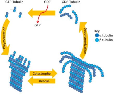
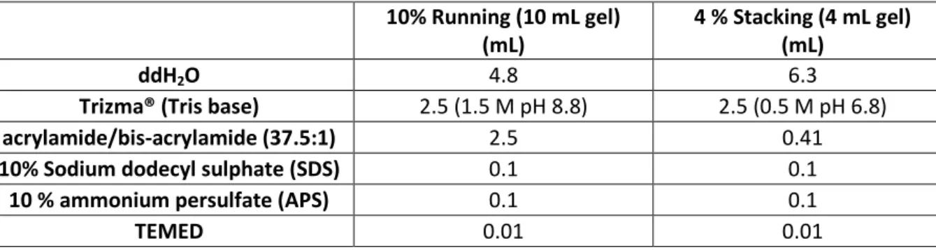
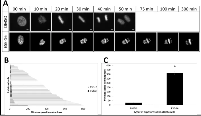
Figure 1.1: The phases of the cell cycle ... 6 Figure 1.2: The spindle assembly checkpoint ... 12 Figure 1.3: The intrinsic and extrinsic apoptotic pathways ... 16 Figure 1.4: Cellular response to radiation-induced DNA double strand breaks ... 21 Figure 1.5: Factors contributing to the development of radiation resistance ... 27 Figure 1.6: Microtubule dynamics ... 31 Figure 1.7: Hepatic metabolism of estradiol ... 34 Figure 1.8: In silico-designed sulphamoylated 2-methoxyestradiol analogues ... 39 Figure 1.9: LIM kinase protein structure ... 39 Figure 1.10: Regulation of the cytoskeleton by LIM kinase ... 41 Figure 1.11: Chemical structure of 9-benzoyloxy-5,11-dimethyl-2H,6H-pyrido[4,3-b]carbazol- 1-one (Pyr1). ... 44 Figure 1.12: Cellular mechanisms involved in Paclitaxel resistance ... 46 Figure 3.1: Experimental setup ... 63 Figure 4.1: Binding of tritiated colchicine to tubulin in the presence of various anti-mitotic compounds ... 92 Figure 4.2: Time-lapsed imaging of HeLa Kyoto cells exposed to the anti-mitotic agent,
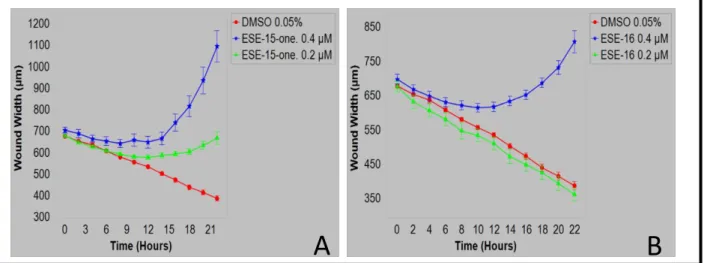
ESE-16... 93 Figure 4.3: Real time IncuCyte® Cytotox Green fluorescence in HeLa cells exposed to two anti-mitotic compounds indicating the number of dead HeLa cells ... 95 Figure 4.4: Graphical representation of IncuCyte® Cytotox Green fluorescence in HeLa cells exposed to ESE-15-one and ESE-16 ... 96 Figure 4.5: Wound closure in HeLa cells exposed to ESE-15-one and ESE-16 indicating the effect on cell migration ... 97 Figure 4.6: Graphical representation of wound closure in HeLa cells exposed to ESE-15-one and ESE-16 ... 98
xxi
Figure 4.7: Dose response curves of breast cancer cells exposed to various microtubule regulators and T4, a compound that sensitizes cells to non-lethal doses of
paclitaxel ... 99 Figure 4.8: Response of the cytoskeleton of BT-20 cells exposed to various microtubule
regulating agents for 24 hours... 102 Figure 4.9: Response of the cytoskeleton of MCF-7 cells exposed to ESE-16 and LimPyr1 ... 103 Figure 4.10: Response of the cytoskeleton of MDA-MB-231 cells exposed to ESE-16 and
LimPyr1 for 24 hours ... 104 Figure 4.11: Representative flow cytometric dot plots of annexin V-FITC indicating apoptosis in BT-20 cells exposed to various treatment modalities ... 107 Figure 4.12: Graphical representation of annexin V-FITC quantification in BT-20 cells exposed to various treatment conditions ... 109 Figure 4.13: Representative flow cytometric dot plots of annexin V-FITC indicating apoptosis in MCF-7 cells exposed to various treatment modalities ... 111 Figure 4.14: Graphical representation of annexin V-FITC quantification in MCF-7 cells
exposed to various treatment conditions ... 113 Figure 4.15: Representative flow cytometric dot plots of annexin V-FITC indicating apoptosis in MDA-MB-231 cells exposed to various treatment modalities... 114 Figure 4.16: Graphical representation of annexin V-FITC quantification in MDA-MB-231 cells exposed to various treatment conditions ... 116 Figure 4.17: Histograms representing the cell cycle distributions of BT-20 cells exposed to various treatment conditions... 119 Figure 4.18: Graphical representation of cell cycle analysis in BT-20 cells exposed to various treatment conditions ... 122 Figure 4.19: Histograms representing the cell cycle distributions of MCF-7 cells exposed to various treatment conditions... 124 Figure 4.20: Graphical representation of cell cycle analysis in MCF-7 cells exposed to various treatment conditions ... 126 Figure 4.21: Histograms representing the cell cycle distributions of MDA-MB-231 cells
xxii
Figure 4.22: Graphical representation of the cell cycle analysis of MDA-MB-231 cells exposed to various treatment conditions ... 129 Figure 4.23: Representative overlay histograms of superoxide detection in MCF-7 cells ... 132 Figure 4.24: Graphical representation of superoxide quantification in MCF-7 cells after
exposure to different treatment conditions ... 134 Figure 4.25: Representative overlay histograms of superoxide detection in MDA-MB-231 cells. ... 136 Figure 4.26: Graphical representation of superoxide quantification in MDA-MB-231 cells after exposure to the different treatment conditions ... 138 Figure 4.27: Representative overlay histograms of superoxide detection in BT-20 cells ... 140 Figure 4.28: Graphical representation of superoxide quantification in BT-20 cells after
exposure to different treatment conditions ... 142 Figure 4.29: Representative overlay histograms of Mitocapture™ detection in MCF-7-,
MDA-MB-231- and BT-20 cells exposed to appropriate controls and various treatment conditions ... 145 Figure 4.30: Graphical representation of Mitocapture™ quantification in MCF-7-,
MDA-MB-231- and BT-20 cells after exposure to different treatment
conditions ... 147 Figure 4.31: Western blots visualizing Bax and cleaved caspase 3 protein expressions in
MCF-7-, MDA-MB-231- and BT-20 cells exposed to various treatment
conditions ... 150 Figure 4.32: Graphical representation of Bax expression in MCF-7 cells exposed to different treatment conditions ... 153 Figure 4.33: Graphical representation of Bax and cleaved caspase 3 proteins in MDA-MB-231 cells exposed to different treatment conditions ... 154 Figure 4.34: Graphical representation of Bax and cleaved caspase 3 proteins in BT-20 cells exposed to different treatment conditions ... 155 Figure 4.35: Western blots visualizing phosphorylated Akt and FoxO1/3a protein levels in MCF-7-, MDA-MB-231- and BT-20 cells exposed to various treatment
xxiii
Figure 4.36: Graphical representation of Akt and FoxO1/3a phosphorylation in MCF-7 cells exposed to different treatment conditions ... 161 Figure 4.37: Graphical representation of Akt and FoxO1/3a phosphorylation in MDA-MB-231 cells exposed to different treatment conditions ... 162 Figure 4.38: Graphical representation of Akt and FoxO1/3a phosphorylation in BT-20 cells exposed to different treatment conditions ... 163 Figure 4.39: Cyclin B1 quantification in MCF-7-, MDA-MB-231- and BT-20 cells exposed to various treatment conditions... 165 Figure 4.40: PlasDIC micrographs of MCF-7 cells exposed to various treatment conditions .. 168 Figure 4.41: PlasDIC micrographs of MDA-MB-231 cells exposed to various treatment
conditions ... 169 Figure 4.42: PlasDIC micrographs of BT-20 cells exposed various treatment conditions ... 170 Figure 4.43: Cellular proliferation 14 days after radiation in MCF-7-, MDA-MB-231- and
BT-20 cells exposed to various treatment conditions ... 172 Figure 4.44: Graphical representation of long term cellular proliferation 14 days after
radiation in MCF-7-, MDA-MB-231- and BT-20 cells exposed to various
treatment conditions ... 174 Figure 4.45: Representative overlay histograms of γH2AX detection in MCF-7 cells ... 178 Figure 4.46: Graphical representation of γH2AX quantification in MCF-7 cells exposed to different treatment conditions ... 180 Figure 4.47: Representative overlay histograms of γH2AX detection in MDA-MB-231 cells ... 181 Figure 4.48: Graphical representation of γH2AX quantification in MDA-MB-231 cells exposed to different treatment conditions. ... 183 Figure 4.49: Representative overlay histograms of γH2AX detection in BT-20 cells... 184 Figure 4.50: Graphical representation of γH2AX quantification in BT-20 cells exposed to
different treatment conditions ... 186 Figure 4.51: Graphical representation of micronuclei quantification in MCF-7 cells exposed to various treatment conditions ... 191 Figure 4.52: Graphical representation of micronuclei quantification in MDA-MB-231 cells exposed to various treatment conditions ... 193
xxiv
Figure 4.53: Graphical representation of micronuclei quantification in BT-20 cells exposed to various treatment conditions... 195 Figure 4.54: Distribution of micronuclei in MCF-7-, MDA-MB-231- and BT-20 cells exposed to various treatment condition ... 197 Figure 4.55: Western blots illustrating Ku70 protein expression in MCF-7-, MDA-MB-231- and BT-20 cells 2 and 24 hours after radiation ... 200 Figure 4.56: Graphical representation of Ku70 protein expression in MCF-7 cells 2 and 24 hours after irradiation ... 202 Figure 4.57: Graphical representation of Ku70 protein expression in MDA-MB-231 cells 2 and 24 hours after irradiation ... 204 Figure 4.58: Graphical representation of Ku70 protein expression in BT-20 cells 2 and 24 hours after irradiation ... 206 Figure 4.59: Representative overlay histograms of superoxide detection in untreated BT-20 cells propagated in conditioned media obtained from cells exposed to
appropriate controls and different treatment conditions for 48 hours ... 208 Figure 4.60: Graphical representation of superoxide quantification in untreated BT-20 cells propagated in conditioned media obtained from cells exposed to various
treatment conditions for 48 hours ... 210 Figure 4.61: Representative overlay histograms of Mitocapture™ detection in untreated BT-20 cells exposed to conditioned media obtained from cells exposed to
appropriate controls and different treatment conditions ... 212 Figure 4.62: Graphical representation of Mitocapture™ detection in untreated BT-20 cells following a 48 hour exposure to conditioned media obtained from cells
exposed to various treatment modalities ... 214 Figure 4.63: Cell proliferation in untreated BT-20 cells propagated in conditioned media for 14 days obtained from cells exposed to various controls and treatment
conditions ... 216 Figure 4.64: Graphical representation of cell survival in untreated BT-20 cells propagated for 14 days in conditioned media obtained from treated cells ... 218
xxv
Figure 4.65: Western blots visualizing γH2AX and Ku70 protein expressions in untreated BT-20 cells propagated in conditioned media obtained from cells exposed to various treatment conditions for 2 hours ... 220 Figure 4.66: Graphical representation of γH2AX and Ku70 expression in untreated BT-20 cells exposed for 2 hours to conditioned media transferred from cells exposed to various treatment conditions ... 221 Figure 4.67: Representative overlay histograms of superoxide detection measured by flow cytometry in BT-20 cells exposed to ESE-16 or LimPyr1, single dose and
fractionated radiotherapy ... 224 Figure 4.68: Graphical representation of superoxide quantification in BT-20 cells exposed to single dose or fractionated radiotherapy ... 226 Figure 4.69: Representative overlay histograms of MitoCapture™ in BT-20 cells exposed to microtubule regulating agents, single dose and fractionated radiotherapy ... 230 Figure 4.70: Graphical representation of MitoCapture™ quantification in BT-20 cells exposed to microtubule regulating agents, single dose or fractionated radiotherapy ... 231 Figure 6.1: Summary of proposed signaling pathways induced by ESE-16 and LimPyr1 to increase the sensitivity of breast cancer cells to radiation ... 279
Additional file 1
Figure 1A: Representative flow cytometric dot plots of annexin V-FITC of breast cancer cells exposed to a radiation dose range ... 355 Figure 1B: Graphical representation of annexin V-FITC analysis of MCF-7-, MDA-MB-231- and BT-20 breast cancer cells exposed to a radiation dose range. ... 357 Figure 1C: Histograms representing the cell cycle distributions of MCF-7-, MDA-MB-231- and BT-20 cells exposed to a radiation dose-response curve ... 360 Figure 1D: Graphical representation of the cell cycle analysis of MCF-7-, MDA-MB-231- and BT-20 cells exposed to a radiation dose range ... 362
xxvi
Additional file 2
Figure 2A: Apoptotic cell death in BT-20 cells exposed to compound dose-response curves as illustrated by flow cytometric dot plots of annexin V-FITC ... 366 Figure 2B: Graphical representation of annexin V-FITC quantification in BT-20 cells exposed to compound dose-response curves ... 368 Figure 2C: Apoptotic cell death in MCF-7- and MDA-MB-231 cells exposed to compound dose-response curves as illustrated by flow cytometric dot plots of
annexin V-FITC ... 370 Figure 2D: Graphical representation of annexin V-FITC quantification in MCF-7- and
MDA-MB-231 cells exposed to compound dose-response curves ... 372 Figure 2E: Histograms representing the cell cycle distributions of BT-20 cells exposed to compound dose-response curves ... 376 Figure 2F: Graphical representation of the cell cycle analysis of BT-20 cells exposed to
compound dose-response curves ... 379 Figure 2G: Histograms representing the cell cycle distributions of MCF-7- and MDA-MB-231 cells exposed to compound dose-response curves ... 380 Figure 2H: Graphical representation of the cell cycle analysis of MCF-7- and MDA-MB-231 cells exposed to compound dose-response curves ... 382
xxvii
List of tables
Table 3.1: Densities seeded for cytotoxicity and radiosensitizing experiments on MCF-7-, MDA-MB-231- and BT-20 cells ... 61 Table 3.2: Dilution series of ESE-15-one, ESE-16, LimPyr1 and T4 that were used during
cytotoxicity studies ... 69 Table 3.3: Reagents required to hand-cast 10% running and 4 % stacking SDS-polyacrylamide (SDS‐PAGE) gels ... 78 Table 4.1: Binding of tritiated colchicine to purified bovine tubulin in competition with
colchicine, vinblastine and ESE-16. ... 92 Table 4.2: Time HeLa Kyoto cells spent in metaphase following exposure to DMSO and an anti-mitotic compound, ESE-16 ... .94 Table 4.3: Half maximum growth inhibitory concentrations (GI50) for ESE-15-one, ESE-16, LimPyr1 and T4 after exposure to BT-20-, MCF-7- and MDA-MB-231 breast
cancer cells for 48 hours ... 100 Table 4.4: Apoptotic induction measured by annexin V-FITC quantification in BT-20 cells exposed to various treatment conditions ... 108 Table 4.5: Apoptotic induction measured by annexin V-FITC quantification in MCF-7 cells exposed to various treatment conditions ... 112 Table 4.6: Apoptotic induction measured by annexin V-FITC quantification in MDA-MB-231 cells exposed to various treatment conditions ... 115 Table 4.7: Cell cycle analysis of BT-20 cells exposed to various treatment conditions ... 121 Table 4.8: Cell cycle analysis of MCF-7 cells exposed to various treatment conditions ... 125 Table 4.9: Cell cycle analysis of MDA-MB-231 cells exposed to the various treatment
conditions ... 128 Table 4.10: Superoxide detection in MCF-7 cells exposed to various treatment conditions... 133 Table 4.11: Superoxide detection in MDA-MB-231 cells exposed to various treatment
conditions ... 137 Table 4.12: Superoxide detection in BT-20 cells exposed to various treatment conditions .... 141
xxviii
Table 4.13: Mitocapture™ quantification in breast cancer cells exposed to various treatment conditions ... 146 Table 4.14: Bax expression in MCF-7-, MDA-MB-231- and BT-20 cells exposed to various treatment conditions ... 151 Table 4.15: Cleaved caspase 3 expression in MDA-MB-231 and BT-20 cells exposed to
various treatment conditions ... 152 Table 4.16: Akt phosphorylation in MCF-7-, MDA-MB-231- and BT-20 cells exposed to
various treatment conditions ... 159 Table 4.17: FoxO1/3a phosphorylation in MCF-7-, MDA-MB-231- and BT-20 cells exposed to various treatment conditions ... 160 Table 4.18: Cyclin B1 quantification in MCF-7-, MDA-MB-231- and BT-20 cells exposed to various treatment conditions ... 166 Table 4.19: Cellular proliferation measure 14 days after radiation in MCF-7-, MDA-MB-231- and BT-20 cells following exposure to various treatment conditions ... 173 Table 4.20: Quantification of γH2AX in MCF-7 cells exposed to various treatment
conditions ... 179 Table 4.21: Quantification of γH2AX in MDA-MB-231 cells exposed to various treatment conditions ... 182 Table 4.22: Quantification of γH2AX in BT-20 cells exposed to various treatment
conditions ... 185 Table 4.23: Micronuclei quantification in MCF-7 cells 2 and 24 hours after radiation ... 190 Table 4.24: Micronuclei quantification in MDA-MB-231 cells 2 and 24 hours after
radiation ... 192 Table 4.25: Micronuclei quantification in BT-20 cells 2 and 24 hours after radiation ... 194 Table 4.26: Ku70 expression in MCF-7 cells 2 and 24 hours after irradiation ... 201 Table 4.27: Ku70 expression in MDA-MB-231 cells 2 and 24 hours after irradiation ... 203 Table 4.28: Ku70 expression in BT-20 cells 2 and 24 hours after irradiation ... 205 Table 4.29: Superoxide detection in untreated breast cancer cells exposed to conditioned media obtained from treated cells for 48 hours ... 209 Table 4.30: Mitocapture™ detection in untreated breast cancer cells following a 48 hour exposure to conditioned media obtained from treated cells... 213
xxix
Table 4.31: Cellular survival in untreated BT-20 cells exposed to conditioned media obtained from BT-20 cells exposed to various controls and treatment conditions ... 217 Table 4.32: Expression of DNA damage (γH2AX) and DNA repair (Ku70) proteins in untreated BT-20 via the bystander effect 2 hours post transfer ... 220 Table 4.33: Superoxide detection by means of flow cytometry employing hydroethidine in BT-20 cells exposed to single dose and fractionated radiotherapy ... 225 Table 4.34: MitoCapture™ quantification in breast cancer cells exposed to single dose and fractionated radiotherapy... 229
Additional file 1
Table 1A: Annexin V-FITC analysis of MCF-7-, MDA-MB-231- and BT-20 breast cancer cells following exposure to a radiation dose range ... 356 Table 1B: Cell cycle analysis of three breast cancer cell lines after exposure to a radiation dose-response curve ... 361
Additional file 2
Table 2A: Compound dose-response curve concentrations exposed to BT-20-, MCF-7- and MDA-MB-231 cells ... 364 Table 2B: Annexin V-FITC quantification in BT-20 cells exposed to compound dose-response curves.. ... 367 Table 2C: Annexin V-FITC quantification in MCF-7- and MDA-MB-231 cells exposed to
compound dose-response curves ... 371 Table 2D: Flow cytometric analysis of BT-20 cells exposed to compound dose-response
curves.. ... 378 Table 2E: Flow cytometric analysis of MCF-7- and MDA-MB-231 cells exposed to compound dose-response curves ... 381
xxx
Appendix A
Table A1: IncuCyte® Cytotox Green fluorescence in HeLa cells exposed to ESE-15-one and ESE-16, indicating cell death ... 384 Table A2: Wound healing in HeLa cells exposed to ESE-15-one and ESE-16, indicating cell migration. ... 386
Appendix B
Table B1: Micronuclei quantification in breast cancer cells pre-treated with ESE-16. ... 387 Table B2: Micronuclei quantification in breast cancer cells pre-treated with LimPyr1. ... 388
xxxi
List of abbreviations
+TIP Plus-end tracking proteins [3H] colchicine Tritiated colchicine
°C Degrees Celsius µg Microgram µL Microliter µm Micrometer µM Micromolar 17β-HSD 17β-hydroxysteriod dehydrogenase 2-ME 2-Methoxyestradiol 53BP1 p53-binding protein 1 ABC ATP-binding cassette ADF Actin-depolymerizing factor ADP Adenosine diphosphate AIF Apoptosis inducing factor
Akt v‐Akt murine thymoma viral oncogene also known as protein kinase B AMP Adenosine monophosphate
AMPK 5′ AMP-activated protein kinase
ANOVA Analysis of variance
Apaf Apoptotic protease activating factor
APC/C Anaphase promoting complex/cyclosome
APS Ammonium persulfate
ATCC American Type Culture Collection
ATM Ataxia–telangiectasia-mutated
ATP Adenosine triphosphate
ATR Ataxia telangiectasia and Rad3-related
Bad Bcl-2-associated death promoter
Bak Bcl-2 homologous antagonist-killer
Bax Bcl-2 associated X protein
BC Before Christ Bcl B-cell lymphoma Bcl-XL Bcl-extra large BH Bcl-2 homology domain Bid BH3 interacting-domain death agonist
Bim Bcl-2-like protein 11
BMPRII Bone morphogenetic protein receptor II
BRCA Breast cancer gene
BSA Bovine serum albumin
Bub Budding uninhibited by benzimidazoles
CA California
xxxii CAII Carbonic anhydrase II
CAIX Carbonic anhydrase XI
CAK Cyclin activating kinase
CANSA Cancer Association of South Africa
CARD Caspase recruitment domain
Cdc Cell division cycle
CDK Cyclin dependent kinases
CDKI Cyclin-dependent kinase inhibitor
CDT1 Chromatin licensing and DNA replication factor 1
CE Common Era cGy Centigray CIN Chronophin
Cip/Kip CDK interacting protein/Kinase inhibitory protein
Ckh Checkpoint kinase
CLIP-170 Cytoplasmic linker protein
cm Centimeter
CNRS Le Centre National de la Recherche Scientifique
CO2 Carbon dioxide COMT Catechol-O-methyltransferase COX Cyclooxygenase
CYP Cytochrome P450 family
Da Dalton
DAPI 4′,6-Diamidino-2-phenylindole
dATP Deoxyadenosine triphosphate
ddH2O Double-distilled water
DDR DNA damage response
DEAE Diethylaminoethyl
DED Death effector domain
DISC Death-inducing signalling complex
DMEM Dulbecco's Modified Eagle's medium
DMSO Dimethyl sulfoxide
DNA Deoxyribonucleic acid
DNA-PK DNA dependent protein kinase
DNA-PKcs DNA-dependent protein kinase catalytic subunit
DR Death receptor
DSB DNA double strand breaks
E2F Elongation factor 2
EB3 Microtubule-associated protein RP/EB family member 3
ECL Enhanced chemiluminescence
EDTA Ethylenediaminetetraacetic acid
EGF Epidermal growth factor
EGFR Epidermal growth factor receptor
EGTA Ethylene glycol-bis(β-aminoethyl ether)-N,N,N',N'-tetraacetic acid
xxxiii EMT Epithelial–mesenchymal transition
EndoR Endoplasmic reticulum
eNOS Endothelial nitric oxide synthase ER Estrogen receptor
ERK Extracellular signal-regulated kinases
ESE-15-one 2-ethyl-3-O-sulphamoyl-estra-1,3,5(10),15-tetraen-17-one ESE-16 2-ethyl-3-O-sulphamoyl-estra-1,3,5(10)16-tetraene
F-actin Filamentous actin
FADD Fas-associated protein with death domain FasL Fas ligand
FC Fold change
FCS Fetal calf serum
FEI Field Electron and Ion Company FITC Fluorescein isothiocyanate FKBP FK506-binding protein
FL Florida
FoxO Forkhead box proteins
FRAP FKBP-rapamycin-associated protein
g Gram
G0 phase Quiescent phase
G1 phase Gap 1 phase
G2 phase Gap 2 phase
GADD DNA damage inducible gene GCP γ-Tubulin complex protein GDP Guanosine diphosphate GFP Green fluorescent protein
GI50 Half maximum growth inhibitory concentration
GJIC Gap junctional intercellular communication
GSH Glutathione GSK3β Synthase kinase-3β GSSG Glutathione disulphide GTP Guanosine triphosphate Gy Gray H2O2 Hydrogen peroxide HCl Hydrochloric acid
Her2 Human epidermal growth factor receptor 2 HIF Hypoxia-inducible factor
HR Homologous recombination HRP Horseradish peroxidase
IAB Institute for Advanced Biosciences
IgG Immunoglobulin G
IL Interleukin
INK4 Inhibitors of the cyclin-dependent kinase 4
xxxiv JNK c-Jun N-terminal kinases
KCl Potassium chloride kDa Kilodalton keV Kiloelectronvolts kg Kilogram KH2PO4 Potassium dihydrogen phosphate
Ki Inhibitor constant
Kif Kinesin heavy chain member
Ku Ku70-Ku80 heteromere
L Liter
LET Linear energy transfer
Lig4 DNA ligase IV
LIM Lin-11, Isl-1 and Mec-3
LIMK1 LIM kinase 1
LIMK2 LIM kinase 2
LTC4 Glutathione conjugate of leukotriene C4 M phase Mitotic phase
M% Averaged percentage
MA Massachusetts
Mad Mitotic arrest deficiency protein MAPK Mitogen-activated protein kinases MAPs Microtubule-associated proteins MCAK Mitotic centromere-associated kinesin MCC Mitotic checkpoint complex
MCM complex Minichromosome maintenance protein complex MDH Malate dehydrogenase
Mdm2 Mouse double minute 2 homolog MDR Multiple drug resistance
MDRP Multidrug resistant protein
mETC Mitochondrial electron transport chain
mg Milligram
MgCl2 Magnesium chloride
MGMT O6-MeG-DNA methyltransferase miRNAs micro-ribonucleic acids
mL Milliliter MLH1 MutL homolog 1 mm Millimeter mM Milimolar MO Missouri MPF Mitosis promoting factor MRN Mre11-Rad50-Nbs1 complex mROS Mitochondrial ROS
MTOC Microtubule organizing centres mTOR Mammalian target of rapamycin
xxxv MTT 3-(4,5-dimethylthiazol-2-yl)-2,5-diphenyltetrazolium bromide MU Monitor unit MutS Mutator S Na2HPO4 Disodium hydrogen phosphate
NaCl Sodium chloride
NADH Nicotinamide adenine dinucleotide
NADPH Nicotinamide adenine dinucleotide phosphate
NF-κB Nuclear factor kappa-light-chain-enhancer of activated B cells NHEJ Non-homologous end joining
nm Nanometer
NMSC Non-melanoma skin cancer
NO Nitric oxide
NPC Nasopharyngeal carcinoma
NRF National Research Foundation
O6-MeG O6-Methylguanine OH- Hydroxide ion OH Ohio OR Oregon
ORC Origin recognition complex
P% Percentage viable cells
PA Pennsylvania
PAK p21 activated kinases
PARP Poly-ADP-ribose polymerase
PBS Phosphate buffered saline
PDT Population doubling time
PDZ Post-synaptic density protein 95 drosophila disc large tumour suppressor zona occludens 1 PerCP Peridinin chlorophyll protein complex
PgP P-glyco-protein pumps PhD Doctor of Philosophy
PI Propidium iodide
PI3K Phosphatidylinositol 3-kinase
PIPES Piperazine-N,N′-bis(2-ethanesulfonic acid)
PlasDIC Polarization-optical transmitted light differential interference contrast microscopy PLK-1 Polo-like kinase-1
PME PIPES-MgCl2-EGTA
PMS2 PMS1 homolog 2, mismatch repair system component PR progesterone receptor
pRB Retinoblastoma protein pre-RC Pre-replication complex
PRKDC Protein kinase DNA-activated catalytic polypeptide PS Phosphatidylserine
psi Pounds per square inch
PUMA p53 upregulated modulator of apoptosis PVDF Polyvinylidene difluoride
xxxvi Pyr1 9-Benzoyloxy-5,11-dimethyl-2H,6H-pyrido[4,3-b]carbazol-1-one
qPCR Quantitative real-time polymerase chain reaction R point Restriction point
Rac1 Ras-related C3 botulinum toxin substrate 1 RBBP8 Retinoblastoma binding protein 8
RESCOM Research Committee (School of Medicine) of the University of Pretoria RFP Red fluorescent protein
RIPA Radioimmunoprecipitation assay RNA Ribonucleic acid
RNS Reactive nitrogen species
ROCK Rho-associated protein kinase
ROS Reactive oxygen species
RPMI Roswell Park Memorial Institute
S phase Synthesis phase
SAC Spindle assembly checkpoint
Sc Absolute absorption of compound‐exposed cells SD Standard deviations
SDS Sodium dodecyl sulfate
SDS‐PAGE Sodium dodecyl sulfate polyacrylamide gel electrophoresis SEM Standard error of the mean
Ser Serine
SERMs Selective estrogen receptor modulators SHBG Sex hormone-binding globulin
siRNA Small interfering ribonucleic acids
Smac Second mitochondria-derived activator of caspases SMTT DMSO Absolute absorption of the DMSO control
SMTT SDS Absolute absorption of the SDS control SOD Superoxide dismutase
SSH Slingshot family
SSH-IL Slingshot phosphatases
STDV Standard deviation
tBid Truncated form of Bid
TBS Tris-buffered saline
TEM Transmission electron microscopy TEMED N,N,N',N'‐tetramethylethylenediamine TGF Transforming growth factor
Thr Threonine
TNF Tumour necrosis factor receptor TP53 Tumor protein p53
TPPP Tubulin polymerization–promoting protein TRAIL TNF-related apoptosis-inducing ligand Tris Trisaminomethane
TSC Tuberous sclerosis complex
TUNEL Terminal deoxynucleotidyl transferase deoxyuridine triphosphate nick end labeling
Tyr Tyrosine
xxxvii U/mL Units per milliliter
UGA Université Grenoble Alpes
UK United Kingdom
uPA Urokinase type plasminogen activator uPAR Urokinase receptor
USA United States of America UVB Ultraviolet B V Volt v/v Volume/volume VA Virginia VEGF Vascular endothelial growth factor
VT Vermont VTN Vitronectin WA Washington x g Times gravity XFL XRCC4-like factor
XKCM1 Xenopus kinesin catastrophe modulator-1 XRCC X-ray repair cross-complementing protein
α Alpha β Beta γ Gamma γH2AX Phosphorylated H2AX
1
1. Introduction and literature study
“Nothing in life is to be feared. It is only to be understood. Now is the time to understand more, so that we may fear less.” -Marie Curie-
1.1 Breast cancer
“If thou examinst a man having bulging tumors on his breast, and if thou puttst thy hand upon his breast upon these tumors, and thou finds them very cool, there being no fever at all when thy hand touches him, they have no granulation, they form no fluid, they do not generate secretions of fluid, and they are bulging to thy hand. Thou should say concerning him: One having bulging tumors. An ailment with which I will not contend”. This is one of the earliest references to a breast tumor as translated from the Edwin Smith Surgical Papyrus dated from 1600 Before Christ (BC) (1). In this piece of ancient Egyptian literature eight cases of breast tumors were described (2). These tumors were treated by cauterisation (3), a futile method as stated by the author: "There is no treatment."
For centuries breast cancer has been diagnosed and treated according to Galenic principles based on the theory by Hippocrates (460 BC) and Galen (200 Common Era (CE)) that ascribed breast cancer to the accumulation of black bile produced by the liver (4). The tumors were described to have a ‘crab-like’ appearance due to dark veins surrounding them (5) and were often treated with castor oil, sulphuric acid and opium (6). Hippocrates strongly advised against surgery as the procedure was more life threating than receiving no treatment (5). The Catholic Church discouraged the surgical removal of tumors during the Middle Ages, thus surgery was only introduced in the 18th century as a form of breast cancer treatment (6). As the depth of our understanding regarding the cellular nature of cancer
2
increased novel treatment regimens were developed for the treatment and management of breast cancer. Chemotherapy, radiotherapy, targeted and hormonal therapies are successfully used to increase patient life expectancy and quality, or even induce remission (7). However, the development of chemo and radiation resistance as well as the treatment of triple receptor-negative breast cancers poses yet unsolved challenges.
Breast cancer is increasing as a cause of morbidity and mortality among men and women. Globally, breast cancer was ranked second for cancer incidence (2.09 million cases) and fifth for cancer deaths (627 000 deaths) in 2018 according to the World Health Organization (8). Africa carries a large percentage of the breast cancer burden due to the socioeconomical constraints of the continent. Thus a more time and cost-effective treatment regime with fewer side effects is imperative and remains a priority on the research agenda.
Breast cancer is the development of a malignant tumor resulting from the uncontrolled growth of breast tissue cells. Gradually these cancer cells invade healthy breast tissue, infiltrate the lymph nodes and can metastasize to other parts of the body. Breast cancer is caused by genetic mutations. However, only 5-10% of patients diagnosed with breast cancer inherit the breast cancer gene 1 (BRCA1) or -2 (BRCA2) mutation (9). A BRCA1 mutation increases the probability of developing breast cancer with 55-65%, whereas a 45% increase in probability is observed with a BRCA2 mutation (10). The proteins encoded by BRCA1 and BRCA2 are tumor suppressor proteins and contribute to deoxyribonucleic acid (DNA) repair. A mutation in these genes decreases the production of the tumor suppressor proteins, resulting in increased genetic aberrations and cancer development (10). Genetic mutations can also be acquired as a result of the aging process. A total of 85-90% of breast cancer patients do not inherit the genetic mutations, but acquire them as a result of every day ‘wear and tear’ (9). Additionally being of Caucasian race, increased age, obese or from high socioeconomic status also increased the risk for breast cancer development (11), as does a mutation in the tumor suppressor protein p53 (12). It has also been reported that some
3
environmental factors and lifestyle choices such as caffeine consumption, cigarette smoke and radiation exposure contribute to cancer development (13).
1.1.1 Classification of breast cancer
Breast cancer is classified and treated based on the breast cancer type, grade and stage, as well as gene expression. Breast cancer types are categorized based on the location and aggressiveness of the neoplasm. Cancer that develops in the connective tissue of the breast is referred to as breast sarcoma (14), while a carcinoma refers to cancer located in the breast ducts and lobules (15). Further classification is based on whether the cancer is invasive or in situ (not invasive). Histologically breast cancer can further be classified based on the tumor grade. The latter refers to the biological appearance of the neoplastic cells. A grade 1 tumor refers to neoplastic cells that are well differentiated and have a morphology similar to that of healthy breast cells (16). The morphology of grade 2 breast cancers is moderately differentiated, whereas a grade 3 classification refers to fast growing poorly differentiated cells (16). Breast cancer is classified into five stages. Breast cancer is classified as stage 0 when neoplastic cells are non-invasive (17). Stage 1 is reserved for locally invasive breast cancers that are less than 2 centimeters (cm) in size and have not spread to the lymph nodes (1A) or has formed isolated small clusters of cancerous cells (2 millimeters (mm)) in the lymph nodes (1B). Once the cancer has spread to the auxiliary lymph nodes the patient is diagnosed with stage 2 breast cancer (17). Stage 3 is termed ‘locally advanced breast cancer’ and has spread to numerous lymph nodes as well as tissue surrounding the breast such as muscle and skin. Once the cancer has spread to distant sites, such as the lung, liver or bones, the patient has stage 4 disease (17). Lastly, breast cancer can be divided into four groups based on gene expression. Using a 50-gene quantitative real-time polymerase chain reaction (qPCR) assay (PAM50 test), breast cancer can be classified as: Luminal A (estrogen receptor (ER) and/or progesterone receptor (PR) positive; human epidermal growth factor receptor 2 (Her2) negative), luminal B (ER positive and/or PR positive, Her2 positive), triple receptor-negative (ER negative, PR negative, Her2 negative), and Her-2 enriched or negative (18).
4 1.1.2 Treatment options for breast cancer
Various treatment options are available to combat breast cancer. Treatment may include surgery, radiotherapy (radiation therapy) and/or chemotherapy. Treatment regimens are based on the type and stage of the cancer; whether the cancerous cells express certain receptors making them sensitive to hormone therapy; and the health status of the patient. Surgical removal of the cancerous mass may be included in the primary treatment strategy. Various surgical procedures are available. A lumpectomy is the surgical removal of the breast cancer, sparing the breast tissue, whereas the entire breast is removed during a mastectomy (7). A mastectomy may be coupled with an axillary lymph node dissection where lymph nodes that drain from the breast are removed. Lastly, a less invasive option includes a sentinel node biopsy where a controlled number of lymph nodes are identified, removed and examined for the presence of cancer cells (7). This form of treatment is successful in removing tumors of early staged breast cancer, however surgical treatment of invasive tumors remains inadequate. Additionally, several surgical and postsurgical risks such as stroke, infection, pulmonary complications and cardiac arrhythmias are associated with breast cancer surgery (19).
Breast cancer surgery is generally used in combination with radiotherapy and/or chemotherapy. Radiation therapy is used for localized tumors and uses ionizing radiation to induce DNA damage in genes associated with cell growth and division, resulting in cell death (20). Unfortunately, radiotherapy is associated with long-term side effect such as anatomical aberrations, surrounding organ and tissue dysfunction and myelosupression, limiting the radiation dose (20). Chemotherapy is commonly used in a combination with surgery for the treatment of localized and metastasized tumors. Various anti-cancer agents such as cytotoxic drugs, hormones and targeted antibodies are used as chemotherapeutic agents to induce apoptosis in rapidly dividing cells such as cancer cells (7). However, rapidly dividing non-cancerous cells such as hair cells and mucosal cells lining the digestive tract are also negatively affected. To potentially decrease these side effects (amongst others) chemotherapeutics can be prescribed in lower doses as part of a combination approach (7).
5
Neoadjuvant chemotherapy is used to simplify the surgical procedure by shrinking the tumor prior to surgery, whereas adjuvant systemic chemotherapy decreases the recurrence possibility by targeting micro-metastasis (7).
Breast cancer cells that express estrogen or progesterone receptors can be treated with hormone therapy. Selective estrogen receptor modulators (SERMs), such as Tamoxifen, prevent hormone attachment to the cancer cells, resulting in a decreased growth rate and increased cancer cell death (7). Additionally, hormone-dependent breast cancer can also be treated with aromatase inhibitors. Formestrone and anastrozole are examples of aromatase inhibitors and are used to prevent the conversion of androsterone to estrogen (21,22). This decreases the available systemic estrogen levels. Due to the lack of hormonal receptors on triple receptor-negative breast cancers, these hormonal therapies are ineffective. Thus novel innovative approaches for treating these patients are imperative.
1.2 The cell cycle
The pathological deregulation of cancer can only be comprehended once the intrinsic mechanisms of normal cell growth and death are understood. The cell cycle is a tightly regulated complex process through which genetic material is accurately duplicated into identical chromosomal copies and segregated into two daughter cells (23,24). The cell cycle is divided into two stages: interphase, which is comprised of the G1 (gap 1) phase, the
synthesis or S phase and the G2 (gap 2) phase; and the mitotic or M phase (Figure 1.1) (25).
Transition through these different cell cycle phases is regulated by different cellular proteins. Cyclin dependent kinases (CDK) are a family of serine/threonine kinases and associate with regulatory proteins called cyclins in order to initiate cell cycle progression (23). CDK 1, -2, -4 and -5 are involved in the regulation of the cell cycle and are the catalytic subunits of the cyclin-CDK complex (26,27). Activation of CDK during different cell cycle stages depends upon their activation by cyclin A, -B, -D or -E (Figure 1.1) (28).