Publisher’s version / Version de l'éditeur:
Pulmonary Research and Respiratory Medicine, 1, 1, pp. 21-31, 2014-12-09
READ THESE TERMS AND CONDITIONS CAREFULLY BEFORE USING THIS WEBSITE.
https://nrc-publications.canada.ca/eng/copyright
Vous avez des questions? Nous pouvons vous aider. Pour communiquer directement avec un auteur, consultez la première page de la revue dans laquelle son article a été publié afin de trouver ses coordonnées. Si vous n’arrivez pas à les repérer, communiquez avec nous à PublicationsArchive-ArchivesPublications@nrc-cnrc.gc.ca.
Questions? Contact the NRC Publications Archive team at
PublicationsArchive-ArchivesPublications@nrc-cnrc.gc.ca. If you wish to email the authors directly, please see the first page of the publication for their contact information.
This publication could be one of several versions: author’s original, accepted manuscript or the publisher’s version. / La version de cette publication peut être l’une des suivantes : la version prépublication de l’auteur, la version acceptée du manuscrit ou la version de l’éditeur.
For the publisher’s version, please access the DOI link below./ Pour consulter la version de l’éditeur, utilisez le lien DOI ci-dessous.
https://doi.org/10.17140/PRRMOJ-1-104
Access and use of this website and the material on it are subject to the Terms and Conditions set forth at
Microarray analysis identifies pathways in progression of early stage
lung adenocarcinoma: the importance of focal adhesion and
ECM-receptor interactions
Douglas, Susan E.; Bethune, Drew C.; Xu, Zhaolin
https://publications-cnrc.canada.ca/fra/droits
L’accès à ce site Web et l’utilisation de son contenu sont assujettis aux conditions présentées dans le site LISEZ CES CONDITIONS ATTENTIVEMENT AVANT D’UTILISER CE SITE WEB.
NRC Publications Record / Notice d'Archives des publications de CNRC:
https://nrc-publications.canada.ca/eng/view/object/?id=96a5364f-7d9d-44fc-ad13-771a7976b12d https://publications-cnrc.canada.ca/fra/voir/objet/?id=96a5364f-7d9d-44fc-ad13-771a7976b12d
Microarray Analysis Identiies Pathways In
Progression of Early Stage Lung
Adenocarcinoma: The Importance of
Focal Adhesion and ECM-Receptor
Interactions
Susan E. Douglas1#*, Drew C. Bethune2# and Zhaolin Xu3#
#equally contributed
1National Research Council Halifax, 1411 Oxford Street, Halifax, NS, B3H 3Z1, Canada 2Department of Surgery, Queen Elizabeth II Health Sciences Centre and Dalhousie University,
1278 Tower Rd, Halifax, NS, B3H 2Y9, Canada
3Department of Pathology, Queen Elizabeth II Health Sciences Centre and Dalhousie
Univer-sity, 5788 University Ave., Halifax, NS, B3H 1V8, Canada
*Corresponding author:
Susan E. Douglas, PhD
Principal Research Oficer
Human Health Therapeutics Portfolio National Research Council Halifax 1411 Oxford Street, Halifax, NS, Nova Scotia, B3H 3Z1, Canada E-mail: susan.douglas@nrc-cnrc.gc.ca Article History: Received: October 22nd, 2014 Accepted: December 7th, 2014 Published: December 9th, 2014 Citation:
Douglas SE, Bethune DC, Xu Z. Mi-croarray analysis identiies pathways in progression of early stage lung adenocarcinoma: the importance of focal adhesion and ECM-receptor interactions. Pulm Res Respir Med
Open J. 2014; 1(1): 21-31.
Copyright:
© 2014 Douglas SE. This is an open access article distributed under the Creative Commons Attribution Li-cense, which permits unrestricted use, distribution, and reproduction in any medium, provided the origi-nal work is properly cited.
Volume 1 : Issue 1
Article Ref. #: 1000PRRMOJ1104
Research
ABSTRACT
Recurrence after lung cancer surgery is high, even among Non-Small Cell Lung Can-cer (NSCLC) adenocarcinoma patients diagnosed early as Stage I, where there has been no spread to lymph nodes. Understanding the biological underpinnings of aggressivity and recur-rence in this subset of tumours may enable the identiication of patients who would beneit from adjuvant therapy. The purpose of this study was to identify differentially expressed molecular biomarkers that might underlie recurrence of Stage I tumours by comparing gene expression in later-stage tumours with those expressed in early-stage tumours. Gene expression in tissue biopsy samples from ive Stage I and ive Stage II/III NSCLC adenocarcinoma patients was analysed using an oligonucleotide microarray containing 17,000 probes printed in duplicate. Analyses were performed on total RNA isolated from tumour tissue of each patient using uni-versal human RNA as a reference. Compared to normal tissues, the transcriptome of Stage I NSCLC adenocarcinomas showed enrichment in general pathways in cancer, whereas in Stage II/III more speciic cancer pathways such as focal adhesion and ECM-receptor interaction path-ways were enriched and components of the PPAR signalling pathway were depleted. Relative to early-stage NSCLC, Stage II/III adenocarcinomas showed up-regulation of genes of the ba-sic transcriptional and translational machinery, particularly the “cancer testis antigen” PASD1 transcription factor. The actin cytoskeleton re-organisation and interleukin-6 pathways were also up-regulated whereas there was a generalized down-regulation of immune effectors and genes involved in immune system development. This small-scale transcriptome study provides important information about the pathways and molecules likely to be involved in the more metastatic propensity of those Stage I NSCLC adenocarcinomas that recur.
KEYWORDS: Adenocarcinoma; Biomarkers; Microarray; Non-small cell lung cancer;
Recur-rence; Transcriptome
ABBREVIATIONS: MMP: Matrix Metalloproteases; NSCLC: Non-Small Cell Lung Cancer;
INTRODUCTION
Lung cancer remains the leading cause of cancer-related death worldwide accounting for over a quarter (27%) of all can-cer deaths each year.1 Non-Small Cell Lung Cancer (NSCLC)
comprises ~80% of all lung cancers, and the majority of patients are diagnosed at an advanced stage that is associated with a poor clinical outcome and low survival (<16% overall 5-year surviv-al.1 Even for patients classiied as Stage I, the rate of
post-opera-tive recurrence is high, with a 5-year survival of only 52%. Risk factors for completely resected Stage IA tumours include poor differentiation, vascular invasion, wedge resection and minimal margins.2 Existing methods of classiication and staging such as
Tumour, Node, Metastasis (TNM)3 have great prognostic
util-ity; however, patients with tumours of identical histology, dif-ferentiation, location and stage classiications may differ widely in their survival time or response to therapy.4 Misclassiication
of Stage I tumours can occur when lymph node metastases are small and escape detection. There is a need to incorporate mo-lecular proiling with standardized criteria from radiology and medical oncology into the classiication of NSCLC.5
Character-ization of the basic underlying molecular alterations that occur during progression of early-stage NSCLC is urgently needed.
Various approaches have been attempted to aid in the classiication, diagnosis and prognosis of NSCLC, includ-ing assessment of cells and nucleic acids (DNA, mRNA and microRNA) in sputum, bronchial biopsies, bronchial washing and brushing specimens, bronchial lavage luid, blood, pleural effusions and solid tumour biopsies.6 Several cellular tumour
markers have shown potential in predicting survival in NSCLC.7
Positive immunohistochemical staining of mTOR has been pro-posed as a marker of poor outcome in early stage NSCLC.8 Apo
lipoprotein E is over-expressed in lung adenocarcinomas with malignant pleural effusion and is associated with poorer survival in these patients.9 SOX2 is over-expressed in the subset of Stage
I adenocarcinomas from patients with poorer outcome and may help predict recurrence.10
Circulating plasma nucleic acid is elevated in lung can-cer patients relative to controls and when detected at the time of diagnosis has been shown to be prognostic for poorer survival.11
RT-PCR assays have detected KRT19 and TTF-1 mRNA12 and
TRIM2813 transcripts in peripheral blood of NSCLC patients,
in-dicative of circulating tumour cells. However, circulating nucle-ic acid markers lack organ specinucle-icity, have poor sensitivity and there is often overlap between markers originating from benign and malignant tumours.
Transcriptome analyses have proved useful in the di-agnosis and prognosis of NSCLC. The transcriptomic signature differs between malignant, normal and lung metastatic tumours of non-pulmonary origin. In addition, transcriptomics can be used to distinguish between different histological subtypes and
stages of lung cancer, to improve prediction of clinical outcome and response to therapy.14 However, recent reviews of
transcrip-tomic studies found that there was little consensus when it came to prognostic signatures,2,14 although a meta-analysis of seven
different microarray studies did reveal a set of 64 genes whose expression was associated with survival of Stage I NSCLC pa-tients.15 Most biomarker studies in NSCLC comprise patient
cohorts including all stages and/or all histological subtypes of lung cancer;16 there are few microarray studies only comparing
patients with early stage adenocarcinoma.17-18 One recent study
compared three Stage IA and thirteen Stage IB NSCLC samples and found there was only one down-regulated gene (DSG3; des-moglein3) that discriminated between them.19
Biomarkers that predict metastatic potential of NSCLC at an early stage could signiicantly improve survival by identi-fying patients at high risk for recurrence and/or metastasis who may beneit from adjuvant chemotherapy.20 Since the pattern
of gene expression in higher stage tumours can be informative for predicting risk of recurrence in Stage I tumours,21 we
per-formed a pilot study that compared the transcriptomic proiles of tumours from patients with Stage I and Stage II/III adenocarci-noma and related these indings to the biological processes that may be involved in early recurrence/metastasis.
MATERIALS AND METHODS Study population
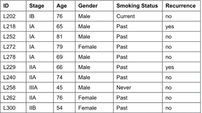
Patient biopsy samples from ive Stage I (with no lymph node involvement) and ive Stage II/III (with lymph node involvement) patients were selected from the Queen Elizabeth II Health Sciences Center Lung Tumour Bank based on the fol-lowing criteria: adenocarcinoma, age >45 years; all but one were current or past smokers (Table 1).
This study was approved by the Capital Health Re-search Ethics Board (CDHA-RS/2004-336), and all participat-ing individuals signed informed consent.
ID Stage Age Gender Smoking Status Recurrence
L202 IB 76 Male Current no
L218 IA 65 Male Past yes
L252 IA 81 Male Past no
L272 IA 79 Female Past no
L278 IA 69 Male Past no
L229 IIA 66 Male Past yes
L240 IIA 74 Male Past no
L258 IIIA 45 Male Never no
L262 IIA 76 Female Past no
L300 IIB 54 Female Past no
RNA extraction
Frozen lung tissue (50 mg) was pulverized in a Multi Sample Bio Pulverizer (BioSpec) and homogenized in 1 mL TRIZOL® (Life Technologies) using a FastPrep®24 (MP
Bio-medical). Total RNA was extracted according to the manu-facturer’s protocol, treated with TURBO DNase (Applied Bio systems), puriied with the Total RNA Puriication Kit (Norgen Biotech Corp.) and quantiied on a NanoDrop-1000 (Nano Drop Products).
Microarray analysis
Equal amounts (1 μg) of Universal Human RNA (Invit-rogen) and lung tumor RNA samples were converted to oligo-modiied cDNA using the 3DNA Array 900 Kit (Genisphere). cDNA samples were co-hybridized at 56 oC for 16 h on an
oli-gonucleotide microarray containing 17,000 probes printed in duplicate (Atlantic Cancer Research Institute, Moncton, NB). A second hybridization was performed to bind the 3DNA Cap-ture Regents, at 56 oC for 4 h. After washing, microarrays were
scanned in an Axon GenePix 4200A scanner (Molecular De-vices), gridded using GenePix software (version 6.0), and the .gpr output iles were loaded into the ArrayPipe22 server at the
National Research Council Halifax. Control spots and low qual-ity spots were lagged, the remaining spots were background-corrected using the limma normexp option with a cutoff of 50, intensity data were normalized using the limma loess sub grid option, and duplicate spot data were merged. Spots that had a normalized corrected intensity value of >100 in either channel 1 or channel 2 were analysed using Limma’s empirical Bayes moderated t-test to identify spots that were signiicantly (p<0.05) differentially expressed between tumour and control reference RNA. Empirical Bayesian methods are used to provide stable results even when the number of arrays is small23 as in this study.
The Student’s t-test module of MeV24 was then used to identify
spots that were signiicantly differentially expressed between the Stage I and Stage II/III samples. Gene enrichment analysis to de-termine signalling pathways and biological processes involved in early stage NSCLC progression was performed of genes that were greater than two-fold differentially expressed using Web Gestalt (http://bioinfo.vanderbilt.edu/webgestalt) and DAVID (http://david.abcc.ncifcrf.gov/).
RESULTS AND DISCUSSION
In order to minimize effects of individual genetic differences and potential confounding signals that can arise from residual cancer cells in the normal tissue surrounding a tumours,25-26 RNAs from tumour samples were compared to
con-trol universal RNA rather than adjacent normal tissue and then differences in these expression ratios were compared between stages. After background correction, normalization and iltering, 16752 genes could be compared in the microarrays from Stage I and Stage II/III NSCLC tumours. The data are available in GEO
under the accession number GSE28956.
In Stage I tumours, 647 genes were up-regulated and 711 genes were down-regulated and in Stage II/III tumours, 868 genes were up-regulated and 1051 genes were down-regulated more than two-fold relative to the control universal reference RNA (p<0.05; Supplemental Tables 1 and 2).
Genes differentially expressed in Stage I and Stage II/III NSCLC adenocarcinomas
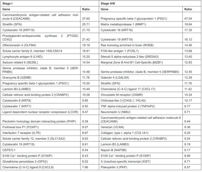
Cytokeratins are valuable markers of lung cancer and have long been used to differentiate between different sub-types of this cancer; more recently they have shown promise as prog-nostic markers in lung cancer. They are major components of the cytoskeletal system and play a role in cell migration, invasion and metastasis. As expected, cytokeratins 7, 8, 16 and 19 (typical markers of NSCLC) were up-regulated in both stages relative to control universal reference RNA (Table 2; Supplemental Tables 1 and 2).
Interestingly, KRT19 was expressed almost two-fold higher in Stage II/III compared to Stage I NSCLC tumours. Con-sistent with our results, high serum concentrations of KRT19 fragments (CYFRA 21-1) are prognostic of poor outcome in adenocarcinoma and tumours from patients with high pre-op-erative CYFRA 21-1 are larger and more poorly differentiated, indicative of a more aggressive nature.27
Cellular adhesion molecules (CEACAM1, 5, 6, 7, 8) and extracellular matrix proteins (LAMB3, LY6D, OLFM4, PSG1, SFN, VCAN, COL1A1, NAPSB, PKP) were also highly expressed in tumours relative to normal. Adhesion pathways are associated with recurrence28 and elevated levels of
extra-cellular matrix proteins in the serum are associated with poor prognosis.29 A recent microarray study identiied cell adhesion
molecule 1 (CADM1) to be signiicantly associated with poor survival in NSCLC, particularly Stage I adenocarcinoma, and its prognostic value was veriied by immunohistochemistry in tis-sue microarrays.30 In our study CADM1 showed 3.2-fold higher
expression in Stage 1 tumours than normal (Supplemental Table 1). CEACAMs are important regulators of invasion and metasta-sis and CEACAM6 inhibits cell-cell contact inhibition mediated by CEACAM1 in A549 lung adenocarcinoma cells,31 inducing
cellular proliferation.32 CEACAM6 was up-regulated 37-fold
in Stage I and 9-fold in Stage II/III tumours; interestingly, both CEACAM6 and surfactant proteins that it interacts with are tar-gets of transcription factor TTF-133 and all are up-regulated.
The putative oncogene RHEB34 plays a role in growth
and cell cycle progression through the AKT/MTOR pathway; it was up-regulated over 14-fold in Stage II/III tumours. Induc-tion of angiogenesis, one of the hallmarks of cancer, is a ma-jor contributor to solid tumour development. CHI3L1, which
promotes tumour angiogenesis35 and has been shown to be
prog-nostic for low survival in NSCLC,36,37 was up-regulated more
than 10-fold in Stage II/III tumours and 6-fold in Stage I tu-mours. Several galectins (LGALS), which are also implicated in tumour angiogenesis as well as progression, were up-regulated in tumours of both stages.
The S100 family are calcium-binding proteins with varied roles in cancer invasion, metastasis and recurrence. They regulate a host of intracellular processes including enzyme ac-tivities, components of cytoskeleton, motility and cell cycle. An earlier microarray study showed calcium-binding proteins S100P and S100A2, and trypsinogens TRY6 and PRSS3 to be correlated with progression to metastasis in NSCLC.20 In
our study, expression of S100P was more than 8-fold higher in both Stage I and Stage II/III tumours but S100A2 was up-regulated only in Stage II/III tumours (5.3-fold). Other S100 proteins such as S100A10, which is essential for migration of tumour-promoting macrophages into tumor sites,38 showed
in-creased gene expression in Stage II/III tumours. Although TRY6
and PRSS3 trypsinogens were not differentially regulated in our study, matrix metalloproteases (MMPs) were dramatically
up-regulated in Stage I and Stage II/III tumours, particularly the latter. MMPs also play a crucial role in metastasis by degrading extracellular matrix thereby allowing cell migration. Proteomics and immunohistochemical staining have shown that members of the annexin family (ANX) promote cancer cell invasion and metastasis in cancer, particularly ANXA1, ANXA2, ANXA4 and ANXA5.31 We found expression of ANXA1, ANXA2, and
ANXA3 was higher in tumour tissue than normal.
Many of the up-regulated genes in the Table 2 such as LY6D, GZMB, IL7R, CXCL9, CXCL17, MZB1, and LGALS4, may be derived from tumour-iniltrating immune cells. Such im-mune genes may be prognostic in NSCLC, and IL7R and CXCR4 were identiied as key players in the tumour microenvironment by gene proiling.39 In our study, CXCR4 showed a 2.8-fold
in-crease in gene expression in Stage II/III tumours compared to Stage I. Interestingly, several of the genes were also included in a list of the top 20 up-regulated genes in the lung cancer
Stage I Stage II/III
Gene Ratio Gene Ratio
Carcinoembryonic antigen-related cell adhesion
mol-ecule 6 (CEACAM6) 37.65 Pregnancy speciic beta-1-glycoprotein 1 (PSG1) 47.04
Stratiin (SFN) 25.71 Matrix metalloprotease 1 (MMP1) 19.64
Cytokeratin 16 (KRT16) 21.75 Cytokeratin 16 (KRT16) 17.35
Prostaglandin-endoperoxide synthase 2 (PTGS2;
COX2) 21.42 Cytokeratin 19 (KRT19) 16.12
Olfactomedin 4 (OLFM4) 18.18 Ras homolog enriched in brain (RHEB) 14.46
Solute carrier family 6, member 14SLC6A14 16.81 FOS-like antigen 1 (FOSL1) 13.68
Lymphocyte antigen 6 (LY6D) 15.20 Steroid 5 alpha-reductase 2-like (SRD5A3) 13.45
Seizure related 6 (SEZ6L) 14.54 Marginal Zone B And B1 Cell-Speciic (MZB1) 12.83
Serine protease inhibitor, clade B, member 5
(SER-PINB5) 14.46 Serine protease inhibitor, clade B, member 5 (SERPINB5) 12.45
Granzyme B (GZMB) 11.78 Galectin 4 (LGALS4) 12.29
Pregnancy speciic beta-1-glycoprotein 1 (PSG1) 10.50 Stratiin (SFN) 11.78
Laminin B3 (LAMB3) 10.49 Chemokine (C-X-C) ligand 17 (CXCL17) 11.42
Cellular retinoic acid binding protein 2 (CRABP2) 10.06 Oncostatin M receptor (OSMR) 10.34
Cytokeratin 8 (KRT8) 9.85 Chitinase-like 3 (CHI3L1; YKL40) 10.17
Cytokeratin 7 (KRT7) 9.50 TNF alpha-induced protein 2 (TNFAIP2) 9.77
Ligand dependent nuclear receptor corepressor (LCOR) 9.47 Neuromedin U (NMU) 9.71
Pleckstrin homology domain interacting protein (PHIP) 9.38
Carcinoembryonic antigen-related cell adhesion molecule 6
(CEACAM6) 9.44
Forkhead box P1 (FOXP1) 9.07 Versican (VCAN) 9.36
Interleukin 7 receptor (IL7R) 8.67 Collagen, type I, alpha 1 (COL1A1) 9.25
Solute carrier family 12, member 2 (SLC12A2) 8.63 Cellular retinoic acid binding protein 2 (CRABP2) 9.24
Cytokeratin 19 (KRT19) 8.61 Laminin B3 (LAMB3) 9.19
CEP57L1 8.44 Napsin B (NAPSB) 9.17
S100 Ca++ binding protein P (S100P) 8.43 S100 Ca++ binding protein P (S100P) 8.89
Glutathione peroxidase 2 (GPX2) 8.02 X (inactive)-speciic transcript (XIST) 8.71
Chemokine (C-X-C) ligand 9 (CXCL9) 7.96 Plakophilin 3 (PKP) 8.57
Table 2: Top 25 up-regulated genes in Stage I and Stage II/III NSCLC adenocarcinomas relative to control RNA (identiied using Limma’s empirical Bayes moderated t-test at p<0.05).
transcriptome derived by RNAseq.40 These included KRT16,
MMP1, Plakophilin (PKP), and Cellular Retinoic Acid Binding Protein 2 (CRABP2), the latter of which was proposed as a puta-tive biomarker for lung adenocarcinoma. There was also consid-erable overlap with the down-regulated genes (data not shown). A recent RNASeq whole transcriptome study of six normal, ad-enocarcinoma in situ and invasive adad-enocarcinoma samples also identiied CRABP2 as up-regulated in adenocarcinoma in situ compared to normal lung,41 strongly implicating it in NSCLC
progression.
Pathways differentially expressed in Stage I and Stage II/III NSCLC adenocarcinomas
To correlate differential gene expression with the bio-logical pathways that are affected, gene enrichment analysis was performed. For Stage I tumours (Table 3) two major
up-regu-lated KEGG pathways were identiied: 34 genes in the cancer pathway and 13 in the more restricted small cell lung cancer pathway.
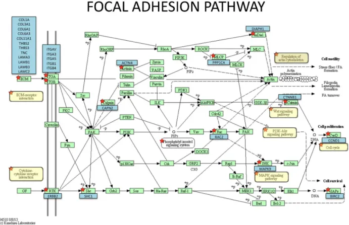
For Stage II/III tumours, two additional more speciic cancer pathways were implicated: ECM-receptor interaction and focal adhesion, indicating the increased emphasis on cell migration and invasion in the later stage tumours. These path-ways included many structural proteins (collagens, integrins, laminins, actinin4 ACTN4, tenascin C) as well as signalling molecules such as ERBB2, thrombopsondins 2 and 3, WNT5B, KRAS, BIRC2, cyclin D1, MAP kinase 9, protein phosphatase 1 PP1CA) and beta catenin (Supplemental Table 3). The effect on WNT, KRAS, PI3K-AKT, and MAPK signalling pathways would result in increased cell proliferation whereas increased ERBB2 signalling and anti-apoptotic protein BIRC2 would re-sult in increased cell survival see Figure 1.
Table 3: Enrichment analysis using Web Gestalt (WG) and DAVID of differentially regulated genes in Stage I and Stage II/III NSCLC adenocarcinomas relative to
control RNA. #, number of genes; p, p value
(A) 647 genes twofold up-regulated in Stage I.
KEGG analysis # p DAVID p WG
Pathways in cancer 34 2.11E-06 3.29e-05
Small cell lung cancer 13 1.91E-04 9.0e-04
(B) 711 genes twofold down-regulated in Stage I.
KEGG analysis # p DAVID p WG
PPAR signaling pathway 15 1.98E-06 1.49E-05
Parkinson’s disease 18 6.24E-05 5.15E-05
Ascorbate and aldarate metabolism 7 7.02E-05 2.0E-04
Drug metabolism 12 9.44E-05 4.0E-04
Pentose and glucuronate interconversions 7 1.01E-04 2.0E-04
Oxidative phosphorylation 17 2.55E-04 2.0E-04
Glycolysis / Gluconeogenesis 11 3.36E-04 1.0E-03
Huntington’s disease 20 5.11E-04 7.0E-04
(C) 868 genes twofold up-regulated in Stage II/III.
KEGG analysis # p DAVID p WG
Pathways in cancer 43 1.07E-06 4.56e-06
ECM-receptor interaction 18 6.10E-06 5.66e-06
Small cell lung cancer 17 2.57E-05 3.23e-05
Focal adhesion 28 4.47E-05 3.23e-05
(D) 1051 genes twofold down-regulated in Stage II/III.
KEGG analysis # p DAVID p WG
PPAR signaling pathway 14 1.67E-04 6.5e-03
Ascorbate and aldarate metabolism 7 2.96E-04 6.5e-03
Drug metabolism 12 8.99E-04 1.66e-02
Figure 1: Focal adhesion pathway showing genes up-regulated in Stage II/III NSCLC adenocarcinomas relative to control RNA. The underlying KEGG pathway was generated
by DAVID and components of the pathway containing enriched genes are indicated by red stars. The relevant genes (blue boxes) are superimposed over it.
In addition, up-regulation of ACTN4, PP1CA and DI-APH1 gene expression would positively impact regulation of the actin cytoskeleton and cell motility.
Both Stage I and Stage II/III tumours showed enrich-ment in down-regulated genes involved in PPAR signalling and various metabolic pathways (Table 3). PPAR
γ
has been impli-cated as a tumour suppressor in NSCLC, and xenograft models of lung cancer show that it inhibits lung tumour cell proliferation and growth through a variety of metabolic activities.42Down-regulation of the anti-tumour PPAR signalling may enhance the ability of NSCLC tumours to grow and metastasize. PPAR li-gands are under development as potential therapeutic agents for lung cancer.42
Genes differentially expressed between Stage I and Stage II/III NSCLC adenocarcinomas
Between Stage I and II/III tumours, there were 48 sig-niicantly differentially regulated genes (26 up-regulated and 22 down-regulated; p<0.01) (Figure 2; Supplemental Table 4).
Figure 2: Heat map of differentially regulated genes (p<0.01) between Stage I (blue bar) and Stage II/III (purple bar) adenocarcinomas. Individual tumor samples are represented in the columns and genes in the rows. Red is up-regulated, green is down-regulated, and
PASD1 exhibited the greatest up-regulation between groups (9.4-fold) whereas the remainder were between 1.4 and 2.9-fold up-regulated. PASD1 is an immunogenic “cancer tes-tis antigen” that is thought to function as a transcription fac-tor and is associated with cancer of the small intestine, colon, lung, head and neck as well as hematopoietic malignancies.43 It
shows promise as a target for immunotherapy against various hematopoietic cancers. PASD1 levels were signiicantly higher in H1299, a commonly used NSCLC cell line, than other cell lines44 and our results conirm that targeting it could also be a
promising therapeutic approach in NSCLC. On the other hand, LCOR showed the greatest down-regulation (5.9-fold) between groups with the remaining 21 genes showing between 1.6 and 4-fold down-regulation. LCOR is a ligand-dependent
co-repres-sor of various nuclear hormone receptors and has recently been shown to regulate transcription of CDKN1A, which encodes the cell cycle regulator p21, and the cell adhesion molecule, E-cadherin;45 reduced expression would promote transcription
of cancer-related genes. Although this effect of LCOR on CD-KN1A and E-cadherin has only been reported in cell lines and not yet for lung cancer, our studies suggest that this regulatory network is important in progression of NSCLC.
At a less stringent cut-off (p<0.05), there were 373 sig-niicantly differentially regulated genes between Stage I and II/ III tumours (178 up-regulated and 195 down-regulated; Supple-mental Table 5) of which the top 25 are shown in Table 4.
Up-regulated Down-regulated
Gene Ratio Gene Ratio
Surfactant, pulmonary-associated protein A2 (SFTPA2) 9.94 Ubiquilin-like (UBQLNL) 0.006
PAS domain containing 1 (PASD1) 9.41 Zinc inger protein 367 (ZNF36) 0.15
Nuclear receptor co-activator 7 (NCOA7) 4.93 Ligand dependent nuclear receptor corepressor (LCOR) 0.17 Surfactant, pulmonary-associated protein D (SFTPD) 3.92 Keratin associated protein 21-1 (KRTAP21-1) 0.17
Matrix Gla protein (MGP) 3.37 Tescalcin (TESC) 0.18
Member RAS oncogene family (RAB2A) 3.33 Pleckstrin homology domain interacting protein (PHIP) 0.19
PHD inger protein 11 (PHF11), variant 2 3.19
Tumor necrosis factor receptor superfamily, member 6b,
decoy (TNFRSF6B) 0.20
Src-like-adaptor (SLA), variant 3 3.06 Dihydrolipoamide branched chain transacylase E2 (DBT) 0.20
Synovial sarcoma, X breakpoint 1 (SSX1) 3.05 Family with sequence similarity 101, member B (FAM101B) 0.20
Tubulin, alpha 1a (TUBA1A) 3.04
Solute carrier family 52, ribolavin transporter, member 1
(GPR172B) 0.20
Ras-related associated with diabetes (RRAD) 3.03
Solute carrier family 30 (zinc transporter), member 7
(SLC30A7) 0.24
Keratin associated protein 19-2 (KRTAP19-2) 2.98 ATP/GTP binding protein-like 5 (AGBL5) 0.24
Heat shock protein 90kDa alpha (cytosolic), class A
member 1 (HSP90AA1), variant 2 2.96 Solute carrier family 2, member 4 regulator (SLC2A4RG) 0.24
Archaemetzincins-2 (AMZ2), variant 1 2.94 Neuralized homolog (NEURL) 0.24
Ribosomal protein S4, X-linked (RPS4X) 2.85 Uridine-cytidine kinase 2 (UCK) 0.24
Pinin, desmosome associated protein (PNN) 2.81 Multiple EGF-like-domains 11 (MEGF11) 0.25
Leptin receptor overlapping transcript (LEPROT) 2.81 Growth differentiation factor 5 (GDF5) 0.25
Leukotriene A4 hydrolase (LTA4H) 2.77 Granzyme B (GZMB) 0.25
Calpain 2, (m/II) large subunit (CAPN2) 2.74
Angiotensin I converting enzyme (peptidyl-dipeptidase A) 1
(ACE) 0.025
Peptidylglycine alpha-amidating monooxygenase (PAM),
variant 1 2.73 Forkhead box C2 (FOXC2) 0.26
Niemann-Pick disease, type C2 (NPC2) 2.69 HNF1 homeobox B (HNF1B) 0.26
Dynein, light chain, LC8-type 1 (DYNLL1), variant 3 2.68
CDC42 effector protein (Rho GTPase binding) 5
(CD-C42EP5; CEP5) 0.26
Histone cluster 1, H2bk (HIST1H2BK) 2.66 Hippocalcin (HPCA) 0.28
LSM1 homolog, U6 small nuclear RNA associated
(LSM1) 2.66
Signal transducer and activator of transcription 5B
(STAT5B) 0.28
Inositol 1,4,5-trisphosphate 3-kinase C (ITPKC) 2.65 GC-rich sequence DNA-binding factor 1 (GCFC1) 0.28
Published gene expression studies have indicated that aggressive lung adenocarcinomas have higher levels of cell pro-liferation-related genes including cyclins, transcription factor TTF-1, surfactant SFTPB, and those involved in immunological function.21 As mentioned above, TTF-1 regulates the
transcrip-tion of CEACAM6 and surfactant-associated proteins. Both SFTPA2 and SFTPD were in the top ive genes up-regulated in Stage II/III compared to Stage I tumours (Table 4) and cyclin I was up-regulated 2.56-fold in Stage II/III compared to Stage I tumours. MGP is a mesenchymal gene encoding an extracellular matrix protein and RT-PCR, ELISA and immunohistochemistry have shown that is over-expressed in recurrent gliomas and as-sociated with poor outcome.46 It has not thus far been studied in
lung cancer, and may provide a novel biomarker for lung recur-rence. Immunohistochemistry, qRT-PCR, Western blot analysis and RNA interference have shown that LSM1 functions as an oncogene in the progression of lung cancer47 and members of
the Ras and Src oncogene families (RAB2A, RRAD and SLA) were also up-regulated. Breast tumours shown to over-express RRAD by immunohistochemistry and Western blotting are of higher grade, size and nodal involvement and have poor progno-sis.48 SLA mediates cell migration and invasion through integrin
signalling by Src and FAK tyrosine kinases and in vitro over-expression studies have shown that it is also a negative regula-tor of T and B cell-mediated responses,49 which are crucial for
anti-tumour immunity. Microarray, reverse phase protein array, ELISA and immunohistochemistry analyses have shown that patients with lung adenocarcinomas that express genes associ-ated with an active immune response, such as RANTES, MIP-1-beta and STAT5 have better outcomes.50-51 Consistent with this is
the 2.7-fold lower STAT5B expression in Stage II/III than Stage I tumours in our study.
Pathways differentially expressed between Stage I and Stage II/ III NSCLC adenocarcinomas
Later-stage tumours showed enrichment in pathways mostly involved in transcription, translation, mitochondrial electron transport chain, and actin cytoskeleton organization (Supplemental Table 6A). This relects the overall higher me-tabolism and growth rate of more advanced cancers. Interest-ingly, there was also enrichment in genes in the IL-6 signaling pathway; these included phospholipase C gamma subunit (PL-CG1), FYN oncogene, ras-related small GTP-binding protein (RAC1), heat shock protein 90kDA alpha A1 (HSP90AA1), and protein phosphatase2 regulatory subunit B, gamma (PPP2R2C). FYN is a tyrosine kinase that has been implicated in the control of cell growth. HSP90AA1 is a chaperone for tyrosine kinases EGFR, MET and ALK,52 all of which are oncogenic drivers of
lung cancer. PLCG1 is responsible for intracellular transduc-tion of receptor-mediated tyrosine kinase activators. RAC1 is involved in control of cell growth/cytoskeletal reorganization, and PPP2R2C is also involved in tumour signal transduction pathways. IL-6 is present in the tumour microenvironment and
induces tumour proliferation, angiogenesis and resistance to chemotherapy; it has recently been shown by tissue microarray analysis to be a novel prognostic biomarker in NSCLC.53 CD88,
which is a high-afinity receptor for C5a that is widely expressed on lung epithelial cells, is up-regulated in Stage II/III (3.6-fold) compared to Stage I (2.3-fold) tumours. Over-expression of this receptor, assessed by tissue microarray analysis, correlates with down-regulation of E-cadherin expression and lymph node me-tastasis in NSCLC patients.54
Stage II/III vs. Stage I tumours did not show statisti-cally signiicant enrichment in any pathways and there was only borderline enrichment in some GO biological processes (Sup-plemental Table 6B).
CONCLUSIONS
By comparing the transcriptomes of early and later-stage NSCLC adenocarcinomas, we have uncovered information on pathways that are involved in recurrence in early-stage lung cancer. Many of the hallmarks of cancer55 such as proliferative
signalling, angiogenesis, invasion and metastasis were evident in the sets of genes we identiied. A signiicant number of the differentially regulated genes participated in closely related pro-cesses, helping to validate our results. Transcript data relected the overall higher growth rate of more advanced cancers and the involvement of pathways involved in actin cytoskeleton remod-elling and cell migration. While the small number of samples analysed in this study makes it dificult to make strong conclu-sions, we have nonetheless found concordant results with other reports that used a variety of complementary molecular tech-niques and uncovered possible pathways underlying recurrence. This study provides interesting novel leads to be followed up on in larger prospective investigations.
ACKNOWLEDGEMENT
We gratefully acknowledge the technical assistance of Jeffrey Gallant, Evelyn Teh and Jason Williams and bioinformatics analysis by Susanne Penny, National Research Council (NRC). We thank John Fris, database manager of the Lung Tumor Bank at Capital District Health Authority, for collating clinical anno-tations. Funding was provided by NRC and Dalhousie Univer-sity Departments of Pathology and Surgery.
REFERENCES
1. Siegel R, Naishadham D, Jemal A. Cancer statistics, 2012. CA
Cancer J Clin. 2012; 62: 10-29. doi: 10.3322/caac.20138
2. Subramanian J, Simon R. Gene expression-based prognostic signatures in lung cancer: ready for clinical use? J. Natl. Cancer
3. Tsim S, O’Dowd CA, Milroy R, Davidson S. Staging of non-small cell lung cancer (NSCLC): A review. Respir. Med. 2010; 104: 1767-1774. doi: 10.1016/j.rmed.2010.08.005
4. Chen HY, Yu SL, Li KC, Yang PC. Biomarkers and transcrip-tome proiling of lung cancer. Respirology. 2012; 17: 620-626. doi: 10.1111/j.1440-1843.2012.02154.x
5. Travis WD, Brambilla E, Noguchi M, et al. International Association for the Study of Lung Cancer/American Thoracic Society/European Respiratory Society international multidisci-plinary classiication of lung adenocarcinoma. J Thorac Oncol. 2011; 6: 244-285. doi: 10.1097/JTO.0b013e318206a221
6. Holdenrieder S, Stieber P. Circulating apoptotic markers in the management of non-small cell lung cancer. Cancer
Biomark-ers. 2009; 6: 197-210. doi: 10.3233/CBM-2009-0130
7. Daigo Y, Nakamura Y. From cancer genomics to thoracic on-cology: discovery of new biomarkers and therapeutic targets for lung and esophageal carcinoma. Gen Thorac Cardiovasc Surg. 2008; 56: 43-53. doi: 10.1007/s11748-007-0211-x
8. Dhillon T, Mauri FA, Bellezza G, et al. Overexpression of the mammalian target of rapamycin: a novel biomarker for poor sur-vival in resected early stage non-small cell lung cancer. J. Thorac.
Oncol. 2010; 5: 314-319. doi: 10.1097/JTO.0b013e3181ce6604
9. Su WP, Chen YT, Lai WW, Lin CC, Yan JJ, Su WC. Apo-lipoprotein E expression promotes lung adenocarcinoma pro-liferation and migration and as a potential survival marker in lung cancer. Lung Cancer. 2011; 71: 28-33. doi: 10.1016/j. lungcan.2010.04.009
10. Sholl LM, Barletta JA, Yeap BY, Chirieac LR, Hornick JL. Sox2 protein expression is an independent poor prognostic indi-cator in stage I lung adenocarcinoma. Am J Surg Pathol. 2010; 34: 1193-1198. doi: 10.1097/PAS.0b013e3181e5e024
11. van der Drift MA, Hol BEA, Klaassen CHW, et al. Circulat-ing DNA is a invasive prognostic factor for survival in non-small cell lung cancer. Lung Cancer. 2010; 68: 283-287. doi:
10.1016/j.lungcan.2009.06.021
12. Yoon SO, Kim YT, Jung KC, Jeon YK, Kim BH, Kim CW. TTF-1 mRNA-positive circulating tumor cells in the peripheral blood predict poor prognosis in surgically resected non-small cell lung cancer patients. Lung Cancer. 2010; 71: 209-216. doi:
10.1016/j.lungcan.2010.04.017
13. Liu L, Zhao E, Li C, et al. TRIM28, a new molecular marker predicting metastasis and survival in early-stage non-small cell lung cancer. Cancer Epidemiol. 2013; 37: 71-78. doi: 10.1016/j. canep.2012.08.005
14. Singhal S, Miller D, Ramalingam S, Sun SY. Gene expres-sion proiling of non-small cell lung cancer. Lung Cancer. 2008; 60: 313-324. doi: 10.1016/j.lungcan.2008.03.007
15. Lu Y, Lemon W, Liu PY, Yang P, You M. A gene expression signature predicts survival of patients with stage I non-small cell lung cancer. PLoS Med. 2006; 3: e467. doi: 10.1371/journal. pone.0030880
16. Ohgino K, Soejima K, Yasuda H, et al. Expression of ibro-blast growth factor 9 is associated with poor prognosis in pa-tients with resected non-small cell lung cancer. Lung Cancer. 2014; 83: 90-96. doi: 10.1016/j.lungcan.2013.10.016
17. Meyerson M, Carbone D. Genomic and proteomic proiling of lung cancers: lung cancer classiication in the age of target-ed therapy. J. Clin. Oncol. 2005; 23: 3219-3226. doi: 10.1200/ JCO.2005.15.511
18. Zhu CQ, Ding K, Strumpf D, et al. Prognostic and predic-tive gene signature for adjuvant chemotherapy in resected non-small-cell lung cancer. J. Clin. Oncol. 2010; 28: 4417-4424. doi:
10.1200/JCO.2009.26.4325
19. Sanchez-Palencia A, Gomez-Morales M, Gomez-Capilla JA, et al. Gene expression proiling reveals novel biomarkers in nonsmall cell lung cancer. Int. J. Cancer. 2011; 129: 355-364. 20. Diederichs S, Bulk E, Steffen B, et al. S100 family mem-bers and trypsinogens are predictors of distant metastasis and survival in early-stage non-small cell lung cancer. Cancer Res. 2004; 64: 5564-5569.
21. Shedden K, Taylor JM, Enkemann SA, et al. Gene expres-sion-based survival prediction in lung adenocarcinoma: a multi-site, blinded validation study. Nat. Med. 2008; 14: 822-827. doi:
10.1038/nm.1790
22. Hokamp K, Roche FM, Acab M, et al. ArrayPipe: a lexible processing pipeline for microarray data. Nucleic Acids Research. 2004; 32: W457-W459. doi: 10.1093/nar/gkh446
23. Smyth GK. Linear models and empirical Bayes methods for assessing differential expression in microarray experiments.
Sta-tistical Applications in Genetics and Molecular Biology. 2004;
3(3). doi: 10.2202/1544-6115.1027
24. Saeed AI, Sharov V, White J, et al. TM4: a free, open-source system for microarray data management and analysis.
BioTech-niques. 2003; 34: 374-378.
25. Kadara H, Lacroix L, Behrens C, et al. Identiication of gene signatures and molecular markers for human lung cancer prognosis using an in vitro lung carcinogenesis system.
Cancer Prev. Res. 2009; 2: 702-711. doi: 10.1158/1940-6207. CAPR-09-0084
26. Mwenifumbo JC, Marra MA. Cancer genome-sequencing study design. Nat Rev Genet. 2013; 14: 321-332. doi: 10.1038/ nrg3445
27. Park SY, Lee JG, Kim J, et al. Preoperative serum CYFRA 21-1 level as a prognostic factor in surgically treated adenocar-cinoma of lung. Lung Cancer. 2013; 79: 156-160.
28. Nanjundan M, Byers LA, Carey MS, et al. Proteomic pro-iling identiies pathways dysregulated in non-small cell lung cancer and an inverse association of AMPK and adhesion path-ways with recurrence. J Thorac Oncol. 2010; 5: 1894-1904. doi:
10.1097/JTO.0b013e3181f2a266
29. Gogali A, Charalabopoulos K, Zampira I, et al. Soluble ad-hesion molecules E-cadherin, intercellular adad-hesion molecule-1, and E-selectin as lung cancer biomarkers. Chest. 2010; 138: 1173-1179. doi: 10.1378/chest.10-0157
30. Botling J, Edlund K, Lohr M, et al. Biomarker Discovery in Non-Small Cell Lung Cancer: Integrating Gene Expression Proiling, Meta-analysis, and Tissue Microarray Validation. Clin
Cancer Res. 2013; 19: 194-204. doi: 10.1158/1078-0432.CCR-12-1139
31. Deng S, Wang J, Hou L, et al. Annexin A1, A2, A4 and A5 play important roles in breast cancer, pancreatic cancer and la-ryngeal carcinoma, alone and/or synergistically. Oncol Lett. 2013; 5: 107-112.
32. Singer BB, Scheffrahn I, Kammerer R, Suttorp N, Ergun S, Slevogt H. Deregulation of the CEACAM expression pattern causes undifferentiated cell growth in human lung adenocarci-noma cells. PLoS One. 2010; 5: e8747. doi: 10.1371/journal. pone.0008747
33. Chapin C, Bailey NA, Gonzales LW, Lee JW, Gonzalez RF, Ballard PL. Distribution and surfactant association of carcino-embryonic cell adhesion molecule 6 in human lung. Am J
Phys-iol Lung Cell Mol PhysPhys-iol. 2012; 302: L216-225. doi: 10.1152/ ajplung.00055.2011
34. Tamborero D, Gonzalez-Perez A, Perez-Llamas C, et al. Comprehensive identiication of mutational cancer driver genes across 12 tumor types. Sci Rep. 2013; 3: 2650. doi: 10.1038/
srep02650
35. Shao R, Hamel K, Petersen L, et al. YKL-40, a secreted gly-coprotein, promotes tumor angiogenesis. Oncogene. 2009; 28: 4456-4468. doi: 10.1038/onc.2009.292
36. Choi IK, Kim YH, Kim JS, Seo JH. High serum YKL-40 is a poor prognostic marker in patients with advanced non-small cell lung cancer. Acta Oncol. 2010; 49: 861-864. doi:
10.3109/02841861003631503
37. Thom I, Andritzky B, Schuch G, et al. Elevated pretreatment serum concentration of YKL-40-An independent prognostic bio-marker for poor survival in patients with metastatic nonsmall cell lung cancer. Cancer. 2010; 116: 4114-4121. doi: 10.1002/
cncr.25196
38. Phipps KD, Surette AP, O’Connell PA, Waisman DM. Plas-minogen receptor S100A10 is essential for the migration of tumor-promoting macrophages into tumor sites. Cancer Res. 2011; 71: 6676-6683. doi: 10.1158/0008-5472.CAN-11-1748
39. Suzuki K, Kachala SS, Kadota K, et al. Prognostic immune markers in non-small cell lung cancer. Clin Cancer Res. 2011; 17: 5247-5256. doi: 10.1158/1078-0432.CCR-10-2805
40. Han SS, Kim WJ, Hong Y, et al. RNA sequencing identiies novel markers of non-small cell lung cancer. Lung Cancer. 2014; 84: 229-235. doi: 10.1016/j.lungcan.2014.03.018
41. Morton ML, Bai X, Merry CR, et al. Identiication of mR-NAs and lincRmR-NAs associated with lung cancer progression us-ing next-generation RNA sequencus-ing from laser micro-dissected archival FFPE tissue specimens. Lung Cancer. 2014; 85: 31-39. doi: 10.1016/j.lungcan.2014.03.020
42. Han SW, Roman J. Anticancer actions of PPARgamma li-gands: Current state and future perspectives in human lung can-cer. World J Biol Chem. 2010; 1: 31-40.
43. Khan G, Denniss F, Mills KI, Pulford K, Guinn B. PASD1: A promising target for the immunotherapy of haematologi-cal malignancies. J. Genet. Syndr. Gene Ther. 2013; 4. doi:
10.4172/2157-7412.1000186
44. Guinn BA, Bland EA, Lodi U, et al. Humoral detection of leukaemia-associated antigens in presentation acute myeloid leukaemia. Biochem Biophys Res Commun. 2005; 335: 1293-1304. doi: 10.1016/j.bbrc.2005.08.024
45. Calderon MR, Verway M, An BS, et al. Ligand-depen-dent corepressor (LCoR) recruitment by Kruppel-like factor 6 (KLF6) regulates expression of the cycldependent kinase in-hibitor CDKN1A gene. J Biol Chem. 2012; 287: 8662-8674. doi:
10.1074/jbc.M111.311605
46.Mertsch S, Schurgers LJ, Weber K, Paulus W, Senner V. Ma-trix gla protein (MGP): an overexpressed and migration-pro-moting mesenchymal component in glioblastoma. BMC Cancer. 2009; 9: 302. doi: 10.1186/1471-2407-9-302
47. Watson PM, Miller SW, Fraig M, Cole DJ, Watson DK, Boylan AM. CaSm (LSm-1) overexpression in lung cancer and mesothelioma is required for transformed phenotypes. Am J
Re-spir Cell Mol Biol. 2008; 38: 671-678. doi: 10.1165/rcmb.2007-0205OC
48. Tseng YH, Vicent D, Zhu J, et al. Regulation of growth and tumorigenicity of breast cancer cells by the low molecular weight GTPase Rad and nm23. Cancer Res. 2001; 61: 2071-2079. 49. Holland SJ, Liao XC, Mendenhall MK, et al. Functional cloning of Src-like adapter protein-2 (SLAP-2), a novel inhibitor of antigen receptor signaling. J Exp Med. 2001; 194: 1263-1276. doi: 10.1084/jem.194.9.1263
50. Moran CJ, Arenberg DA, Huang CC, et al. RANTES expres-sion is a predictor of survival in stage I lung adenocarcinoma.
Clin Cancer Res. 2002; 8: 3803-3812.
51. He Y, Zhou Z, Hofstetter WL, et al. Aberrant expression of proteins involved in signal transduction and DNA repair pathways in lung cancer and their association with clinical pa-rameters. PLoS One. 2012; 7: e31087. doi: 10.1371/journal. pone.0031087
52. Koizumi H, Yamada T, Takeuchi S, et al. Hsp90 inhibition overcomes HGF-triggering resistance to TKIs in EGFR-mutant lung cancer by decreasing client protein expression and angiogenesis. J Thorac Oncol. 2012; 7: 1078-1085. doi: 10.1097/ JTO.0b013e3182519a2c
53. Pei BX, Sun BS, Zhang ZF, Wang AL, Ren P. Interstitial tu-mor-associated macrophages combined with tumor-derived col-ony-stimulating factor-1 and interleukin-6, a novel prognostic biomarker in non-small cell lung cancer. J Thorac Cardiovasc
Surg. 2014; 148: 1208-1216. doi: 10.1016/j.jtcvs.2014.05.003
54. Gu J, Ding JY, Lu CL, et al. Overexpression of CD88 pre-dicts poor prognosis in non-small-cell lung cancer. Lung Cancer . 2013; 81: 259-265. doi: 10.1016/j.lungcan.2013.04.020
55. Hanahan D, Weinberg RA. Hallmarks of cancer: the next generation. Cell. 2011; 144: 646-674. doi: 10.1016/j. cell.2011.02.013