HAL Id: hal-02292671
https://hal.archives-ouvertes.fr/hal-02292671
Submitted on 20 Sep 2019
HAL is a multi-disciplinary open access
archive for the deposit and dissemination of
sci-entific research documents, whether they are
pub-lished or not. The documents may come from
teaching and research institutions in France or
abroad, or from public or private research centers.
L’archive ouverte pluridisciplinaire HAL, est
destinée au dépôt et à la diffusion de documents
scientifiques de niveau recherche, publiés ou non,
émanant des établissements d’enseignement et de
recherche français ou étrangers, des laboratoires
publics ou privés.
morphological neoplastic transformation potential on
Balb/3T3 cells by pristine and remediated titania and
zirconia nanoparticles
Andrea Stoccoro, Sebastiano Di Bucchianico, Chiara Uboldi, Fabio Coppedè,
Jessica Ponti, Claudia Placidi, Magda Blosi, Simona Ortelli, Anna Costa,
Lucia Migliore
To cite this version:
Andrea Stoccoro, Sebastiano Di Bucchianico, Chiara Uboldi, Fabio Coppedè, Jessica Ponti, et al.. A
panel of in vitro tests to evaluate genotoxic and morphological neoplastic transformation potential on
Balb/3T3 cells by pristine and remediated titania and zirconia nanoparticles. Mutagenesis, Oxford
University Press (OUP), 2016, 31 (5), pp.511-529. �10.1093/mutage/gew015�. �hal-02292671�
© The Author 2016. Published by Oxford University Press on behalf of the UK Environmental Mutagen Society.
All rights reserved. For permissions, please e-mail: journals.permissions@oup.com. 1
Mutagenesis, 2016, 00, 1–19
doi:10.1093/mutage/gew015 Original Manuscript Advance Access Publication Date: xx xxxx xxxx
Original Manuscript
A panel of in vitro tests to evaluate genotoxic
and morphological neoplastic transformation
potential on Balb/3T3 cells by pristine and
remediated titania and zirconia nanoparticles
Andrea Stoccoro
1,2, Sebastiano Di Bucchianico
1,6, Chiara Uboldi
1,7,
Fabio Coppedè
1, Jessica Ponti
3, Claudia Placidi
4, Magda Blosi
5,
Simona Ortelli
5, Anna Luisa Costa
5and Lucia Migliore
1,*
1Laboratory of Medical Genetics, Department of Translational Research and New Technologies in Medicine and
Surgery, University of Pisa, Via Roma 55, Pisa 56126, Italy, 2Doctoral School in Genetics, Oncology and Clinical
Medicine, Department of Medical Biotechnologies, University of Siena, 53100 Siena, Italy, 3European Commission,
Joint Research Centre (JRC), Institute for Health and Consumer Protection (IHCP), Nanobiosciences (NBS) Unit, via E. Fermi 2749, 21027 Ispra (VA), Italy, 4Department of Pathology, Ospedale di Circolo and Department of Human
Morphology, University of Insubria,via Ottorino Rossi 9, 21100 Varese, VA, Italy and 5Institute of Science and
Technology for Ceramics (CNR-ISTEC), National Research Council of Italy, Via Granarolo 64, 48018 Faenza, RA, Italy
6Present address: Division of Biochemical Toxicology, Institute of Environmental Medicine, Karolinska Institutet,
Stockholm, Sweden.
7Present address: Biogenotoxicology, Human Health and Environment Unit, Mediterranean Institute of Marine and
Terrestrial Biodiversity and Ecology (IMBE), Aix-Marseille University, Marseille, France.
*To whom correspondence should be addressed. Tel:+39 050 2218549; Fax: +39 050 2210624; Email: lucia.migliore@med.unipi.it Received 12 January 2016; Revised 25 February 2016; Accepted 7 March 2016.
Abstract
The FP7 Sanowork project was aimed to minimise occupational hazard and exposure to engineered nanomaterials (ENM) through the surface modification in order to prevent possible health effects. In this frame, a number of nanoparticles (NP) have been selected, among which zirconium (ZrO2) and titanium (TiO2) dioxide. In this study, we tested ZrO2 NP and TiO2 NP either in their pristine (uncoated) form, or modified with citrate and/or silica on their surface. As benchmark material, Aeroxide® P25 was used. We assessed cytotoxicity, genotoxicity and induction of morphological neoplastic transformation of NP by using a panel of in vitro assays in an established mammalian cell line of murine origin (Balb/3T3). Cell viability was evaluated by means of colony-forming efficiency assay (CFE). Genotoxicity was investigated by cytokinesis-block micronucleus cytome assay (CBMN cyt) and comet assay, and by the use of the restriction enzymes EndoIII and Fpg, oxidatively damaged DNA was detected; finally, the morphological neoplastic transformation of NP was assayed in vitro by cell transformation assay (CTA). Our results show that the surface remediation has not been effective in modifying cyto- and genotoxic properties of the nanomaterials tested; indeed, in the case of remediation of zirconia and titania with citrate, there is a tendency to emphasise the toxic effects. The use of a panel of assays, such as those we have employed, allowing the evaluation of multiple endpoints, including cell transformation, seems particularly advisable especially in the case of long-term exposure effects in the same cell type.
at Orta Dogu Teknik University Library (ODTU) on May 19, 2016
http://mutage.oxfordjournals.org/
Introduction
The research on engineered nanomaterials (ENM) and the design of synthetic methods for their production has seen an enormous improvement during the last decade, originating from potential applications of these systems in a wide variety of attractive fields, ranging from communications and microelectronics to medical diag-nostics and therapy (1).
Despite of the large amount of available literature on these themes, the production of engineered nanoparticles (NP) with a high control degree over their chemicophysical and functional properties remains a hot topic in materials science (2).
The possible impact on human health of NP is until now not well understood. Several authors have suggested that NP can be toxic to various human organs and systems, inducing complex pathologies such as cancer (3) and neurodegenerative diseases (4).
The FP7 Sanowork project was aimed to minimise occupational hazard and exposure to ENM through their surface modification in order to prevent possible deleterious health effects.
Different ‘design option’-based risk remediation strategies focused on ENM surface engineering have been proposed and inte-grated within various ENM manufacturing processing lines, becom-ing process extra-steps to be evaluated in terms of risk and expected performances.
In this frame, a number of ENM have been selected, among which ZrO2 and TiO2 NP have been collected, modified and investi-gated in this study.
In order to control and decrease the potential toxicity and emis-sion of ZrO2 and TiO2 NP, two different approaches for surface
modification were employed: an inorganic inert coating (SiO2 NP), added by means of a heterocoagulation and an organic coating citrate-based, applied by using a self-assembling strategy. The pro-cess line involving ZrO2 NP was focused on the washing step of
industrial reactors employed for synthesising ZrO2 by sol gel reac-tion. The critical step to be monitored for its potential exposure was referred to the washing of the synthetic reactor. During this step, the proposed risk remediation strategies had the potentiality to improve the washing efficiency by improving ZrO2 water dispersion and so decreasing environmental emission potential. The target TiO2 NP
sample studied was involved in the processing line consisting in the application of TiO2 nanosols to ceramic substrates through spray
coating, one of the most flexible methods for the application of nanostructured suspensions, allowing rapid and simple production changes. The critical step for health hazard and exposure assess-ment was actually the spray coating operation which could cause nanoaereosolisation and induce inhalation of NP. SiO2 and citrate coatings were proposed for both nano metal oxides investigated in the present study for their potential to improve powder dispersabil-ity and hydrophylicdispersabil-ity, positively impacting on mechanism driving cellular toxicity (5).
To investigate the cytotoxic, genotoxic and morphological neoplastic potential of the pristine and modified ZrO2 and TiO2 NP, we used a panel of assays including the colony formation effi-ciency test (CFE), the cytokinesis-block micronucleus cytome assay (CBMN cyt), the comet assay and the cell transformation assay (CTA) in Balb/3T3 mouse fibroblasts. The choice of the cell line was driven by the availability of the in vitro CTA limited to few cell types. After exposure to carcinogens, Balb/3T3 cells become tumo-rigenic forming morphologically transformed colonies (foci type III). Their ability to induce cancer in vivo is detectable by injecting the transformed cells in nude mice and observing their growth as sarcomas (6,7).
Methods
Preparation and characterisation of ZrO2 and
TiO2 NP
Commercial ZrO2 and TiO2 (84% anatase, 16% brookite crystal phase composition, 8), NP were provided by PlasmaChem GmbH (Germany) as nanopowder and by Colorobbia Italia SpA as colloidal nanosuspension (nanosol), respectively.
For ZrO2 and TiO2 NP, the remediation process consisted in silica and sodium citrate coating. The following panel of NP were thus studied: pristine ZrO2, citrate-coated ZrO2 (low and high cit-rated), silica-coated ZrO2, pristine TiO2, citrate-coated TiO2 and
silica-coated TiO2. Furthermore, TiO2 Aeroxide® P25 were used as benchmark material.
ZrO2 nanosols preparation
The process line involving ZrO2 NP is focused on the washing step of industrial reactors employed for synthesizing ZrO2 by sol gel
reac-tion. Two kinds of surface modifications were aimed at improving ZrO2 NP dispersion: an inorganic inert coating (SiO2 NP) by means
of a heterocoagulation approach and an organic coating citrate-based by using a self-assembling strategy.
ZrO2 NP provided by PlasmaChem as nanopowder was pended in water with a solid loading of 3% wt. The resulting sus-pension (pristine), highly stable, was used as starting material for the subsequent modifications.
Modified sample labelled as ZrO2 silica coated NP, was obtained by mixing pristine ZrO2 nanosol with commercial colloidal SiO2
(Ludox S40) in a proper weight ratio (SiO2:ZrO2 = 4:1) keeping a total solid loading of 3% wt. Then, in order to promote the hetero-coagulation process, the so prepared sample was ball milled for 24 h. Samples modified with the organic layer were prepared by add-ing trisodium citrate dihydrate to the pristine then ball milladd-ing for 24 h. Two different weight ratios were produced (ZrO2:Cit = 1:0.01
and 1:1) keeping the total solid concentration at 3% wt, the so pre-pared samples were named, respectively, low-citrate and high-citrate coated NP.
TiO2 nanosols preparation
The silica modified TiO2 NP were produced following the hetero-coagulation route: opposite charged TiO2 NP and SiO2 NP were
diluted and then mixed together at room temperature, maintaining a SiO2/TiO2 weight ratio equal to 3 and a total solid content of 3% wt.
The obtained mixtures were ball milled for 24 h using 5 mm diameter zirconia spheres as milling media to obtain slightly cloudy solutions.
The citrate modified TiO2 NP were produced by self-assembly strategy: trisodium citrate dihydrate was added to a previously diluted TiO2 NP suspension at room temperature and employing a citrate/TiO2 NP weight ratio equal to 0.83. The reaction mixture was
stirred overnight resulting in a transparent and stable suspension.
Sol state characterisation of ZrO2 and TiO2 NP
The chemicophysical characterisation of the pristine and modified ZrO2 and TiO2 samples was performed by means of transmission electron microscopy (TEM), dynamic light scattering (DLS) and Z-potential.
The morphological analysis on pristine and modified samples was performed by FEI titan TEM operating at an acceleration volt-age of 300kV. One drop of diluted NP suspension in deionised water (30 μg/ml) was deposited on a film-coated copper grid and characterised.
at Orta Dogu Teknik University Library (ODTU) on May 19, 2016
http://mutage.oxfordjournals.org/
Hydrodynamic diameter of ZrO2 and TiO2 (pristine and modi-fied forms) were investigated for samples dispersed both in deion-ised water and complete cell culture medium (125 μg/ml) using light scattering optical technique (Zetasizer nano ZSP model ZEN5600; Malvern Instruments, UK).
NP hydrodynamic diameter (d DLS) was obtained from DLS data, setting the measurement angle to 173° and the measurement duration on automatic. After 2 min temperature equilibration step, 1 ml of sample volume was subjected to three consecutive measure-ments performed at 25°C and particle size distributions by intensity were obtained by averaging these measurements.
The colloidal stability in medium was evaluated by repeating measurements at t = 0–72 h. The standard operating procedure described hereafter was followed for all samples. At t = 0 h, each stock suspension was sonicated for 15 min and aliquots were first added to 0.05% (v/v) BSA/PBS to obtain an intermediate which was then added to MEM (Minimum Essential Medium, Gibco) culture medium supplemented with 10% (v/v) serum to reach a final ZrO2 and TiO2 concentration of 125 μg/ml. For analyses at t = 24, 48, 72 h, samples were thermostated at 37°C.
After particle size determination, samples underwent Zeta poten-tial measurement by electrophoretic light scattering (ELS). The Smoluchowski approximation, consistent with the high dielectric constant of water that is the main component of the all above speci-fied solvents, was applied to convert the electrophoretic mobility to zeta potential. Zeta potential measurements were performed on 700 µl of volume sample at 25°C. Before and after zeta potential analyses a size measurement was performed to check that the sam-ples have not changed. The measurement data of samsam-ples dispersed in MEM culture medium were collected in monomodal mode due to the high medium conductivity (approximately thus obtaining a mean zeta potential value). The amount of BSA added was calculated as well as a function of powder surface, carrying out specific surface area (BET) measurements on the dried powders using N2 as adsorp-tive gas Sorpty 1750 (Carlo Erba, Italy). The specific surface area (SSA) measurements were determinated by Brunauer–Emmett–Teller (BET) method. It is a well-recognised approach to determine the spe-cific surface area of nanoporous materials which is based on physical adsorption of gas molecules (N2) onto the material interface (9,10). The specific surface area (BET) measurements were carried out using N2 as adsorptive gas Sorpty 1750 (Carlo Erba, Italy).
Zeta potential of nanosols as a function of pH (experimental uncertainty: 3 mV for Z potential and 0.03 for pH) was meas-ured with an electroacoustic technique (AcoustoSizer II, Colloidal Dynamics, Australia, equipped with an automatic titrating system) that allowed identify the point of zero Z-potential (isoelectric point, i.e.p.). The measurements were carried out on aqueous solutions at 10 mg/mL. KOH 1M and HCl 1M were used to modify pH within 2–8 pH range.
Cell culture conditions
Balb/3T3 mouse fibroblasts were supplied at passage 13 from the Hatano Research Institute, Japan. Experimental cultures were pre-pared from deep-frozen stock vials and always kept in a subcon-fluent state. They were maintained in complete culture medium prepared using Minimum Essential Medium with Earle’s salts and l-glutamine (MEM) supplemented with 1% v/v of penicillin/strepto-mycin (104 Units/ml), 10% v/v of Fetal Bovine Serum New Zeland
Origin (Moregate, lot. number 47827101), and passaged at 1:15 ratio, using trypsin-EDTA (Gibco). Cell preparations were main-tained in standard cell culture conditions (37°C, 5% CO2 and 95% humidity) no longer than 10 passages.
In order to identify the most suitable concentrations to use in genotoxicity assays and in CTA assay, we previously have inves-tigated the effect of a wide range of NP doses on Balb/3T3 cell viability by means of CFE assay. For this purpose, we treated the cells with NP concentrations ranging from 1.25 to 80 µg/cm2,
corresponding to a range of 8.3–266.6 µg/ml, a range of doses representative of realistic human exposure (11). In this way, the concentrations of 10, 20 and 40 µg/cm2 were selected, excluding the
concentration of 80 µg/cm2 since at this concentration NP caused a
marked turbidity of the medium. For cellular uptake study, we have chosen the intermediate time and concentration used in genotoxic-ity and cell transformation investigations.
ZrO2 and TiO2 NP internalisation in Balb/3T3 cells
To study NP cell internalisation, Balb/3T3 cells were seeded in 75cm2
flasks (Corning Costar, Italy) at a density of 2.5 × 10⁵ cells in 10 ml
of complete cell culture medium. After 24 h, cells were exposed to 20 µg/cm2 of ZrO
2 and TiO2 NP for 48 h. After exposure, medium
was removed and cells were thoroughly washed with PBS, detached using 1 ml trypsin-EDTA (Invitrogen, Italy) and centrifuged at 200×g for 5 min to obtain the cell pellet. The supernatant was removed and cells were fixed using a Karnovsky 2% v/v solution (glutaraldehyde and paraformaldehyde in 0.05 M cacodilate buffer at pH 7.3, Sigma Aldrich, Italy) over night. Cells were then washed three times with 0.05 M cacodilate, pH 7.3 (Sigma Aldrich, Italy) and post-fixed in osmium tetroxide solution in 0.1 M pH 7.3 cacodilate (Sigma Aldrich, Italy) for 1 h; after 3 additional washes in 0.05 M cacodilate of 10 min each cells were dehydrated in a graded series of ethanol solution in MilliQ water (30%; 50%; 75%; 95% for 15 min each, and 100% for 30 min), incubated in absolute propylene oxide (Sigma Aldrich, Italy) for 20 min (2 changes of 10 min each) and embedded in a solution of 1:1 epoxy resin (Sigma Aldrich, Italy) and propyl-ene oxide for 90 min. This mixture was rpropyl-enewed with pure epoxy resin (Sigma Aldrich, Italy) over night at room temperature, and later polymerised at 60°C for 48 h. Ultrathin sections (60–90 nm) were obtained using Leica UCT ultramicrotome (Leica, Italy) and stained for 25 min with uranyl acetate (Sigma Aldrich, Italy) and lead citrate for 20 min, washed and dried. Ultrathin sections were imaged under 80 kV Philips 208 TEM.
Concurrent cytotoxicity and morphological transformation assay
The Balb/3T3 CTA has been carried out by assessing the concurrent cytotoxicity and morphological neoplastic transformation in terms of colony-forming efficiency (CFE) and formation of type-III foci, respectively.
Colony-forming efficiency assay
CFE assay was performed as detailed elsewhere (12) to study the cytotoxicity induced by ZrO2 and TiO2 NP at 24, 48 and 72 h of exposure. Cells were seeded at the density of 200 cells per dish in 3 ml complete culture medium (60 × 15 mm Petri dish, 20 cm2
bot-tom surface area, BD Falcon™). After 24 h, NP suspensions were directly added to the cell culture to obtain the appropriate final ZrO2 and TiO2 NP concentrations ranging from 1.25 to 80 µg/cm2,
cor-responding to a range of 8.3–266.6 µg/ml. Negative (untreated cells) and positive (Na2CrO4 1000µM) were also included. After 24, 48 or 72 h of exposure, the medium was changed with fresh complete cul-ture medium, and 7 days later, the cells were fixed with 3.7% (v/v) of formaldehyde solution (Sigma-Aldrich, Milan, Italy) in phosphate-buffered saline (PBS) (1×) without calcium, magnesium and sodium
at Orta Dogu Teknik University Library (ODTU) on May 19, 2016
http://mutage.oxfordjournals.org/
bicarbonate (Gibco), and stained with 0.4% (v/v) Giemsa solution (Sigma-Aldrich) in ultrapure water. Colonies were manually scored under a stereomicroscope.
The results are expressed as CFE (%) = [(average of treatment colonies/average of control colonies) × 100] and the corresponding standard error mean [SEM % = SD/√(number of treatments)].
CTA
CTA was performed to investigate if tested NP were able to induce morphological neoplastic transformation in Balb/3T3 fibroblasts. CTA is considered a powerful tool to investigate in vitro the mor-phological neoplastic transformation induced by both genotoxic and non-genotoxic carcinogens as reported in OECD guidelines (13).
CTA was carried out as reported elsewhere (12,14) on Balb/3T3 cells treated with ZrO2 and TiO2 NP at concentration of 10, 20 and 40 µg/cm2, corresponding to 91.6, 183.3 and 366.6 µg/ml,
respec-tively. Non-treated Balb/3T3 cells and cells exposed to 4 µg/ml methylcholanthrene (MCA, Sigma–Aldrich; Saint Louis, MO, USA), which is a well-known carcinogenic compound, were used as nega-tive and posinega-tive controls, respecnega-tively.
On day zero, 2 × 10⁴ Balb/3T3 cells were seeded in 100 mm-Petri
dish (100 × 20 mm, 55 cm2 bottom surface area, BD Falcon™) in 6 ml
of complete fresh medium Minimum Essential Medium (MEM) 1× supplemented with 10% (v/v) fetal bovine serum New Zeland ori-gin (Moregate, lot. number 47827101) and 1% v/v antibiotics (10 000 U/ml penicillin and 10 000 µg/ml streptomycin); 5 replicates per concentration were prepared. At the end of exposure (72 h), medium containing NP was removed and replaced with Dulbecco’s modified Eagle’s medium/F12 (DMEM/F12) with high glucose, l-glu-tamine (365 mg/l) and sodium bicarbonate (1200 mg/l) (Invitrogen; Carlsbad, CA, USA) supplemented with 2% (v/v) fetal bovine serum New Zeland origin (Moregate, lot. number 47827101), 2 µg/ml insu-lin (Sigma-Aldrich; Saint Louis, MO, USA) and 1% v/v antibiotics (10 000 U/ml penicillin and 10 000 µg/ml streptomycin) (Invitrogen; Carlsbad, CA, USA). The culture medium was changed twice a week, and on days 31–35 cells were fixed with 4% (v/v) of formaldehyde in PBS. Subsequently, cells were stained with a 10% (v/v) Giemsa solution in ultrapure water. The dishes were air-dried and observed using a stereomicroscope (Olympus; Milan, Italy) in order to detect and count the morphologically transformed colonies (type-III foci), as described from the International Agency for Cancer Research (IARC) Working-Group (W.g. IARC/NCI/EPA, 1985).
Transformation results were expressed as transformation fre-quency (Tf) (five replicates per concentration, three experiments performed) using the following formula: Tf = A/B; where: A = total number of type III foci counted in each treatment, B = number of surviving cells corresponding to [(2 × 104) × 5 dishes × CFE%]/100 ×
(plating efficiency/100), where: 2 × 104 = number of cells seeded per
dish and plating efficiency = (number of colonies formed in the con-trol/number of cells seeded in each CFE dish) × 100.
Cytokinesis-block micronucleus cytome assay
CBMN cyt was performed according to the procedure described by Di Bucchianico and coauthors (15). A total of 7.5 × 10⁵ cells were
seeded in six-well cell culture plates (9.6 cm2 bottom surface area,
Falcon ™) and after 24 h of culture, the cells were exposed to ZrO2 and TiO2 NP at the concentrations of 10, 20 and 40 μg/cm2,
corre-sponding to 32, 64 and 128 µg/ml, respectively, for 48 h. Mitomycin C (0.1 μg/ml; MMC, Kyowa Hakko Kogyo Co, Chiyoda, Tokyo, Japan) as positive control was used. Cytochalasin B (6 μg/ml) was
added after 44 h to block the cytokinesis process and cells were har-vested after 72 h.
The parameters evaluated were the cytokinesis-block prolifera-tion index (CBPI) and the replicaprolifera-tion index (RI) to test the cytostasis and the apoptotic and necrotic indices to investigate the cytotoxicity exerted by ZrO2 and TiO2 NP in the first 500 cells counted. The gen-otoxic potential was evaluated by scoring the micronucleus (MN) frequency on 1000 binucleated cells. On these 1000 binucleated cells other parameters such as nucleoplasmic bridges (NPB), a biomarker of DNA misrepair and/or telomere end-fusions, and nuclear buds (NBUD), a biomarker of elimination of amplified DNA and/or DNA repair complexes, were also scored as previously described (15,16).
Comet assay
The modified alkaline comet assay was carried out on Balb/3T3 cell cultures seeded at a concentration of 7 × 10⁴ cells/cm2 in
six-well cell culture plates (9.6 cm2 bottom surface area, Falcon ™) by
using the Comet Assay kit following the manufacturer’s instructions (Trevigen, Gaithersburg, MD, USA). After 24 h, cells were treated with ZrO2 and TiO2 NP at 10, 20 and 40 µg/cm2, corresponding
to 32, 64 and 128 µg/ml, respectively, for 2, 24, 48 and 72 h. The percentage of total DNA fluorescence in tail in a total of 100 ran-domly selected cells per sample (two replicates, each with 50 cells/ slide) was used as a measure of the amount of primary DNA dam-age. Untreated cells (negative control) and cells exposed for 5 min to H2O2 (50 µM) as positive control were used. To determine the
Table 1. Mean hydrodynamic size by intensity and Zeta potential
for 125 μg/ml of uncoated, silicated and citrated ZrO2 NP, dispersed in deionized water and complete culture medium
ZrO2 NP Deionized water
pH Z-pot (mV) Mean size (nm) PdI Uncoated 3.6 41.0 ± 2.0 261 ± 79 0.36 ± 0.08 Silicated 4.6 −40.4 ± 1.8 276 ± 85 0.38 ± 0.08 Low citrated 3.4 40.3 ± 2.7 83 ± 32 0.76 ± 0.22 High citrated 6.5 −47.0 ± 0.0 173 ± 13 0.39 ± 0.08 Complete culture medium
Uncoated 0 h 7.4 −11.7 ± 0.3 1537 ± 84 1.00 ± 0.00 24 h −10.9 ± 0.4 1376 ± 460 0.84 ± 0.28 48 h −10.8 ± 0.4 941 ± 141 1.00 ± 0.00 72 h −11.0 ± 0.4 444 ± 50 0.88 ± 0.21 Silicated 0 h 7.3 −11.0 ± 0.5 244 ± 9 0.99 ± 0.02 24 h −10.2 ± 0.4 172 ± 3 1.00 ± 0.00 48 h −11.2 ± 0.5 141 ± 3 0.59 ± 0.04 72 h −10.9 ± 0.7 101 ± 1 0.54 ± 0.01 Low citrated 0 h 7.3 −11.9 ± 0.6 1950 ± 1018 0.99 ± 0.02 24 h −10.9 ± 0.3 1713 ± 236 0.79 ± 0.20 48 h −11.0 ± 1.4 1530 ± 293 0.80 ± 0.21 72 h −11.2 ± 0.2 1239 ± 49 0.96 ± 0.07 Ultrafiltereda 8.20 −22.1 ± 0.7 High citrated 0 h 7.4 −10.3 ± 0.4 26 ± 0 0.68 ± 0.01 24 h −11.1 ± 0.1 36 ± 0 0.47 ± 0.00 48 h −11.20 ± 0.55 55 ± 1 0.47 ± 0.04 72 h −11.20 ± 0.50 62 ± 1 0.42 ± 0.06
Data are reported as the mean ± SD. PdI, polydispersity index.
aZrO
2 NP extracted by ultrafiltration from complete culture medium and
redispersed in water.
at Orta Dogu Teknik University Library (ODTU) on May 19, 2016
http://mutage.oxfordjournals.org/
presence of oxidised pyrimidine and purine bases, after 2 and 24 h of treatment, Endonuclease III (EndoIII) and Formamidopyrimidine-DNA Glycosylase (Fpg) enzymes were used. To determine the num-ber of enzyme-sensitive sites, the difference between the value of the percent of DNA fluorescence in tail obtained after digestion with each enzyme and with the buffer only was calculated. Three inde-pendent experiments were performed for each treatment by avoid-ing direct light exposures of preparavoid-ing slides. Analysis was carried out by using a Comet Image Analysis System, version 5.5 (Kinetic Imaging, Nottingham, UK).
Statistical analysis
Statistical analysis was performed using GraphPad Prism software. Data of toxicological assays were presented as the mean ± SEM. Since no difference was observed between negative and solvent con-trol (ultrapure water) for NP, statistical analysis was performed only to evaluate differences between negative control and NP treated cells. The approach to test statistical significance of differences between negative control and NP-treated groups for the CFE assay, CBMN cyt assay and comet assay was one-way analysis of variance (ANOVA) followed by the Dunnett multiple comparison test. For the CTA assay, experimental data were analysed by Fisher’s exact test considering the number of type III foci in the treatments and the surviving cells compared to the corresponding negative control or DMSO solvent control for MCA. Results were considered statisti-cally significant at P < 0.05.
Results
Characterisation of ZrO2 and TiO2 NP
The experiments were carried out using time, temperature, con-centration and medium that closely simulates the experimental conditions used for cellular tests. Table 1 illustrates the results of NP dispersions (sol) state characterisation (pH, Zeta potential and
mean hydrodynamic diameter by intensity) of uncoated, silicated and citrated ZrO2 NP dispersed in deionised water and complete culture medium, at 0.125 mg/ml. The absorption of BSA of culture medium on powder surfaces is evident. In fact, Z potential assessed for ZrO2 samples dispersed in deionised water showed different val-ues, according to Z potential-pH curves, while in culture medium negative values were measured for all the samples.
Concerning the hydrodynamic diameters, for the water media the citrate acts as dispersant, producing a diameter decreasing, particu-larly if added in a low amount. For higher citrate amount, instead, the hydrodynamic diameter increases probably due to the steric effect of the anionic chelant.
Uncoated ZrO2 in the culture media buffered at pH 7.4 showed a strong aggregation, the hydrodynamic diameter decrease observed over time is consistent with the sample precipitation on the bottom of the cuvette, leaving the smallest particles to float.
In the culture media, the same effect was observed for the low citrated sample, where the citrate added is too poor to change the surface charge and to induce stability changes. Instead, no aggrega-tion was evidenced for the silicated and the high citrated samples, which probably at the buffered pH of 7.4 are more stable and far from their isoelctric points than in deionised water.
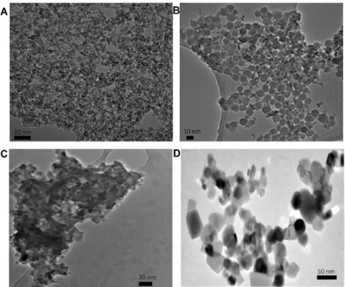
TEM imaging of ZrO2 NP are reported in Figure 1. TEM image of pristine ZrO2 NP shows a very fine primary nanostructure in accordance with wet chemical sol–gel process employed by manu-facturer for the synthesis. Due to higher values of surface energy in comparison with TiO2, ZrO2 nucleation requires higher energy for the formation of stable crystalline nuclei that can grow upper the critical radius. The presence of coating agents did not seem to influence the pristine structure that have an high tendency to form aggregates.
Table 2 illustrates the results of NP dispersions (sol) state char-acterisation (pH, Zeta potential and mean hydrodynamic diameter by intensity) of uncoated, citrated, silicated TiO2 NP and Aeroxide®
Figure 1. TEM images of ZrO2 NP. (A) Pristine; (B) silica coated; (C) low-citrate coated; (D) high-citrate coated.
at Orta Dogu Teknik University Library (ODTU) on May 19, 2016
http://mutage.oxfordjournals.org/
P25 dispersed in deionised water and complete culture medium. Zeta potential of ultrafiltered particles from dispersion in culture medium is also reported, in order to discriminate between contribute of par-ticles and medium components not adsorbed onto parpar-ticles surface.
The comparison of sol state characterisation between deion-ised water and culture medium clearly evidenced the absorption of BSA and more generally of protein components of culture medium on powder surfaces. TiO2 NP dispersed in water at their natu-ral pH, in fact, showed different values, in accordance with those reported on Z potential versus pH titration curves (Figure 2), with small deviations in absolute Z potential values, due to different concentration of TiO2 NP used for light scattering and electroa-coustic technique.
TiO2 NP transferred in complete culture medium, due to a buffer effect, meet neutral pH ranging from 7.25 to 7.74 units and showed a slightly negative Z potential, as expected by BSA and other protein components at neutral pH (BSA isoelectric point ≅ 5) (17). In this condition, silicated and citrated TiO2 NP samples exhibited a good dispersion, whilst uncoated and Aeroxide® P25 dispersions turned instantaneously cloudy. In particular pristine TiO2 NP partially pre-cipitated, as reflected in the size distribution data obtained which are not readily amenable to interpretation. The observed behaviour is easily explainable by considering that uncoated TiO2 NP cross the isoelectric point by reversing Z potential sign from positive to negative, with an expected destabilisation of the sample. The same occurred for P25 and silica coated samples, nevertheless the lower increase of hydrodynamic diameter observed is due, in the case of silica to its dispersing ability that prevented a further aggregation, whilst in case of P25 the already aggregated structure in water, revealed by high values of hydrodynamic size, is preserved also in culture medium. Citrated TiO2 NP did not show any destabilisation effect because maintained a negative Z potential and a good electro-steric stabilisation due to the presence of citrate coating. In order to test if the acid base behaviour shown by all TiO2 NP samples was due to medium components adsorbed onto their surfaces or to proteins freely dispersed in the medium, samples were ultrafiltered, redis-persed in water and Z potential re-measured. The results confirm that all the TiO2 NP surfaces were coated by proteic components, as explained by the well-known protein corona paradigm (18). Z potential of all ultrafiltered samples was leveled off around −20 mV. In particular, the high deviations shown by uncoated and citrated samples, comparing Z potential in the absence and in the presence of culture medium at pH 8, confirmed the presence of protein coating that masks all TiO2 NP surfaces. In order to confirm the results, the concentration of BSA was normalised for the surface of TiO2 sam-ples, as calculated by BET analysis. The results (Table 3) agreed with the formation of BSA coating, being the BSA concentration in all
Table 2. Mean hydrodynamic size by intensity and Zeta
poten-tial for 125 μg/ml of uncoated, citrated, silicated TiO2 NP and Aeroxide® P25 dispersed in deionized water and complete culture medium
TiO2 NP Deionized water
pH Z-pot (mV) Mean size (nm) PdI Uncoated 2.33 41.2 ± 0.5 83.5 ± 10.4 0.48 ± 0.09 Citrated 5.47 −34.2 ± 1.2 57.5 ± 2.6 0.68 ± 0.05 Silicated 2.84 32.2 ± 4.1 155.6 ± 22.1 0.28 ± 0.01 Aeroxide® P25 4.13 37.4 ± 0.9 489.5 ± 130.5 0.30 ± 0.04 Complete culture medium
Uncoated 0 h 7.25 −10.6 ± 1.0 1608 ± 211 1.00 ± 0.01 24 h −11.4 ± 0.6 1829 ± 99 0.64 ± 0.05 48 h −10.9 ± 0.6 1962 ± 147 0.98 ± 0.03 72 h −10.8 ± 0.4 1318 ± 85 0.73 ± 0.22 Ultrafiltereda 8.10 −20.9 ± 0.2 Citrated 0 h 7.69 −10.7 ± 0.3 68.3 ± 1.2 0.27 ± 0.01 24 h −10.9 ± 0.2 137.6 ± 0.9 0.21 ± 0.01 48 h −10.7 ± 0.5 148.6 ± 0.4 0.21 ± 0.01 72 h −11.6 ± 0.5 157.3 ± 0.4 0.22 ± 0.01 Ultrafiltereda 8.20 −20.1 ± 0.8 Silicated 0 h 7.56 −10.6 ± 0.4 563.2 ± 84.0 0.80 ± 0.11 24 h −10.8 ± 0.6 478.6 ± 44.2 0.77 ± 0.15 48 h −11.0 ± 0.4 619.1 ± 60.1 0.83 ± 0.13 72 h −11.6 ± 0.7 501.6 ± 81.6 0.89 ± 0.09 Ultrafiltereda 8.20 −22.1 ± 0.7 Aeroxide® P25 0 h 7.74 −10.2 ± 0.2 477.0 ± 8.1 0.47 ± 0.01 24 h −11.5 ± 0.4 468.1 ± 16.1 0.43 ± 0.01 48 h −10.8 ± 0.4 531.6 ± 16.5 0.53 ± 0.11 72 h −11.5 ± 0.3 489.7 ± 19.8 0.49 ± 0.09 Data are reported as the mean ± standard deviation. PdI, polydisperisity index.
aTiO
2 NP extracted by ultrafiltration from complete culture medium and
redispersed in water.
Figure 2. Z Potential versus pH titration curves of TiO2 NP.
at Orta Dogu Teknik University Library (ODTU) on May 19, 2016
http://mutage.oxfordjournals.org/
cases higher than the BSA adsorption at the maximum concentration reported in literature, which is about 50 ng/cm2 (19).
TiO2 NP TEM imaging are reported in Figure 3. The first evi-dence arising by comparing different TiO2 preparation with TiO2 P25 is the thinner structure of TiO2 nanosols primary particles (A) as obtained by wet chemical sol–gel process in comparison with P25 that is produced by high temperature flame hydrolysis synthesis (aerosol process, Evonik) (D). Moreover in the silicated sample (B) a homogeneous dispersion of TiO2 NP (mean diameter ~ 5nm) within SiO2 NP (mean diameter ~ 15 nm) can be clearly observed. Finally, in the presence of the organic modifier (C), TiO2 NP resulted totally embedded in the matrix of citrate, as confirmed by Z potential data.
ZrO2 and TiO2 NP internalisation in Balb/3T3 cells
Internalisation results were obtained after Balb/3T3 exposure for 48 h to ZrO2 and TiO2 NP.
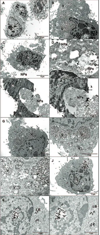
Cells in complete culture medium (negative control) showed normal morphology of live cells with well-defined nuclei and organelles. The same morphology was observed for most of the cells exposed to NP where no evident signs of toxicity were present. Cells exposed to high citrate coated ZrO2 (Figure 4H–I), pristine TiO2 (Figure 5B–D) and citrate coated TiO2
(Figure 5G–I) showed increasing number of enlarged vacuoles and amor-phous material in the cytosol indicating a starting of a possible inflamma-tory process. However, no apoptosis or cell necrosis was found.
In all the analysed samples, ZrO2 and TiO2 NP were observed both inside and outside cells in form of agglomerates/aggregates.
A possible mechanism of phage-endocytosis with autophagic lysosome transport and release of NP in the cytosol was observed in each sample. Lamellar bodies, probably involved in secretory pro-cess, were found after exposure to P25 (Figure 5K–L).
Cell viability
As shown in Figure 6, ZrO2 NP did not affect the Balb/3T3 viability cells after 24, 48 and 72 h of exposure. After 24 h treatment, only the low-est dose of low citrated ZrO2 NP showed a statistical significant effect (P < 0.05), however as the effect falls in the sub-toxic range, the result can be considered due to biological variability. After 72 h, only the higher dose of pristine ZrO2 NP showed a statistical significant effect (P <0.05). Figure 7 shows the dose-response curves for TiO2 NP after 24, 48 and 72 h of exposure. The data obtained showed that all NP induced a cytotoxic effect in Balb/3T3 starting from a time of exposure of 24 h.
Cytostasis and cell death
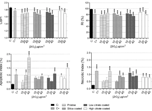
Cytostasis was measured by CBPI and RI, and cell death by apop-totic and necrotic indices. CBPI and RI showed a cytostatic effect for both pristine and remediated ZrO2 NP on Balb/3T3 cells compared to the untreated control (Figure 8). TiO2 NP induced a mild effect (Figure 9). Only the higher doses of pristine TiO2, 10 and 20 µg/cm2
of citrated and 20 µg/cm2 of P25 induced a statistically significant
effect. The same effects were observed by the RI.
The apoptotic and necrotic indices showed that all ZrO2 NP were cytotoxic to Balb/3T3 (Figure 8). Regarding TiO2 NP apoptotic cells were induced by citrated NP and by P25, while necrotic cells were induced by all the NP tested (Figure 9).
Figure 3. TEM images of TiO2 NP. (A) pristine; (B) Silica coated; (C) citrate coated; (D) P25.
Table 3. Amount of BSA added, normalized for the TiO2 NP surface area
SSA (g/m2)a ng BSA/cm2 TiO 2b
Uncoated TiO2 NP 154 97
Citrated TiO2 NP 156 96
Silicate TiO2 NP 86 174
Aeroxide® P25 60 250
aSSA, specific surface area determined by BET analysis.
bAmounts of BSA (ng) adsorbed on TiO
2 NP surface (cm2).
at Orta Dogu Teknik University Library (ODTU) on May 19, 2016
http://mutage.oxfordjournals.org/
Chromosomal damage
As shown in Figure 10, the highest doses of pristine, silicate coated and high citrate coated as well as the 10 µg/cm2 dose of high citrate
coated ZrO2 NP, significantly induced micronuclei formation. NPB
were induced by all the tested doses of the pristine ZrO2 NP and by the 10 and 20 µg/cm2 of the low citrated coated NP. NBUD
forma-tion was induced in a statistically significant manner only following exposure with the higher dose of pristine ZrO2 NP (Figure 10).
Also TiO2 NP induced a mild formation of micronuclei respect to negative control. Only the citrated TiO2 NP and the lowest tested
concentration of P25 induced a statistically significant formation of micronuclei (Figure 11). TiO2 NP exposure did not increase the NPB
frequency (Figure 11). Similarly many of the experimental condi-tions tested did not induce NBUD formation (Figure 11), except for the 40 µg/cm2 citrated and 10 µg/cm2 of Aeroxide® P25, which
enhanced significantly NBUD frequency.
Comet assay
Following 2 h treatment, each concentration of all the tested ZrO2
NP induced a significant increase of primary DNA damage in Balb/3T3 (Figure 12). After 24 h of treatment quite all the tested concentrations induced an increase of DNA fragmentation, although to a lesser extent (Figure 12). Following 48 and 72 h of exposure, there is a reduction of the DNA damage exerted by ZrO2 NP. Figure 4. TEM images of Balb/3T3 cells cultured for 48 h in complete cell culture medium (A, negative control) and exposed for 48 h to 20 µg/cm2 of: pristine ZrO2 (B, C magnification of B); silicated ZrO2 (D, E = magnification of D); low-citrated ZrO2 (F, G = magnification of F); high-citrated ZrO2 (H, I, J = increasing magnifications). N: nucleus; V, vesicles with NP inside; NP, nanoparticles.
at Orta Dogu Teknik University Library (ODTU) on May 19, 2016
http://mutage.oxfordjournals.org/
Oxidative comet assay showed that only the lower concentra-tion of pristine ZrO2 NP induced a statistically significant effect after 2 h, while following 24 h of exposure only 20 and 40 µg/cm2 of the
silica coated ones slightly increased oxidised pyrimidines, Endo-III sensitive sites (Figure 13). Oxidation of purines, Fpg sensitive sites,
increased significantly at all the tested concentrations of silica-coated ZrO2 NP at 2 h treatment, but this effect was lost after 24 h of treat-ment. The low citrate-coated NP induced a significant effect only at the highest concentration (2 h treatment), and at 20 µg/cm2 after 24 h
of exposure (Figure 13).
The genotoxicity exerted by TiO2 in Balb/3T3 cells investigated by comet assay showed that after both 2 and 24 h treatment all the NP induced a significant increase of primary DNA damage while, as shown for ZrO2 NP, after 48 and 72 h the level of DNA damaged was very low (Figure 14).
Both oxidised pyrimidines and purines increased significantly at quite all the tested concentrations of TiO2 NP as compared to base-line values, following the 2 and 24 h of treatment (Figure 15).
Morphological neoplastic transforming potential
As shown in Figure 16A, the pristine and both low and high cit-rate coated ZrO2 NP induced statistically significant morphological transformation in Balb/3T3 cells at all the tested concentrations.
Regarding TiO2 NP, only the citrate coated and the P25 induced type-III foci in Balb/3T3 cells (Figure 16B).
Discussion
In this study internalisation analysis showed that both ZrO2 and TiO2 NP were localised both inside and outside of the Balb/3T3 cells in form of agglomerates/aggregates after 48 h of treatment at a concentration of 20 µg/cm2 (Figures 4 and 5). The majority of the
cells exposed to NP did not show signs of toxicity. However the high-citrate coated ZrO2 NP and pristine and citrate coated TiO2 NP induced increasing number of enlarged vacuoles and amorphous material in the cytosol, likely indicating the beginning of a possi-ble inflammatory process, however without signs of cell death as increase of apoptotic or necrotic cells was not found.
CFE assay showed that ZrO2 NP did not affect Balb/3T3 clono-genicity. On the other hand, by scoring the apoptotic and necrotic cells, we observed that ZrO2 NP were able to induce cell death. Furthermore CBPI and RI indices were significantly lower in cells treated with ZrO2 NP respect to the negative control.
Starting by a treatment of 24 h, CFE showed that TiO2 NP were able to induce cytotoxic effects. From the results of CBMN-cyt assay, it is evident that only the citrated titania and P25 were able to exert apoptosis while all NP were able to induce necrosis, although only the P25 at all the tested doses. Cytostatic effects were induced only by the pristine, the citrated NP and by the P25.
This apparent discordance regarding the cytotoxicity results obtained with the CFE test and cytome assay, in particular regard-ing ZrO2 NP, could be due to the fact that these assays measure cell proliferation by taking into account distinct endpoints and by using different experimental designs. In fact the cytome assay measures the frequency of cell division and the evaluation occurs directly after the end of the NP exposure. On the contrary, the CFE assay does not refer to a specific mechanism of cytotoxicity, and colonies can origin from any surviving cell following 7 days of culture and after 4 days by the end of the exposure period (20). Taken together these data suggest that ZrO2 NP are able to induce cell death, however cells not too heavily damaged remain viable, continue to replicate themselves and are able to form colonies. Hence ZrO2 NP, in our experimental design, do not seem to possess a strong cytotoxic property and this is in agreement with literature data. In fact some studies reported no capacity by ZrO2 NP to induce cytotoxic effects (21–23) and only few studies reported a moderate cytotoxic potential (24,25). Figure 5. TEM images of Balb/3T3 cells cultured for 48 h in complete cell
culture medium (A, negative control) and exposed for 48 h to 20 µg/cm2 of: pristine TiO2 (B–D = magnification of C); silicate TiO2 (E, F = magnification of E); citrated TiO2 (G–I = increasing magnifications); P25 (J–L = increasing magnifications). N: nucleus; V: vesicles with NP inside; NP, nanoparticles; LB, lamellar bodies.
at Orta Dogu Teknik University Library (ODTU) on May 19, 2016
http://mutage.oxfordjournals.org/
The effects of TiO2 NP on cell viability are reported by several studies available in the literature. Regarding the toxic effects of anatase TiO2 NP, the crystalline form used in this study, literature results are conflicting. Cytotoxic potential of TiO2 NP was observed in human bronchial epithelial BEAS-2B cells (26), in IMR90 human bronchial fibroblasts cells (27), in human umbilical vein endothelial cells (28), in rat and human glial cells (29) and in Syrian Hamster Embryo Cells (SHE cells) where an EC50 value at the dose of 10 μg/ cm2 was obtained (30). However some researchers failed to find
cytotoxic potential of anatase TiO2 NP in human peripheral blood lymphocytes (PBL) (31), in human hepatoma HepG2 cells (32) and in A549 human lung carcinoma cells (33). Obviously possible vari-ability source in the results reported in literature may derive by the different cell type employed, the presence of serum, different char-acteristics of NP and the tested doses. Another point to take into account is the ability of NP to interfere with the classical colorimet-ric tests used for the study of cytotoxicity, such as WST-1, MTT or Neutral Red (34,35). In this context, CFE is an in vitro method for Figure 6. Cell viability in Balb/3T3 cells exposed to ZrO2 NP evaluated by CFE assay. Cells were exposed to increasing concentrations (1.25–80 µg/cm2) of NP for 24, 48 and 72 h. Data are plotted as mean %CFE values normalized to the untreated control (C−; black bar) ± standard error of the mean (SEM); n = 6. *P < 0.05, **P< 0.01, ***P < 0.001. C+: 1µM Na2CrO4 that induced 0% CFE (data not shown).
at Orta Dogu Teknik University Library (ODTU) on May 19, 2016
http://mutage.oxfordjournals.org/
the assessment of basal cytotoxicity which is noncolorimetric and nonfluorescent, avoiding thus possible interferences of NP with the toxicity assessment. In this context the European Commission’s Joint Research Centre (JRC) coordinated an interlaboratory comparison study on CFE for assessing the cytotoxicity of NP and the results obtained showed that CFE assay is a suitable and robust in vitro method to assess cytotoxicity of NP (36).
Genotoxicity evaluated by means of MN frequency demonstrated that ZrO2 and TiO2 NP possess a low capacity to induce micronu-clei. Our data are in agreement with previous studies in SHE cells (30) and in A549 cells (33) treated with pristine TiO2 NP. However some researchers found that TiO2 NP induced micronuclei forma-tion in PBL (37) and in human epithelial cells (38). In human embry-onic kidney (HEK293) and NIH/3T3 cells a statistically significant Figure 7. Cell viability in Balb/3T3 cells exposed to TiO2 NP evaluated by CFE assay. Cells were exposed to increasing concentrations (1.25– 80 µg/cm2) of NP for 24, 48 and 72 h. Data are plotted as mean %CFE values normalized to the untreated control (C−; black bar) ± standard error of the mean (SEM); n = 6. *P < 0.05, **P < 0.01, ***P < 0.001. C+: 1 µM Na2CrO4 that induced 0% CFE (data not shown).
at Orta Dogu Teknik University Library (ODTU) on May 19, 2016
http://mutage.oxfordjournals.org/
induction of micronuclei was observed when cells were exposed to 1000 µg/ml (a concentration much higher than that used in our study) of two anatase NP (39). One of the factors that could contrib-ute to the differences observed in those studies is that the induction of micronuclei by TiO2 NP is affected by the characteristics of the culture medium including its capacity to decrease the agglomeration and to increase the NP–cell interaction (39,40). It is also evident from some studies that the formation of micronuclei is correlated to the size of the NP; for example, it was observed an increased MN frequency after exposure of SHE cells to TiO2 NP of 20 nm, but not of 200 nm (41) as well as in BEAS-2B cells exposed to TiO2 NP of 10 and 200 nm, but not to TiO2 NP> 200 nm (42).
To the best of our knowledge, there is no available literature con-cerning micronuclei induction by ZrO2 NP.
Comet assay revealed that after 2 h exposure all the tested ZrO2 NP at all the doses were able to induce DNA damage, and after 24 h only the higher doses of the pristine, low-citrate and high citrate-coated NP did not induce significant effects. On the other hand after 48 and 72 h of exposure, minor DNA damage was found. Similar results were observed for TiO2 NP. For this rea-son, we decided to apply the modified protocol of comet assay to determine the presence of oxidised pyrimidine and purine bases in cells exposed for 2 and 24h to NP. Inclusion of EndoIII enzyme in the protocol revealed that only pristine and silica-coated ZrO2 NP were able to induce oxidation of pyrimidines while oxidation of purines was induced by silica-coated and low citrate-coated ZrO2 NP. On the other hand, all TiO2 NP were able to induce both pyrimidine and purine oxidation.
Only few studies investigated the genotoxic potential of ZrO2 NP. In PBL and cultured human embryonic kidney (HEK293) cells, exposure to ZrO2 NP did not induce significant primary and oxida-tive DNA damage (22). In an in vivo study, ZrO2 NP did not show genotoxic potential in the wing spot assay of Drosophila
mela-nogaster (43). Conflicting results about the capacity of anatase TiO2 NP to induce primary and oxidative DNA damage are reported. Positive results were obtained in HepG2 cells (32), in HEK293 and NIH/3T3 cell lines (39), and in SHE cells (30). Negative results were observed in IMR 90 and BEAS-2B cells (27), in cells of the human nasal mucosa and in PBL (31,44). Jugan and collaborators observed that TiO2 NP of 12 and 142 nm induced DNA strand breaks after 4 h of exposure in A549 cells and that after 24 h only the 12 nm sized NP induced DNA damage, while after 48 h the frequency of breaks drastically decreased in exposed cells (33). The results of the study by Jugan and collaborators are similar to our results that show the capacity of comet assay to detect DNA strand breaks is strictly dependent on the time of exposure, and that shorter is the time of exposure, higher is the damaged DNA detectable, likely due to the fact that cells have not enough time to repair most of the damage induced. Other factors could explain the time-related geno-toxic effects of NP observed with comet assay such as an adaptive response of the cells or a dilution effect of NP following cell replica-tions (45).
Carcinogenic potential of ZrO2 and TiO2 NP was evaluated by CTA assay, an in vitro test that has attracted attention within the field of alternative methods due to its potential to reduce the number of animals sacrificed to assay carcinogenicity in vivo (46).
Figure 8. Cytostatic (CBPI and RI) and cytotoxic (apoptotic and necrotic index) effects induced by ZrO2 NP. Cells were exposed to increasing concentrations (10–40 µg/cm2) of NP for 48 h. Data are plotted as mean values ± standard error of the mean (SEM); n = 4. *P< 0.05, **P< 0.01, ***P< 0.001. As positive control 0.10 μg/ml mitomycin-C was employed.
at Orta Dogu Teknik University Library (ODTU) on May 19, 2016
http://mutage.oxfordjournals.org/
Figure 9. Cytostatic (CBPI and RI) and cytotoxic (apoptotic and necrotic index) effects induced by TiO2 NP. Cells were exposed to increasing concentrations (10–40 µg/cm2) of NP for 48 h. Data are plotted as mean values ± standard error of the mean (SEM); n = 4. *P < 0.05, **P < 0.01, ***P < 0.001. As positive control, 0.10 μg/ml mitomycin-C was employed.
Figure 10. Chromosomal damage induced by ZrO2 NP. Balb/3T3 cells were exposed to 10–40 µg/cm2 NP for 48 h and micronuclei, NPB and NBUD were manually
scored using an inverted microscope (400× magnification). Data are plotted as mean values ± SEM; n = 4; statistical analysis performed by one-way ANOVA and Dunnet post-test. *P < 0.05, **P < 0.01, ***P < 0.001. As positive control 0.10 μg/ml mitomycin-C was employed.
at Orta Dogu Teknik University Library (ODTU) on May 19, 2016
http://mutage.oxfordjournals.org/
Figure 11. Chromosomal damage induced by TiO2 NP. Balb/3T3 cells were exposed to 10–40 µg/cm2 NP for 48h and micronuclei, NPB and NBUD were manually scored using an inverted microscope (400× magnification). Data are plotted as mean values ± SEM; n = 4; statistical analysis performed by one-way ANOVA and Dunnet post-test. *P < 0.05, **P < 0.01, ***P < 0.001. As positive control 0.10 μg/ml mitomycin-C was employed.
Figure 12. Primary DNA damage induced by ZrO2 NP. Balb/3T3 were treated with increasing concentrations (10–40 µg/cm2) of NP for 2, 24, 48 and 72 h. Each data point represents the mean ±SEM of two independent experiments. C+: positive control (50 μM H2O2); C−: untreated control. Statistical analysis performed by one-way ANOVA and Dunnet post-test. * P < 0.05; **P < 0.01; ***P < 0.001.
at Orta Dogu Teknik University Library (ODTU) on May 19, 2016
http://mutage.oxfordjournals.org/
Figure 13. Oxidised DNA lesions induced by ZrO2 NP. Balb/3T3 were treated with increasing concentrations (10–40 µg/cm2) of NP for 2 and 24 h. The level of enzyme-sensitive sites was obtained by subtracting the value of % of DNA fluorescence in tail obtained after digestion with each enzyme and with the buffer only. Each data point represents the mean ± SEM of two independent experiments. C+: positive control (50 μM H2O2); C−: untreated control. Statistical analysis performed by one-way ANOVA and Dunnet post-test. *P< 0.05; **P< 0.01; ***P< 0.001.
Figure 14. Primary DNA damage induced by TiO2 NP. Balb/3T3 were treated with increasing concentrations (10–40 µg/cm2) of NP for 2, 24, 48 and 72 h. Each data point represents the mean ± SEM of two independent experiments. C+: positive control (50 μM H2O2); C−: untreated control. Statistical analysis performed by one-way ANOVA and Dunnet post-test. *P < 0.05; **P < 0.01; ***P < 0.001.
at Orta Dogu Teknik University Library (ODTU) on May 19, 2016
http://mutage.oxfordjournals.org/
Regarding ZrO2 NP, only the silica-coated form did not induce transformation of Balb/3T3 suggesting that the presence of silica coating is able to protect Balb/3T3 from the formation of type-III foci and therefore from morphological transformation. These results are consistent with a previous publication which reported that SiO2 NP did not induce the formation of type III foci in Balb/3T3
cells (14). Moreover high performance zirconia toughened alumina
(ZTA) materials did not induce DNA damage, mutagenicity and car-cinogenicity in C3H/10T1/2 mouse fibroblasts (47) and zirconia
poly-crystals did not elicit mutagenic or transforming effects on irradiated C3H/10T1/2 cells (48).
CTA results on TiO2 NP showed that pristine and silica-coated NP did not induce cell transformation, but both citrate coated and P25 induced the formation of type III foci. To date, the current Figure 15. Oxidised DNA lesions induced by TiO2 NP. Balb/3T3 were treated with increasing concentrations (10–40 µg/cm2) of NP for 2 and 24 h. The level of enzyme-sensitive sites was obtained by subtracting the value of % of DNA fluorescence in tail obtained after digestion with each enzyme and with the buffer only. Each data point represents the mean ± SEM of two independent experiments. C+: positive control (50 μM H2O2); C−: untreated control. Statistical analysis performed by one-way ANOVA and Dunnet post-test. *P < 0.05; **P < 0.01; ***P < 0.001.
Figure 16. ZrO2 (A) and TiO2 (B) NP CTA results. Balb/3T3 cells were exposed for 72 h to increasing concentrations of NP. To evaluate the carcinogenicity of ZrO2 NP, the transformation frequency was calculated and data were compared to the untreated control cells (C−). The statistical significance was evaluated by Fisher exact test in respect to untreated control. Data are presented as mean values ± SEM; n = 3; *P < 0.05, **P <0.01, ***P < 0.001. C+: 3 µg/ml methylcholanthrene.
at Orta Dogu Teknik University Library (ODTU) on May 19, 2016
http://mutage.oxfordjournals.org/
literature about TiO2, that in 2010 has been recognised as a
pos-sible carcinogen for humans (group 2B, 49), does not present studies on the carcinogenic potential of TiO2 NP tested by CTA. However,
the ability to induce cell transformation in human embryonic kid-ney (HEK293) and in mouse embryonic fibroblast (NIH/3T3) cells exposed to TiO2 NP for 3 weeks was shown (40). Also in BEAS-2B cells treated for up to 4 weeks with TiO2 a significant increase in the
number of clones growing in an anchorage-independent way were observed (50). Changes in cell morphology, enhanced cell prolifera-tion and growth on soft agar, accompanied with increased chromo-somal instability, in NIH-3T3 cells after 12 weeks of exposure were also observed (51).
As discussed above, according to our findings the remediation of ZrO2 and TiO2 NP has not proven effective in modifying cyto- and genotoxic properties of the tested nanomaterials. Regarding CTA results, only the presence of silica coating has been effective in prevent-ing the type III foci formation of pristine ZrO2 NP, possibly making
safer the nanomaterial. On the other hand, both TiO2 and ZrO2 NP coated with citrate induced a strong transforming effect. These results are indicative that citrate itself could possess genotoxic and cell trans-forming properties. In this regard, Huk and collaborators (52) coated silver NP (Ag NP) with trisodium citrate in order to provide a negative charge on NP. They observed that the coated NP induced HPRT gene mutations in V79-4 cells, and that either sodium citrate alone was able to induce mutations analogously to those induced by Ag NP coated with citrate. Regarding remediated NP investigated in current study, in literature are reported only toxic effects of silica coated TiO2 NP
showing conflicting results. In an in vivo study the airway exposure to silica coated TiO2 NP, but not to the pristine one, induced pulmonary
neutrophilia in BALB/c mice with concomitant increase of expression of tumor necrosis factor-alpha and neutrophil-attracting chemokine CXCL1 in the lung tissue (53). Conversely, in rats intratracheally instilled with silica and alumina surface coated TiO2 NP, a lower
induction of adverse pulmonary health effects respect to uncoated NP was observed (54). In vitro, silica-coating improved the biocompat-ibility of TiO2 NP in a mouse fibroblast cell line (55) while in dorsal root ganglion cells after exposure to several types of TiO2 NP, among
which two NP coated with silica, NP induced apoptosis with the only exception of the uncoated rutile TiO2 (56).
Our results, taken together with the conflicting results reported in literature about TiO2 and ZrO2 NP toxicity, suggest that integrated
testing strategies are required for an adequate assessment of the impact of NP on human health and the environment. It is important, when assessing the hazard associated with NP, to establish standard testing procedures and thorough strategies to consider the different conditions relevant to possible exposures (57,58).
Conclusions
In Table 4, we report a summary of the results obtained with the various endpoints studied, by taking into account comparable doses and time treatment of the NP tested.
From this table, it is evident that each NP is able to induce some toxic effects, although none of the nanomaterials tested is able to induce significant effects for all toxic endpoint investigated.
The NP remediation strategy adopted in this study did not prove to change the toxic effects induced by pristine NP, although the coat-ing with SiO2 seems to prevent Balb/3T3 morphological transforma-tion induced by ZrO2 NP. In order to identify any potential impact of NP on human health, it is essential to fully investigate their toxi-cological profiles in different model systems. A test battery designed to evaluate risks to human health of NP should include reliable test assays, with no NP interferences, that are able to give complemen-tary information on the mechanisms of NP toxicity.
Funding
This study was financially supported by the FP7 projects No CP-FP 214478-2, NanoReTox and No 280716, SANOWORK.
Acknowledgements
We would like to thank Dr. Paolo Lucchesi (Department of Clinical and Experimental Medicine, University of Pisa) for assistance with TEM images acquisition. The authors are responsible for writing of the article and report no conflicts of financial, consulting and personal interests.
Conflict of interest statement: None declared.
References
1. George, S., Ho, S. S., Wong, E. S., et al. (2015) The multi-facets of sustain-able nanotechnology - Lessons from a nanosafety symposium.
Nanotoxi-cology, 9, 404–406.
2. Heiligtag, F. J. and Niederberger, M. (2013) The fascinating world of nano-particle research. Materialstoday, 16, 262–271.
3. Kumar, A. and Dhawan, A. (2013) Genotoxic and carcinogenic potential of engineered nanoparticles: an update. Arch. Toxicol., 87, 1883–1900. 4. Migliore, L., Uboldi, C., Di Bucchianico, S. and Coppedè, F. (2015)
Nano-materials and neurodegeneration. Environ. Mol. Mutagen., 56, 149–170. 5. Nel, A. E., Mädler, L., Velegol, D., Xia, T., Hoek, E. M., Somasundaran,
P., Klaessig, F., Castranova, V. and Thompson, M. (2009) Understanding biophysicochemical interactions at the nano-bio interface. Nat. Mater., 8, 543–557.
6. Kurzepa, H., Kyriazis, A. P. and Lang, D. R. (1984) Growth characteris-tics of tumors induced by transplantation into athymic mice of BALB/3T3 cells transformed in vitro by residue organics from drinking water. J.
Envi-ron. Pathol. Toxicol. Oncol., 5, 131–138.
Table 4. Summary of results obtained with NP tested with comparable doses and time treatment
Endpoint ZrO2
pristine
ZrO2 silica coated
ZrO2 low cit-rate coated
ZrO2 high citrate coated
TiO2 pristine TiO2 silicate coated TiO2 citrate coated Aeroxide P25 CFE − − − − + − + + Cytostasis + + + + + − − − Apoptosis + + + + − − + + Necrosis + + + + + + − +
Micronuclei, NBUD and NPB + + − + − − + −
Primary Comet Assay − + − − + + − +
Endo III − + − − − + + −
Fpg − + − − + + + −
CTA + − + + − − − +
+ indicates statistically significant effect respect to the negative control; − indicates no statistically significant effect with respect to the negative control.
at Orta Dogu Teknik University Library (ODTU) on May 19, 2016
http://mutage.oxfordjournals.org/