HAL Id: hal-01777834
https://hal.sorbonne-universite.fr/hal-01777834
Submitted on 25 Apr 2018
HAL is a multi-disciplinary open access
archive for the deposit and dissemination of sci-entific research documents, whether they are pub-lished or not. The documents may come from teaching and research institutions in France or abroad, or from public or private research centers.
L’archive ouverte pluridisciplinaire HAL, est destinée au dépôt et à la diffusion de documents scientifiques de niveau recherche, publiés ou non, émanant des établissements d’enseignement et de recherche français ou étrangers, des laboratoires publics ou privés.
Cognitive and neuroimaging features and brain
β-amyloidosis in individuals at risk of Alzheimer’s
disease (INSIGHT-preAD): a longitudinal observational
study
Bruno Dubois, Stéphane Epelbaum, Francis Nyasse, Hovagim Bakardjian,
Geoffroy Gagliardi, Olga Uspenskaya, Marion Houot, Simone Lista, Federica
Cacciamani, Marie-Claude Potier, et al.
To cite this version:
Bruno Dubois, Stéphane Epelbaum, Francis Nyasse, Hovagim Bakardjian, Geoffroy Gagliardi, et al.. Cognitive and neuroimaging features and brain β-amyloidosis in individuals at risk of Alzheimer’s disease (INSIGHT-preAD): a longitudinal observational study. The Lancet Neurology, Elsevier, 2018, 17 (4), pp.335 - 346. �10.1016/S1474-4422(18)30029-2�. �hal-01777834�
Cognitive and neuroimaging parameters and brain amyloidosis in individuals at risk of Alzheimer's disease (INSIGHT-preAD): a longitudinal observational study
Prof Bruno Dubois1 MD, Stephane Epelbaum2 MD, Francis Nyasse3, Hovagim Bakardjian4 PhD,
Geoffroy Gagliardi5, Olga Uspenskaya6 MD, Marion Houot7 PhD, Simone Lista8 PhD, Federica
Cacciamani9, Marie-Claude Potier10 PhD, Anne Bertrand11 MD, Foudil Lamari12 PhD, Habib Benali13
PhD, Jean-François Mangin14 PhD, Olivier Colliot15 PhD, Remy Genthon16 MD, Marie-Odile Habert17
MD, Prof Harald Hampel18 MD
Corresponding author: Bruno Dubois (IM2A, Pavillon François Lhermitte, Hôpital Salpêtrière, 47 Bd de
l’Hôpital, 75013 Paris, France; tel : 33 1 42 16 75 02 ; email: bruno.dubois@aphp.fr)
(1) Centre des Maladies Cognitives et Comportementales, Institut du Cerveau et de la Moelle épinière (ICM), UMR-S975; Université Pierre et Marie Curie-Paris 6, AP-HP, Hôpital de la Salpêtrière, Paris, France ; bruno.dubois@aphp.fr; 2) Centre des Maladies Cognitives et Comportementales, Institut du Cerveau et de la Moelle épinière (ICM), UMR-S975; Université Pierre et Marie Curie-Paris 6, AP-HP, Hôpital de la Salpêtrière, Paris, France ; stephane.epelbaum@aphp.fr; 3) Centre des Maladies Cognitives et Comportementales, Institut du Cerveau et de la Moelle épinière (ICM), UMR-S975 ; Université Pierre et Marie Curie-Paris 6, AP-HP, Hôpital de la Salpêtrière, Paris, France; francis.nyasse@aphp.fr 4) Centre des Maladies Cognitives et Comportementales, Institut du Cerveau et de la Moelle épinière (ICM), UMR-S975 ; Université Pierre et Marie Curie-Paris 6, AP-HP, Hôpital de la Salpêtrière, Paris, France ; h.bakardjian-ihu@icm-institute.org; 5) Institut du Cerveau et de la Moelle épinière (ICM), UMR-S975; Université Pierre et Marie Curie-Paris 6, Hôpital de la Salpêtrière, Paris, France ; gagliardi.geoffroy@gmail.com; 6) QuintilesIMS, 151-161 Bd Victor-Hugo, 93 Saint-Ouen, France ; olga.uspenskaya-cadoz@quintilesims.com; 7) Institute of Memory and Alzheimer’s Disease (IM2A), Centre of excellence of neurodegenerative disease (CoEN), ICM, APHP Department of Neurology, Hopital Pitié-Salpêtrière, University Paris 6, Paris, France; marion.houot@yahoo.fr; 8) Institut du Cerveau et de la Moelle épinière (ICM), UMR-S975; Université Pierre et Marie Curie-Paris 6, Hôpital de la Salpêtrière, Paris, France; s.lista-ihu@icm-institute.org ; 9) Centre des Maladies Cognitives et Comportementales, AP-HP, Hôpital de la Salpêtrière, Paris, France; federica.cacciamani@studio.unibo.it;10) Institut du Cerveau et de la Moelle épinière (ICM), UMR-S975; Université Pierre et Marie Curie-Paris 6, Hôpital de la Salpêtrière, Paris, France; marie-claude.potier@upmc.fr; 11) Neuroradiology Department, AP-HP, Inserm, CNRS, Institut du cerveau et la moelle (ICM), Inria, Aramis project-team, Sorbonne Universités, UPMC Univ Paris 6, Pitié-Salpêtrière Hospital, Paris, France ; anne.bertrand@aphp.fr; 12) Service de Biochimie Métabolique, Hôpital de la Salpêtrière, AP-HP, Paris, France; foudil.lamari@aphp.fr ; 13) Laboratoire d'Imagerie Biomédicale, Inserm U 1146, CNRS UMR 7371 Université Pierre et Marie Curie-Paris 6, Hôpital de la Salpêtrière, Curie-Paris, France ; habib.benali@gmail.com; 14) Neurospin, Institut d’Imagerie BioMédicale, CEA, Gif/Yvette, France ; jfmangin@gmail.com; 15) Sorbonne Universités, UPMC Univ Paris 06, Inserm, CNRS, Institut du cerveau et la moelle (ICM), Inria, Aramis project-team, AP-HP, Departments of Neurology and Neuroradiology, Hôpital Pitié-Salpêtrière, Paris, France ; olivier.colliot@upmc.fr;Aramis, Institut du Cerveau et de la Moelle épinière (ICM), UMR-S975; Université Pierre et Marie Curie-Paris 6, Hôpital de la Salpêtrière, Paris, France ; colliot.olivier@gmail.com; 16) Centre des Maladies Cognitives et Comportementales, AP-HP, Hôpital de la Salpêtrière, Paris, France;
remygenthon@gmail.com; 17) Nuclear Medicine Department, Laboratoire d’Imagerie Biomédicale, Sorbonne Universités, UPMC Univ Paris 06, Inserm U 1146, CNRS UMR 7371, Pitié-Salpêtrière University Hospital, Université Pierre et Marie Curie-Paris 6, AP-HP, Paris, France ; marie-odile.habert@aphp.fr; 18) Centre des Maladies Cognitives et Comportementales, Institut du Cerveau et de la Moelle épinière (ICM), UMR-S975; AXA Chair; Université Pierre et Marie Curie-Paris 6, AP-HP, Hôpital de la Salpêtrière, Paris, France ; Harald.Hampel@med.uni-muenchen.de;
ABSTRACT
Background-. A better understanding is needed concerning the risk factors and markers of disease
progression in preclinical AD. In the Investigation of Alzheimer’s Predictors in subjective memory complainers (INSIGHT-preAD) study, we aimed to investigate the relation between brain amyloidosis and various cognitive and neuroimaging parameters and the progression of cognitive decline in individuals with preclinical AD.
Methods- INSIGHT-preAD is an on going and mono-centric cohort study from the Salpêtrière Hospital,
Paris, France, which started 25th May 2013. The cohort includes cognitively normal individuals, over 70
years, with subjective memory complaints (SMC) but normal cognitive and memory scores according to the Mini Mental State Examination (MMSE≥27), Clinical Dementia Rating (CDR=0) and Free and Cued Selective Reminding Test (Total Recall≥41). Subjects were stratified by brain amyloid status (amyloid positive or amyloid negative) according to the uptake of 18F-Florbetapir. Demographic, cognitive, psycho-behavioural, functional, ApoE status, MRI (anatomical, diffusion, resting state-fMRI, arterial spin labeling sequences), FDG-PET imaging, EEG recordings with resting state and ERP, were performed at baseline with optional Actigraphy and CSF investigations. All subjects participate in follow-up with neuropsychological assessment, EEG, and Actigraphy every year; blood samplings for research on biomarkers, MRI, FDG-PET and amyloid-PET scans every 2 years. We investigated the association between amyloid status and the assessed measures at baseline and month 24, and assessed the clinical status of participants at month 30 to identify the factors associated with progression to prodromal AD, defined as an amnestic syndrome of the hippocampal type.
Findings- At baseline, the 318 participants had a mean age of 76.03 (SD 3.47) years with a mean
MMSE score of 28.67 (SD 0.96) and a high educational level (6.19 [SD 2.05] on a scale of 1-8). A significant positivity of the amyloid tracer 18F-Florbetapir was observed in 88 subjects (28%) whereas 230 were amyloid negative. After adjustment for age, gender and education and correction for multiple comparisons, there was no difference between the A+ and A- subgroups for any behavioural, cognitive (including SMC questionnaires), actigraphy and neuroimaging measures. As expected, ApoE 4 was more frequent in A+ (33 [38%] vs 29 [12.6%]; p<0.0001) and CSF Ab42 levels significantly correlated with mean SUVr (r=-0.62, p<0.0001) and discriminated A+ from A- subjects with high accuracy (AUCs= 0.89 [0.80-0.98] and 0.84 [0.72-0.96], respectively). After 30 months (44 withdrawals), the global cognitive efficiency remained stable on the MMSE (28.34 vs 28.87; p=0.16) and CDR (0.06 vs 0.05; p= 0.79) scales in the A+ participants compared to A- and only four of them progressed to prodromal AD, all from the amyloid-positive group. Compared to the rest of the amyloid-positive participants, at
baseline these subjects were older (80.3 years [SD 4.1] vs 76.9 years [SD 3.4]), with a greater amyloid SUVr (1.46 [SD 0.16] vs 1.02 [SD 0.2]) and ApoE4 allele frequency (n=3 [75%] vs n=33 [38%]) and mild executive dysfunction (FCSRT free recall score: 21.25 [SD 2.75] vs 29.08 (SD 5.44]; FAB total score: 13.25 [SD 1.50] vs 16.05 (SD 1.68]).
.
Interpretation- Brain amyloidosis was not associated with differences in cognition and behaviour and it
was not sufficient alone, even in this aged population, to define a high risk of rapid progression to a prodromal AD within 30 months. Follow-up is needed to establish whether this remains the case over longer periods.
Funding- IHU-A-ICM, Investissement d’Avenir from the Ministry of Research, Fondation
Plan-Alzheimer, Pfizer and AVID
RESEARCH IN CONTEXT Evidence before this study
The PubMed Database and ClinicalTrials.gov were searched for the terms “Preclinical Alzheimer(’s) disease”, “Presymptomatic Alzheimer(’s) disease”, “Asymptomatic Alzheimer(’s) disease” up to June 30th 2016, without any language restriction. This research was published in a recent 2017 systematic review. The same search strategy was further performed between June 2016 and the 4th of July 2017 to
include up to date published data. A meta-analysis on more than three thousand cognitively normal individuals, published in 2015, showed that amyloid PET positivity is a frequent finding even in the middle-aged population, in line with post-mortem studies. However, the longitudinal outcome of cognitively healthy individuals with markers of brain amyloidosis alone (ie, with negative tau or neurodegeneration markers) suggests that the risk of rapid progression to an overt clinical disease may not be high. At present, the natural history of these asymptomatic at risk subjects has not been completely elucidated. deep knowledge of the evolution of AD-related processes is absolutely needed for the successful design of the adequate clinical trials.
We tested in a mono-center cohort of well-defined cognitively normal elderly participants with subjective memory complaints, whether brain amyloidosis, a mandatory marker of preclinical Alzheimer’s disease, is associated to worse cognitive performances as well as brain atrophy on MRI and hypometabolism on Fluorodeoxyglucose PET in a multimodal analysis. We did not evidence any difference on these parameters at baseline and after a 24 months follow-up between amyloid positive versus negative participants after adjusting for age, gender, educational level. Using the occurrence of an amnestic syndrome of the hippocampal type as a clinically relevant marker of progression from preclinical to prodromal stage of AD , 4/88 (5%) A+ participants converted, giving an annual rate of 1.8%, while none of the 230 A- participants declined during the first 30 months of follow-up
Implications of all the available evidence
When strict inclusion criteria are used to warrant normal cognition in studies in preclinical AD, brain amyloidosis alone is not associated even to subtle cognitive changes. The annual rate of progression to a clinical diagnosis of AD of amyloid positive elderly with normal cognition is low in our study maybe in relation with their high educational level (mean 6.2, for a scale from 1 to 8 (max)). This is of major importance for clinical trials targeting preclinical AD and suggests that a large number of participants should be followed for more than 30 months to demonstrate clinical efficacy.
INTRODUCTION
During the last decade substantial progress has been achieved in the field of Alzheimer’s disease (AD). Both the International Working Group (IWG)1,2 and the National Institute on Aging-Alzheimer’s
Association (NIA-AA)3,4,5 conceptualized the disease as a continuum, with the dementia syndrome
representing the late end stage of a long period of cumulative pathological insults in the brain. This allowed for considering the preclinical stage of the disease, in which individuals free of cognitive and behavioural symptoms can now be identified by in vivo evidence of Alzheimer pathology6. The
preclinical AD stage seems particularly important for interventions aiming at preventing progression to the clinical stage, as well as for research into novel biomarkers that might guide therapies with early disease modification. Amyloid brain lesions are necessary for the development of clinical AD, however they may not be sufficient. The progression to clinical AD can result from complex and specific interaction between influencing factors that may favour or decrease the disease progression. In parallel, it may be possible to identify markers of progression announcing or certifying further occurrence of clinical AD.
The objectives of the Investigation of Alzheimer’s Predictors in subjective memory complainers (INSIGHT-preAD) study were to identify both the factors associated with and the markers of progression to clinical AD in asymptomatic at risk subjects. Working on these issues needs to use strict and clinically meaningful definitions of inclusion criteria and outcomes measures. To date, the use of cognitive composite scores to define preclinical AD progression and clinical expression raises the issue of the meaningfulness of these scores in practice, especially as the scores used vary from one study to another. 7,8,9,10,11,12 INSIGHT-preAD was aimed at tackling these objectives by using evidence-based
and clinically meaningful criteria for inclusion and outcomes in a group of 318 cognitively normal older individuals with a defined amyloid status. The follow-up of participants is on going. In this paper we analysed : i) the baseline data, comparing Amyloid positive (A +) and Amyloid negative (A -) subjects in order to investigate the impact of beta-amyloid deposition on several domains including subjective cognitive complaints, neuropsychological performance, fluid biomarkers, specific brain structures on volumetric MRI and regional metabolism on Fluorodeoxyglucose (FDG) – PET; ii) the evolution on all these parameters at month 24; iii) and the outcome for all participants at month 30 and the factors that may have influenced the progression in 4 participants.
METHODS
Study design and participants
INSIGHT-PreAD study (INveStIGation of AlzHeimer’s PredicTors in subjective memory complainers) is a university expert memory clinic based mono-center observational cohort study conducted by the Institute of Memory and Alzheimer’s disease, Pitié-Salpêtrière University Hospital, Paris. To be included, participants must meet the following criteria: age range between 70 and 85; presence of subjective memory complaints; normal Mini Mental State Examination13 (MMSE≥27) and Clinical Dementia Rating 14 (CDR=0) scores; no evidence of episodic memory deficit as documented by a normal Free and Cued
Selective Reminding Test score15 (FCSRT; total score≥41); having visual and auditory acuity adequate
for testing; and no systemic or chronic disease that may interfere with follow-up. The Ethic Committee of the Pitié-Salpêtrière Hospital approved the study protocol and all participants signed an informed consent form, previously explained and given (2 weeks before signature). The subjects were recruited through spontaneous consultation of all people referred at the memory clinic and through announcement of the study through press release and TV coverage. Study participants were recruited between May 25, 2013 and the last on January 20, 2015.
Procedures
Clinical, cognitive, psycho-behavioural and functional assessments (see Panel) were performed every 6 months by the same neuropsychologists (LB, MR, PR) and physicians (ADS, ML).
Brain amyloid PET scans were acquired 50 minutes after injection of 370 MBq (10 mCi) of 18F-Florbetapir33. Brain FDG-PET scans were obtained 30 minutes after injection of 2 MBq/kg of
2-deoxy-2-(18F)fluoro-D-glucose. Reconstructed images were analysed with a pipeline developed by the CATI
(www.cati-neuroimaging.com) (Supplementary Fig 1). For amyloid PET images, standard uptake value ratios (SUVR) were calculated by averaging the mean activity of cortical regions of interest: both left and right precuneus, cingulum posterior, cingulum anterior, and parietal, temporal and orbitofrontal cortex. The reference region was a combination of whole cerebellum and pons regions. The SUVR threshold to determine abnormality uptake was extracted performing linear correlation between our method34 and
the method used by Besson et al 35 using 53 PET scans from another French study, the IMAP cohort36.
This strategy was previously used to study any relationships between different tracers or methods37.
The SUVr threshold of 0.7918 allowed a categorization of our population in Aß positive or Aß negative. (More information are detailed in Supplementary data and Fig 2). Neither the participants nor the investigators were aware of the amyloid status.
The same pipeline was applied to brain glucose metabolism PET images. Cortical metabolic indexes were calculated in four bilateral regions (posterior cingulate cortex, inferior parietal lobule, precuneus and inferior temporal gyrus), specifically affected by AD38 with pons as reference region.
MRI acquisitions (1 hour duration) were performed on a 3T Siemens Magnetom VERIO MRI system (Siemens Medical Solutions, Erlangen, Germany). Scanning sessions included 3D T1-weighted magnetization-prepared rapid gradient echo (MP-RAGE), 2D FLAIR, 2D T2*, DTI acquisition and a T2*-weighted gradient-echo echo-planar imaging scan series for use in the resting-state connectivity analysis and visual task, and a pulsed arterial spin labeling scan for measurement of cerebral blood flow at rest and visual task. Hippocampal volume was measured on 3DT1 sequence using the in-house SACHA software39 and normalized to the mean total intracranial volume. Cortical thickness was
measured in 68 regions of interest (ROI) of the Desikan-Killiany atlas using Freesurfer 5.3.
EEG data were acquired using a 256-channel whole-head EEG System GES 300 (Electrical Geodesics Inc. EGI, Oregon, USA). High-density EEG was recorded: i) during rest while eyes were consecutively closed and open according to an audio cue for 30 s each and repeated twice; and ii) during a cognitive task-memory recall of words, which were previously memorized one hour before the recording with the FCSRT15.
CSF concentrations of total tau protein (t-tau), tau protein phosphorylated at threonine 181 (p-tau181)
and amyloid-β peptide 1-42 (Aβ1-42) were analysed using the double antibody sandwich ELISA method
(Innotest-Fujirebio ®, Courtaboeuf, France)40. The laboratory participates in the European External
quality control program, provided by “The Alzheimer's Association QC program for CSF biomarkers”
(http://neurophys.gu.se/sektioner/psykiatri_och_neurokemi/neurokem/theAlzAssQCprogram)41.
Genomic DNA was prepared from frozen blood samples using the 5Prime ArchivePure DNA purification system (Gaithersburg, MD) according to the manufacturer’s instructions. APOE genotypes were determined for each individual using PCR-based Sanger sequencing. The amplified fragments were then purified and sequenced using the same primers (see Appendix).
All the subjects participate in a follow-up with clinical, cognitive, psycho-behavioural and functional assessments every 6 months, EEG and Actigraphy investigations every year, structural and functional MRI with resting state, FDG-PET and amyloid-PET scans every 24 months. The study will continue until the last participants to be enrolled into the trial have been followed for the prescribed 72 months.
MMSE and FCSRT scores below the threshold for inclusion in the study was indicative of a possible progression to clinical AD at a prodromal stage, defined by a positive amyloid PET and a persistent amnestic syndrome of the hippocampal type according to the IWG-2 criteria41. A low performance in one
visit was not considered sufficient to ascertain a significant progression. In case of a persistent cognitive decline on two consecutive neuropsychological evaluations, an independent and blinded committee composed of two neurologists (BD, SE), a neuropsychologist (GG) and a neuroimaging expert (AB) reviews the medical file. All prodromal incident cases, consisting of an episodic memory deficit with a FCSRT Total recall below 41 together with a positive amyloid PET, therefore fulfilling IWG-2 criteria42,
were further included in a clinical cohort with the same cognitive and neuroimaging investigations as those used in the INSIGHT study.
Statistical analysis
A sample size calculation was performed in order to get a sufficient degree of confidence around a positive likelihood ratio (LR+) and a negative likelihood ratio (LR-)43. The likelihood ratios incorporate
both the sensitivity and specificity of the predictive model providing a direct estimator of how much the combination of predictors would change the odds of a progression to prodromal AD. Based on the figure of 14% of progression over 3 years reported by Rowe et al. in 2013 44 (data available when the study
was designed) and based on the use of a 95% confidence interval (95%CI), 82 subjects are required. Assuming a 8% permanent discontinuation rate during the study, enrolment was to be stopped when the number of 88 PET amyloid positive subjects has been reached.
Cognitive and behavioural tests scores, hippocampal volume, FDG-PET indexes and cortical thickness were compared between amyloid positive and negative subgroups. The t-test was performed on continuous data while the 2 test was utilized for categorical variables. A paired t-test was used for the
comparison between right and left hippocampus volumes. For comparison between amyloid subgroups, linear models were used for continuous variables, Poisson models for discrete variables and logistic models for dichotomous variables in order to control for age, gender and education. FDG indexes were also adjusted for blood glucose. Tests in which a large number of participants scored zero were dichotomized in 0 vs non-0 categories. Group differences were tested using log-likelihood tests. P values were corrected for multiple testing using Benjamini-Hochberg correction. Missing data were not imputed. The same linear model generalized linear models were performed using the amyloid SUVR instead of amyloid group (Supplementary Table 3). Statistical analysis was performed using R 3.3.2.
Role of the funding source
None of the funders of the study participated in the design of the study, data collection, analysis, interpretation or in the writing of the report. The corresponding author had full access to all the data in the study and had final responsibility for the decision to submit for publication.
RESULTS
Out of the 363 successive screened subjects, 318 met the inclusion criteria (figure1). Table 1 shows the characteristics of INSIGHT-PreAD study participants at baseline with a mean age of 76.1 years (SD 3.47), a female predominance (63.2 %) and a high education level (mean 6.2; SD=2.1) for a scale from 1 to 8-max34. Their mean MMSE score was 28.67 (SD=0.96) and FCSRT total recall score was 46.09
out of 48 (SD=1.98). Participants had no deficit in any of the cognitive tests assessing memory, executive and instrumental functions. All the subjects were CDR=0 with a mean score at the FAB of 16.41 (± 1.68) and no naming difficulties (79.21 ± 1.11 out of 80 at the DO 80). Sixty-two subjects (20%) were APOE-ε4 carriers. The mean normalized hippocampal volume (left plus right) was of 2.71 cm3 (0.31), being significantly higher for the right hippocampus (Table 1). The highest means of cortical metabolic activity in FDG PET were found in the right precuneus and parietal inferior region. Concerning optional investigations, 51 subjects (27 men and 24 women) consented to lumbar puncture for AD biomarker investigation and 88 had an actigraphy at baseline.
Of the 318 subjects who underwent an amyloid PET investigation, 88 subjects (27.7%) were considered as positive (A+) (using the threshold of 0.79 - see methods) and 230 (72.3%) as negative (A-). 16/51 subjects who had a lumbar puncture (31%) were classified as (A+) and 45 (69%) as (A-) based on amyloid PET stratification. At baseline, as expected (Table 1) CSF Ab42 levels were lower and total tau and phosphorylated Tau were higher in (A+) compared to (A-) subjects (p<0.0001). Mean SUVr was significantly correlated with CSF Ab42 (r=-0.62, p<0.0001), and with CSF Ab40/Ab42 ratio (r = 0.61; p<0.0001). CSF Ab42 and CSF Ab40/Ab42 ratio discriminated A+ from A- subjects with high accuracy (AUCs= 0.89 [0.80-0.98] and 0.84 [0.72-0.96], respectively). A non-linear correlation was observed with Amyloid PET results, with the best correlation noticed in the lower range values of CSF Ab42 and in the parietal inferior and cingulate posterior areas (Supplementary Fig 3). A+ subjects were on average significantly older with a higher prevalence of APOE-ε4 carriers compared to the A- group (Table 1). At baseline, no difference was found in terms of gender and education between the two groups. The two groups did not significantly differ in any other questionnaires assessing subjective feelings, behaviour, mood and quality of life. The number of subjects at each cognitive, behavioural and neuro-imaging investigation at baseline and at follow-up is given in supplementary Table S1. A+ participants showed significantly lower scores in MMSE and in FAB and a longer TMT B-A time (Table 1). These differences disappeared when the results were adjusted for age, gender and education. There was no difference for the other cognitive tests, including the FCSRT total recall and the Memory Binding Test. There was no significant difference in regional metabolic imaging values between the positive and amyloid-negative subgroups (table 1; supplementary table S3). A significant correlation was observed (p<0.05)
between SUVr and FDG PET values in both cingulate posterior, precuneus and left parietal and temporal inferior regions, which disappeared after adjustment for age, sex and education (Table S3). On structural MRI, a significant decrease was observed at baseline in A+ subjects for each hippocampal volume, the difference remaining significant when adjusting for age, gender and education and correcting for multiple comparisons. Significant differences were observed in cortical thickness of the left temporal pole, left anterior cingulate (rostral) and right pars orbitalis that remained after adjustment for age, sex and education but disappeared after correction for multiple comparisons for the three cortical thickness measures (supplementary Table S2).
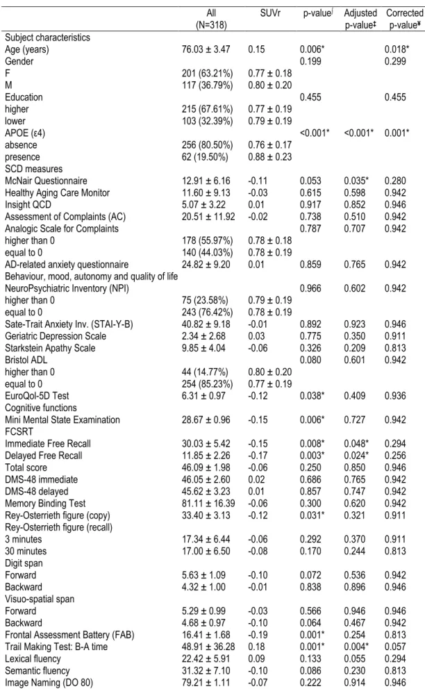
During the follow-up, the A+ participants did not differ from A- participants in any of the main cognitive tests assessing global efficiency (MMSE), episodic memory (FCSRT) and executive functions (FAB and TMTB-A); after 12 months and 24 months (see Table 2; results for all the tests will be reported separately). After 30 months, 274 subjects (out of whom 4 progressed to prodromal AD) were still in the study, 39 subjects have withdrawn (subject decision) and 5 deceased. Their performance remains stable over time (Table 2 and Supplementary Fig 4 for the plots of individual patient results). Resting-state EEG recordings showed significant longitudinal changes in the cortical oscillatory activity in A+ participants compared to A- ones, as shown in Fig 2 for θ/α power ratio changes (in preparation). After 30 months (44 withdrawals), the global cognitive efficiency remained stable on the MMSE (28.3 vs 28.8; 0.53 [0.14; 1.20], p=0.16) and CDR (0.06 vs 0.05; -0.01 [-0.08; 0.06], p= 0.79) scales in the A+ participants compared to A- and only four of them progressed to prodromal AD, all from the amyloid-positive group. Table 2 also shows the characteristics of the 4 participants who progressed to prodromal AD compared to the whole population and to the amyloid positive participants at baseline who did not progress to prodromal AD at M12, M24 and M30: each of them was older than the mean age of the other participants (80.3 years [SD 4.1] vs 76.9 years [SD 3.4]) and they have a higher ApoE4 allele frequency (n=3 [75%] vs n=33 [38%]), a greater amyloid SUVr (1.46 [SD 0.16] vs 1.02 [SD 0.2]), a lower normalized hippocampal volume for both sides and a mild executive dysfunction with a lower free recall score at the FCSRT and a lower FAB scores at baseline. By contrast they did not differ for the MMSE and the Total recall of the FCSRT. In each of the 4 cases, the progression began, in the year preceding prodromal AD by a severe drop in episodic memory: adjusting their cognitive performance to the group with Z scores, they all showed a significant decline of their Total Recall performance on the FCSRT episodic memory (10.75 for a mean decline of the population of 0.5) (Table 2).
DISCUSSION
In cognitively normal subjects with subjective memory complaints, with a mean age of 76 years, no difference in cognitive parameters was found between A+ and A- at baseline and after a 24 months
follow-up and only 4 subjects progressed to a prodromal AD after 30 months. Compared to other on-going cohorts44, the INSIGHT-preAD study presents substantial advantages. It is mono-centric, i.e. each
subject being investigated by the same team of academic, expert neuropsychologists and by the same neuroimaging scanners, therefore substantially reducing variance of data and results. The normal cognitive status of each subject was formally confirmed at baseline and none of them had any evidence of an amnestic MCI based on the FCRST used for the first time as a screening tool in a cohort of people at risk of AD. This is an observational study with no intervention that may modify the follow-up and therefore affect statistical power. A large number of domains are investigated, including and objective measures of cognition and behaviour, different MRI and PET investigations and EEG with resting state and ERP. Among various psychometric methods, the high number of scales investigating the subjective feelings of the subjects and the carers may provide a unique opportunity to evaluate the impact of cortical beta-amyloid deposition on subtle cognitive or behavioural changes.
One of the main results at baseline is that only 28% of subjects with a mean age of 76 years were amyloid positive, a feature that is slightly below the picture of the main on-going multicentre studies45-49.
Reviewing the literature, 27% of subjects were considered as positive for amyloid PET in the main cross-sectional studies in preclinical AD (56;50;51) but this increases to 30.4% for studies when mean age
is above 70 years (mean age of 74.4) according to our previously published systematic review on the cohorts used to study preclinical AD44 (Supplementary Table S4). The comparison between A + and A -
showed several differences at baseline for MMSE, tests of executive functions and hippocampal volume, significance that disappeared after adjustment for age and correction for multiple analyses. This underlines the necessity to control for the confounding effect of age, known to on executive functioning52
and hippocampal volume53. It is noteworthy that: i) in all the published studies on cognitive decline in A+
subjects, these participants are always significantly older than the A- subgroup 7-11 and ii) that the
declineoccurs late, after at least 18 months of follow-up8. In sum, our results suggest that cortical
beta-amyloid deposition have no effect on cognitive, functional and behavioural domains.
The degree of cognitive complaints between A + and A - subjects were similar. All the subjects must have had some memory complaints to be included, but those who were A + did not complain more, suggesting that the intensity of subjective memory complaints may not be a strong candidate marker of preclinical AD as already shown in AIBL aging study54,55. Moreover, the presence of amyloid brain
lesions was associated with a low cognitive awareness in our participants. 34 This result may appear in
contradiction with some recent data56 but it is noteworthy that the subjects were only complainers and
investigation of their subjective feelings was performed, and to our knowledge, INSIGHT-preAD is the first study with such a comprehensive evaluation of different aspects of cognitive complaints, including 6 questionnaires with a total of 88 items all of which administered with the same investigators in participants who were cognitively healthy at study entry.
The overall cognitive performance does not decline over time for the whole group and for the A + subgroup after exclusion of the 4 progressors (Table 2). This surprising result suggests that age-related changes in A- subjects on the one hand and cortical beta-amyloid deposition in the A+ subjects on the other hand are either not severe enough to impact cognitive functioning or are compensated by brain changes and/or reserve. The increase of resting-EEG alpha oscillations with a stronger change in frontal activation over a period of 2 years in the A+ subgroup (p < 0.03) is in favour of a possible cognitive control compensation58 (figure 2). These changes indicate that EEG is able to capture the neuronal
dynamics associated with the beginning stages of brain amyloidosis and over time. To conclude, the fact that cognitive performance remains stable in the A+ participants and that they marginally benefit from a practice effect (see Table 2) favour the hypothesis of a compensated state in asymptomatic at risk subjects (decoupling between structural lesions and maintenance of cerebral functioning) that precedes the decompensation in a clinical disease rather than a slow decline in a progressive continuum with no clear barriers between the asymptomatic and symptomatic states7,9 (see figure 3).
Strict inclusion criteria, short delay of observation, exclusion for the analysis of those subjects who further progressed to prodromal AD, and adjustment for age difference between subgroups (A+ vs A-) may explain the absence of decline.
The rate of progression to a clinically defined AD is surprisingly low despite the mean age of 77 year-old for the A + subjects. The follow-up is still on-going and the number of progressors might increase during further analyses according to a recent estimate of prevalence59. However, the number is low and may
result from a possible selection bias. For agreeing to participate in this observational study with a heavy follow-up including several hours of cognitive, behavioural, and functional investigations and several PET and MRI scans, the subjects must have a certain degree of cultural level and interest in supporting research, which is confirmed by their high mean level of education. We may postulate that their cognitive reserve compensated for the effect of brain amyloid lesions and has delayed the entrance in a clinical disease. The analysis of these 4 cases raises the question of the factors that may have facilitated the disease progression. At baseline, they were older, with a higher beta-amyloid deposition, frequently ApoE-ε4 allele carriers, with an evidence of a mild executive dysfunction suggesting saturation of functional mechanisms. By contrast they did not differ from the rest of the participants for
the MMSE and the Total recall of the FCSRT. During the follow-up, the onset of a severe drop in total recall performance during the preceding year is a marker of an on-going progression to a prodromal AD. ApoE4 was also a strong predictor of rapid progression to clinical AD in A+ subjects (3.24% per year [3/37 in 2.5 years]) compared to non-ApoE4 carriers (0.78% per year [1/51 in 2.5 years]).
Taken together, these data suggest that cortical amyloidosis may be relatively clinically silent for a long period of time (see figure 3). It is only when a progression to a prodromal AD is on-going that episodic memory disorders appear, probably in relation with the activation of tau pathology at the level of medial temporal lobe structures. However, the rate of progression at 2.5 years remains weak in our study and in accordance with data from other follow-up studies on preclinical AD48; 8;11. These data are important
with respect to on-going and future clinical trials on preclinical subjects, because the demographic characteristics of the randomized subjects will probably be similar with the same bias of selection. In that case, there is a need to increase considerably either the number of subjects or the duration of the trials as this is the case for instance in the A4 and Tommorrow ongoing studies (1150 and 3494 participants respectively, followed for almost 5 years)60;61. This also underlines the need to determine
the associated factors that influence the decline such as age, ApoE status and initial amyloid burden among others. Besides the short follow-up so far, another limitation of our study is the censoring effect due to the inclusion criterion of age over 70 years old. Another related issue is to define new markers of disease progression that are less rigid. If the onset of a prodromal AD is indisputably a formal outcome for the study of efficacy of a disease modifier, it would be interesting to identify some surrogate markers that predict such an event before its occurrence and that may help to distinguish the progressors from those A+ subjects that remain stable over time. Our analysis of patients who converted to a prodromal AD suggests that a recent decrease in cued recall performance may be a marker of progression. This is not surprising as it indicates a progression of brain AD lesions. The next follow-up of the study should help to confirm whether this effect is consistent..
In the field of disease-modifying therapies, there is an upcoming trend to shift from AD dementia stages to the early prodromal stages. It will be crucial to define clearly the dynamic processes that precede the progression to a clinical disease. The INSIGHT-preAD study, designed for identifying the best multimodal biomarkers combination for predicting the secondary occurrence of clinical AD, will constitute a valuable repository of clinical, cognitive, neuroimaging, neurophysiological, and biological data to be shared with the scientific community. Our data suggest that brain amyloidosis has no impact on behaviour and cognition at baseline and after a follow-up of 30 months suggesting that
compensatory mechanisms are present for maintaining a normal brain functioning and that amyloidosis alone is not sufficient to define a high risk of rapid progression to a clinical AD.
Panel: Assessments used in INSIGHT-PreAD
Subjective feelings about memory and cognition (more information in the supplementary data)
15-item version of the McNair Frequency of Forgetting Questionnaire (modified from16)
Healthy Age Brain Care Monitor (HABC-M)17
INSIGHT Questionnaire of Cognitive Decline (IQCD)* Assessment of Complaints (AC)*
Analogic Scale for Complaints (ASC)*
AD-related Anxiety Questionnaire (AD-NOS)*
Psycho-behaviour, mood, autonomy and quality of life
Neuropsychiatric Inventory (NPI)18
State-Trait-Anxiety Inventory Y (STAI-Y-B)19
Geriatric Depression Scale (GDS)20
Starkstein Apathy Scale21
Bristol Activities of Daily Living (Bristol ADL)22
Amsterdam IADL Questionnaire EuroQol 5D test (EQ-5D-3)23
Cognitive functions
For global assessment of cognitive functioning Mini Mental State Examination (MMSE)13 †
Clinical Dementia Rating (CDR)14
For episodic memory:
Free and Cued Selective Reminding (FCSRT)15 †
DMS-48 (immediate and delayed)24
Rey-Osterrieth Complex Figure (3-min and 30-min recall)25
For working memory and executive functions:
Forward and backward Digit and Visuo-spatial span27
Frontal Assessment Battery (FAB)28 †
Trail Making Test (TMT)29 †
Lexical Fluency (P words in 2 minutes)30
For instrumental functions
Semantic Fluency (animals in 2 minutes)30
Image Naming (DO 80)31
Praxis assessment32
Rey-Osterrieth Complex Figure (copy)25
Brain imaging
Brain amyloid PET with 18F-Florbetapir33
Brain FDG-PET 2-deoxy-2-(18F)fluoro-D-glucose
MRI: 3D T1-weighted magnetization-prepared rapid gradient echo (MP-RAGE), 2D FLAIR, 2D T2*, DTI acquisition and a T2*-weighted gradient-echo echo-planar imaging
Pulsed ASL scan. Hippocampal volume Cortical thickness.
Neural dynamics EEG with resting state and ERP
High-density EEG during rest
High-density EEG during a cognitive task-memory recall of words
CSF biomarkers
total tau protein (t-tau),
tau protein phosphorylated at threonine 181 (p-tau181)
ApoE genotyping
All assessments were done at baseline. All participants have neuropsychological assessment, EEG and actigraphy every 12 months, and blood sampling MRI, FDG-PET, and amyloid-PET scans every 2 years. Here, we report baseline data for all variables except for Amsterdam IADL, which will be presented in a separate paper.
† For 12 and 24 months we present results of main cognitive tests assessing cognitive global efficiency
(MMSE), episodic memory (FCSRT) and executive functions (FAB and TMTB-A), with other test results to be reported in more detail in subsequent follow-up reports.
* Information can be found in Reference34
Funding- The study was promoted by INSERM in collaboration with ICM, IHU-A-ICM and Pfizer and
has received a support within the “Investissement d’Avenir” (ANR-10-AIHU-06). The study was realized in collaboration with the “CHU de Bordeaux” (coordination CIC EC7), the promoter of Memento cohort, funded by the Foundation Plan-Alzheimer. The IHU-A-ICM (Investissement d’Avenir from the Ministry of Research) and Pfizer gave funds that were used for the recruitment of Clinical Research Assistants, neuropsychologists and MD. The Fondation Plan-Alzheimer and Memento project participate at the payment of the MRI and FDG-PET. Avid provided 18F-Florbetapir ligand for the amyloid PET.
Acknowledgment- Doctor Carole DUFOUIL, member of the MEMENTO study, and Doctor Rachel
Schindler from Pfizer participated in the elaboration of the protocol.
Each of the authors has participated in the INSIGHT-preAD study either in the elaboration of the protocol (BD, FN, HB, OU, MCP, FL, HB, OC, MOH), the collection of the data (SE, HB, GG, AB, JFM), their analysis (BD, SE, FN, HB, GG, MH, SL, FC, MCP, FL, OC, RG, HH). They also contributed in the writing and the revision of the paper and have approved the final version. The corresponding author had full access to all the data in the study and had final responsibility for the decision to submit for publication.
Conflict of interest statements
1. Bruno Dubois reports consultancy fees from Boehringer-Ingelheim, Eli Lilly, Biogen, and MedAvante; he received grants for his institution from Merck, Pfizer and Roche.
2. Stéphane Epelbaum – reports grants from Eli Lilly and consultant fees from Astellas Pharma. 3. Francis Nyasse no personal conflict of interest.
4. Hovagim Bakardjian reports speaker fees from Roche.
5. Geoffroy Gagliardi reports grants from France Alzheimer during the conduct of the study. 6. Olga Uspenskaya is a IQVIA (formerly Quintiles IMS) employee.
7. Marion Houot reports no personal conflict of interest.
8. Simone Lista reports speaker fees from Roche, outside the submitted work. 9. Federica Cacciamani reports no personal conflict of interest.
10. Marie-Claude Potier reports grants from Pfizer, Roche, Fondation Vaincre Alzheimer, and Laboratoires Servier.
11. Anne Bertrand reports no personal conflict of interest. 12. Foudil Lamari reports no personal conflict of interest.
13. Habib Benali reports no personal conflict of interest in relation with the current study. 14. Jean-François Mangin reports no personal conflict of interest.
15. Olivier Colliot - reports speaker fees from Roche and grants to his institution from Air Liquide Medical Systems, Qynapse and myBrainTechnologies.Prior to 2 years ago: O.C. has received lecture fees from Lundbeck and consulting fees from Guerbet. O.C.'s laboratory has received funding from EISAI.
16. Remy Genthon is a former employee of Sanofi and reports stock options in Sanofi
17. Marie-Odile Habert reports personal fees from Lilly, personal fees from Piramal, outside the submitted work.
18. Harald Hampel reports grants from Pfizer and Avid paid to his institution; personal fees from Jung Diagnostics and Anavex; personal fees and non-financial support from Roche, GE Healthcare, Eli Lilly, Cytox Ltd, Axovant Sciences, Takeda, Zinfandel Pharmaceuticals Inc, and Oryzon Genomics; holds patents for in vitro determination methods (8916388; 20100062463; 7547553; 20080199966) and in vitro procedures (8298784; 20100035286; 20090263822) for diagnosis and early diagnosis of neurodegenerative disorders, neurodegenerative markers for psychiatric conditions (20120196300; 20080131921), and CSF diagnostic in vitro method for diagnosis of dementias and neuroinflammatory diseases (20080206797).
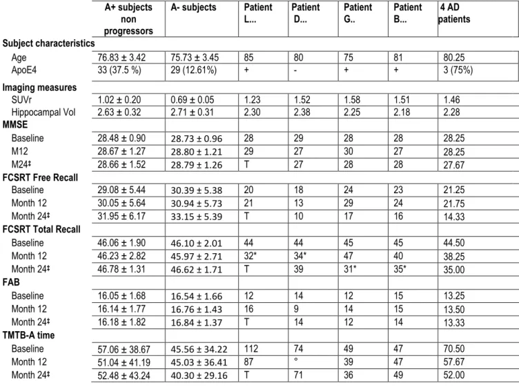
Table 1. Characteristics and test performance of A+ and A- subjects at baseline
(N = 318) All (n=88; 27.67%) (n=230; 72.33%) A+ subjects A- subjects p-value ∫ Adjusted p-value‡ Corrected p-value¥ Subject characteristics Age (years) 76.03 ± 3.47 76.83 ± 3.42 75.73 ± 3.45 0.0111* 0.0332* Gender (male) 117 (36.79%) 32 (36.36%) 85 (36.96%) 1.0000 1.0000 Education (high§) 215 (67.61%) 53 (60.23%) 162 (70.43%) 0.1082 0.1623 APOE (ε4) 62 (19.50%) 33 (37.50%) 29 (12.61%) <0.0001* <0.0001* 0.0001* SCD measures McNair Questionnaire 12.91 ± 6.16 12.24 ± 5.39 13.16 ± 6.41 0.2014 0.1918 0.6818 Healthy Aging Care Monitor 11.60 ± 9.13 11.28 ± 8.19 11.72 ± 9.48 0.6957 0.5534 0.9739 Insight QCD 5.07 ± 3.22 5.39 ± 3.14 4.95 ± 3.25 0.2755 0.2853 0.9130 Assessment of Complaints (AC) 20.51 ± 11.92 21.01 ± 12.73 20.32 ± 11.62 0.6577 0.9431 0.9739 Analogic Scale for Complaints 140 (44.03%) 40 (45.45%) 100 (43.48%) 0.8482 0.6274 0.9739 AD-related anxiety questionnaire 24.82 ± 9.20 25.99 ± 9.02 24.37 ± 9.24 0.1745 0.1893 0.6818
Behaviour, mood, autonomy and quality of life
NeuroPsychiatric Inventory (NPI) 243 (76.42%) 61 (69.32%) 182 (79.13%) 0.0898 0.0850 0.5942 Sate-Trait Anxiety Inv. (STAI-Y-B) 40.82 ± 9.18 41.27 ± 9.66 40.69 ± 9.09 0.8025 0.8269 0.9739 Geriatric Depression Scale 2.34 ± 2.68 2.41 ± 2.72 2.32 ± 2.69 0.8379 0.7615 0.9739 Starkstein Apathy Scale 9.85 ± 4.04 9.32 ± 3.28 10.05 ± 4.28 0.1092 0.0910 0.5942 Bristol ADL 254 (85.23%) 64 (81.01%) 190 (86.76%) 0.2942 0.3194 0.9293 EuroQol-5D Test 6.31 ± 0.97 6.22 ± 0.99 6.35 ± 0.96 0.3622 0.6033 0.9739
Cognitive functions
Mini Mental State Examination 28.67 ± 0.96 28.48 ± 0.90 28.74 ± 0.97 0.0302* 0.8151 0.9739 FCSRT
Immediate Free Recall 30.03 ± 5.42 29.08 ± 5.44 30.39 ± 5.39 0.0574 0.1432 0.6545
Delayed Free Recall 11.85 ± 2.26 11.44 ± 2.43 12.00 ± 2.18 0.0607 0.1114 0.5942
Total score 46.09 ± 1.98 46.06 ± 1.90 46.10 ± 2.01 0.5961 0.9942 0.9942 DMS-48 immediate 46.05 ± 2.60 46.05 ± 3.35 46.05 ± 2.25 0.2934 0.9065 0.9739 DMS-48 delayed 45.62 ± 3.23 45.95 ± 1.98 45.49 ± 3.59 0.3963 0.4947 0.9739 Memory Binding Test 81.11 ± 16.39 81.10 ± 16.25 81.11 ± 16.48 0.9945 0.8759 0.9739
Rey-Osterrieth figure (copy) 33.40 ± 3.13 32.96 ± 3.56 33.57 ± 2.94 0.0802 0.6190 0.9739 Rey-Osterrieth figure (recall)
3 minutes 17.34 ± 6.44 17.08 ± 5.62 17.43 ± 6.72 0.6481 0.8184 0.9739 30 minutes 17.00 ± 6.50 16.62 ± 5.69 17.14 ± 6.78 0.4955 0.9081 0.9739 Digit span Forward 5.63 ± 1.09 5.53 ± 1.00 5.67 ± 1.12 0.4026 0.8830 0.9739 Backward 4.32 ± 1.00 4.38 ± 0.93 4.30 ± 1.02 0.4505 0.5300 0.9739 Visuo-spatial span Forward 5.29 ± 0.99 5.30 ± 1.02 5.29 ± 0.98 0.5290 0.7791 0.9739 Backward 4.68 ± 0.97 4.58 ± 1.00 4.72 ± 0.96 0.2923 0.7989 0.9739 Frontal Assessment Battery (FAB) 16.41 ± 1.68 16.05 ± 1.68 16.54 ± 1.66 0.0064* 0.5151 0.9739 Trail Making Test: B-A time 48.91 ± 36.28 57.06 ± 38.67 45.83 ± 34.93 0.0200* 0.0613 0.5942 Lexical fluency 22.42 ± 5.91 22.98 ± 5.97 22.21 ± 5.88 0.3138 0.1015 0.5942 Semantic fluency 31.32 ± 7.10 30.60 ± 6.10 31.60 ± 7.44 0.2285 0.6093 0.9739 Image Naming (DO 80) 79.21 ± 1.11 79.22 ± 1.08 79.20 ± 1.12 0.9529 0.9435 0.9739
Amyloid PET imaging
Standardized uptake value ratios
(SUVr) 0.78 ± 0.19 1.02 ± 0.20 0.69 ± 0.05 FDG-PET imaging Cingulum Posterior L 2.44 ± 0.28 2.40 ± 0.27 2.46 ± 0.29 0.1051 0.1088 0.2149 Cingulum Posterior R 2.53 ± 0.29 2.49 ± 0.31 2.54 ± 0.29 0.1570 0.1376 0.2149 Parietal Inferior L 2.45 ± 0.26 2.41 ± 0.25 2.47 ± 0.26 0.0809 0.1196 0.2149 Parietal Inferior R 2.58 ± 0.27 2.54 ± 0.28 2.60 ± 0.27 0.0925 0.1088 0.2149 Precuneus L 2.52 ± 0.29 2.49 ± 0.28 2.54 ± 0.29 0.1706 0.2206 0.2942 Precuneus R 2.58 ± 0.29 2.54 ± 0.28 2.60 ± 0.29 0.1156 0.1433 0.2149 Temporal Inferior L 2.15 ± 0.20 2.13 ± 0.21 2.16 ± 0.20 0.2789 0.3794 0.4139 Temporal Inferior R 2.36 ± 0.24 2.33 ± 0.24 2.36 ± 0.24 0.2928 0.3105 0.3726
Magnetic Resonance Imaging
Normalized hippocampal volume 2.71 ± 0.31 2.63 ± 0.32 2.74 ± 0.31 0.0052* 0.0175* 0.1047 - Left hippocampal volume 2.65 ± 0.32 2.59 ± 0.33 2.68 ± 0.31 0.0250* 0.0624 0.2149 - Rght hippocampal volume 2.77 ± 0.33 2.67 ± 0.35 2.81 ± 0.32 0.0010* 0.0027* 0.0325* CSF Biomarkers (n=51) Number of subjects 51 16 (31.37%) 35 (68.63%) Age (years) 76.01 ± 3.40 76.34 ± 3.27 75.86 ± 3.50 0.0164* Gender (male) 27 (52.94%) 4 (25.00%) 23 (65.71%) 0.6478 Amyloid peptide 1-42 (pg/ml) 918.75 ± 365.64 612.50 ± 201.29 1058.74 ± 338.26 <0.0001* Ratio Amyloid peptide1-42/1-40 20.17 ± 10.16 28.52 ± 12.02 16.35 ± 6.33 0.0001* Total tau (pg/ml) 295.2 ± 122.0 382.3 ± 114.6 255.4 ±104.3 0.0009* Phosphorylated-tau (pg/ml) 50.49 ± 15.97 62.25 ± 12.30 45.11 ± 14.62 0.0003*
Note. Counts, percentages, means and standard deviations are shown for the whole INSIGHT-PreAD sample and for the
two groups, as well as p-values, to indicate statistically significant group differences. Values are expressed as Mean values ± Standard Deviation
§ Equal to or higher than high-school diploma
∫ p-values using the t-test for continuous variables and chi-square test for qualitative variables
‡ p-values adjusted for age, gender and education and blood glucose only for FDG indexes using generalized linear models ¥ adjusted p-values corrected for multiple testing using Benjamini-Hochberg correction
* Statistically significant at p < 0.05
Legend. A+: amyloid positive subjects; A-: amyloid negative subjects; McNair Frequency of Forgetting Questionnaire; Insight
QCD: INSIGHT Questionnaire of Cognitive Decline; Bristol ADL: Bristol Instrumental Activities of Daily Living; FCSRT: Free and Cued Selective Reminding Test; PET: Positon Emission Tomography; FDG: Fluoro-deoxyglucose ; L: left; R: right
Table 2. Comparative data at baseline, 12 months and 24 months between the participants Amyloid (-), Amyloid (+) non progressors and the 4 subjects that progressed to prodromal AD
A+ subjects non progressors
A- subjects Patient
L... PatientD... PatientG.. PatientB... 4 AD patients Subject characteristics Age 76.83 ± 3.42 75.73 ± 3.45 85 80 75 81 80.25 ApoE4 33 (37.5 %) 29 (12.61%) + - + + 3 (75%) Imaging measures SUVr 1.02 ± 0.20 0.69 ± 0.05 1.23 1.52 1.58 1.51 1.46 Hippocampal Vol 2.63 ± 0.32 2.71 ± 0.31 2.30 2.38 2.25 2.18 2.28 MMSE Baseline 28.48 ± 0.90 28.73 ± 0.96 28 29 28 28 28.25 M12 28.67 ± 1.27 28.80 ± 1.21 29 27 30 27 28.25 M24‡ 28.66 ± 1.52 28.79 ± 1.26 T 27 28 28 27.67 FCSRT Free Recall Baseline 29.08 ± 5.44 30.39 ± 5.38 20 18 24 23 21.25 Month 12 30.05 ± 5.64 30.94 ± 5.73 21 13 29 24 21.75 Month 24‡ 31.95 ± 6.17 33.15 ± 5.39 T 10 17 16 14.33 FCSRT Total Recall Baseline 46.06 ± 1.90 46.10 ± 2.01 44 44 45 45 44.50 Month 12 46.23 ± 2.82 45.97 ± 2.71 32* 34* 47 40 38.25 Month 24‡ 46.78 ± 1.31 46.62 ± 1.71 T 39 31* 35* 35.00 FAB Baseline 16.05 ± 1.68 16.54 ± 1.66 12 14 12 15 13.25 Month 12 16.14 ± 1.77 16.76 ± 1.43 16 9 14 15 13.50 Month 24‡ 16.18 ± 1.82 16.84 ± 1.37 T 14 12 14 13.33 TMTB-A time Baseline 57.06 ± 38.67 45.56 ± 34.22 112 74 49 47 70.50 Month 12 51.04 ± 41.19 45.03 ± 36.41 87 ° 39 47 57.67 Month 24‡ 52.48 ± 43.24 40.30 ± 29.16 T 71 36 49 52.00
Note. Percentages, means and standard deviations are shown for A+ subjects and A- subjects as well as value for the 4
prodromal AD patients.
‡ Results without the 4 subjects that progressed to prodromal AD
Legend. A+: amyloid positive subjects; A-: amyloid negative subjects; SUVr : Standardized uptake value ratios; MMSE: Mini Mental State Examination; FCSRT: Free and Cued Selective Reminding Test; FAB: Frontal Assessment Battery; TMT: Trail Making Test; T: data not available (patient deceased); ° missing data
* The asterisk underlines the drop in the Total recall score of the FCSRT in the year that precedes the progression to prodromal AD
FIG 1 - On going Flow Diagram of INSIGHT-preAD Study
363 screened
318 included at baseline
45 screening failures:
- 23 Inclusion criteria unfilled (3 MMSE, 2 FCSRT, 6 MMSE and FCSRT, 1 MMSE+ FCSRT and CDR, 7 MRI Contraindication, 4 PET not available).
-22 Patient’s decisions.
21 withdrew:
- 4 Changes of place of residence - 17 Patient’s decisions.
297 with month 12 data
10 withdrew:
- 1 Change of place of residence - 5 Patient decision
- 1 converted - 3 Deaths 287 with month 24 data
13 withdrew:
- 9 Patient’s decisions. - 2 converted
- 2 Deaths (1 converted) 274 with month 30 data
FIG 2- Longitudinalθ/α power ratio changes of A+ participants in EEG at-rest
Legend: a frontal activation in amyloid-positive elderly subjects over time is suggested by the
FIG 3 – Two hypothetical models of the natural history of AD
Legend: Two models of the natural history of preclinical to clinical AD transition.
Model 1 refers to the dominant view that cognition is progressively impaired in a continuous
Cognition AD dementia Aß+ Stage 1 Stage 2 Stage 3 MCI due to AD Tau+ Subtle cognitive changes MODEL ONE A progressive deterioration Unspecific clinical phenotype Preclinical AD AD dementia AD brain lesions Prodromal AD Cognition Compensated state Preclinical AD MODEL TWO The preclinical compensation
Specific clinical phenotype
fashion from preclinical stage (separated into three different stages according the type of underlying brain lesion such as Aβ and tau3) to the clinical stages of AD (MCI to dementia). In
model 2, we propose an alternative view, based on our data on brain amyloidosis. Cognition
remains stable in the preclinical phase of the disease despite the underlying AD brain lesions until brain compensatory mechanisms are overwhelmed, leading to the the clinical disease.
A+: abnormal amyloid peptide levels; Tau+: abnormal Tau protein levels; MCI: mild cognitive impairment; AD: Alzheimer’s disease
REFERENCES
1. Dubois B, Feldman HH, Jacova C, et al. Research criteria for the diagnosis of Alzheimer's disease: revising the NINCDS-ADRDA criteria. Lancet Neurol 2007; 6: 734–46.
2. Dubois B, Feldman HH, Jacova C, et al. Revising the definition of Alzheimer's disease: a new lexicon. Lancet Neurol 2010; 9: 1118–27.
3. Sperling RA, Aisen PS, Beckett LA, et al. Toward defining the preclinical stages of Alzheimer’s disease: recommendations from the National Institute on Aging-Alzheimer’s Association workgroups on diagnostic guidelines for Alzheimer’s disease. Alzheimers Dement 2011; 7: 280–92.
4. Albert MS, DeKosky ST, Dickson D, et al. The diagnosis of mild cognitive impairment due to Alzheimer’s disease: recommendations from the National Institute on Aging-Alzheimer’s Association workgroups on diagnostic guidelines for Alzheimer’s disease. Alzheimers Dement 2011; 7: 270–9.
5. McKhann GM, Knopman DS, Chertkow H, et al. The diagnosis of dementia due to Alzheimer’s disease: recommendations from the National Institute on Aging-Alzheimer’s Association workgroups on diagnostic guidelines for Alzheimer’s disease. Alzheimers Dement 2011; 7: 263–9.
6. Dubois B, Hampel H, Feldman HH, et al. Preclinical Alzheimer's disease: Definition, natural history, and diagnostic criteria. Alzheimers Dement 2016; 12: 292–323.
7. Clark, LR, Racine AM, Koscik RL, et al.. "Beta-amyloid and cognitive decline in late middle age: Findings from the Wisconsin Registry for Alzheimer's Prevention study." Alzheimers Dement 2016; 2: 805-814.
8. Donohue, M. C., R. A. Sperling, D. P. Salmon, D. M. Rentz, R. Raman, R. G. Thomas, M. Weiner, P. S. Aisen, B. Australian Imaging, A. Lifestyle Flagship Study of, I. the Alzheimer's Disease Neuroimaging and S. the Alzheimer's Disease Cooperative (2014). "The preclinical Alzheimer cognitive composite: measuring amyloid-related decline." JAMA Neurol 71(8): 961-970.
9. Langbaum, J. B., S. B. Hendrix, N. Ayutyanont, K. Chen, A. S. Fleisher, R. C. Shah, L. L. Barnes, D. A. Bennett, P. N. Tariot and E. M. Reiman (2014). "An empirically derived composite cognitive test score with improved power to track and evaluate treatments for preclinical
Alzheimer's disease." Alzheimers Dement 10(6): 666-674.
10. Lim, Y. Y., P. Maruff, R. H. Pietrzak, D. Ames, K. A. Ellis, K. Harrington, N. T. Lautenschlager, C. Szoeke, R. N. Martins, C. L. Masters, V. L. Villemagne, C. C. Rowe and A. R. Group (2014). "Effect of amyloid on memory and non-memory decline from preclinical to clinical Alzheimer's disease." Brain 137(Pt 1): 221-231.
11. Lim, Y. Y., P. J. Snyder, R. H. Pietrzak, A. Ukiqi, V. L. Villemagne, D. Ames, O. Salvado, P. Bourgeat, R. N. Martins, C. L. Masters, C. C. Rowe and P. Maruff (2016). "Sensitivity of
composite scores to amyloid burden in preclinical Alzheimer's disease: Introducing the Z-scores of Attention, Verbal fluency, and Episodic memory for Nondemented older adults composite score." Alzheimers Dement (Amst) 2: 19-26.
12. Mormino EC, Papp KV, Rentz DM, Donohue MC, Amariglio R, Quiroz YT, Chhatwal J, Marshall GA, Donovan N, Jackson J, Gatchel JR, Hanseeuw BJ, Schultz AP, Aisen PS, Johnson KA, Sperling. RA Early and late change on the preclinical Alzheimer's cognitive composite in clinically normal older individuals with elevated amyloid-β. Alzheimers Dement. 2017 Feb 28. pii: S1552-5260(17)30044-4. doi: 10.1016/j.jalz.2017.01.018. [Epub ahead of print] PMID: 28253478
13. Folstein MF, Folstein SE, McHugh PR. “Mini-mental state”. A practical method for grading the cognitive state of patients for the clinician. J Psychiatr Res. 1975;12(3): 189-198.
14. Morris JC. The Clinical Dementia Rating (CDR): current version and scoring rules. Neurology 1993;43:2412-2414.
15. Grober E, Buschke H, Crystal H, Bang S, Dresner R. Screening for dementia by memory testing. Neurology 1988;38:900-903.
16. McNair M, Kahn R. Self-assessment of cognitive deficits. In: Crook T, Ferris S, Bartus (Eds.). Assessment in geriatric psychopharmacology. New Canaan, Connecticut, USA: Mark Powley Associates. 1983. pp. 137-144.
17. Monahan PO, Boustani MA, Alder C, et al. Practical clinical tool to monitor dementia symptoms: the HABC-Monitor. Clin Interv Aging 2012; 7: 143–57.
18. Cummings JL, Mega M, Gray K, Rosenberg-Thompson S, Carusi DA, Gornbein J. The Neuropsychiatric Inventory: comprehensive assessment of psychopathology in dementia.
Neurology 1994; 44: 2308–14.
19. Spielberger CD, Gorsuch RL, Lushene R, Vagg PR, Jacobs GA. Manual for the State-Trait
Anxiety Inventory. Palo Alto, CA, USA: Consulting Psychologists Press. 1983.
20. Yesavage JA, Brink TL, Rose TL, et al. Development and validation of a geriatric depression screening scale: a preliminary report. J Psychiatr Res 1982-1983; 17: 37–49.
21. Starkstein SE, Migliorelli R, Manes F, et al. The prevalence and clinical correlates of apathy and irritability in Alzheimer’s disease. Eur J Neurol 1995; 2: 540–6.
22. Bucks RS, Ashworth DL, Wilcock GK, Siegfried K. Assessment of activities of daily living in dementia: development of the Bristol Activities of Daily Living Scale. Age Ageing 1996; 25: 113–20.
23. Brooks R. EuroQol: the current state of play. Health Policy 1996; 37: 53–72..
24. Barbeau E, Didic M, Tramoni E, et al. Evaluation of visual recognition memory in MCI patients.
Neurology 2004;62:1317-1322.
25. Osterrieth PA. « Le test de copie d’une figure complexe: Contribution à l’étude de la perception et de la mémoire », Archives de Psychologie, 30, 1944, p. 286-35619.
26. Buschke H, Mowrey WB, Ramratan WS et al Memory Binding Test Distinguishes Amnestic Mild Cognitive Impairment and Dementia from Cognitively Normal Elderly. Arch Clin
Neuropsychol. 2017 Feb;32(1):29-39.
27. Wechsler D. Wechsler Memory Scale. New-York, NY: Psychological Corporation, 1975.
28. Dubois B, Slachevsky A, Litvan I, Pillon B. The FAB: a Frontal Assessment Battery at bedside.
Neurology 2000;55:1621-1626.
29. Tombaugh TN. Trail Making Test A and B: normative data stratified by age and education.
Archives of clinical neuropsychology : the official journal of the National Academy of
Neuropsychologists 2004;19:203-214.
30. Thurstone LL. Psychophysical analysis. By L. L. Thurstone, 1927. American J Psychol 1987;100:587-609.
31. Deloche G, Hannequin D, Dordain M, et al. Picture confrontation oral naming: performance differences between aphasics and normals. Brain and language 1996;53:105-120.
32. Peigneux P, Van der Liden M. Presentation d'une batterie neuropsychologique et cognitive pour l'evaluation de l'apraxie gestuelle. 2000: 311-362.
33. Clark CM, Schneider JA, Bedell BJ, et al. Use of florbetapir-PET for imaging beta-amyloid pathology. JAMA 2011; 305: 275–83.
34. Cacciamani F, Tandetnik C, Gagliardi G, Bertin H, Habert MO, Hampel H, Boukadida L, Révillon M, Epelbaum S, Dubois B; INSIGHT-PreAD study group. Low Cognitive Awareness,
but Not Complaint, is a Good Marker of Preclinical Alzheimer's Disease. J Alzheimers Dis. 2017;59(2):753-762. doi: 10.3233/JAD-170399.
35. Besson FL, La Joie R, Doeuvre L, et al. Cognitive and Brain Profiles Associated with Current Neuroimaging Biomarkers of Preclinical Alzheimer's Disease. J Neurosci 2015; 35: 10402–11. 36. La Joie R, Perrotin A, Barré L, Hommet C, Mézenge F, Ibazizene M, et al. Region-specific
hierarchy between atrophy, hypometabolism, and β-amyloid (aβ) load in Alzheimer's disease dementia. J Neurosci 2012, Nov 14;32(46):16265–73.
37. Landau SM, Thomas BA, Thurfjell L, Schmidt M, Margolin R, Mintun M, et al. Amyloid PET imaging in Alzheimer's disease: A comparison of three radiotracers. Eur J Nucl Med Mol
Imaging 2014, Jul;41(7):1398-407.
38. Jack CR Jr, Knopman DS, Weigand SD, et al. An operational approach to National Institute on Aging-Alzheimer's Association criteria for preclinical Alzheimer disease. Ann Neurol 2012; 71: 765–75.
39. Chupin M, Hammers A, Liu RS, et al. Automatic segmentation of the hippocampus and the amygdala driven by hybrid constraints: method and validation. Neuroimage 2009; 46: 749–61.
40. Blennow K, Hampel H, Weiner M, Zetterberg H. Cerebrospinal fluid and plasma biomarkers in Alzheimer disease. Nat Rev Neurol 2010; 6: 131–44.
41. Epelbaum S, Genthon R, Cavedo E, et al. Preclinical Alzheimer's disease: A systematic review of the cohorts underlying the concept. Alzheimers Dement 2017. doi: 10.1016/j.jalz.2016.12.003.
42. Dubois B, Feldman HH, Jacova C, Hampel H, Molinuevo JL, Blennow K, et al. Advancing research diagnostic criteria for Alz- heimer’s disease: the IWG-2 criteria. Lancet Neurol 2014;13:614–29.
43. Simel DL, Samsa GP, Matchar DB. Likelihood ratios with confidence: sample size estimation for diagnostic test studies. J Clin Epidemiol 1991; 44: 763–70.
44. Rowe C, Ames D, Ellis K, Bourgeat P, Brown B, Salvado O, Szoeke C, Woodward M, Macaulay L, Maruff P, Martins R, Masters C, Villemagne V. Biomarker-based prediction of cognitive decline in 270 nondemented older individuals: Three-year follow-up results from the Australian Imaging Biomarkers and Lifestyle Study of Aging (AIBL) 2013 Alzheimer Dementia July 2013Volume 9, Issue 4, Supplement, Page P12
45. Jansen WJ, Ossenkoppele R, Knol DL, et al. Prevalence of cerebral amyloid pathology in persons without dementia: a meta-analysis. JAMA 2015; 313: 1924-38.
46. Duyckaerts C. Tau pathology in children and young adults: can you still be unconditionally baptist? Acta Neuropathol 2011; 121: 145–7.
47. Villemagne VL, Pike KE, Chételat G, et al. Longitudinal assessment of Aβ and cognition in aging and Alzheimer disease. Ann Neurol 2011; 69: 181–92.