HAL Id: hal-00492628
https://hal-polytechnique.archives-ouvertes.fr/hal-00492628
Submitted on 16 Jun 2010
HAL is a multi-disciplinary open access
archive for the deposit and dissemination of
sci-entific research documents, whether they are
pub-lished or not. The documents may come from
teaching and research institutions in France or
abroad, or from public or private research centers.
L’archive ouverte pluridisciplinaire HAL, est
destinée au dépôt et à la diffusion de documents
scientifiques de niveau recherche, publiés ou non,
émanant des établissements d’enseignement et de
recherche français ou étrangers, des laboratoires
publics ou privés.
First reductive dimerization of a polycyclic azine
Jean Claude Berthet, Pierre Thuéry, Cécile Baudin, Bruno Boizot, Michel
Ephritikhine
To cite this version:
Jean Claude Berthet, Pierre Thuéry, Cécile Baudin, Bruno Boizot, Michel Ephritikhine. First
reduc-tive dimerization of a polycyclic azine. Dalton Transactions, Royal Society of Chemistry, 2009, 37,
pp.7613. �10.1039/b909547k�. �hal-00492628�
COMMUNICATION
www.rsc.org/dalton
| Dalton Transactions
First reductive dimerization of a polycyclic azine†
Jean-Claude Berthet,*
aPierre Thu´ery,
aC´ecile Baudin,
bBruno Boizot
band Michel Ephritikhine*
aReceived 1st April 2009, Accepted 20th July 2009
First published as an Advance Article on the web 31st July 2009
DOI: 10.1039/b909547k
The tptz molecule is reduced by potassium into its
anion-radical in the compound K(tptz)
2(1), whereas it
is reductively coupled by SmI
2and UI
3(py)
4into the
bis-triazinide ligand in the dinuclear complexes [Sm
2(tptz–
tptz)(DMF)
8][I]
4·3.5DMF (2·3.5DMF) and U
2I
6(tptz–
tptz)(MeCN)
2·2MeCN (3·2MeCN) where each metal ion
occupies a pentadentate N
5cavity of the [tptz–tptz]
2–ligand.
Reductive coupling reactions of unsaturated substrates promoted
by low-valent metal species are of considerable interest in
or-ganic synthesis, and lanthanide complexes occupy a prominent
position in these processes.
1While various metal reagents were
found to be efficient in the coupling of imines,
2examples of
reductive dimerization of azines are very rare, being limited to
those of pyridine, pyridazine and benzaldehyde azine mediated
by divalent lanthanide or titanium complexes.
3,4Reactions of
Yb(C5Me5)2(OEt2) and U(C5H4R)3
(R
=
tBu or SiMe3) with
aromatic azines also involved metal-to-ligand electron transfer
and gave dinuclear complexes where the two ytterbium(
III) and
uranium(
IV) centres are bridged by the non-coupled nitrogen
ligand in its dianionic form, as illustrated with
[Yb(C5Me5)2]2(2,2¢-bipyrimidine)
5and [U(C5H4R)3]2(m-pyrazine).
6In other cases,
such reactions afforded 1 : 1 adducts with a radical-anionic ligand
which did not undergo further dimerization; representative
exam-ples of these complexes are given by Sm
III(C5Me5)2(bipy
–∑)
3aand
the series of M
III(C5Me5)2(terpy
–∑) derivatives (M
= U, Ce,
7Sm,
Yb
8) which are of interest for their reactivity
7and their electronic
and magnetic properties.
9Here we report on the synthesis and
X-ray crystal structures of the complexes obtained by the
reaction of 2,4,6-tris(2-pyridyl)-1,3,5-triazine (tptz) with SmI2
and UI3(py)4, representing the first reductive dimerization of a
polycyclic azine; we also present the synthesis and crystal structure
of the potassium salt of the tptz anion-radical.
Although the tptz ligand is present in a large number of d and
f element complexes, no data on the preparation and structure of
the anion-radical tptz
–∑were available up until now. Treatment of
tptz with a slight excess of potassium or K(Hg) in pyridine led
rapidly to a dark blue suspension. The analytically pure powder
of K(tptz)2
(1) was isolated in 57% yield after extraction of the
reaction mixture in pyridine, and dark blue crystals of 1 were
obtained upon slow diffusion of pentane into a pyridine solution
(Scheme 1).‡ The free anion-radical nature of 1 in the solid state
aCEA, IRAMIS, SIS2M, CNRS URA 331, 91191 Gif-sur-Yvette, France.
E-mail: jean-claude.berthet@cea.fr, michel.ephritikhine@cea.fr; Fax: +33 169 08 66 40; Tel: +33 169 08 60 42
bCEA, IRAMIS, LSI, Ecole Polytechnique, Aile 5, Route de Saclay, 91128
Palaiseau, France
† Electronic supplementary information (ESI) available: EPR spectra. CCDC reference numbers 726184–726186. For ESI and crystallographic data in CIF or other electronic format see DOI: 10.1039/b909547k
Scheme 1 Synthesis of the complexes. Note that the radical in 1 is fully delocalized on both tptz ligands.
was demonstrated by EPR measurements; the mean paramagnetic
centre is characterized by an axial type tensor (g^
= 2.0038, g//
=
2.0025; 298 K, 0.01 mW). At 123 K and at a higher applied
microwave power (10 mW), the spectrum is far more complicated
with apparition of hyperfine and/or superhyperfine structures
resulting from the interaction with the nitrogen neighbours (ESI†).
A view of the 1D structure of 1 is shown in Fig. 1 together with
selected bond lengths and angles.§ The potassium ions occupy the
terdentate sites of two tptz ligands which are symmetry-related by
a two-fold axis, and are linked to the nitrogen atoms of the third
pyridyl group of two adjacent tptz ligands to form infinite chains.
The coordination geometry of the metal centre can be described as
a very distorted square anti-prism with the square bases N1–N2–
N3–N6¢¢ and N1¢–N2¢–N3¢–N6¢¢¢ (rms deviation 0.463 A
˚ ) forming
a dihedral angle of 8.57(13)
◦. The tptz ligand is planar (rms
deviation 0.081 A
˚ ); the two ligands attached to the same K atom
form a dihedral angle of 37.10(2)
◦and the metal is at 0.3703(10) A
˚
from these planes. The K–N distances in the terdentate site
which average 2.93(2) A
˚ are smaller than the K–N6 distance of
3.311(2) A
˚ ; these values can be compared with those found in the
Fig. 1 View of the 1D structure of 1. The H atoms have been omitted. Displacement ellipsoids are drawn at the 40% probability level. Symmetry codes:¢= 2 - x, y, 3/2 - z; ¢¢ = x, 2 - y, z + 1/2; ¢¢¢ = 2 - x, 2 - y, 1- z. Selected bond lengths (A˚ ) and angles (◦): K–N1 2.946(2), K–N2
2.942(2), K–N3 2.902(2), K–N6¢¢ 3.311(2); N1–K–N2 55.94(6), N2–K–N3 56.73(6), N1–K–N6¢¢ 80.50(6), N2–K–N6¢¢ 77.87(6), N3–K–N6¢¢ 80.64(6), N6¢¢–K–N6¢¢¢ 143.22(8).
cationic eight-coordinate complexes [K(phen)3(H2O)]2[ClO4]2
10aand [K(phen)3]2[BPh4]2
10bwhich vary from 2.800(5) to 3.167(6) A
˚ .
The data are not precise enough to determine if there is, or not,
a difference between the C–C and C–N bond lengths and those
measured in the free
11aor coordinated
11bneutral tptz molecule.
In contrast to its reaction with potassium, treatment of tptz
with SmI2
in pyridine or acetonitrile led to the dimerization of
the anion-radical tptz
–∑and formation of the dinuclear species
[Sm2(tptz–tptz)][I]4
(2) isolated as an orange powder in good
yield (
> 80%). Crystallization of this compound from a mixture
of dimethylformamide and diethyl ether gave orange crystals of
[Sm2(tptz–tptz)(DMF)8][I]4·3.5DMF (2·3.5DMF).
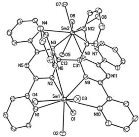
The crystal structure of the cation of 2 (Fig. 2) exhibits a pseudo
two-fold axis of symmetry passing through the middle of the new
C–C bond, C13–C31, between the two triazinide fragments of
the [tptz–tptz]
2–ligand. This ligand contains two pentadentate
cavities, N1–N2–N6–N9–N10 and N3–N4–N7–N8–N12, which
are occupied by the Sm1 and Sm2 ions, respectively. These ions
are also bound to four DMF molecules and their environment
can be seen as a distorted tricapped trigonal prism; around Sm1,
the trigonal faces are defined by O2–O3–O4 and O1–N2–N9,
while N1, N6 and N10 are in capping positions. Each half of
the tptz–tptz molecule acts simultaneously as a tridentate and
bidentate ligand, adopting a
m-k
3:
k
2ligation mode similar to that
encountered with tptz itself in dinuclear complexes like
(CuCl2)2(m-tptz)·MeOH
12aand [Hg(CO2CF3)2]2(m-tptz).
12bThe C13 and C31
atoms are at 0.483(5) and 0.429(5) A
˚ , respectively, from the
planar pentadienide-like C2N3
fragment of the corresponding
triazinide units (rms deviations 0.044 and 0.032 A
˚ ). The C–N
dis-tances at C13 and C31, which average 1.463(9) A
˚ , are typical
single-bond values while the remaining eight C–N distances narrowly
spread in the double-bond range with a mean value of 1.33(2) A
˚ ;
the angles around C13 and C31, which vary from 106.1(3) to
114.0(3)
◦, are close to the ideal tetrahedral value of 109.28
◦. The
pyridyl groups attached to C13 and C31 form a dihedral angle of
40.37(12)
◦and dihedral angles of 82.73(12) and 76.86(12)
◦with
the mean planes of the corresponding triazinide rings while the
other pyridyl groups are rotated with respect to the C3N3
rings
by 3.8(2)–17.7(2)
◦. These structural features of the tris(pyridyl)
Fig. 2 View of the cation of 2. The H atoms have been omitted and only the O atoms of the DMF ligands are represented. Displacement ellipsoids are drawn at the 30% probability level. Selected bond lengths (A˚ ) and angles (◦): Sm1–N1 2.657(3), Sm1–N2 2.465(3), Sm1–N6 2.694(3), Sm1–N9 2.518(3), Sm1–N10 2.653(3), Sm2–N3 2.543(3), Sm2–N4 2.653(3), Sm2–N7 2.656(3), Sm2–N8 2.477(3), Sm2–N12 2.651(3), C13–C31 1.584(5), C13–N2 1.469(5), C13–N3 1.455(5), C31–N8 1.472(4), C31–N9 1.456(5); N1–Sm1–N2 62.44(10), N2–Sm1–N9 63.38(10), N9–Sm1–N10 62.63(10), N1–Sm1–N6 103.03(10), N2–Sm1–N6 62.30(10), N9–Sm1–N6 71.81(9), N10–Sm1–N6 116.83(10).
triazinide moieties are similar to those found in the rhenium(
I)
compound Re(CO)3(m-tptz–OMe)ReBr(CO)3
where tptz–OMe
results from the nucleophilic attack of methanol on the triazine
ring.
13The Sm–N(triazinide) distances are smaller than the Sm–
N(pyridyl) distances, with average values of 2.50(3) and 2.66(1) A
˚ ,
respectively; these distances can be compared with those measured
in Sm(
III) tptz complexes, 2.61(2) A
˚ in Sm(tptz)(NO3)3·H2O
14and
2.61(2) A
˚ in Sm(tptz)(H2O)4Fe(CN)6·8H2O.
14bWhile CeI3
reacted with 1 or 2 mol equivalents of tptz
in acetonitrile or pyridine to give the Lewis base adducts
CeI3(tptz)(MeCN)2
and [CeI2(tptz)2L2]I (L
= MeCN, py),
15the
1 : 1 mixture of UI3(py)4
and tptz in acetonitrile afforded a
pale brown powder of U2I6(tptz–tptz) which recrystallized as
orange crystals of U2I6(tptz–tptz)(MeCN)2·2MeCN (3·2MeCN).
Such clear differentiation between lanthanide(
III) and uranium(
III)
complexes involving U
III→ U
IVoxidation was previously revealed
with the metallocenes M(C5H4R)3
(M
= Ce, U; R =
tBu or SiMe3)
in their reaction with pyrazine which gave Ce(C5H4R)3(pyrazine)
and [U(C5H4R)3]2(m-pyrazine), respectively;
6in that case, the azine
molecule was not coupled and was transformed into its dianionic
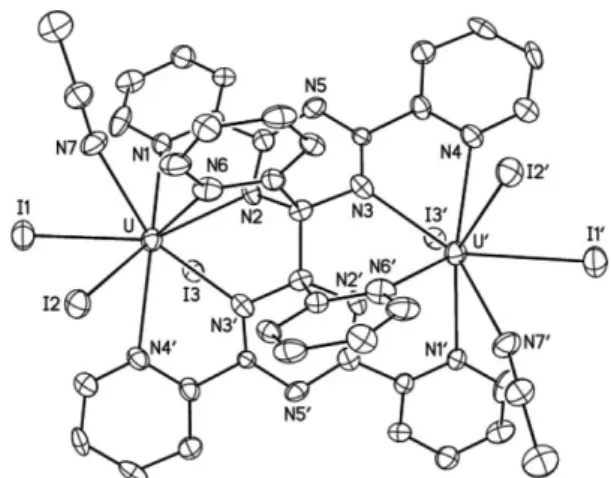
form. Although the quality of the refinement for 3 is low, the
overall structure of the complex was clearly established and shows
the presence of the coupled tptz molecule which adopts the same
coordination mode and geometry as in 2 (Fig. 3).
In conclusion, the reactions of tptz with the reducing species K,
SmI2
and UI3(py)4
follow different pathways. While the free
anion-radical K(tptz)2
is isolated from the reduction with potassium
metal, reactions of tptz with SmI2
or UI3(py)4
provide the first
examples of reductive coupling of a polycyclic azine molecule. The
syntheses of 2 and 3 are likely to involve an electron transfer from
the Sm
IIor U
IIIion to the aromatic nitrogen ligand, as expected
Fig. 3 View of 3. The H atoms have been omitted. Displacement ellipsoids are drawn at the 20% probability level. Symmetry code:¢= 2 - x, y, 1/2 - z. Selected bond lengths (A˚ ) and angles (◦): U–N1 2.662(17),
U–N2 2.412(16), U–N3¢ 2.466(19), U–N4¢ 2.660(18), U–N6 2.750(19), C13–C13¢ 1.49(4), C13–N2 1.51(3), C13–N3 1.45(3); N1–U–N2 63.2(6), N2–U–N3¢ 62.9(6), N3¢–U–N4¢ 63.2(6), N1–U–N6 97.0(6), N2–U–N6 59.7(6), N3¢–U–N6 73.5(6), N4¢–U–N6 122.4(6).
from the reduction potential of the tptz molecule (-1.41 V vs.
Ag/AgCl in MeCN),
16followed by dimerization of the
anion-radical tptz
–∑. The various factors which determine the outcome of
the reduction of an azine molecule by a low-valent metal species are
intricate, as previously noted with the reaction of pyridazine and
M(C5Me5)2
which afforded the radical-anion for M
= Yb
3band the
dianionic coupled ligand for M
= Sm,
3awhile 2,2¢-bipyrimidine
was transformed into the corresponding dianion in the presence
of Yb(C5Me5)2.
3bIn relation with the nature of the metal, the
azine molecule and the solvent, the lifetime and localization of the
radical should have a major influence on its reactivity.
Notes and references
‡ Synthesis and characterizing data. All manipulations were carried out under argon. K(tptz)2(1): (a) a flask was charged with tptz (100.0 mg,
0.32 mmol) and K (15.0 mg, 0.38 mmol) in pyridine (5 mL). Rapidly the solution turned dark blue, and a blue suspension was obtained after 15 h at 20◦C. The solid was extracted in pyridine for 20 h by a soxhlet, and was then washed with pyridine (5 mL) and a 2 : 1 mixture of THF : pyridine (5 mL). After drying under vacuum, 1 was isolated as a dark blue powder (65 mg, 57%). Found: C, 63.87; H, 3.73; N, 25.13; K, 5.75. KC36N12H24
(M= 663.78) requires C, 65.14; H, 3.64; N, 25.32; K, 5.89%. (b) In an NMR tube, tptz (10.0 mg, 0.032 mmol) was reacted with 2% K(Hg) (65 mg, 0.032 mmol of K) in pyridine (1 mL) for 1 h at 20◦C. After filtration, slow diffusion of pentane (1.5 mL) into the dark blue solution led to the formation of dark blue crystals of 1.
[Sm2(tptz–tptz)][I]4: (a) a flask was charged with SmI2 (25.0 mg,
0.062 mmol), tptz (19.3 mg, 0.062 mmol) and freshly distilled MeCN (1 mL). The orange suspension was heated at 100◦C for 10 h. After filtration, the orange powder of [Sm2(tptz–tptz)][I]4 was washed with
MeCN (1 mL) and dried under vacuum (39.0 mg, 88%). Found: C, 29.86; H, 1.76; N, 11.95. Sm2C36H24I4N12 (M = 1433.07) requires C,
30.17; H, 1.69; N, 11.73%. Diffusion of Et2O into a DMF solution of the
powder afforded orange crystals of [Sm2(tptz–tptz)(DMF)8][I]4·3.5DMF
(2·3.5DMF). (b) The same reaction with SmI2(200 mg, 0.49 mmol) and
tptz (154.5 mg, 0.49 mmol) in pyridine (20 mL) gave an orange solution with yellow microcrystals. Evaporation of the solvent and addition of ~5 mL MeCN led to an orange suspension after 12 h stirring (287 mg, 81%). Found: C, 29.79; H, 1.83; N, 11.72. The poor solubility of the complex in the usual organic solvents precluded recording of the NMR spectra.
U2I6(tptz–tptz): a flask was charged with UI3(py)4(150 mg, 0.160 mmol),
tptz (50.1 mg, 0.160 mmol) and acetonitrile (20 mL). The solution immediately turned red and deposited a brown powder. After 2 h at 100◦C, the powder of U2I6(tptz–tptz) was filtered off, washed with Et2O
(10 mL) and dried under vacuum (137.6 mg, 69%). Found: C, 24.41; H, 1.47; N, 9.20. U2I6C36N12H24requires C, 23.22; H, 1.30; N, 9.03%. Orange
crystals of U2I6(tptz–tptz)(MeCN)2·2MeCN (3·2MeCN) were obtained by
crystallization from acetonitrile. Complex 3 in pyridine is1H NMR and
EPR silent (in the solid state and in a frozen solution).
§ Crystal data for 1: C36H24KN12, M= 663.77, monoclinic, space group C2/c, a = 17.7227(18), b = 10.9183(12), c = 16.0694(17) A˚, b =
104.505(6)◦, V = 3010.4(6) A˚3, Z= 4, T = 100(2) K. Refinement of
222 parameters on 2822 independent reflections out of 10 673 measured reflections (Rint= 0.092) led to R1= 0.054 (observed data), wR2= 0.124
(all data), S= 1.021, Drmin= -0.41, Drmax= 0.26 e A˚-3.
Crystal data for 2: C70.5H104.5I4N23.5O11.5Sm2, M= 2273.58, triclinic, space
group P¯1, a= 12.9092(3), b = 14.1646(6), c = 25.1108(11) A˚, a = 85.764(2),
b= 89.355(3), g = 80.132(2)◦, V= 4511.3(3) A˚3, Z= 2, T = 100(2) K.
Refinement of 1049 parameters on 17 063 independent reflections out of 160 534 measured reflections (Rint= 0.044) led to R1= 0.032 (observed
data), wR2= 0.079 (all data), S = 1.034, Drmin= -1.40, Drmax= 2.42 e A˚-3.
Crystal data for 3: C44H36I6N16U2, M= 2026.35, monoclinic, space group C2/c, a= 25.314(2), b = 13.4164(9), c = 16.0957(10) A˚, b = 90.106(8)◦,
V= 5466.5(7) A˚3, Z= 4, T = 100(2) K. Refinement of 337 parameters
on 5005 independent reflections out of 18 972 measured reflections (Rint=
0.151) led to R1= 0.077 (observed data), wR2= 0.214 (all data), S = 1.008,
Drmin= -1.91, Drmax= 1.34 e A˚-3.
Data were collected on a Nonius Kappa-CCD area-detector diffractometer and processed with HKL2000.17Absorption effects were corrected with
DELABS (PLATON)18 or SCALEPACK.17 The structures were solved
by direct methods and refined by full-matrix least-squares on F2 with
SHELXTL.19 All non-hydrogen atoms were refined with anisotropic
displacement parameters. In 2, one solvent DMF molecule is disordered around an inversion centre and it was refined with restraints on bond lengths and displacement parameters. Restraints were also applied in 3, particularly for the very badly resolved acetonitrile solvent molecules. The carbon-bound hydrogen atoms were introduced at calculated positions. The drawings were done with SHELXTL.19
1 (a) G. A. Molander and C. R. Harris, Chem. Rev., 1996, 96, 307; (b) W. J. Evans, Polyhedron, 1987, 6, 803; (c) H. B. Kagan and J. L. Namy,
Tetrahedron, 1986, 42, 6573.
2 (a) M. Kim, B. W. Knettle, A. Dahl´en, G. Hilmersson and R. A. Flowers, Tetrahedron, 2003, 59, 10397; (b) T. Kawaji, K. Hayashi, I. Hashimoto, T. Matsumoto, T. Thiemann and S. Mataka, Tetrahedron
Lett., 2005, 46, 5277.
3 (a) W. J. Evans and D. K. Drummond, J. Am. Chem. Soc., 1989, 111, 3329; (b) T. Zippel, P. Arndt, A. Ohff, A. Spannenberg, R. Kempe and U. Rosenthal, Organometallics, 1998, 17, 4429.
4 (a) I. L. Fedushkin, V. I. Nevodchikov, M. N. Bochkarev, S. Dechert and H. Schumann, Russ. Chem. Bull., 2003, 52, 154; (b) F. Jaroschik, F. Nief, X. F. Le Goff and L. Ricard, Organometallics, 2007, 26, 3552.
5 D. J. Berg, J. M. Boncella and R. A. Andersen, Organometallics, 2002, 21, 4622.
6 T. Mehdoui, J. C. Berthet, P. Thu´ery and M. Ephritikhine, Eur. J. Inorg.
Chem., 2004, 1996.
7 T. Mehdoui, J. C. Berthet, P. Thu´ery, L. Salmon, E. Rivi`ere and M. Ephritikhine, Chem.–Eur. J., 2005, 11, 6994.
8 J. M. Veauthier, E. J. Schelter, C. N. Carlson, B. L. Scott, R. E. Da Re, J. D. Thompson, J. L. Kiplinger, D. E. Morris and K. D. John, Inorg.
Chem., 2008, 47, 5841.
9 C. N. Carlson, J. M. Veauthier, K. D. John and D. E. Morris, Chem.–
Eur. J., 2008, 14, 422.
10 (a) J. H. N. Buttery, Effendy, S. Mutrofin, N. C. Plackett, B. W. Skelton, C. R. Whitaker and A. H. White, Z. Anorg. Allg. Chem., 2006, 632, 1851; (b) G. Bombieri, G. Bruno, M. D. Grillone and G. Polizzotti, Acta Crystallogr., Sect. C: Cryst. Struct. Commun., 1984, 40, 2011.
11 (a) M. G. B. Drew, M. J. Hudson, P. B. Iveson, M. L. Russell and C. Madic, Acta Crystallogr., Sect. C: Cryst. Struct. Commun., 1998, 54, 985; (b) H. Sch ¨odel, T. T. H. Van and H. Bock,
Acta Crystallogr., Sect. C: Cryst. Struct. Commun., 1995, 51,
12 (a) T. Glaser, T. L ¨ugger and R. Fr ¨ohlich, Eur. J. Inorg. Chem., 2004, 394; (b) J. Halfpenny and R. W. H. Small, Acta Crystallogr., Sect. B:
Struct. Crystallogr. Cryst. Chem., 1982, 38, 939.
13 X. Chen, F. J. Femia, J. W. Babich and J. A. Zubieta, Inorg. Chem., 2001, 40, 2769.
14 (a) S. A. Cotton, V. Franckevicius, M. F. Mahon, L. L. Ooi, P. R. Raithby and S. J. Teat, Polyhedron, 2006, 25, 1057; (b) H. Zhao, N. Lopez, A. Prosvirin, H. T. Chifotides and K. R. Dunbar, Dalton Trans., 2007, 878.
15 J. C. Berthet, F. Gupta, P. Thu´ery, and M. Ephritikhine, unpublished results.
16 P. Paul, B. Tyagi, M. M. Bhadbhade and E. Suresh, J. Chem. Soc.,
Dalton Trans., 1997, 2273.
17 Z. Otwinowski and W. Minor, Methods Enzymol., 1997, 276, 307.
18 A. L. Spek, J. Appl. Crystallogr., 2003, 36, 7.
19 G. M. Sheldrick, Acta Crystallogr., Sect. A: Fundam. Crystallogr., 2008, 64, 112.

