HAL Id: tel-01127099
https://tel.archives-ouvertes.fr/tel-01127099
Submitted on 24 Mar 2015HAL is a multi-disciplinary open access
archive for the deposit and dissemination of sci-entific research documents, whether they are pub-lished or not. The documents may come from teaching and research institutions in France or abroad, or from public or private research centers.
L’archive ouverte pluridisciplinaire HAL, est destinée au dépôt et à la diffusion de documents scientifiques de niveau recherche, publiés ou non, émanant des établissements d’enseignement et de recherche français ou étrangers, des laboratoires publics ou privés.
The Role of DIAPH1 in the Megakaryopoiesis
Jiajia Pan
To cite this version:
Jiajia Pan. The Role of DIAPH1 in the Megakaryopoiesis. Hematology. Université Paris Sud - Paris XI, 2014. English. �NNT : 2014PA11T074�. �tel-01127099�
3
Abstract
Megakaryocytes (MKs) are the highly specialized precursor cells that produce platelets via cytoplasm extensions called proplatelets. Proplatelet formation (PPF) requires profound changes in microtubule and actin organization. Formins are a family of highly conserved eukaryotic proteins with multidomains that govern dynamic remodeling of the actin and microtubule cytoskeletons. Most formins are Rho-GTPase effectors proteins. DIAPH1, a member of the formin family, is a mammalian homolog of Drosophila diaphanous gene that works as an effector of the small GTPase Rho and regulates the actomyosin cytoskeleton as well as microtubules. It contains the Rho-binding domain in the N-terminal and two distinct regions of formin homology, FH1 in the center and FH2 in the C-terminus. DIAPH coordinates microtubules and actin cytoskeleton through its FH2 and FH1 regions respectively, making DIAPH an ideal candidate in cell functions that depend closely on the cooperation between the actin and microtubule cytoskeletons.
The objective of the project was to decipher the role of DIAPH1 in megakaryopoiesis. At the end of the MK maturation, PPF and MK migration are associated with profound changes in cytoskeleton organization. Due to its dual function in actin polymerization and microtubule stabilization, DIAPH1 was an obvious candidate to play an essential role in PPF and MK migration.
Our results showed that DIAPH1 expression increased during MK differentiation, whereas DIAPH2 and DIAPH3 expression decreased suggesting that DIAPH1 may play a more important role than DIAPH2 and DIAPH3 in the late stages of MK differentiation. Immunostaining showed that DIAPH1 co-localized with F-actin, tubulin and myosin IIa along the plasma membrane and proplatelet. Using a knockdown strategy with shRNA and expression of an active form of DIAPH1, we showed that DIAPH1 is an important effector of Rho that negatively regulates PPF by remodeling actin and microtubule cytoskeletons. A previous work of our team has shown that Rho-ROCK also negatively regulates PPF by inhibiting myosin IIa activation. By the double inhibition of the DIAPH1 and the ROCK/Myosin pathway, we showed that DIAPH1 and ROCK played additive roles in the negative regulation of PPF.
These observations suggest that the cooperation between DIAPH1 and ROCK is required for the formation of cell structures dependent on actomyosin, such as the stress fibers and the contractile ring. Collectively, these results strongly suggest that cooperation of DIAPH1/microtubules and ROCK/Myosin may regulate PPF by modifying the balance between actomyosin and microtubules.
4
Résumé
Les mégacaryocytes sont les précurseurs cellulaires hautement spécialisés qui produisent des plaquettes via des extensions cytoplasmiques appelées proplaquettes. La formation des proplaquettes exige de profonds changements dans l‟organisation du cytosquelette: microtubules et actine. Les formines sont une famille de protéines hautement conservées chez les eucaryotes composées de plusieurs domaines qui régulent le remodelage et la dynamique du cytosquelette d'actine et des microtubules. La plupart des formines sont des effecteurs protéiques des Rho-GTPase. DIAPH1, un membre de la famille des formines, est un homologue chez les mammifères du gène diaphanous de la drosophile qui fonctionne comme un effecteur de la petite GTPase Rho et régule le cytosquelette d'actomyosine ainsi que les microtubules. Il contient le domaine de liaison à Rho (Rho-binding domain) dans sa partie amino-terminale et deux régions distinctes d‟homologie aux formines, FH1 localisée au centre de la protéine et FH2 dans la partie carboxy-terminale. DIAPH1 co-régule le cytosquelette des microtubules et d'actine à travers respectivement ses régions de FH2 et FH1. DIAPH1 est donc un gène candidat idéal dans toutes les fonctions cellulaires qui exigent une coopération étroite entre cytosquelettes d‟actine et de microtubules.
L'objectif de ce projet de thèse était d‟étudier le rôle de DIAPH1 dans la mégacaryopoïèse. A la fin de la maturation des mégacaryocytes, la formation des proplquettes et la migration sont associées à des modifications importantes de la structure du cytosquelette. Nous avons émis l‟hypothèse que grâce à la sa double fonction dans la polymérisation de l'actine et la stabilisation des microtubules, DIAPH1 pourrait jouer un rôle essentiel dans les temps terminaux de la différenciation mégacaryocytaire.
Nos résultats ont montré qu‟au cours de la différenciation mégacaryocytaire, l‟expression de DIAPH1 augmente, alors que celles de DIAPH2 et DIAPH3 diminuent, ce qui suggère que DIAPH1 pourrait jouer un rôle plus important que DIAPH2 et DIAPH3 dans les stades tardifs de la différenciation mégacaryocytaire. Les études en immunomarquage montrent que DIAPH1 co-localise avec l‟actine F, la tubuline et la myosine IIa en niveau de la membrane plasmique et des proplaquettes. Nous avons étudié la fonction de DIAPH1 par des stratégies d‟invalidation (knockdown) et de surexpression d‟une forme active de DIAPH1. Les résultats montrent que DIAPH1 est un effecteur important de Rho, pour réguler négativement la formation des proplaquettes en remodelant le cytosquelette d‟actine et les microtubules. Le travail antérieur de notre équipe avait montré que Rho-ROCK régulait aussi négativement la formation des proplaquettes, en inhibant l‟activation de la myosine IIa. En inhibant simultanément DIAPH1 et ROCK/myosine, nous avons montré que ces deux voies jouent un rôle additif dans l‟inhibition de la formation des proplaquettes.
Ces résultats suggèrent que la coopération entre les voies DIAPH1 et ROCK/myosine est nécessaire pour la formation de structures cellulaire dépendant de l'actomyosine, telles les fibres de stress et l'anneau contractile en agissant à la fois sur le remodelage du cytosquelette et en assurant un équilibre entre l'actomyosine et microtubules.
Mots-clés: Mégacaryocytes, Formation des proplaquettes, DIAPH1, Myosine, Actine, Microtubules,
5
Table of contents
Abstract ... 3 Résumé ... 4 Table of contents ... 5 List of figures: ... 8 Abbreviation ... 9 Chapter I: Introduction ... 11 1 Megakaryopoiesis ... 111.1 Transcriptional regulation of megakaryopoiesis ... 14
1.1.1 GATA-1/FOG-1 related complex ... 14
1.1.2 MYB ... 15
1.1.3 ETS family... 16
1.1.4 RUNX1 ... 18
1.1.5 SRF/MAL ... 19
1.1.6 NF-E2 ... 20
1.2 TPO/MPL signaling in megakaryopoiesis ... 23
2 Cytoskeleton and MK differentiation ... 28
6
2.1.1 Endomitosis/polyploidization ... 28
2.2 Proplatelet formation and platelet release ... 32
2.2.1 DMS or IMS ... 36
2.2.2 Microtubules ... 37
2.2.3 Actin ... 38
2.3 Rho-GTPase during megakaryopoiesis ... 40
2.3.1 RhoA during megakaryopoiesis ... 42
2.3.2 Cdc42 during megakaryopoiesis ... 43
2.3.3 Rac1 during megakaryopoiesis ... 45
3 Formin family ... 47
3.1 Formin family ... 47
3.1.1 Molecular structure of formins ... 47
3.1.2 Formin functions in the regulation of actin cytoskeleton ... 51
3.1.3 Formin functions in regulation of microtubule cytoskeleton ... 53
3.2 Formin associated with megakaryopoiesis ... 54
3.3 DRFs-related diseases ... 56
Chapter II: Results and Conclusion ... 59
7
Article1 ... 63
Article2 ... 74
2 Results about karyokinesis in endomitosis ... 80
Article 3 ... 82
3 DIAPH1 and proplatelet formation by megakaryocytes ... 88
Article 4 ... 95
Chapter III: Discussion and Perspectives ... 130
Reference ... 140
Acknowledgement ... 172
8
List of figures:
Figure 1... 13 Figure 2... 22 Figure 3... 26 Figure 4... 31 Figure 5... 35 Figure 6... 41 Figure 7... 50 Figure 8... 91 Figure 9... 92 Figure 10... 93 Figure 11... 1349
Abbreviation
ADP, Adenosine diphosphate AML, Acute myeloid leukemia
AMKL, Acute megakaryoblastic leukemia APC, Adenomatous polyposis coli
Cdc42, Cell division control protein 42 CLP, common lymphoid progenitor CMP, Common myeloid progenitor CXCR4, CXC chemokine receptor type 4 DAD, Diaphanous auto-regulatory domain DID, Diaphanous-inhibitory domain DMS, Demarcation membrane system DRFs, Diaphanous-related formins EB3, End-binding protein three ECM, Extracellular matrix
Erk1/2, Extracellular signal-related kinase 1/2 ERG, ETS-related gene
ETS, E26 transformation-specific or E-twenty six FH, Formin homology
FGF-4, Fibroblast growth factor-4 FLI-1, Friend leukemia integration 1 FOG-1, Friend of GATA-1
GABPα, GA-binding protein alpha GAPs, GTPase-activating proteins GBD, GTPase-binding domain
GEFs, Guanine-nucleotide exchange factors GFI-1B, Growth factor independent 1B HSCs, Hematopoietic stem cells IF, Immunofluorescence
IL, Interleukin
IMS, Invaginated membrane system JAKs, Janus family of protein kinases KO, Knockout
10 MAL, Megakaryocytic acute leukemia
MAPK, Mitogen-activated protein kinase mDia, mammalian Diaphanous-related formins MDS, Myelodysplastic syndromes
MEP, Megakaryocyte-erythroid progenitor MLC, Myosin light chain
MK, Megakaryocyte
MPL, Myeloproliferative leukemia virus oncogene MPPs, Multipotent progenitors
MPD, myeloproliferative disorder MRLC, Myosin regulatory light chain
MRTFs, Myocardin-related transcription factors MT, Microtubule
mTOR, Mammalian target of rapamycin MYH, Myosin heavy chain
MYB, Myeloblastosis
NF-E2, Nuclear factor erythroid 2 PAK1/2, p21-activated kinase PF4, Platelet factor 4
PI3K, Phosphoinositide-3 kinase PKC, Protein kinase C
PPF, Proplatelet formation
SDF-1, Stromal cell-derived factor SOCS, Suppressor of cytokine signaling SRF, Serum response factor
STATs, Signal transducers and activators of transcription ROCK, Rho-associated protein kinase
RUNX, Runt-related transcription factor TPO, Thrombopoietin
vWF, von Willebrand Factor
WASP, Wiskott Aldrich syndrome protein WB, Western blot
11
Chapter I: Introduction
All blood cells are derived from hematopoietic stem cells (HSCs). HSCs reside in the bone marrow and are the only long life cells of the hematopoietic system, capable to self renew and to differentiate towards all hematopoietic cells. One of the lineages derived from HSCs is the megakaryocytic cell line; the entire process of diffrentiation of this cell line is called megakaryopoiesis or thrombopoiesis and gives rise to platelets. Megakaryocyte (MK) is the bone marrow cell that has the ability to produce circulating platelets (Kaushansky, 2008).
During hematopoiesis, HSCs give rise to multipotent progenitors (MPPs), a cell that has lost its self-renewal capacities, but has retained the capacity to differentiate towards all hematopoietic multi-lineages (Figure1). Generally, it is believed that the multipotent progenitors will subsequently give rise to two types of progenitors, the common lymphoid progenitor (CLP) (Kondo et al., 1997) and the common myeloid progenitor (CMP) (Akashi et al., 2000). CLP then will give rise to al lymphoid cells including natural killer cells, T and B cells and dendritic cells, while CMP will be at the origin of all myeloid lineages including the granulocyte/macrophage progenitor and the MK-erythroid progenitor (MEP) (Debili et al., 1996; Szalai et al., 2006). MEPs are bipotential precursors that give rise to cells of both erythroid and megakaryocytic lineages. Nevertheless, recent studies have suggested that the MEP may directly derive from the HSCs to yield either the erythroid or megakaryocytic lineages without the CMP intermediate (Adolfsson et al., 2005; Forsberg et al., 2006). Furthermore, it has also been suggested the presence of MK biased HSCs expressing the CD41 or the vWF (von Willebrand Factor), which allow a rapid platelet recovery in case of bone marrow transplantation. However this classical model is presently challenged by a model in which committed progenitors play a central role in producing blood cells in homeostatic conditions.
1 Megakaryopoiesis
Megakaryopoiesis is a complex and integrated differentiation process, which is highly regulated at multiple stages. The MK differentiation from MK progenitors includes the switch from mitosis to endomitosis and proplatelet formation (PPF). After proliferation by mitosis at the level of progenitors, MKs enter into endomitosis and increase their ploidy, then they develop an extensive internal demarcation membrane system (DMS), now called invaginated membrane system (IMS), which serves as a membrane reservoir and then mature to release platelets through the formation of long pseudopods, which are called proplatelet (Italiano et al., 1999a). Platelets are shed into vascular
12
sinusoids within the bone marrow by these processes. Platelets are essential in homeostasis and thrombosis and also in other processes such as the development of innate immunity, angiogenesis and metastasis. MK differentiation is regulated both positively and negatively by transcription factors and by the cytokine signaling.
13 Figure 1: The hematopoietic hierarchy
Overview of the differentiation of hematopoietic stem cells (HSCs) towards the different hematopoietic lineages.
HSCs are both multipotent and capable of self-renewal. LT-HSCs (long-term hematopoietic stem cell) represent the „true‟ stem cells that have important self-renewal capacities and produce ST-HSCs (short-term hematopoietic stem cell) with limited self-renewal capacities and multipotent potential and subsequently the MPPs (multipotent progenitor) without self-renewal capacity. MPPs give rise to CLPs (common lymphoid progenitor) and CMPs (common myeloid progenitor). The CLPs then give rise to lymphocytes including T cells and B cells, while CMPs give rise to GMPs (granulocyte-macrophage progenitor) that produce granulocytes and (granulocyte-macrophages and MEPs (megakaryocyte-erythrocyte progenitor) that produce (megakaryocyte-erythrocytes and megakaryocytes/platelets. LT-HSCs can directly give rise to MEPs without the CMP intermediate (Figure adopted and modified from (Sharpless and DePinho, 2007) ).
14
1.1 Transcriptional regulation of megakaryopoiesis
A lot of transcription factors are involved in the regulation of megakaryopoiesis (Chang et al., 2007b; Goldfarb, 2007). During megakaryopoiesis, series of transcription factors coordinately regulate the MK-specific gene expression.
Through loss-of-function studies in mice and analysis of human diseases, various transcription factors have been identified, which are involved in MK differentiation, polyploidization, proplatelet formation (PPF) and platelet shedding. Multiple transcription factors, including RUNX1 (AML1), GATA-1/GATA-2, FOG-1 (Friend of GATA-1), GFI1b, NF-E2, FLI-1 (Friend leukemia integration1) and MYB, form large protein complexes that regulate the MK differentiation both positively and negatively (Figure 2) (Chang et al., 2007b). Some important examples are developped below.
1.1.1 GATA-1/FOG-1 related complex
GATA-1 is a member of the GATA transcription factor family and is involved in cell growth and malignancy. GATA-1 protein regulates the expression of nearly all erythroid and MK specific genes. It also plays a role in erythroid development by regulating the switch from fetal to adult hemoglobin. GATA-1 plays also a crucial role in MK development acting as either activator or repressor depending of the protein complexes (Chang et al., 2007b). GATA-1 regulates the main MK-specific genes such as GPIIB, PF4 (platelet factor 4), GPIbα, β-TG, GPIX or GPV.
FOG-1 (Friend of GATA-1) is one of the most important co-factors of GATA-1. The GATA-1/FOG-1 complex is critical in promoting MK/Erythroid lineage differentiation. The X chromosome-linked GATA-1 contributes to the differentiation of hematopoietic progenitors into erythroid and MK cells, and also to the development of eosinophil and mast cells. GATA-1 and FOG-1 physically interact and their complex is essential for GATA-1 functions in different cellular contexts. FOG-1 contributes to the regulation of cell type-specific gene expression in erythroid and MK differentiation (Tsang et al., 1997). Mice lacking FOG-1 have an absence of megakaryopoiesis and an arrest in erythropoiesis (Tsang et al., 1998). Loss of the GATA-1/FOG-1 interaction leads to an obvious decrease in membrane protein expression and to an increase in reactive oxygen species accumulation, which disrupts the function of GATA-1 in erythrocyte development (Hasegawa et al., 2012).
15
GATA-1 mutations in the site of interaction with FOG-1 lead to an inherited human disorder characterized by an X-linked dyserythropoietic anemia associated with a macrothrombocytopenia: platelets are giant and immature with abnormal membrane complexes (Freson et al., 2001; Shivdasani et al., 1997). GATA-1 deficient MKs fail to undergo terminal differentiation and exhibit a significant hyperproliferation in vitro. gata-1 knockout (KO) mice die at the embryonic stage because of a failure in erythroid maturation with a blockage at the proerythroblast stage (Fujiwara et al., 1996; Fujiwara et al., 2004). GATA-1 and GATA-2 have overlapping functions at the yolk sac stage required for normal hematopoiesis, loss of either leads to embryonic lethality in KO mice due to a failure in erythroid maturation and the expansion of progenitors, suggesting that either GATA-1 or GATA-2 is essential to initiate blood formation in the embryo (Fujiwara et al., 2004).
Mutations in exon 2 leading to a short form of GATA-1 (GATA-1s) are associated with all cases of Down syndrome-associated transient myeloproliferative disorder or transient leukemia (TL) (Greene et al., 2003), which may progress to acute megakaryoblastic leukemia (AMKL) (Rainis et al., 2003; Wechsler et al., 2002). GATA-1 mutations are present in utero demonstrating that it is an early event in leukemogenesis. GATA-1 mutation at birth may serve as a biomarker for an increased risk of Down syndrome-related AMKL (Pine et al., 2007; Shimada et al., 2004b).
GATA-1 can interact with the myeloid PU.1 transcription factor and can repress PU.1-dependent transcription and myeloid differentiation (Nerlov et al., 2000). The competition between GATA-1 and PU.1 play an important role in determining the hematopoietic cell fate in CMP (Chou et al., 2009). Huang and colleagues have demonstrated that GATA-1 can regulate not only MK maturation by activation of platelet-specific gene expression, but also MK polyploidization by modulating STAT1 (Huang et al., 2007).
GFI-1B (growth factor independent 1B), a zinc-finger proto-oncogene acting as transcription repressor, is essential for erythroid and MK lineage development by interacting with GATA-1 (Saleque et al., 2002). Taken together, all these findings emphasize the requirement of GATA-1 for MK development and platelet biogenesis.
1.1.2 MYB
c-MYB proto-oncogene protein, known as a transcriptional activator, is a member of the MYB (myeloblastosis) family of transcription factors. MYB family proteins contain three domains: an N-terminal DNA-binding domain, a central transcriptional activation domain and a C-N-terminal regulatory
16
domain involved in transcriptional repression. c-Myb is highly expressed in multipotent hematopoietic progenitors and plays an important role in the regulation of hematopoietic cell differentiation and in oncogenesis (Oh and Reddy, 1999).
Loss of c-Myb results in an embryonic lethality due to a failure of hematopoietic development. Suboptimal level of c-Myb expression induces the commitment of MPP toward the MK lineage and macrophage, while high level may control the erythropoiesis and lymphopoiesis (Emambokus et al., 2003). In addition, c-Myb mutant mice exhibit a myeloproliferative phenotype with an important MK expansion and a major increased platelet production without requiring TPO signaling (Carpinelli et al., 2004; Garcia et al., 2009).
Studies have shown that c-Myb activity may affect the balance between MK lineage differentiation and other hematopoietic lineages (Emambokus et al., 2003; Frampton et al., 1995). In another study, it has been shown that a transgenic insertion inducing a marked decrease of c-Myb expression in the MEP, leads to an increase in MK number and a decrease in erythroid progenitors (Mukai et al., 2006). Expression of the c-Myb in the MEP favors erythropoiesis, and is down-regulated during megakaryopoiesis (Metcalf et al., 2005). These findings indicate that c-Myb is a crucial regulator of the MK/Erythroid lineage fate.
Furthermore, studies have shown that MYB is a target of the microRNA-150 (miR-150) in human and is negatively regulated by miR-150. MYB and miR-150 interaction is essential for embryonic development and probably oncogenesis (Lin et al., 2008). By gain- and loss-of-function studies, Lu J and colleagues have shown that miR-150 is expressed in MK development and in a post-transcriptional manner decreases c-Myb expression and directs MEP differentiation toward the MK lineage (Lu et al., 2008). Another study suggests that TPO regulates the level of miR-150 and favors MK engagement of the MEP by down-regulating c-Myb (Barroga et al., 2008).
Collectively, c-MYB has a complex involvement in the regulation of proliferation and establishment in the hematopoietic hierarchy, but inhibits MK differentiation.
1.1.3 ETS family
ETS (E26 transformation-specific or E-twenty six) family is one of the largest families of transcription factors. All ETS family members are identified by a highly conserved DNA binding domain, which is called ETS domain with a winged helix-turn-helix structure (Lee et al., 2005; Yordy and
Muise-17
Helmericks, 2000). In addition to DNA binding function, ETS domain is also involved in protein-protein interaction. ETS family protein-proteins are implicated in a wide range of functions including the regulation of cell differentiation, cell cycle control, cell migration, cell proliferation, apoptosis and angiogenesis. ETS-domain proteins function as either transcriptional repressors or transcriptional activators. 29 ETS genes in humans and 28 genes in the mouse have been described; among them, some are expressed in megakaryopoiesis and play a role in the development of erythroid and megakaryocytic lineages, such as FLI-1, GABPα, ETS1, ETS2 and ERG (ETS-related gene).
FLI-1 is the most investigated ETS family member and is required for normal MK maturation by controlling MK-specific gene expression. It can cooperate with GATA-1 and FOG-1 to activate the expression of late MK genes such as GPIX, GPIbα, and PF4. fli-1 KO affects both vascular development and megakaryopoiesis, which leads to embryonic lethality at day 11.5 of embryogenesis with loss of blood vessel integrity and a partial block in MK differentiation (Hart et al., 2000). Another group has shown that fli-1 mutant mice have a profound hematopoietic phenotype and hematopoiesis is severely impaired at mid-gestation, which provide in vivo evidence for the role of Fli-1 in the regulation of hematopoiesis and hemostasis (Spyropoulos et al., 2000). In addition, FLI-1 and GATA-1 work in cooperation to activate the expression of MK-specific genes (Eisbacher et al., 2003): FLI-1 enhances the GATA-1 activity at MK promoters and represses the activity of erythroid factors on erythroid promoters (Starck et al., 2003).
In human, the Paris Trousseau syndrome and Jacobsen syndrome have similar congenital anomalies with a thrombocytopenia and giant platelet α-granules, which are the consequence of a FLI-1 haploinsufficiency due to a chromosome 11 deletion at 11q23.3 (Breton-Gorius et al., 1995; Raslova et al., 2004). However, fli-1 deletion in adult mice induces a mild thrombocytopenia with a defect of bone marrow MK maturation and modifies several myeloid lineage commitment decisions and accelerates proliferation arrest and terminal erythroid differentiation (Starck et al., 2010).
ERG is an ETS family member extremely homologous with FLI-1. One study has shown that ERG is essential for definitive hematopoiesis in the embryo, adult HSC regulation and maintenance of the normal number of blood platelets (Loughran et al., 2008). Evidence from genetic approaches suggests that FLI-1 is required for megakaryopoiesis and ERG, more for normal adult HSC function. Double heterozygous KO mice for fli-1 and erg display a more dramatic phenotype than those with a single deletion, concerning the thrombocytopenia, defect in MK numbers and MK maturation (Kruse et al., 2009). These results suggest that FLI-1 and ERG act in synergy and may regulate common target genes.
18
In addition, some other members of the ETS family are also involved in the regulation of megakaryopoiesis, such as TLE-1, ETS1 and GABPα (GA-binding protein alpha). GABPα-deficient mice show a decrease in immature MKs and an increase in mature MKs. In addition, the ratio between GABPα/FLI-1 decreases during MK maturation. In agreement with their pattern of expression, it has been shown that GABPα regulates early stages of MK differentiation, particularly the expression of early MK-specific genes such as GPIIb and MPL (Pang et al., 2006).
Furthermore, FLI-1 can interact with RUNX-1 by direct protein-protein interaction and results in a synergistic transcriptional activation of the MPL promoter. They are associated in a transcriptional protein complex that also includes GATA-1 and FOG-1. FLI-1 dephosphorylation affects RUNX-1 binding and inhibits in vitro fetal liver differentiation of MK. These data underscore the interaction of FLI-1 and RUNX-1 during MK development (Huang et al., 2009).
1.1.4 RUNX1
RUNX1, also known as AML1 (acute myeloid leukemia 1) or CBFA2 (core-binding factor subunit alpha-2), is a member of the RUNX (Runt-related transcription factor) family. RUNX-1 is a DNA-binding subunit of the core DNA-binding transcription factor complex, which is expressed in almost all hematopoietic cells and contributes to the commitment of hematopoietic stem and progenitor cells by regulating or interacting with other elements (Okuda et al., 2001).
RUNX1 may interact and cooperate with GATA-1 in MK differentiation and during hematopoietic development in mammals (Elagib et al., 2003; Waltzer et al., 2003). RUNX1 is one of the rare transcription factors that is involved in MK differentiation, but inhibits erythroid differentiation, suggesting that RUNX1 plays an essential role in the MK/Erythroid lineage commitment (Niitsu et al., 1997). An inducible runx-1 KO leads to a marked defect in megakaryopoiesis with a thrombocytopenia, highlighting its important role in MK differentiation (Ichikawa et al., 2004; Sun and Downing, 2004).
It has been shown that runx1 haploinsufficiency is associated with alterations in the early mesoderm development of ES cell differentiation (Lacaud et al., 2004), as well as it influences the temporal and spatial generation of HSC in mouse embryos (Cai et al., 2000). In the hematopoietic system, it induces an increase in myeloid progenitors and may lead to leukemogenesis (Yamashita et al., 2005).
19
Several RUNX1 target genes have been identified during MK differentiation, such as CBF, MPL, IEX-1, MYL9 (myosin light chain) (Hamelin et al., 2006; Heller et al., 2005; Jalagadugula et al., 2010;
Song et al., 1999), as well as p19INK4D, which is involved in the endomitotic arrest and MK maturation (Gilles et al., 2008). Germ-line heterozygous mutations and somatically point mutations are found in
RUNX-1, all resulting in loss of transcriptional activity. Germ line mutations induce familial platelet
disorder with predisposition to develop acute myeloid leukemia (AML) (Harada et al., 2004; Heller et al., 2005; Song et al., 1999).
The role of RUNX1 on the control of HSC fate may be achieved by regulating the MPL promoter both positively and negatively, this dual activity being related to the binding partner according to the cell types (Satoh et al., 2008). The RUNX1 mutation related thrombocytopenia may result from a low MPL receptor expression in MK although low MPL expression is usually associated with a thrombocytosis in several murine models, but more obviously from altered expression of numerous genes encoding the actomyosin cytoskeleton (Heller et al., 2005).
1.1.5 SRF/MAL
SRF (serum response factor), a member of the MADS (MCM1, Agamous, Deficiens and SRF) domain-containing transcription factor, binds to the serum response element in the promoter/enhancer region of many target genes and confers the recruitment of other transcriptional cofactors. SRF is widely expressed and plays an important role in regulating the activity of many genes including immediate early genes and genes involved in cytoskeleton and muscle development, as well as genes involved in cell cycle regulation, growth and differentiation (Chai and Tarnawski, 2002; Miano, 2010; Morita et al., 2007; Schratt et al., 2002).
MAL (megakaryocytic acute leukemia, also known as MKL1, BSAC and MRTF-A), a member of the MRTF (myocardin-related transcription factor) family, is one of the most important cofactors of SRF (Vartiainen et al., 2007). There are different extracellular stimuli that can activate SRF. One is the MAPK (mitogen-activated protein kinase) pathway, which acts through the ternary complex factors, the other is the small GTPase pathway acting through the MRTF family proteins (Cen et al., 2003; Hill et al., 1995). The MAL/SRF interaction is regulated by the Rho-actin pathway (Miralles et al., 2003; Morita et al., 2007). A recent study has shown that SRF and MAL regulate the myofibroblast differentiation by responding to both TGF-β1 and RhoA signaling (Small, 2012).
20
SRF and MAL play a crucial role in MK terminal differentiation including PPF. Mrft-A (Mal) KO mice exhibit a decrease of platelet count in peripheral blood and a reduced ploidy in bone marrow MKs, suggesting the involvement of MAL in MK differentiation (Cheng et al., 2009). MAL knockdown resulted in dysmorphic MKs with disorganized DMS and α-granules heterogeneously scattered in the cytoplasm, as well as a decreased expression of MYL9 and MMP9 (Gilles et al., 2009).
Srf KO mice exhibit a similar phenotype with an abnormal DMS, a reduced platelet count and a
down-regulation of actin cytoskeleton gene expression as well as an abnormal stress fiber formation and actin distribution (Halene et al., 2010). These indicate that MAL/SRF complex is involved in normal MK maturation and PPF by regulating MYL9 and MMP9.
MAL is shuttling from the cytoplasm to the nucleus. When localized in the nucleus, it activates SRF target genes transcription, suggesting that MAL subcellular localization is one important mechanism to control SRF activity (Vartiainen et al., 2007). A recent study has shown that MAL localization is disrupted by drugs inhibiting RhoA activity or actin polymerization, revealing that MAL subcellular localization and function is dependent on Rho GTPase family and actin organization (Smith et al., 2013). These data widen the study of the molecular mechanisms that regulate MK differentiation. MAL16 (also known as MKL2, MRTF-B), a homologue of MAL, is also expressed in MK and participates in MK differentiation. By a double KO strategy, it has been demonstrated that MAL and MAL16 play a redundant and crucial role in MK maturation and platelet formation (Smith et al., 2012). Together, these findings confirm that SRF/MAL is necessary for MK maturation and platelet production, partly by regulating cytoskeleton genes.
1.1.6 NF-E2
The transcription factor NF-E2 (nuclear factor erythroid 2), belongs to the basic-leucine zipper family. It is a heterodimer complex containing two basic subunits, a widely expressed 18-kDa (p18NF-E2) subunit (Andrews et al., 1993b) related to chicken Maf proteins and a tissue-restricted 45-kDa (p45
NF-E2
) subunit (Andrews et al., 1993a), whose expression is restricted to erythroid cells, MKs and mast cells.
The NF-E2 complex is essential in erythroid and MK maturation and differentiation. p45NF-E2 KO mice are lethal at birth as a consequence of a profound thrombocytopenia with an increased number of MKs, which present evident defect in both DMS development and α-granules distribution and an absence of PPF (Shivdasani et al., 1995). Other genetic evidence has indicated a crucial role for p45NF-E2 and small Maf in the terminal MK maturation and platelet production. The double KO mice for the two
21
NF-E2 partners, MafG and MafK show a similar thrombocytopenia as p45NF-E2 KO mice with an absence of proplatelets (Lecine et al., 1998; Motohashi et al., 2000; Onodera et al., 2000). Furthermore, these mutant mice develop a severe anemia accompanied by an abnormal erythrocyte morphology and modifications in the membrane protein composition. These findings provide evidences that NF-E2 plays an important regulatory role in erythropoiesis (Onodera et al., 2000), more particularly by regulating globin gene transcription.
Several genes have been established as regulated by p45NF-E2 in MKs directly or indirectly, such as Thromboxane synthase, β1-tubulin, Rab27b, 3β-HSD (3beta-hydroxysteroid dehydrogenase) and Lims1 (Chen et al., 2007a; Deveaux et al., 1997; Lecine et al., 2000; Nagata et al., 2003; Tiwari et al., 2003), all of which are involved in PPF and platelet biogenesis. Recently, Selp and Myl9, two other genes have been identified as direct p45NF-E2 targets, which are also implicated in platelet production and function (Fujita et al., 2013). All these observations provide strong evidence that NF-E2 is essential for establishing normal platelet function as well as for generating the proper number of platelets.
Some other transcription factors also participate in hematopoiesis, MK differentiation and platelet production, such as zinc finger protein GFI-1b (growth factor independent 1B transcription repressor). GFI-1b is a crucial proto-oncogenic transcriptional regulator necessary for MK/Erythroid lineage development and differentiation. Dependent on the promoter and cell type context, GFI-1b functions as transcriptional repressor or transcriptional activator. It may also regulate cytokine signaling by repressing the activity of some promoters.
22
Figure 2: Overview of the transcription factors involved in megakaryopoiesis
Multiple transcription factors regulate megakaryopoiesis at different developmental stages. HSC, hematopoietic stem cell; MPP, multipotent progenitor; CLP, common lymphoid progenitor; CMP, common myeloid progenitor; GMP, granulocyte-monocyte progenitor; Eo, eosinophil; Ba, basophil; Ne, neutrophil; Mo/Ma, monocyte/macrophage; MEP, megakaryocyte/erythrocyte progenitor; BFU-MK, burst-forming unit megakaryocyte; CFU-BFU-MK, colony-forming unit megakaryocyte; PMKB, promegakaryoblast; MK, megakaryocyte; PfMK, proplatelet forming megakaryocyte; BFU-E, burst-forming unit erythrocyte; NK, natural killer cell; DC, dendritic cell (figure from (Chang et al., 2007b) ).
23
1.2 TPO/MPL signaling in megakaryopoiesis
Cytokines and multiple compounds of bone marrow microenvironment are also involved in the regulation of MK differentiation. Numerous cytokines can stimulate MK progenitor proliferation, such as GM-CSF (Granulocyte-macrophage colony-stimulating factor), IL (interleukin)-3, 6, 11, IL-12 and erythropoietin. Nevertheless, other cytokines participate in the regulation of MK maturation and platelet release, such as IL-1α and leukemia inhibitory factor (LIF) (Vainchenker et al., 1995). Thrombopoietin (TPO) is the most important cytokine for MK differentiation, more particularly in homeostatic conditions, as well as it plays a central role in HSC regulation (Kaushansky, 2006). TPO is also known as megakaryocytic growth and development factor or MPL (Myeloproliferative leukemia virus oncogene) ligand. TPO is a glycoprotein which functions as a hormone and is produced by the liver and at a lesser extent by kidney and regulates platelet production in homeostasis. TPO acts throughout all stages of megakaryopoiesis, including MK progenitor proliferation and MK polyploidization. It also increases MK size by promoting protein synthesis. The TPO receptor, MPL has been identified as the proto-oncogene of the viral oncogene v-mpl, a member of the hematopoietic receptor super-family that was identified as the transformation factor of the murine myeloproliferative leukemia virus.
The TPO/MPL signaling pathway regulates platelet production and conversely the platelet number in a feedback manner negatively regulates TPO level. TPO is constitutively produced and the circulating TPO level is regulated both by platelets, its end products and MKs. TPO binds to MPL expressed by platelets and is endocytosed before being degraded. The blood platelet number thus regulates the TPO level. Low platelet level leads to higher level of plasmatic TPO that increases the number of MKs enhancing their further maturation. On the other hand, high platelet level decreases the availability of TPO for MKs. The discovery of TPO has contributed profoundly to platelet biology because it allows the development of cultures that generate MKs in vitro. This has greatly facilitated studies of MK differentiation and platelet related disorders. TPO and now synthetic MPL ligands are potential thrombopoietic and hematopoietic agents that are used as therapeutic drugs to stimulate expansion of HSC ex vivo and to treat some thrombocytpenia (Basser, 2002).
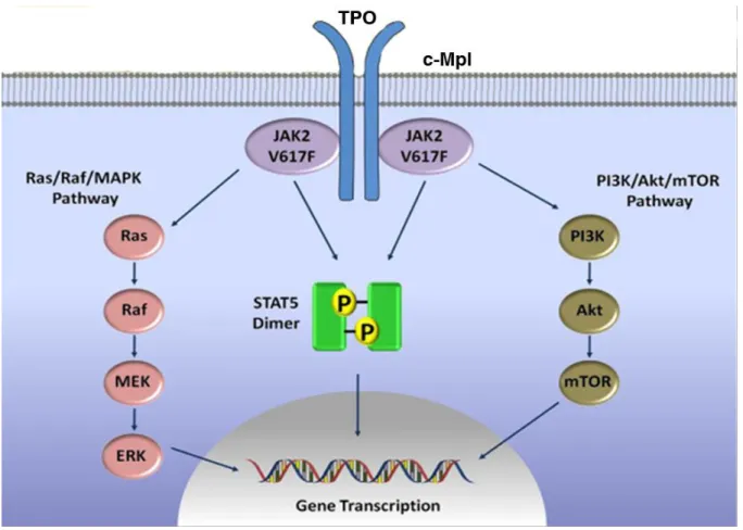
TPO/MPL has remarkable functions in MK differentiation and thrombopoiesis through the TPO-dependent signaling pathways (Figure 3) (Geddis, 2010; Santos and Verstovsek, 2011). These pathways include the Janus family of protein kinases (JAKs)/signal transducers and activators of
24
transcription (STATs) pathway (Schulze et al., 2000), the Ras/Raf/mitogen-activated protein kinase (MAPK) pathway (Rojnuckarin et al., 1999), the phosphoinositide-3 kinase (PI3K)/Akt/mTOR pathway (Nakao et al., 2008; Pasquet et al., 2000; Soda et al., 2008) and the nuclear factor kappa B (NF-κB) pathway (Zhang et al., 2002). The important role of TPO has been demonstrated by animal experiments. TPO and MPL are essential for MK growth and development in mouse models, KO of one of them induces reduces numbers of MKs and of circulating platelets to approximately 10% of the normal (Gurney et al., 1994; Murone et al., 1998).
MPL does not have intrinsic kinase activity, which is dependent on the cytoplasm tyrosine kinase JAK2. JAK2 kinase actives the downstream signaling pathways, such as the STATs, PI3K and the MAPK that activate and mediate the cellular response to TPO (Geddis et al., 2002). This cascade signaling allows cell proliferation and differentiation. MPL and JAK2 are involved in multiple inherited and malignant disorders leading to thrombocytosis, thrombocytopenia and aplastic anemia. TPO binding changes MPL conformation leading to JAK2 phosphorylation. Activated JAK2 kinase phosphorylates MPL, which induces the binding of the different STAT family members and their subsequent phosphorylation (Schulze et al., 2000). This leads to the dimerization of the activated STAT proteins and then to their translocation to the nucleus where they act as transcription factors by binding to different STAT-response DNA sequences. Constitutive activation of JAK2/STAT pathway induces a cytokine-independent growth and transformation, such as the V617F mutant of JAK2 in myeloproliferative disorders, TEL-JAK2 in leukemia or a constitutively active STAT5 in different leukemia cell lines (Harir et al., 2007; Najfeld et al., 2007).
In addition, JAK2 can activate the small Ras-GTPase and the MAPK pathway to stimulate the activation of extracellular signal-related kinase 1/2 (ERK1/2). Studies have shown that TPO-induced MAKP signaling pathway has important role although controversial in normal MK differentiation (Rojnuckarin et al., 1999). TPO signaling activates Ras-GTPase based on the binding of the adaptor protein SHC to the phosphorylated MPL and the assembly of a complex consisting of the adaptor protein GRB2 (Growth factor receptor-bound protein 2) and the guanine nucleotide exchange factors (GEFs) like SOS (Son of sevenless), and then RAS activates RAF-1, ERK1/2 and MEK (mitogen-induced extracellular kinase) (Avruch et al., 2001).
The PI3K signaling pathway also participates downstream of JAK2 in megakaryopoiesis. It has been shown that TPO up-regulates platelet α-granule secretion and aggregation induced by thrombin through PI3K signaling (Kojima et al., 2001). TPO stimulates PI3K phosphorylation and activation of
25
the threonine/serine kinase AKT (also known as protein kinase B, PKB) that enhances the TPO-induced survival and proliferation of MK (Nakao et al., 2008; Soda et al., 2008). mTOR (mammalian target of rapamycin), a downstream target of the PI3K/AKT pathway, is involved in the growth and the ploidy of mature MKs. Inhibition of mTOR by rapamycin reduces MK size and ploidy, through an inhibition of the G1/S transition, via a decrease of p21 and cyclin D3, and a delay in MK maturation preventing PPF. These results suggest that mTOR plays an important role in MK proliferation and maturation (Raslova et al., 2006).
Furthermore, TPO has a major function in the regulation of HSC and progenitors in vivo and in vitro (Bruno et al., 2003; Kaushansky, 2006; Pick et al., 2002). Studies of c-mpl KO mice have shown that TPO/MPL is associated with early hematopoietic progenitor development, while HSCs also express MPL and depend on TPO signaling for their maintenance and expansion (Fox et al., 2002). Studies have shown that TPO plays an important role in maintaining HSC quiescence in the bone marrow niche and in DNA gene repair after double strand DNA breaks by stimulating NHEJ (de Laval et al., 2014). tpho and c-mpl KO mice show not only a thrombocytopenia, but also a decrease in HSCs and in early progenitors of all hematopoietic lineages (Murone et al., 1998). Thus, the TPO/MPL axis plays a crucial role for hematopoiesis and megakarypoiesis. Interestingly, in human, hereditary homozygous or composite heterozygous loss of function mutations of MPL lead to a thrombocytopenia with an absence of MK, which subsequently progresses to aplastic anemia, demonstrating that the TPO/MPL axis is also crucial for the maintenance of human HSCs.
26
Figure 3: TPO-induced signaling pathways in megakaryopoiesis
TPO binds to its receptor Mpl and the conformational changes on the homodimer induced by TPO binding result in activation of JAK2 by transphosphorylation, which initiates and activates downstream signaling pathways including the STATs, PI3K/Akt/mTOR and Ras/Raf/MAPKs pathways. TPO, thrombopoietin; MPL, Myeloproliferative leukemia virus oncogene; JAK, Janus family of protein kinase; PI3K, Phosphoinositide-3 kinase; mTOR, mammalian target of rapamycin; MAKP, mitogen-activated protein kinase (Figure adopted from (Santos and Verstovsek, 2011) ).
27
Recent data suggest that the roles of TPO and MPL/JAK2 on MK differentiation are more complex than previously thought. First, it has been shown that a strong signaling through MPL/JAK2 induces an arrest in proliferation and a „senescence‟ like phenotype. Second, jak2 and c-mpl KO during MK differentiation induce a thrombocytosis associated with high TPO level. This suggests that the role of TPO is indispensable in early stages of hematopoiesis and MK differentiation, but is not required for terminal differentiation. MPL plays an important role in late MK differentiation stages, not as a signaling receptor, but more as a receptor involved in TPO clearance.
As the main cytokine regulating MK development and platelet production, TPO is critical to maintain the homeostatic balance and prevent a hematopoietic deficiency, such as thrombocytopenia and aplastic anemia. TPO/MPL signaling is also negatively regulated in order to maintain this homeostatic balance. For instance, the JAK/STAT pathway activation induces the transcription of members of the suppressor of cytokine signaling (SOCS) family, which can inhibit JAK signaling in different ways according to the member (Alexander and Hilton, 2004). TPO signaling and JAK2 activity can be inhibited by TPO induced SOCS1 and SOCS3, respectively (Hookham et al., 2007; Wang et al., 2000). LNK is an adaptor protein that binds JAK2 and regulates its activity. LNK negatively regulates growth of HSCs, erythroid and megakaryocytic cells by inhibiting TPO/MPL signaling pathway (Seita et al., 2007; Tong and Lodish, 2004).
Overall, TPO is the most important hematopoietic cytokine for MK differentiation and platelet production; both a positive and a negative regulation of TPO signaling are necessary for homeostasis and hematopoietic development.
Chemokines and cellular interactions are also involved in megakaryopoiesis and thrombocytopoiesis. The two chemokines that have crucial roles on megakaryopoiesis and platelet production are SDF-1 (Stromal cell-derived factor1, also known as CXCL12) and PF4 (CXCL4). SDF-1 belongs to the CXC family, which is involved in MK differentiation and in homing of HSCs to the bone marrow. SDF-1 and fibroblast growth factor-4 (FGF-4) can restore platelet production in thpo-/- and c-mpl-/- mice by mediating interaction of MK progenitors with the bone marrow vascular niche (Avecilla et al., 2004). CXCR4 (CXC chemokine receptor type 4) is the SDF-1 receptor, which is expressed during the entire MK differentiation from early progenitor to platelet production. SDF-1-induced migration of mature MK through endothelial cell layers leads to an increase in platelet production (Lane et al., 2000). A study has shown that molecular upregulation of CTAPIII in MKs, a CXC cytokine, is associated with maturation and is involved in cellular interactions with extracellular matrix (ECM) and platelet production (Deutsch et al., 2000).
28
2 Cytoskeleton and MK differentiation
MK differentiation is associated with multiple cell changes at the level of the cytoplasmic membrane and of secretory granules and in the cytoskeleton. Reorganization of cytoskeleton including MT (microtubule), actin filaments and myosin, plays an essential role in MK differentiation and migration, particularly in PPF and platelet release. Because of the dramatic morphological changes that occur during proplatelet production, the cytoskeleton mechanics that drive these processes have become a central issue in studies of megakaryopoiesis.
2.1 MK maturation
The late stages of MK differentiation include the switch from mitosis to endomitosis and PPF. After proliferation by mitosis, the MK performs endomitosis to increase its ploidy and then matures to release platelets through the formation of long pseudopods, which are called proplatelets (Deutsch and Tomer, 2006; Italiano et al., 1999a). The former process by inducing MK polyploidization increases the cell volume, while the latter is essential for platelet production. At last, the mature MK migrates to the vascular niche where platelets are directly released into the blood stream.
2.1.1 Endomitosis/polyploidization
When MKs progressively differentiate from HSCs, they loss their ability of proliferation and become polyploid through a process called endomitosis (Bluteau et al., 2009). The MK endomitotic cell cycle appears to be nearly identical to a normal proliferative cell cycle, but lacking cytokinesis (Figure 4). During differentiation, diploid premegakaryoblasts give rise to tetraploid megakaryoblasts and then larger and more polyploid pro-MKs and MKs by rounds of DNA replication.
During cell cycle, MKs firstly undergo a proliferative 2N stage which is the same as for other hematopoietic cells and subsequently begin endomitosis and accumulate successively a DNA content of 4N, 8N, 16N, 32N, 64N and eventually 128N in a single polylobulated nucleus before proceeding with their final maturation and subsequent PPF (Figure4) (Chang et al., 2007b; Zimmet and Ravid, 2000). The hallmark of a mature MK is to be a large cell containing a single, multilobulated and polyploid nucleus (Geddis, 2010). Mature MKs may have a diameter of up to 80µM and are the largest hematopoietic cells in the bone marrow. This massive size allows each MK to produce several thousands of platelets.
29
Though not completely known, the mechanisms regulating MK endomitosis begin to be better understood. Endomitosis is a form of “endoreduplication” process, in which the cell cycle is regulated as in the mitotic process. During polyploidization, MKs undergo normal cell cycle progression i.e., G1, S, G2 and M phases, but the M phase is incomplete (Ravid et al., 2002). After M phase, MKs re-enter into a G1 phase to initiate a subsequent cell cycle in order to increase their DNA content. Initial studies have suggested that the endomitotic cell cycle continues till anaphase A and then skips anaphase B and cytokinesis, because the subsequent late stages of mitosis were not observed (Nagata et al., 1997; Vitrat et al., 1998). Later using time-lapse microscopy, it was observed the presence of a telophase and the onset of a cytokinesis, but endomitotic MKs were unable to complete the late cytokinesis (Geddis and Kaushansky, 2006; Lordier et al., 2008).
In order to explain the defects in late stages of the mitosis in endomitosis, studies have been first focused on the central spindle. Studies have shown that two components of the central spindle, Aurora B and survivin, are absent in endomitotic MKs (Kawasaki et al., 2001; Zhang et al., 2001; Zhang et al., 2004). Nevertheless, in human MKs, Aurora B and survivin are present and are normally localized including in the midbody (Bluteau et al., 2009; Geddis and Kaushansky, 2004). Aurora B is functional both at the metaphase/anaphase transition and during the late stages of endomitosis (Lordier et al., 2010). However, in contrast to normal mitosis, survivin and Aurora B are dispensable for polyploidization. Several studies have demonstrated that endomitosis is associated with a defect cleavage furrow, which cannot confer the abscission forces required for separation of two daughter cells. Following G1 and S phases, endomitotic MKs enter M phase and at anaphase, they separate the two pairs of chromosomes and begin to form a cleavage furrow (Geddis et al., 2007; Vitrat et al., 1998). However, nuclei fail to completely divide and cleavage furrows regress before cytokinesis completion, leading to the generation of a single cell with a multilobulated nucleus (Geddis et al., 2007). In higher ploidy MKs, cleavage furrow formation and ingression are also observed, but at much lower level than in the 2N/4N stages.
Endomitosis is a failure in cytokinesis and appears to be related to a defect in contractile forces related to the actin/myosin II complex. RhoA is one important regulator of cytokinesis and of the contractile ring assembly. RhoA is present in the cleavage furrow of MKs and is partially functional because inhibition of RhoA or ROCK increases MK ploidy (Lordier et al., 2008). The late failure of cytokinesis in endomitotic MKs seems to be related to a defect in RhoA activation/deactivation and/or to a defect in myosin II present at the cleavage furrow. Both mechanisms have been demonstrated. On the one hand, it has been shown that there is no accumulation of MYH9 in the contractile furrow of MKs and that MYH10, which can accumulate in the cleavage furrow of MKs, is transcriptionally silenced by RUNX1 during endomitosis (Lordier et al., 2012a). On the other hand, a defect of RhoA
30
activation in the cleavage furrow has been directly illustrated due to a downregulation of two RhoA-GEF (RhoA-GEF-H1 and ECT2) preventing the localization and activation of RhoA in the contractile ring (Gao et al., 2012).
A defect in karyokinesis is also observed in MK in addition to a cytokinesis failure (Lordier et al., 2012b). Recent study has identified two protein-tyrosine phosphatases Shp1 and Shp2 as important regulators of MK differentiation, showing that Shp1 and Shp2 function in endomitosis, and emphasize the importance of Shp1 and Shp 2 in MK maturation and ultimately in platelet production (Mazharian et al., 2013).
Several other regulators have been demonstrated to be involved in MK endomitosis process, such as cyclin D1/D3 and cyclin E. MKs express very high level of cyclin D3 in endomitosis, while overexpression of cyclin D1 leads to an increase in MK ploidy level, suggesting that the D-type cyclins function in MK endomitosis and polyploidization (Sun et al., 2001; Wang et al., 1995). In addition, cyclin E KO mice have a significant defect in MK polyploidization without altering proliferation of the other hematopoietic cells (Geng et al., 2003). MK endomitosis process can stop DNA duplication at any ploidy level between 2N and 128N, thus one possibility to explain platelet heterogeneity may be that platelets originating from different classes of polyploid MKs have different functions. However, there is some evidence that polyploidization may not affect per se gene expression, but is mostly involved in the regulation of gene expression in a differentiation-related manner (Raslova et al., 2007). This implies that polyploidization may be directly integrated into the MK differentiation program.
The majority of neonatal MKs derived from cord blood have a low ploidy, suggesting they have a reduced capacity to produce platelets (Mattia et al., 2002). Recent studies have suggested that neonate platelets have a prolonged survival that counteracts this relative defect in production (Liu et al., 2014). In contrast to other cells that become polyploid in response to stress, MK polyploidization occurs during normal homeostatic differentiation. Polyploidy is a manner of increasing platelet production, as polyploidy is associated with an increase in DNA and protein synthesis leading to an augmentation in the MK cytoplasm volume. In addition, polyploidization may be a way to significantly increase metabolic pathways and to modify the level of gene expression, as MK polyploidization results in a functional gene amplification, all alleles being transcriptionally active except for those localized on the X chromosome (Raslova et al., 2003). Polyploidization is also a way to partially counterbalance a haploinsufficiency in case of mutations in key MK genes and thus to better keep a genomic integrity. This is particularly important as MK terminal differentiation is associated with high ROS levels, which may promote double DNA strain breaks.
31 Figure 4: Scheme of the megakaryocyte endomitosis
The overview of the endomitotic process in megakaryocyte, which results in polyploidization (Figure adopted from (Chang et al., 2007b) ).
32
2.2 Proplatelet formation and platelet release
Each day, 2x10¹¹ platelets are produced in humans by a highly regulated mechanism. The biology of platelet formation is unique, as platelets arise from cytoplasm fragmentation of their marrow precursors, the MKs. Circulating blood platelets are specialized cells that function to prevent bleeding and minimize blood vessel injury. Platelets circulate in a non-activated form, when stimulated, they change their form and spread on the affected tissue to generate a physical barrier that prevents blood loss. Platelets are essential for homeostasis. Thrombocytopenia is a main clinical disorder across several pathological conditions, including immune thrombocytopenic purpura, hematological malignancies, particularly myelodysplastic syndrome, leukemia and other malignant bone marrow infiltration, chemotherapy or drug toxicity, intravascular coagulation, aplastic anemia and viral infection including HIV and Ebola. A better understanding of the platelet formation mechanisms will give the chance to improve therapy of thrombocytopenia.
The mechanism of platelet biogenesis has been studied for many years; up to now, there are two models proposed to explain platelet production from mature MKs, which are not mutually exclusive. In the fragmentation model, within the MK cytoplasm, the prepackaged platelets are released by fragmentation within DMS. This hypothesis is supported by electron microscopy analysis of platelet producing from MKs explosive-fragmentation within internal membranes (Mori et al., 1993). When cultured on subendothelial extracellular matrix, mature MKs are stimulated to release platelets by a highly efficient explosive fragmentation of the entire cytoplasm (Caine et al., 1986; Eldor et al., 1986). Alternatively, in the PPF model, mature MKs in the bone marrow develop numerous and long branching cytoplasm processes that extend into the marrow sinusoidal blood vessels, where they fragment and release individual platelets into the circulation (Figure 5) (Italiano et al., 1999a; Junt et al., 2007; Patel et al., 2005a). Using live cell microscopy, Italiano and colleagues have observed the development of a network of branching proplatelets in cultured MK, then platelet assembly and their bud off at the end of each proplatelet (Italiano et al., 1999a). Another group has extended these results
in vivo by dynamic imaging of the bone marrow compartment and show that fluorescently-labeled
MKs form proplatelets into marrow vascular sinusoids and release large fragments making a link between PPF and their rupture by shear forces (Junt et al., 2007). PPF is thus an essential intermediate in platelet biogenesis, but is not yet sure that it is the only manner for MKs to produce platelets. The platelet production process is a dynamic and organized process beginning after the end of the endomitosis and the increase of cell size by organelles and granules synthesis. The platelet release in contrast to all the other steps of megakaryopoiesis takes place in the blood stream and is regulated by
33
the shear stress. The MT cytoskeleton undergoes a profound remodeling during PPF. In immature MKs, MTs translocate from the center to the cell cortex. Cortical MTs organize into thick bundles situated beneath the plasma membrane. These tubules further grow and form repeated branching structures, which develop a beaded appearance at short intervals along their length. The whole MK cytoplasm is consumed in proplatelets, except a nuclear mass surrounded by a thin rim of cytoplasm that consequently is degraded by apoptosis and phagocytosed by macrophages. During proplatelet evagination, the MT bundles turn and bring opposing bundles in contact and then interlink together in the proplatelet shaft. MTs sliding in the shaft extends the proplatelet and then MTs detach from the shaft to further fragment and release platelets (Hartwig and Italiano, 2006). After the whole MK cell body has been converted into proplatelet, the nucleus is eventually extruded and degraded, while individual platelets are released from the proplatelet ends.
Apoptosis is the process of programmed cell death that may occur in multicellular organisms and confer advantages for organism life cycle. The role of apoptosis is a controversial issue concerning MK development and functions, especially in platelet production (Kile, 2014). There are two distinct apoptotic pathways in mammals: the intrinsic pathway and extrinsic pathway, they seem to be convergent and both might be implicated in megakaryopoiesis and thrombopoiesis (Youle and Strasser, 2008). These two apoptotic pathways can be triggered by chemotherapy or infection. The intrinsic apoptotic pathway is controlled by the BCL-2 family proteins, which can be either pro-apoptotic (including Bax, BAD, and Bak) or anti-apoptotic (including Bcl-2, Bcl-xL, Bcl-w). The extrinsic apoptotic pathway can be triggered by ligands binding to death receptors belonging to the tumor necrosis factor receptor family, such as Fas or tumor necrosis factor receptor-1 (Youle and Strasser, 2008).
Concerning apoptosis and PPF there are presently opposite results. On the one hand, it has been demonstrated by several genetic approaches in the mouse that PPF absolutely requires a state of resistance to apoptosis with an excess of anti-apoptotic genes of the BH3 family, Mcl1 and Bcl-xL over the apoptotic members (Kodama et al., 2012). Studies have shown that at the end of MK differentiation, the activation of the intrinsic anti-apoptotic pathway is involved in platelet production (Kile, 2009), and also in platelet survival (Mason et al., 2007; Zhang et al., 2007). Platelet survival is regulated by an apoptotic process, which is dependent of the equilibrium between anti-apoptotic and apoptotic BH3 family members. Indeed a recent study has demonstrated that MKs and platelets possess functional BAK/BAX-mediated intrinsic apoptotic pathway and FasL-inducible extrinsic apoptotic pathway (Josefsson et al., 2014). Both pathways must be restrained during MK growth and development to allow normal platelet production. MKs possess a functional extrinsic apoptotic pathway, which can be activated by FasL, but the activation of apoptotic pathway by FasL does not
34
stimulate platelet production and leads to failure of PPF (Josefsson et al., 2014). Livin is an intracellular anti-apoptotic protein, which belongs to the inhibitor of apoptosis protein family and acts by binding and inhibiting caspases. Recent study has shown that Livin has a role in thrombopoiesis by regulating the apoptotic and anti-apoptotic balance in MK endomitosis and platelet production (Abd-Elrahman et al., 2013).
On the other hand, Bcl-xL over expression in MKs induces an abnormally developed platelet DMS and cell margin extensions, as well as impaired PPF (Kaluzhny et al., 2002). Furthermore, two studies have suggested that members of the apoptotic machinery also contribute to the PPF (Clarke et al., 2003; De Botton et al., 2002). Clarke and colleagues have shown that distinct apoptotic factors accumulate in mature MKs including caspase activation. De Botton et al have shown that this activation of caspase is compartmentalized in the cytoplasm and recent studies suggest that it occurs in the mitochondria. Caspase inhibitors are able to inhibit PPF (Clarke et al., 2003; De Botton et al., 2002). Presently, the caspase substrates are not yet known and may be components of the cytoskeleton. However it remains possible, as none of the caspase inhibitors used is totally specific that other proteases close to caspases are required for PPF. Indeed in the mouse, caspase 9 KO does not inhibit PPF.
It remains that this controversy is not completely solved, but it is not excluded that platelet release may occur by different mechanisms: in vivo during the homeostatic production platelets may be formed by the standard PPF which requires a strong anti-apoptotic machinery; in vitro, in the shear or in case of acute thrombocytopenia when platelet release is accelerated, it may require additional mechanisms involving a caspase activation that may be initially independent of apoptosis. However, after platelet release, apoptosis is involved in the destruction of the MK nucleus that remains surrounded by a rim of cytoplasm. Further studies are required to elucidate the function of apoptosis in MK differentiation and to further investigate the role of the apoptotic machinery in platelet release with the hypothesis that the apoptotic machinery is used for platelet production in a different way than apoptosis.
Platelet shedding must occur directly in the circulation. During maturation, MKs originally located in the marrow close to the osteoblasts migrates to the sub-endothelium region near sinusoids. Proplatelets must cross the endothelial barrier to enter into the marrow sinusoids. In vivo imaging has shown that platelets can be shed from proplatelet extension directly in marrow sinusoids (Junt et al., 2007). There are three main determinants that are associated with platelet production, including DMS or IMS, MT and actin filaments. The process and regulation of PPF is based on both MT and actin cytoskeleton (Patel-Hett et al., 2008; Patel-Hett et al., 2011; Patel et al., 2005b; Thon et al., 2012; Thon et al., 2010).
35
Figure 5: Overview of platelets formation from mature megakaryocytes
Successive events occur during MK transition from immature cells (A) to individual platelet shedding (E). (B) MKs undergo rounds of endomitosis, organelle synthesis, and cytoplasm maturation and expansion, as well as centrosomal MT array. (C) Before the onset of proplatelet formation, centrosomes disassemble and microtubules translocate to the cell cortex. PPF begins with the thick pseudopodia formation. (D) Sliding of overlapping microtubules drives proplatelet elongation; while organelles are tracked into proplatelet ends to give rise to nascent platelets. Proplatelet elongation continues expansion, bending and branching to amplify proplatelet ends. (E) The entire megakaryocytic cytoplasm is converted into proplatelets, which are released from the cell. The nucleus is eventually extruded from proplatelets, and individual platelets are released from proplatelet ends (Figure is from (Patel et al., 2005a) ).
36 2.2.1 DMS or IMS
Proplatelet elongation requires an increase in plasma membrane, which is performed by the DMS or IMS. The network of DMS contains the membrane cisternae and tubules originally thought to fragment the cytoplasm into channelized areas, from where platelets are shed. The process of MK maturation is associated with formation of secretory granules, increased cytoskeleton proteins and development of a complex membrane system. DMS or IMS arises from the invagination of the MK plasma membrane and forms a network of membrane channels that serves as a membrane reservoir for platelet formation (Radley and Haller, 1982; Schulze et al., 2006). At all development stages, the DMS remains continuous on the cell surface. A recent study has demonstrated that the DMS biogenesis from the plasma membrane occurs by four successive steps, but a part of the DMS directly originates from the Golgi system (Eckly et al., 2014). The DMS is also associated with both the MT and the actin filaments during PPF, and is evaginated to form pseudopodial processes during PPF (Patel et al., 2005b; Schulze et al., 2006). A recent study has shown that CIP4 (Cdc42-interacting protein 4) is involved in the formation of plasma membrane and platelet production in MKs by impairing membrane-cytoskeleton remodeling (Chen et al., 2013).
During development, numerous different types of granules including lysosomes, α- and dense granules are formed. These granules as well as other MK organelles such as mitochondria and ribosomal RNAs are transferred into the nascent platelets. α-granules are formed in the Golgi and contain both endogenously produced proteins and proteins derived from the extracellular environment through receptor-mediated endocytosis or pinocytosis. The DMS directly participates in platelet production, whereas special platelet organelles such as α- and dense granules through their content play important roles in platelet function, but also in more general phenomenon such as bone marrow remodeling and formation of the hematopoietic niche, innate immunity and endothelium activation. PF4 and vWF are component of -granules that are synthesized by MKs and follow the normal route for packaged proteins. They are detected early in MK differentiation in the endoplasmic reticulum and Golgi, then their expression increases during differentiation and they localize into α-granules (Schmitt et al., 2001). Some platelet proteins, such as the GPIIb/IIIa, are synthesized and directed to the MK surface membrane and then to -granules, may be through an endocytic pathway. Other proteins such as fibrinogen or immunoglobulins are not synthesized by MKs, but are endocytosed to the α-granules by their respective receptors GPIIb/IIIa or the Fc receptor. This may imply that a part of the GPIIb/IIIa is activated in MKs to bind fibrinogen. Individual organelles including granules migrate from the cell body to the proplatelets, with approximately 30% of them in motion at any given time (Richardson et al., 2005).


![[PDF] Débuter avec la Méthode de Conception des Systèmes d'Information Merise | Cours informatique](data:image/gif;base64,R0lGODlhAQABAIAAAP///wAAACH5BAEAAAAALAAAAAABAAEAAAICRAEAOw==)