HAL Id: hal-03066940
https://hal.archives-ouvertes.fr/hal-03066940
Submitted on 15 Dec 2020HAL is a multi-disciplinary open access
archive for the deposit and dissemination of sci-entific research documents, whether they are pub-lished or not. The documents may come from teaching and research institutions in France or abroad, or from public or private research centers.
L’archive ouverte pluridisciplinaire HAL, est destinée au dépôt et à la diffusion de documents scientifiques de niveau recherche, publiés ou non, émanant des établissements d’enseignement et de recherche français ou étrangers, des laboratoires publics ou privés.
Skull optical clearing for longitudinal non invasive
optical imaging
H Soleimanzad, M Juchaux, D Crepin, H Gurden, F Pain
To cite this version:
H Soleimanzad, M Juchaux, D Crepin, H Gurden, F Pain. Skull optical clearing for longitudinal non invasive optical imaging. SPIE West BIOS 2020, Feb 2020, San Francisco, United States. �hal-03066940�
Skull optical clearing for longitudinal non invasive optical imaging
H
. Soleimanzad
a,, M Juchaux
a, D Crepin
a, H Gurden
b, F Pain*
a,c.
a
Université Paris-Saclay, CNRS, Imagerie et Modélisation en Neurobiologie et Cancérologie,
F-91406 Orsay, France ;
bUniversité de Paris, CNRS, BFA, F-75013 Paris, France ;
cUniversité
Paris-Saclay, IOGS, Laboratoire Charles Fabry, F-91 Palaiseau, France
We have evaluated in vitro and in vivo the efficiency and practicability of a recently proposed approach for minimally invasive, longitudinal imaging of the rodent brain: reversible optical clearing of the skull1. Use of this approach for rodent
brain imaging would allow to suppress the need of time consuming and prone to defect chronic optical windows2,3. In
addition, the method would allow to preserve completely the dura matter and brain tissues thus unaltering the physiology of the tissues. Firstly, in vitro experiments have been conducted on resected mice skulls to determine whether a two steps protocol including calcium chelation followed by topical application of an index matching solution is better than solely the direct optical index matching strategy4. The time course of the effect of different chemicals on the optical properties of
the skull have been monitored quantitative to provide with an optimal protocol for in vivo skull optical clearing. Secondly, in vivo validation was carried out for mapping of brain blood flow using multiple exposure laser speckle imaging.
Keywords: clearing, skull, longitudinal imaging, laser speckle imaging, cerebral blood flow, mice, brain,
METHODS
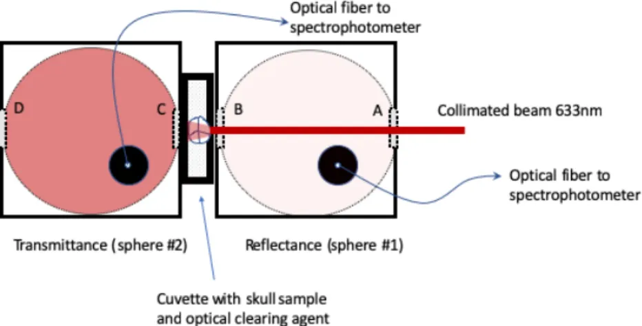
In vitro experiments have been conducted on mice skull samples from 20 mice of varying ages, using a two integrating sphere approach depicted on figure 1. Integrating sphere #1 is used to measure total reflectance and integrating sphere#2 is used to measure total transmittance. Both measurements are carried out simultaneously and acquired dynamically. This allows to study the time course of the clarification process while the skull sample is immersed in either calcium chelating or index matching solutions. For further analysis, the thickness of the skull samples was determined using OCT imaging.
Figure 1. In vitro set-up for the dynamic study of mice skull clarification process.
RESULTS
Representative results of the in vitro experiments are shown on figure 2. The optical clearing agents used were glycerol 80%, EDTA -10% followed by Glycerol 80% or PEG-400. All protocols show a significant increase in transmittance and decrease of reflectance following optical clearing treatment.
Figure 2 Transmittance (filled symbols) and Reflectance (open symbols) for different skull optical clearing protocols. The optical clearing agents are introduced between 0 and 1 minute.
For all protocols, the data show a relatively rapid time course of optical clearing as about 90% of the maximum transmittance is obtained after 7 minutes. The kinetics is significantly faster for clearing with PEG-400 compared to glycerol-80%. The optimal clearing was obtained for the skull pre-treated with EDTA-10% as a calcium chelator, followed by glycerol 80%. A transmittance of about 65% is obtained combined with a negligible (<1%) reflectance. As it is knowns that the optical properties of skull changes with age in mice5, further analysis will be presented regarding the influence of
mice age on the skull thickness and the clarification process. In vivo validation have been carried out for longitudinal imaging. A representative study is shown on figure 3 where the same mice was imaged twice with a one-month interval between the two sessions. Laser speckle contrast imaging was performed using the multiple exposure approach6.
Figure 3 In vivo longitudinal laser speckle contrast imaging after skull optical clearing in mice
Altogether this study proposes a systematic methodology and a quantitative framework for the choice of skull clarification protocols. The in vivo validation demonstrates the practicability of skull optical clearing for longitudinal optical imaging of the mice brain which is of great interest for a vast community of researchers.
REFERENCES
1. Y.-J. Zhao et al., “Skull optical clearing window for in vivo imaging of the mouse cortex at synaptic resolution,” Light Sci. Appl. 7, 17153 (2018) [doi:10.1038/lsa.2017.153].
2. G. Silasi et al., “Intact skull chronic windows for mesoscopic wide-field imaging in awake mice,” J. Neurosci. Methods 267, 141–149 (2016) [doi:10.1016/j.jneumeth.2016.04.012].
3. G. J. Goldey et al., “Removable cranial windows for long-term imaging in awake mice,” Nat. Protoc. 9(11), 2515–2538 (2014) [doi:10.1038/nprot.2014.165].
4. A. N. Bashkatov et al., “Measurement of tissue optical properties in the context of tissue optical clearing,” J. Biomed. Opt. 23(9), 1–31 (2018) [doi:10.1117/1.JBO.23.9.091416].
5. H. Soleimanzad, H. Gurden, and F. Pain, “Optical properties of mice skull bone in the 455- to 705-nm range,” J. Biomed. Opt. 22(1), 010503 (2017) [doi:10.1117/1.JBO.22.1.010503].
6. H. Soleimanzad et al., “Multiple speckle exposure imaging for the study of blood flow changes induced by functional activation of barrel cortex and olfactory bulb in mice,” Neurophotonics 6(1), 015008 (2019) [doi:10.1117/1.NPh.6.1.015008].