HAL Id: hal-01586102
https://hal.sorbonne-universite.fr/hal-01586102
Submitted on 12 Sep 2017HAL is a multi-disciplinary open access archive for the deposit and dissemination of sci-entific research documents, whether they are pub-lished or not. The documents may come from teaching and research institutions in France or abroad, or from public or private research centers.
L’archive ouverte pluridisciplinaire HAL, est destinée au dépôt et à la diffusion de documents scientifiques de niveau recherche, publiés ou non, émanant des établissements d’enseignement et de recherche français ou étrangers, des laboratoires publics ou privés.
Development of anti-hepatitis B surface (HBs)
antibodies after HBs antigen loss in HIV-hepatitis B
virus co-infected patients
Anders Boyd, Laetitia Canini, Joël Gozlan, Caroline Lascoux-Combe, Patrick
Miailhes, Laurent Fonquernie, Pierre-Marie Girard, Karine Lacombe
To cite this version:
Anders Boyd, Laetitia Canini, Joël Gozlan, Caroline Lascoux-Combe, Patrick Miailhes, et al.. De-velopment of anti-hepatitis B surface (HBs) antibodies after HBs antigen loss in HIV-hepatitis B virus co-infected patients. Journal of Clinical Virology, Elsevier, 2017, 95, pp.55 - 60. �10.1016/j.jcv.2017.08.008�. �hal-01586102�
Development of anti-hepatitis B surface (HBs) antibodies after HBs antigen loss in HIV-hepatitis B virus co-infected patients
Anders Boyd1, Laetitia Canini2, Joël Gozlan3,4, Caroline Lascoux-Combe5, Patrick Miailhes6,7, Laurent Fonquernie8, Pierre-Marie Girard8,9, Karine Lacombe8,9.
Author affiliations:
1INSERM, UMR_S1136, Institut Pierre Louis d’Epidémiologie et de Santé Publique, Paris,
France;
2Centre for Immunity, Infection and Evolution, University of Edinburgh, Edinburgh, United
Kingdom;
3Laboratoire de Virologie, Hôpital Saint-Antoine, AP-HP, Paris, France; 4UPMC UMRS CR7; INSERM U1135 CIMI, Paris, France;
5Service des Maladies Infectieuses et Tropicales, Hôpital Saint-Louis, AP-HP, Paris, France; 6Centre de Recherche sur le Cancer de Lyon, Equipes 15 et 16, INSERM, Unité 1052, CNRS,
UMR 5286, Lyon, France;
7Service des Maladies Infectieuses et Tropicales, Hôpital de la Croix-Rousse, Hospices Civils de
Lyon, Lyon, France;
8Service des Maladies Infectieuses et Tropicales, Hôpital Saint-Antoine, AP-HP, Paris, France; 9Sorbonne Universités, INSERM, UPMC Univ Paris 06, Institut Pierre Louis d’épidémiologie et
de Santé Publique (IPLESP UMRS 1136), Paris, France.
Corresponding Author: Dr. Anders Boyd
Services des Maladies Infectieuses et Tropicales; Hôpital Saint-Antoine 184 rue du Fbg. St. Antoine
75571 Paris Cedex 12; France Telephone: +33 1 71 97 05 17 Fax: +33 1 49 28 21 49 Email: anders.boyd@upmc.fr
ABSTRACT
Background: Hepatitis B surface antigen (HBsAg)-seroconversion, or loss of HBsAg and acquisition of anti-hepatitis B surface (HBs) antibodies, defines functional cure of chronic
hepatitis B virus (HBV) infection. After HBsAg-loss, little is known regarding the development of anti-HBs antibodies and even less so in individuals co-infected with HIV.
Objectives: To determine anti-HBs antibody kinetics after HBsAg-loss and explore determinants of HBsAg-seroconversion in HIV-HBV co-infected patients.
Study design: Patients enrolled in the French HIV-HBV cohort were included if they had >1 study visit after HBsAg-loss. Individual patient kinetics of anti-HBs antibody levels were
modeled over time using mixed-effect non-linear regression, whereby maximum specific growth rate and maximal level of antibody production were estimated from a Gompertz growth equation. Results: Fourteen (4.6%) of 308 co-infected patients followed in the cohort exhibited HBsAg-loss, all of whom were undergoing antiretroviral therapy. Nine (64.3%) of these patients achieved HBsAg-seroconversion during a median 3.0 years (IQR=1.1-5.1) after HBsAg-loss. Across individuals with HBsAg-seroconversion, the fastest rates of antibody growth ranged between 0.57-1.93 year-1 (population maximum growth rate=1.02) and antibody production plateaued between 2.09-3.66 log10 mIU/mL at the end of follow-up (population maximal antibody
levels=2.66). Patients with HBsAg-seroconversion had substantial decreases in HBV DNA viral loads (P=0.03) and proportion with elevated ALT levels (P=0.02) and HBeAg-positive serology (P=0.08). No such differences were observed in those without HBsAg-seroconversion. Conclusions: Most co-infected patients with HBsAg-seroconversion produced and maintained stable antibody levels, yet kinetics of anti-HBs production were much slower compared to those observed post-vaccination or after clearance of acute HBV-infection.
KEYWORDS: anti-HBs antibodies; HBsAg-seroconversion; antiretroviral therapy; kinetics; logistic growth model.
HIGHLIGHTS
- Studies have yet to fully examine anti-HBs antibody development after HBsAg-loss. - Nine HIV-HBV co-infected patients with HBsAg-loss and extensive viral suppression were prospectively followed.
- Rate of maximum antibody growth and level of maximum antibody development were much lower compared to post-vaccination or acute HBV.
- The modeling strategy presented herein could be useful for clinical trials evaluating novel agents and observational cohorts of HBV-infected patients.
BACKGROUND
The most ideal therapeutic goal in patients chronically infected with hepatitis B virus (HBV) is loss of hepatitis B surface antigen (HBsAg) and appearance of anti-hepatitis B surface (HBs) antibodies, or HBsAg-seroconversion [1]. HBsAg-seroconversion is associated with decreased viral activity in the liver and regression of liver inflammation and fibrosis [2,3], hence why HBsAg-seroconversion is considered the hallmark of “functional cure” [4]. While almost all studies on the clinical advantages of HBsAg-seroconversion have been conducted in HBV mono-infected patients, similar trends have been noted in HIV-HBV co-infection [5].
Unfortunately, acquiring anti-HBs antibodies is an uncommon event for HIV-HBV co-infected individuals. HBsAg-loss itself occurs in roughly 1-2% of patients per year during treatment with potent nucleoside/nucleotide analogues [6,7]. In this group, an even smaller yet variable proportion converts to anti-HBs antibody-positive serology [5,6]. Co-infected patients are also known to exhibit fluctuating anti-HBs antibody profiles, which is noteworthy considering the improved survival linked to consistently detectable anti-HBs antibodies from the beginning of antiretroviral therapy (ART) [5].
Understanding of the development, persistence, and potential disappearance of anti-HBs antibodies could be further clarified by examining the kinetics of antibody levels after HBsAg-loss. Nevertheless, few studies to date have explored anti-HBs antibody levels in vivo during chronic hepatitis B infection, with none emphasizing the HIV-HBV co-infected population.
OBJECTIVES
The aim of this study was to define the kinetics of anti-HBs antibodies, including the longitudinal determinants of HBsAg-seroconversion, in a small cohort of HIV-HBV co-infected patients exhibiting HBsAg-loss.
STUDY DESIGN
Study participants
Patients were selected from the French HIV-HBV Cohort Study as described previously [6]. Briefly, this prospective study recruited 308 patients from seven centers located in Paris and Lyon, France during May 2002-May 2003. Patients were included if they had HIV-positive serology confirmed by western blot and HBsAg-positive serology for at least six months. All data collection continued until 2010-2011.
For this study, subjects were selected if they achieved HBsAg-seroclearance and had at least two available anti-HBs antibody measurements thereafter. All patients provided written informed consent to participate in the cohort and the protocol was approved by the Hôpital
Pitié-Salpêtrière and Hôpital Saint-Antoine Ethics Committees (Paris, France).
Assessing HBV-related parameters
Plasma HBV DNA viral load was quantified using a commercial PCR-based assay [6]. Due to varying detection limits, undetectable HBV DNA was defined at <60 IU/mL.
Qualitative HBsAg and HBeAg were detected using a commercial enzyme-linked immunoassay at baseline and each yearly visit [6]. Anti-HBs antibodies were quantified during the same visits using one of the following commercially-available immunoassays: LIASON Anti-HBs Plus (measuring range=5-1000 mIU/mL; DiaSorin, Antony, France), Monolisa anti-HBs Plus
(measuring range=0.01-1000 mIU/mL; Bio-Rad, Marnes-la-Coquette, France), or Architect anti-HBs (measuring range=0.01-1000 mIU/mL; Abbott Diagnostics, Rungis, France). High between-assay correlation and linearity over quantification ranges have been described for these between-assays [8,9].
Alanine aminotransferase (ALT) levels were quantified using standard methods. Liver fibrosis was assessed yearly by a non-invasive method: Fibrotest® and/or Fibroscan® (Echosens, Paris, France). Cutoffs for METAVIR F3 fibrosis were applied (Fibrotest®, F3=0.59 and Fibroscan®, F3=7.6 kPa) [10,11].
Statistical analysis
HBsAg-seroclearance was defined as HBsAg-loss (no longer HBsAg-positive) during follow-up and HBsAg-seroconversion was defined as HBsAg-loss and acquiring anti-HBs antibody titers >10 mIU/mL. Statistical analysis was performed using STATA statistical software (v12.1, College Station, TX) and R 3.3.1 and a P-value of <0.05 was considered significant.
We initially described the evolution of biological and clinical parameters during overall follow-up in the cohort study to determine if there were any particular differences between patients with and without HBsAg-seroconversion. Patient characteristics were compared between inclusion visit and last follow-up visit. A paired-sample Wilcoxon signed-rank test was used for continuous variables and Pearson χ² test or Fisher’s exact test for categorical variables.
In subsequent analysis, we defined baseline as the study visit prior to HBsAg-seroclearance and follow-up as every yearly visit until last available anti-HBs antibody level, end of follow-up, study discontinuation, or death; whichever occurred first. In vivo kinetics of anti-HBs antibody after HBsAg-seroclearance were examined. Previous kinetic models have been mostly drawn from studies on immune responses post-vaccination [12], lending to the assumption that anti-HBs antibody levels would have an immediate increase upon vaccination with an exponential decay shortly thereafter [13]. These models might not fully reflect anti-HBs antibody levels during clearance of HBV infection, where antigen-independent proliferation of memory B cells and plasma cells likely reach a state of homeostatic equilibrium and continuously produce antibodies at a specific threshold [14]. Consequently, we modeled anti-HBs antibody kinetics using a Gompertz growth equation [15], defined as follows:
𝑦𝑦 = 𝐴𝐴 ∙ 𝑒𝑒−𝑘𝑘1𝑒𝑒−𝑘𝑘2𝑡𝑡
where A describes maximal antibody levels, k1 the delay in anti-HBs antibody response, and k2
the maximum growth rate per year. Anti-HBs antibody levels <10 mIU/mL and >1000 mIU/mL were left- and right-censored, respectively. Population and individual parameters were
estimated with nonlinear mixed-effect models taking into account inter-individual variability (IIV) (Supplementary Materials).
RESULTS
Of the 308 patients in the cohort study, 17 with documented HBsAg-seroclearance were
considered. Three of these patients had only one anti-HBs antibody measurement after HBsAg-seroclearance and were excluded. In total, 14 patients were included in analysis.
Patients were predominately male (93%), coming from an intermediate or high zone of HBV-prevalence (43%), with a median age of 42 years (IQR=38-52). From the start of the cohort study, median duration of known HIV-infection was 6 years (IQR=4-13) and median CD4+ cell count at 440/mm3 (IQR=303-554), while three patients (21.4%) had presented with an AIDS-defining illness. One and two patients also had positive serology for hepatitis C and D virus, respectively.
Rates and determinants of HBsAg-seroconversion
HBsAg-seroclearance occurred after a median 4.6 years (IQR=2.1-7.4) of follow-up in the cohort. Nine (64.3%) patients achieved HBsAg-seroconversion during a median 3.0 years (IQR=1.1-5.1) after HBsAg-seroclearance. HBsAg-seroconversion was identified either at the same visit as HBsAg-seroclearance (n=5) or 0.9-2.3 years after the first HBsAg-negative result (n=4). Two patients fluctuated back to anti-HBs antibody-negative serology once during follow-up while ending with anti-HBs antibody-positive serology, one of whom had an HBsAg/anti-HBs antibody-positive profile before achieving HBsAg-seroclearance.
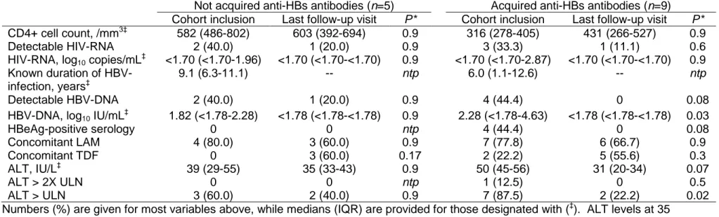
Table 1 summarizes the clinical differences between cohort inclusion and last follow-up visits, while stratified between those with versus without HBsAg-seroconversion. At cohort inclusion, patients with HBsAg-seroconversion were more likely to have HBeAg-positive serology, higher ALT levels and HBV-DNA viral loads; yet none of these differences was significant. During follow-up, significant changes in HBV-DNA viral loads (P=0.03) and proportion with elevated
ALT levels (>35 IU/L, P=0.02) were observed in patients with HBsAg-seroconversion. In contrast, there were no significant changes in HIV- or HBV-related parameters from cohort inclusion to last follow-up visit in those without HBsAg-seroconversion. ART was administered in all 14 patients during follow-up, with no noticeable difference between seroconversion groups (Supplementary Material).
Liver fibrosis levels were stable for most patients during follow-up (Supplementary Material). No cases of portal hypertension, decompensated liver, end-stage liver disease, hepatocellular carcinoma, or liver-related death occurred in any of the included patients.
Kinetics of anti-HBs antibodies after HBsAg-loss
In total, 54 samples were available for 9 patients after HBsAg-loss. We selected a model without IIV for k1. The estimate for k1 was 2.49 (RSE=26%) and maximum growth rate, k2, was
1.02 years-1 (RSE=30%) showing 47% of IIV. Population maximum anti-HBs antibody titer was 2.71 log10 mIU/mL (relative standard error, RSE=10%) showing 20% of IIV. The additive error
term, measuring the difference between predicted and observed values, was 0.35 (RSE=1%) log10 mIU/mL.
Model parameters of anti-HBs antibody kinetics after HBsAg-seroclearance are given for each patient in Table 2, noting that one patient had an insufficient number of time-points to obtain estimates. Model fit was good (Figure 1). Individual maximum growth rate somewhat varied between patients (0.57-1.93 year-1). Strong yet non-significant correlations were observed between k2 and nadir CD4+ (Spearman’s rho=-0.48, p=0.2) and concomitant CD4+ cell count
(rho=-0.53, p=0.18) at the moment of HBsAg-seroconversion. Five patients had a maximum level of log10 antibody units estimated between 2.09-2.66 log10 mIU/mL and two patients >3.0
log10 mIU/mL. There were no significant correlations between maximum levels of antibody units
and ALT (Spearman’s rho=0.15, p=0.7), CD4+ cell count (rho=-0.07, p=0.9), and nadir CD4+ cell count (rho=-0.32, p=0.4) at HBsAg-seroconversion.
In order to understand the projected development of anti-HBs antibodies over eight years, we simulated anti-HBs antibody trajectories of 1000 subjects by drawing individual parameters from the population parameter distribution among those with HBsAg-seroconversion. As shown in Figure 2, we predicted that 300 (30.0%) simulated patients would eventually reach the upper limit of quantification (>1000 mIU/mL) after a median 3.3 years (IQR=2.4-4.3). At the end of eight years, 657 (65.7%) simulated patients would reach >100-1000 mIU/mL, while the remaining 43 (4.3%) would have levels persisting between >10-100 mIU/mL.
DISCUSSION
In this small group of patients exhibiting loss, 64% progressed further to HBsAg-seroconversion during a median three years of follow-up. This proportion was slightly higher compared to other cohorts of treated co-infected patients (14-50%) [5,7,16,17] and somewhat similar to HBV mono-infected patients (50-71%) [18–21]. Nevertheless, comparisons to other cohorts are oftentimes difficult, as most have insufficient follow-up and the low rates of HBsAg-seroclearance in general give rise to equally small sample sizes.
Anti-HBs antibody production is important for several reasons. First, anti-HBs antibodies, especially those targeted to the preS1 domain, have the capacity to neutralize circulating virus [22]. Modeling data from in vitro experiments have also shown that anti-HBs antibodies can block the release of HBV and HBsAg sub-particles from infected hepatocytes [23]. These
antiviral mechanisms could be beneficial even during viral suppression under anti-HBV agents, such as TDF, when virus is known to circulate at low levels below detection thresholds of most conventional PCR assays [24]. Second, levels of hepatitis B core antigen-specific CD8+ T cells, associated with cell-mediated clearance of HBV infection, are almost completely undetectable decades after HBsAg-seroconversion [25]. It would then be assumed that humoral responses linked to antibody production would play a dominant role in preventing HBV reactivation. Finally, HBsAg is expressed on the membrane of infected hepatocytes [26], to which recognition by anti-HBs antibodies could lead to cytotoxicity and clearance of infected liver cells [27]. There is, therefore, an inherent interest in studying how anti-HBs antibody production is achieved after HBsAg-loss.
After applying a sigmoidal model of antibody growth, we were able to characterize several remarkable features of anti-HBs antibody production during HBsAg-seroconversion. When examining the maximum rate of antibody production, there was slight variability between
patients, ranging from 0.57-1.93 year-1. In studies using dynamic modeling, the highest rates of antibody production have been observed after vaccination among immunocompetent
populations, with estimates around 0.94 per day [28]. These rates are much lower during clearance of acute infection at 0.96-1.32 per month, while the lower bounds of these estimates are strongly associated with low virus production [14]. We observed a much lower rate of antibody production compared to these previous studies. Whether this attenuated rate is due to the pathophysiological features of chronic infection, immunocompromised status, or a
combination of both are difficult to determine given that no data of this kind are available in HBV mono-infected patients.
The model asymptotes obtained in our study showed that only two patients had anti-HBs antibody levels plateauing consistently above 1000 mIU/mL, whereas the remainder had levels
hovering around 123-457 mIU/mL. How this translates to clinical outcomes is difficult to state given the lack of evidence correlating long-term anti-HBs antibody levels and liver-related morbidity and mortality. Interestingly, these levels are similar to vaccine response or HBsAg-seroconversion in immunocompromised individuals, particularly HIV-positive [29] or liver transplant patients [30,31], and much lower for vaccine response in healthy individuals [32].
Notwithstanding the potential for inadequate power, certain determinants were noted between patients with HBsAg-seroclearance who did versus did not develop anti-HBs antibodies. Patients with HBsAg-seroconversion appeared to have gone from a more active form of infection, with higher levels of replication, higher proportion with HBeAg-positive serology and higher ALT levels before evolving towards inactive infection. The accelerated development of anti-HBs antibodies has been observed in other studies among HIV-HBV co-infected patients, yet this was strongly linked to severe immunocompromised status and immunoreconstitution after ART-initiation [17,33] and thus could represent a type of “immunoreactive” phase of HBV-infection. Indeed, patients with HBsAg-seroconversion did have lower levels of nadir and current CD4+ cell counts prior to HBsAg-loss and maximum antibody growth rate was inversely correlated with nadir CD4 cell count; however, none of these associations were significant. Determining if improvements in common immunological deficiencies related to antibody
acquisition, such as B cell proliferation upon activation [34] and HBsAg-specific T helper 1 cells [35,36], could help establish the components of immunity responsible for these observations in co-infected individuals.
Other genetic determinants could have also explained our results. For instance, specific human leukocyte antigen class II alleles, notably DRB1 and DQB1, have been associated with antibody response to HBV vaccine [37]. The relationship between these alleles and
HBsAg-seroconversion has yet to be studied during the natural history of chronic HBV-infection and as human genetic data were not collected in our study, we cannot infer on its particular role.
Despite the advantages of HBsAg-seroconversion obtained from in vitro findings, the clinical benefit of acquiring anti-HBs antibodies is debatable. In HIV-infected patients with previous exposure to HBV-infection, rates in all-cause mortality were not significantly different between those who did versus did not acquire anti-HBs antibodies [5]. Likewise, we saw no particular improvement in liver fibrosis markers for any of the patients in this cohort, regardless of anti-HBs antibody acquisition. One explanation could be that stable liver fibrosis levels are generally problematic for the HIV-HBV co-infected population, even during antiretroviral therapy containing more potent anti-HBV treatments [38]. Alternatively, as this was a population of HBsAg-negative patients with low prevalence of cirrhosis, especially when considering the FibroScan® alone, no further declines in fibrosis would be anticipated during such a short follow-up period.
There are certain limitations that need to be addressed. First, samples were taken at yearly intervals for almost all patients and hence the exact timing of HBsAg-seroconversion would be challenging. Infrequent sampling also made it difficult to test for oscillations in anti-HBs antibody levels during follow-up. Second, anti-HBs antibody levels were quantified from a variety of assays across centers, with some demonstrating moderate intra-assay variability [39]. Between-patient comparability could have been affected; nevertheless, the uniformity of assays within centers, with the exception of modifications in reagents with the Biorad assay [9], would limit measurement bias in the kinetic parameters obtained from individual patients.
In conclusion, of the two-thirds of patients with HBsAg-seroclerance who continued further to HBsAg-seroconversion, most were able to produce and maintain stable levels of antibodies but some did exhibit vast fluctuations in antibody production. The finding that non-invasive markers
of fibrosis did not subside after HBsAg-seroconversion raises questions on whether this end-point is an adequate representation of decreased risk in liver-disease for HIV-HBV co-infected patients, yet this would need further validation in studies with higher prevalence of pre-treatment cirrhotics and/or longer follow-up. Future research should aim to develop if any benefit in anti-HBs antibody production could extend towards switching to a non-anti-HBV containing
antiretroviral regimen.
CONFLICT OF INTEREST
The authors declare no conflicts of interest relevant to the manuscript.
ETHICAL APPROVAL
All patients provided written informed consent to participate in the cohort and the protocol was approved by the Hôpital Pitié-Salpêtrière and Hôpital Saint-Antoine Ethics Committees (Paris, France) in accordance with the Helsinki Declaration.
ACKNOWLEDGEMENTS
The authors are grateful to all patients and clinical teams for their commitment to the French HIV-HBV Cohort Study. We acknowledge H. Rougier, M. Sébire-Le Cam, and L. Roguet for managing the logistics of the French HIV-HBV Cohort; G. Pannetier and F. Carrat for their help
in data management; and Pr J.-F. Flejou and E. Roux of the Tumorothèque HUEP at Saint-Antoine Hospital for storing samples.
Funding: This work was supported in part by the Institut de Médecine et d’Epidémiologie Appliquée and received additional grants from ANRS (Agence Nationale de Recherche sur le Sida et les Hépatites). Gilead Sciences, Inc. provided an unrestricted grant for the French HIV-HBV cohort and was not involved in any part of the data collection, analysis, and manuscript writing. Post-doctoral fellowships from the ANRS and SIDACTION were awarded to AB. LC was funded by the UK Biotechnology and Biological Sciences Research Council (grant reference 1698:BB/L001330/1).
REFERENCES
[1] European Association For The Study Of The Liver, EASL clinical practice guidelines: Management of chronic hepatitis B virus infection, J. Hepatol. 57 (2012) 167–185. doi:10.1016/j.jhep.2012.02.010.
[2] B. Werle-Lapostolle, S. Bowden, S. Locarnini, K. Wursthorn, J. Petersen, G. Lau, C. Trepo, P. Marcellin, Z. Goodman, W.E. Delaney 4th, S. Xiong, C.L. Brosgart, S.-S. Chen, C.S. Gibbs, F. Zoulim, Persistence of cccDNA during the natural history of chronic hepatitis B and decline during adefovir dipivoxil therapy, Gastroenterology. 126 (2004) 1750–1758. [3] R. Moucari, A. Korevaar, O. Lada, M. Martinot-Peignoux, N. Boyer, V. Mackiewicz, A.
Dauvergne, A.C. Cardoso, T. Asselah, M.-H. Nicolas-Chanoine, M. Vidaud, D. Valla, P. Bedossa, P. Marcellin, High rates of HBsAg seroconversion in HBeAg-positive chronic hepatitis B patients responding to interferon: a long-term follow-up study, J. Hepatol. 50 (2009) 1084–1092. doi:10.1016/j.jhep.2009.01.016.
[4] M.B. Zeisel, J. Lucifora, W.S. Mason, C. Sureau, J. Beck, M. Levrero, M. Kann, P.A. Knolle, M. Benkirane, D. Durantel, M.-L. Michel, B. Autran, F.-L. Cosset, H.
Strick-Marchand, C. Trépo, J.-H. Kao, F. Carrat, K. Lacombe, R.F. Schinazi, F. Barré-Sinoussi, J.-F. Delfraissy, F. Zoulim, Towards an HBV cure: state-of-the-art and unresolved
questions-report of the ANRS workshop on HBV cure, Gut. 64 (2015) 1314–1326. doi:10.1136/gutjnl-2014-308943.
[5] W.-H. Sheng, J.-H. Kao, P.-J. Chen, L.-M. Huang, S.-Y. Chang, H.-Y. Sun, C.-C. Hung, M.-Y. Chen, S.-C. Chang, Evolution of hepatitis B serological markers in HIV-infected patients receiving highly active antiretroviral therapy, Clin. Infect. Dis. 45 (2007) 1221–1229.
doi:10.1086/522173.
[6] A. Boyd, J. Gozlan, P. Miailhes, C. Lascoux-Combe, M.S.-L. Cam, H. Rougier, F. Zoulim, P.-M. Girard, K. Lacombe, Rates and determinants of hepatitis B “e” antigen and hepatitis B surface antigen seroclearance during long-term follow-up of patients coinfected with HIV and hepatitis B virus, AIDS 29 (2015) 1963–1973. doi:10.1097/QAD.0000000000000795. [7] R. Zoutendijk, H.L. Zaaijer, T.E.M.S. de Vries-Sluijs, J.G.P. Reijnders, J.W. Mulder, F.P.
Kroon, C. Richter, A.A. van der Eijk, M.J. Sonneveld, B.E. Hansen, R.A. de Man, M.E. van der Ende, H.L.A. Janssen, Hepatitis B surface antigen declines and clearance during long-term tenofovir therapy in patients coinfected with HBV and HIV, J. Infect. Dis. 206 (2012) 974–980. doi:10.1093/infdis/jis439.
[8] S. Kinn, S. Akhavan, H. Agut, V. Thibault, Performance of the DiaSorin LIAISON(®) anti-HBs II for the detection of hepatitis B surface antibodies: comparison with the Abbott Architect anti-HBs assay, J. Clin. Virol. 50 (2011) 297–302. doi:10.1016/j.jcv.2011.01.009. [9] Direction de l’Evaluation des Dispositifs Médicaux, Département Surveillance du Marché,
Unité Evaluation et Contrôle du Marché-DIV, Rapport du controle de marché des dispositifs médicaux de diagnostic in vitro pour la détection et le dosage des anticorps anti-HBs, Agence française de sécurité sanitaire des produits de santé, 2007.
[10] J. Bottero, K. Lacombe, J. Guéchot, L. Serfaty, P. Miailhes, P. Bonnard, D. Wendum, J.-M. Molina, C. Lascoux-Combe, P.-M. Girard, Performance of 11 biomarkers for liver fibrosis assessment in HIV/HBV co-infected patients, J. Hepatol. 50 (2009) 1074–1083.
doi:10.1016/j.jhep.2009.01.022.
[11] P. Miailhes, P. Pradat, M. Chevallier, K. Lacombe, F. Bailly, L. Cotte, M.-A. Trabaud, A. Boibieux, J. Bottero, C. Trepo, F. Zoulim, Proficiency of transient elastography compared to liver biopsy for the assessment of fibrosis in HIV/HBV-coinfected patients, J. Viral Hepat. 18 (2011) 61–69. doi:10.1111/j.1365-2893.2010.01275.x.
[12] D. Le, J.D. Miller, V.V. Ganusov, Mathematical modeling provides kinetic details of the human immune response to vaccination, Front. Cell. Infect. Microbiol. 4 (2014) 177. doi:10.3389/fcimb.2014.00177.
[13] M.C. Honorati, A. Palareti, P. Dolzani, C.A. Busachi, R. Rizzoli, A. Facchini, A
mathematical model predicting anti-hepatitis B virus surface antigen (HBs) decay after vaccination against hepatitis B, Clin. Exp. Immunol. 116 (1999) 121–126.
[14] S.M. Ciupe, R.M. Ribeiro, A.S. Perelson, Antibody responses during hepatitis B viral infection, PLoS Comput. Biol. 10 (2014) e1003730. doi:10.1371/journal.pcbi.1003730. [15] M.H. Zwietering, I. Jongenburger, F.M. Rombouts, K. van ’t Riet, Modeling of the bacterial
growth curve, Appl. Environ. Microbiol. 56 (1990) 1875–1881.
[16] L. Kosi, T. Reiberger, B.A. Payer, K. Grabmeier-Pfistershammer, R. Strassl, A. Rieger, M. Peck-Radosavljevic, Five-year on-treatment efficacy of lamivudine-, tenofovir- and tenofovir + emtricitabine-based HAART in HBV-HIV-coinfected patients, J. Viral Hepat. 19 (2012) 801–810. doi:10.1111/j.1365-2893.2012.01601.x.
[17] G.V. Matthews, R.J. Ali, A. Avihingsanon, J. Amin, R. Hammond, S. Bowden, S.R. Lewin, J. Sasadeusz, M. Littlejohn, S.L. Locarnini, K. Ruxrungtham, G.J. Dore, Quantitative HBsAg and HBeAg predict hepatitis B seroconversion after initiation of HAART in HIV-HBV coinfected individuals, PloS One. 8 (2013) e61297. doi:10.1371/journal.pone.0061297.
[18] E. Lauret, M.L. González-Diéguez, M. Rodríguez, M. González, S. Melón, L. Rodrigo, M. Rodríguez, Long-term outcome in Caucasian patients with chronic hepatitis B virus infection after HBsAg seroclearance, Liver Int. 35 (2015) 140–147. doi:10.1111/liv.12461. [19] T. Hara, F. Suzuki, Y. Kawamura, H. Sezaki, T. Hosaka, N. Akuta, M. Kobayashi, Y.
Suzuki, S. Saitoh, Y. Arase, K. Ikeda, M. Kobayashi, S. Watahiki, R. Mineta, H. Kumada, Long-term entecavir therapy results in falls in serum hepatitis B surface antigen levels and seroclearance in nucleos(t)ide-naïve chronic hepatitis B patients, J. Viral Hepat. 21 (2014) 802–808. doi:10.1111/jvh.12211.
[20] P. Marcellin, F. Zoulim, C. Hézode, X. Causse, B. Roche, R. Truchi, A. Pauwels, D. Ouzan, J. Dumortier, G.-P. Pageaux, M. Bourlière, G. Riachi, J.-P. Zarski, J.-F. Cadranel, V. Tilliet, C. Stern, P. Pétour, O. Libert, S.M. Consoli, D. Larrey, Effectiveness and Safety of
Tenofovir Disoproxil Fumarate in Chronic Hepatitis B: A 3-Year, Prospective, Real-World Study in France, Dig. Dis. Sci. (2016). doi:10.1007/s10620-015-4027-8.
[21] Y.C. Chen, W.J. Jeng, R.N. Chien, C.M. Chu, Y.F. Liaw, Clinical outcomes after
spontaneous and nucleos(t)ide analogue-treated HBsAg seroclearance in chronic HBV infection, Aliment. Pharmacol. Ther. 43 (2016) 1311–1318. doi:10.1111/apt.13630.
[22] C.Y. Maeng, C.J. Ryu, P. Gripon, C. Guguen-Guillouzo, H.J. Hong, Fine mapping of virus-neutralizing epitopes on hepatitis B virus PreS1, Virology. 270 (2000) 9–16.
doi:10.1006/viro.2000.0250.
[23] A.U. Neumann, S. Phillips, I. Levine, S. Ijaz, H. Dahari, R. Eren, S. Dagan, N.V. Naoumov, Novel mechanism of antibodies to hepatitis B virus in blocking viral particle release from cells, Hepatology. 52 (2010) 875–885. doi:10.1002/hep.23778.
[24] P. Marcellin, E.J. Gane, R. Flisiak, M.P. Manns, K. Kaita, A. Gaggar, L. Lin, K.M. Kitrinos, J.F. Flaherty, M. Subramanian, J.G. McHutchison, H.L.A. Janssen, M. Buti, Evidence for ongoing low-level viremia in patients with chronic hepatitis B receiving long-term
[25] H. Kefalakes, C. Jochum, G. Hilgard, A. Kahraman, A.M. Bohrer, N. El Hindy, F.M. Heinemann, J. Verheyen, G. Gerken, M. Roggendorf, J. Timm, Decades after recovery from hepatitis B and HBsAg clearance the CD8+ T cell response against HBV core is nearly undetectable, J. Hepatol. 63 (2015) 13–19. doi:10.1016/j.jhep.2015.01.030. [26] K.H. Heermann, U. Goldmann, W. Schwartz, T. Seyffarth, H. Baumgarten, W.H. Gerlich,
Large surface proteins of hepatitis B virus containing the pre-s sequence, J. Virol. 52 (1984) 396–402.
[27] T.I. Michalak, J.Y. Lau, B.M. McFarlane, G.J. Alexander, A.L. Eddleston, R. Williams, Antibody-directed complement-mediated cytotoxicity to hepatocytes from patients with chronic hepatitis B, Clin. Exp. Immunol. 100 (1995) 227–232.
[28] J.N. Wilson, D.J. Nokes, G.F. Medley, D. Shouval, Mathematical model of the antibody response to hepatitis B vaccines: implications for reduced schedules, Vaccine. 25 (2007) 3705–3712. doi:10.1016/j.vaccine.2007.01.012.
[29] O. Launay, A.R. Rosenberg, D. Rey, N. Pouget, M.-L. Michel, J. Reynes, D. Neau, F. Raffi, L. Piroth, F. Carrat, ANRS HB03 VIHVAC-B (Trial Comparing 3 Strategies of Vaccination Against the Virus of Hepatitis B in HIV-Infected Patients) Group, Long-term Immune Response to Hepatitis B Virus Vaccination Regimens in Adults With Human
Immunodeficiency Virus 1: Secondary Analysis of a Randomized Clinical Trial, JAMA Intern. Med. 176 (2016) 603–610. doi:10.1001/jamainternmed.2016.0741.
[30] I. Gutierrez Domingo, J.M. Pascasio Acevedo, A. Alcalde Vargas, A. Ramos Cuadra, M.T. Ferrer Ríos, J.M. Sousa Martin, M. Sayago Mota, A. Giráldez Gallego, G. Suárez Artacho, Response to vaccination against hepatitis B virus with a schedule of four 40-μg doses in cirrhotic patients evaluated for liver transplantation: factors associated with a response, Transplant. Proc. 44 (2012) 1499–1501. doi:10.1016/j.transproceed.2012.05.071.
[31] J. Rosenau, T. Kreutz, M. Kujawa, M.J. Bahr, K. Rifai, N. Hooman, A. Finger, G. Michel, B. Nashan, E.R. Kuse, J. Klempnauer, H.L. Tillmann, M.P. Manns, HBsAg level at time of liver
transplantation determines HBsAg decrease and anti-HBs increase and affects HBV DNA decrease during early immunoglobulin administration, J. Hepatol. 46 (2007) 635–644. doi:10.1016/j.jhep.2006.11.022.
[32] A. Floreani, V. Baldo, M. Cristofoletti, G. Renzulli, A. Valeri, C. Zanetti, R. Trivello, Long-term persistence of anti-HBs after vaccination against HBV: an 18 year experience in health care workers, Vaccine. 22 (2004) 607–610.
[33] P. Miailhes, M.-A. Trabaud, P. Pradat, B. Lebouché, M. Chevallier, P. Chevallier, F. Zoulim, C. Trepo, Impact of highly active antiretroviral therapy (HAART) on the natural history of hepatitis B virus (HBV) and HIV coinfection: relationship between prolonged efficacy of HAART and HBV surface and early antigen seroconversion, Clin. Infect. Dis. 45 (2007) 624–632. doi:10.1086/520752.
[34] B. Oliviero, A. Cerino, S. Varchetta, E. Paudice, S. Pai, S. Ludovisi, M. Zaramella, G. Michelone, P. Pugnale, F. Negro, V. Barnaba, M.U. Mondelli, Enhanced B-cell
differentiation and reduced proliferative capacity in chronic hepatitis C and chronic hepatitis B virus infections, J. Hepatol. 55 (2011) 53–60. doi:10.1016/j.jhep.2010.10.016.
[35] W.O. Böcher, E. Galun, H. Marcus, N. Daudi, D. Terkieltaub, D. Shouval, H.F. Löhr, Y. Reisner, Reduced hepatitis B virus surface antigen-specific Th1 helper cell frequency of chronic HBV carriers is associated with a failure to produce antigen-specific antibodies in the trimera mouse, Hepatology. 31 (2000) 480–487. doi:10.1002/hep.510310231.
[36] G.M. Dusheiko, J.H. Hoofnagle, W.G. Cooksley, S.P. James, E.A. Jones, Synthesis of antibodies to hepatitis B virus by cultured lymphocytes from chronic hepatitis B surface antigen carriers, J. Clin. Invest. 71 (1983) 1104–1113.
[37] Z.-K. Li, J.-J. Nie, J. Li, H. Zhuang, The effect of HLA on immunological response to hepatitis B vaccine in healthy people: a meta-analysis, Vaccine. 31 (2013) 4355–4361. doi:10.1016/j.vaccine.2013.06.108.
[38] A. Boyd, J. Bottero, P. Miailhes, C. Lascoux-Combe, H. Rougier, P.-M. Girard, L. Serfaty, K. Lacombe, Liver fibrosis regression and progression during controlled hepatitis B virus infection among HIV-HBV patients treated with tenofovir disoproxil fumarate in France: a prospective cohort study, J. Int. AIDS Soc. 20 (2017) 1–12. doi:10.7448/IAS.20.1.21426. [39] D. Huzly, T. Schenk, W. Jilg, D. Neumann-Haefelin, Comparison of nine commercially
available assays for quantification of antibody response to hepatitis B virus surface antigen, J. Clin. Microbiol. 46 (2008) 1298–1306. doi:10.1128/JCM.02430-07.
FIGURE LEGENDS
Figure 1. Kinetics of anti-HBs antibodies for individual HIV-HBV co-infected patients after hepatitis B surface antigen (HBsAg) seroclearance.
Time starts at the visit prior to HBsAg-seroclerance and continues until end of follow-up. Levels of anti-HBs antibodies are plotted at each time point, with separate panels depicting a different patient. Fitted lines from the Gompertz model, as described in the methods, are also given. The dashed grey lines represent the lower and upper limit of quantification, dots observations
between these limits of quantification, triangles observations below the lower limit of
quantification and squares observations above the upper limit of quantification. One patient with HBsAg-seroconversion is not included as they did not have enough time-points needed to estimate model parameters.
Figure 2. Simulated kinetics of anti-HBs antibodies using individual parameters
Each line represents a simulated patient’s trajectory of anti-hepatitis B surface (HBs) antibody growth over eight years of follow-up. A total of 1000 subjects were simulated by drawing individual parameters from the population parameter distribution (of a Gompertz model) among those with HBsAg-seroconversion.
TABLES
Table 1. Evolution of clinical characteristics during follow-up in HIV-HBV co-infected patients without and with HBsAg- seroconversion
Not acquired anti-HBs antibodies (n=5) Acquired anti-HBs antibodies (n=9) Cohort inclusion Last follow-up visit P* Cohort inclusion Last follow-up visit P* CD4+ cell count, /mm3‡ 582 (486-802) 603 (392-694) 0.9 316 (278-405) 431 (266-527) 0.9
Detectable HIV-RNA 2 (40.0) 1 (20.0) 0.9 3 (33.3) 1 (11.1) 0.6
HIV-RNA, log10 copies/mL‡ <1.70 (<1.70-1.96) <1.70 (<1.70-<1.70) 0.9 <1.70 (<1.70-2.87) <1.70 (<1.70-<1.70) 0.9
Known duration of HBV-infection, years‡
9.1 (6.3-11.1) -- ntp 6.0 (1.1-12.6) -- ntp
Detectable HBV-DNA 2 (40.0) 1 (20.0) 0.9 4 (44.4) 0 0.08
HBV-DNA, log10 IU/mL‡ 1.82 (<1.78-2.28) <1.78 (<1.78-<1.78) 0.9 2.28 (<1.78-4.63) <1.78 (<1.78-<1.78) 0.03
HBeAg-positive serology 0 0 ntp 4 (44.4) 0 0.08 Concomitant LAM 4 (80.0) 3 (60.0) 0.9 7 (77.8) 6 (66.7) 0.9 Concomitant TDF 0 3 (60.0) 0.17 2 (22.2) 5 (55.6) 0.3 ALT, IU/L‡ 39 (29-55) 35 (33-43) 0.9 50 (45-56) 31 (20-34) 0.07 ALT > 2X ULN 0 0 ntp 1 (12.5) 0 0.5 ALT > ULN 3 (60.0) 2 (40.0) 0.9 7 (87.5) 2 (22.2) 0.02
Numbers (%) are given for most variables above, while medians (IQR) are provided for those designated with (‡). ALT levels at 35 IU/L were defined as the upper limit of normal (ULN).
*Significance between groups determined using paired-sample Wilcoxon signed-rank test for continuous variables and Pearson χ² test or Fisher’s exact test for categorical variables. ntp – no test performed
25 Table 2. Detailed description of patients with HBsAg-seroconversion and parameters of anti-HBs antibody kinetics
Patient number
Prior treatment* Sex
Nadir
CD4 /mm3 CD4 /mm3 † CDC stage HIV-RNA (copies/ mL) † ALT (IU/L) † A [log10 mIU/mL k2 (yr-1) tmax (yr) 111 LAM→TDF→LAM/TDF M 324 690 A <50 72 ‡ ‡ 0.9 131 LAM→TDF M 321 522 C <50 32 2.66 0.96 3.0 140 LAM→ADV M 107 262 A <50 55 3.40 0.70 5.1 416 ø F 179 417 A <50 25 3.66 1.59 4.7 516 LAM/TDF M 281 623 A <50 17 2.58 0.57 6.6 526 IFN→LAM M 205 498 B <50 42 2.66 0.87 5.8 527 LAM/Peg-IFN (3X)→LAM/TDF M 194 377 A <50 33 2.09 1.39 6.7 1212 LAM→LAM/TDF M 82 232 C <50 18 2.66 1.93 3.0 1302 LAM→LAM/TDF M 129 220 B <50 52 2.59 1.07 3.0
Model parameters A and k2 are respectively the asymptote (or maximum antibody levels) and maximum growth rate. Tmax is the total
follow-up duration after HBsAg-seroclearance.
*Only anti-HBV treatment leading up to HBsAg-seroconversion: ø, no treatment; ADV, adefovir; IFN, standard interferon-α; LAM, lamivudine; Peg-IFN, pegylated-interferon (only patient with Peg-IFN was treated three times in combination with LAM during a period of ten years); TDF, tenofovir. Underlined treatments were administered exclusively prior to cohort inclusion.
†
At first visit where anti-HBs antibodies appeared.
‡
26 Figure 1.
27 Figure 2.