1
Characterizing Antigen-specific CD4
+T cells
Using HLA-DR Oligomers
by
Thomas O. Cameron
B.A. ChemistryCornell University, Ithaca, NY, 1994 Submitted to the Department of Chemistry in Partial Fulfillment of the Requirements for the Degree of
DOCTOR OF PHILOSOPHY In Biological Chemistry
at the
Massachusetts Institute of Technology February 2002
2002 Massachusetts Institute of Technology All rights reserved
Signature of Author________________________________________________________ Department of Chemistry January, 2002
Certified by _____________________________________________________________ Lawrence J. Stern Associate Professor of Chemistry Thesis Supervisor
Accepted by _____________________________________________________________ Robert W. Field Chairman, Departmental Committee on Graduate Students
2
This doctoral thesis has been examined and approved by a Committee of the Department of Chemistry as follows:
Professor John M. Essigmann _______________________________________________ Committee Chairman
Professor Lawrence J. Stern _________________________________________________ Research Supervisor
Professor Alexander M. Klibanov ____________________________________________ Committee Member
3
Characterizing Antigen-specific CD4+ T cells Using HLA-DR Oligomers
By
Thomas O. Cameron
Submitted to the Department of Chemistry in Partial Fulfillment of the Requirements for the Degree of Doctor of Philosophy in Biological Chemistry, February 2002
ABSTRACT
T cells are activated by the engagement of their surface T cell receptors (TCR) by antigenic peptide bound to major histocompatibility complex (MHC). The success or failure of this TCR to MHC-peptide interaction determines the specificity of T cell action, and thus plays a central role in proper immune function. In this thesis, soluble oligomers of MHC-peptide complex were used to investigate several aspects of the T cell immune response. Soluble fluorescent oligomers of human class II MHC were produced and used to detect CD4+ T cells of particular specificities. The critical parameters of this interaction were determined, and differing behaviors of various T cell clones were observed. The implications of these results are discussed, and MHC oligomers are suggested as powerful tools for the investigation T cell avidity modulation. Using a novel methodology for the analysis of the antigen-specific TCR repertoire which includes identification by MHC oligomers, T cells specific for a peptide derived from influenza were isolated, cloned and sequenced. This pool of sequences was observed to be extremely diverse in both Vβ usage and CDR3 sequence. These results are discussed with regard to the TCR repertoire, structural aspects of TCR/MHC-peptide interaction, and future studies of TCR repertoire analysis. Other studies investigating the triggering mechanism of TCR are summarized and implications of these results for various models of transmembrane activation are discussed. A novel mechanism is proposed involving the reorganization of a receptor oligomer from a specific inhibited state into an
uninhibited state. Future directions of research based on the work presented in this thesis are suggested.
Thesis Supervisor: Lawrence J. Stern Title: Associate Professor of Chemistry
4 Acknowledgements:
Many people have contributed to my graduate career and made this thesis possible.
Larry Stern, my advisor and mentor, has shown patience with me during the past seven years; I know that I haven't been the easiest graduate student in the world. His example, advice, and leadership have been invaluable to my development.
I would also like to thank my thesis committee, Prof. Alexander Klibanov and Prof. John Essigmann, and my undergraduate mentors, Prof. Barbara Baird, and Dr. David Holowka, for their support and advice. I have received generous funding from the NIH Biotechnology Training Grant at MIT, and superlative administrative assistance from Darlene Ray.
Thanks to the various members of the Stern lab over the past few years, Mia, Lihui, Jodi, Aaron, Dikran, Jenny, Stefan, Gus, Ravi, Jen Z., Laura, Carthene, Danny, Greg, Christian, Jen, Zari, Deepali, Betty, Sulabha, Todd, Sasha, and Iwona who have made the lab an exciting place to work.
Over the years I have developed special relationships with several collaborators whom I would like to thank for their insight, assistance, and goodwill: Lucy Wedderburn, Souheil Younes, George Cohen, Philip Norris, Yassine-Diab Bader, Betsy Mellins, and Yuri Naumov.
I extend a personal thanks to the support staff of my private life: my family, Mom, Dad and Sis, and my fiancee, Nummy Wummy.
Finally, a special "oh-my-god-i'm-gonna-miss-you-guys" to Mia and Dikran who have been my constant companions through the good and the bad, the stupid and the stupider, the silly and the inane, for seven years.
5
TABLE OF CONTENTS
page
I. Introduction: The role of T cells in the Immune Response
A. Introduction 10
B. A brief introduction to the immune system 10
1. Innate Immunity
2. The strategy of adaptive immunity
3. Generation of the antigen-specific repertoire
C. T cells and the interaction of TCR with MHC-peptide complex 14 1. Introduction to T cells and APCs
2. MHC allelic polymorphism
3. Peptide Binding to MHC molecules and recognition by TCR 4. The transmembrane signaling mechanism of T cells
D. Placing the work of this thesis into context 21
1. Techniques of antigen-specific T cell identification 2. Overview of thesis
E. Figures 28
II. Detection of antigen-specific CD4+ T cells by HLA-DR1 oligomers Depends on the T cell activation state
A. Introduction 36
B. Materials and Methods 37
1. Peptides
2. Preparation of labeled antibodies and streptavidin 3. Preparation of fluorescent class II MHC oligomers 4. T cell clones and line
5. Flow cytometry
6. Fluorescence microscopy 7. Dynamic light scattering
C. Results 40
1. DR1 oligomers detect antigen-specific CD4+ T cells 2. Oligomer staining is observed only under conditions
permissive for internalization
3. Bound class II MHC oligomers are present in internalized compartments 4. A polyclonal T cell line contains subpopulations with
Different requirements for staining
5. Staining is reduced by treatments that induce a non-responsive state
D. Discussion 45
E. Acknowledgements 47
F. Figures 49
III. Labeling antigen-specific CD4+ T cells with class II MHC oligomers
A. Introduction 54
6 1. Peptides
2. Antibodies and Streptavidin 3. DR subunit bacterial expression 4. Isolation of crude inclusion bodies
5. Ion exchange purification of DR inclusion bodies
6. In vitro folding and purification of DR1-peptide complexes 7. Chemical biotinylation of DR1-cys
8. Enzymatic biotinylation of DR1-cys 9. DR1-cys expression in insect cells 10. Oligomerization
11. T cell clones and lines
12. Flow cytometric staining of T cells
C. Results 67
1. DR1 subunit production in E. coli
2. Production of soluble DR1-peptide complexes from insect cells 3. Oligomerization using fluorescent streptavidin reagents
4. Detection of antigen-specific CD4 T cells in mixed lymphocyte populations
5. In vitro expansion of antigen-specific CD4+ T cells and detection by MHC oligomers
6. Different clones show different temperature dependencies of staining 7. Short-term polyclonal T cell lines show heterogenous staining at 4C
D. Discussion 76
1. Summary of various methodologies for production of biotinylated MHC proteins
2. Interpretation of class II MHC oligomer staining results 3. Temperature sensitivity of staining;
MHC oligomers as probes of T cell avidity
E. Figures 83
IV. Towards TCR Proteomics: Examination of a highly diverse repertoire of CD4+ T cells specific for an influenza peptide bound to HLA-DR1
A. Introduction 92
B. Materials and methods 94
1. Peptides
2. Preparation of fluorescent class II MHC oligomers 3. T cell culture
4. DR1 oligomer staining
5. Activation and IFN-γ surface-capture assay 6. PCR amplification, cloning, and sequencing 7. Statistical analysis
C. Results 99
1. Polyclonal T cell lines can be stained with DR1 oligomers
2. TCRβs can be analyzed in detail by sorting, cloning and sequencing 3. Ha-specific T cells can be identified in once-stimulated
7
4. Ha-specific T cells can be identified by IFN-γ secretion 5. A large number of different TCRβ sequences found in
DR1-Ha specific CD4+ T cells
6. None of these TCRβ sequences match previously published sequences 7. Comparison of Vβ mAb analysis with sequences isolated directly 8. Some TCRβ sequences contain similar CDR3 regions
9. Statistical analysis suggests that most of the TCRβ sequences are reliable 10. Particular Vβ genes are highly utilized by the DR1-Ha-binding repertoire 11. DR1-Ha binding TCRβ sequences use acidic residues in CDR1 and CDR3 12. TCRβ sequences detected by DR1-oligomer staining
and IFN-γ secretion are similar
D. Discussion 111
E. Acknowledgements 114
F. Figures 116
V. Investigation of the T cell activation trigger
A. Current understanding of T cell activation 132
B. Experimental investigation of T cell triggering mechanism 133 1. Transmembrane signal transduction models
2. TCR dimerization is the critical determinant of T cell triggering 3. Receptor proximity, not receptor orientation,
determines T cell triggering
C. TCR losing its inhibitions: a novel mechanism of TCR triggering 135
D. Figures 138
VI. Conclusions and future directions for research
A. Summary of thesis and overview of future directions for research 141
B. T cell activation projects 142
1. T cell avidity modulation
2. Investigation of cytotoxic CD4+ T cells 3. Basis of T cell anergy
C TCR repertoire analysis 148
1. TCR Proteomics
D Improved MHC tetramer technology 149
1. Brighter oligomers 2. Diffusable oligomers VII. References 152 Appendices A.I Protocols 1. Molecular Biology 168
2. Protein expression in E. coli 183
8
4. T cell protocols 198
5. S2 cell protocols 210
6. Peptide synthesis 215
7. Miscellaneous 221
A.II Notebook tables of contents
1. Notebooks 232
2. Spiral-bound FACS data 242
A.III Plasmids and peptides
1. Gene sequences and plasmids 246
2. Clone codes 252
3. Peptide sequences 265
9
A
bbreviations used in this thesis:
APC: AllophycocyaninAPC: Antigen presenting cell BCR: B cell receptor
BSP: Biotin-ligase substrate peptide
CFSE: Carboxyfluorescein diacetate succinimide ester DR1: HLA-DR1
DTT: 1,4-dithiothreitol
FITC: fluorescein isothiocyanate
HABA: 2-hydroxyazobenzene-4’-carboxylic acid HLA: Human leukocyte antigen
IFN-γ: Interferon-gamma IL-2: Interleukin-2
mAb: Monoclonal antibody
MHC: Major Histocompatibility Complex PBMC: Peripheral blood mononuclear cells PBS: Phosphate-buffered saline
PE: R-phycoerythrin SA: Streptavidin
SA-PE: Streptavidin-phycoerythrin conjugate
SDS-PAGE: Sodium dodecyl sulfate-polyacrylamide gel electrophoresis TCR: T cell receptor
10
I. Introduction: The role of T cells in the Immune System
I. A. Introduction
Vertebrates have developed a specialized system, the immune system, to recognize and respond to invading pathogenic microorganisms. In this chapter, I will introduce the major branches of the immune response, discuss the source of antigen-binding diversity in the adaptive immune system, examine the presentation of peptides presented by Major Histocompatibility Complex (MHC) molecules to T cells, review mechanisms of T cell activation, and evaluate techniques for the identification of antigen-specific T cells. For further discussion of these and other topics in immunology I suggest the reader consults one of several available textbooks on the subject (Goldsby et al 2000; Janeway & Travers 2001).
I. B. A brief introduction to the immune system I. B.1 Innate Immunity
One of the most important strategies of the mammalian immune system is the use of multi-layered and potentially redundant defenses, each of which must be separately eluded for an invading pathogen to establish a secure foothold inside the host. The innate immune response consists of mechanisms that identify and clear pathogens according to a limited set of relatively non-specific rules. Included within the innate immune system are the skin and mucosa which exclude pathogens based on their extra-organismal location. Also included are local environmental factors such as gastric pH, body temperature, and oxygen tension which make certain pathways of entry less hospitable. If an invading
11 organism is able to navigate these primary defenses, they encounter some of the more complex mechanisms of the innate immune response based on the recognition of particular lipids and polysaccharides normally absent in vertebrates and known as pathogen-associated molecular patterns (PAMPs). Examples of such structures include lipopolysaccharide, found in most gram-negative bacteria, peptidoglycan, providing the framework of most bacterial cell walls, and zymosan, a carbohydrate component of yeast cell walls (Aderem & Ulevitch 2000). Cells such as macrophages, neutrophils and monocytes attack pathogens by endocytic and phagocytic mechanisms triggered through cell-surface receptors which are not well understood but are a hot topic of current immunological research. Foremost among these receptors are a family of Toll-like receptors (TLRs) which are reviewed in (Akira et al 2001; Medzhitov & Janeway 2000). A group of serum proteins termed complement react with potentially foreign targets, and either lyse them directly or mark them for destruction by phagocytosis or other cellular mechanisms (reviewed in (Song et al 2000)). Finally, the innate response recognizes the footprint of infection by identifying local tissue damage and triggering the inflammatory response. Soluble factors released by dendritic cells in reponse to necrotic or apoptotic damage recruit virtually every kind of immune cell to the region, causing a massive localized immunological bombardment, which hopefully clears the offending pathogen with minimal damage to the local area.
I. B.2 The Strategy of Adaptive Immunity
If a pathogen successfully evades the forces of the innate immune response, it may still be identified by the more scrupulous adaptive immune response. The adaptive
12 immune system maintains constant surveillance of all cells and fluids in the body. It screens any protein encountered anywhere in the body and judges whether it signals infection and requires response.
To accomplish this goal, the adaptive immune response needs the capability to bind foreign proteins (antigens) of which it has no previous knowledge. This feat is accomplished by surveillance clones (B and T lymphocytes), each carrying clonotypic antigen-binding proteins on their surface with randomly mutagenized binding sites. However, it is very important that these randomized proteins not bind to any of the proteins native to the host (so-called self-proteins or self-peptides). Accordingly, during development of the individual, clones are screened against self-peptides and self-reactive clones are deleted from the repertoire. This leaves the adaptive immune system with a repertoire of cells specific for unknown non-self proteins. A byproduct of this method is that most surveillance clones will never meet a cognate antigen, if indeed a cognate antigen even exists. Therefore, those that do encounter antigen within the hosts' lifetime become especially useful to the immune system in case the same pathogen ever invades again. In anticipation of this possibility, any clone which becomes activated will proliferate, and some of the resulting progeny will be maintained as memory clones circulating the immune system at a higher frequency than previously. This provides the immune system a rapid and effective response to re-infection, and is the well-known phenomenon of "immunological memory," providing the basis for prophylactic vaccination.
13 As yet a further example of the multi-layered defenses of the immune system, the above strategy is accomplished in the adaptive immune system by two types of antigen-binding proteins on two surveillance cell types: immunoglobulins (antibodies) on B cells and T cell receptors on T cells. Figure I.1 shows a schematic of the two cells bearing their respective cell-surface receptors, B cell receptor (BCR) and T cell receptor (TCR). The antigen-binding unit of the BCR is the immunoglobulin protein (Ig). At the end of each arm of the Y-shaped Ig is a copy of the antigen-binding site formed by the heavy-chain subunit paired with either the κ or λ light-chain. Once activated, a B cell secretes a soluble version of its immunoglobulin capable of mediating a variety of functions on different cells throughout the immune system. The antigen-binding site of the TCRαβ molecule is formed at the top of the TCRα and TCRβ chains. The antigen-binding site of the more rarely found TCRγδ molecule is formed at the top of the TCRγ and TCRδ chains (TCRγδ is not shown here looks very similar to TCRαβ). While Ig binds directly to proteins in solution, in a virus, or on a cell surface, TCR binds to peptides (derived from any of those sources) carried by Major Histocompatibility Complex molecules found on antigen-presenting cells (APCs).
Immunoglobulins and TCRs generate the vast diversity of their antigen-binding repertoire by several shared mechanisms: permutational diversity achieved by
combinations of a limited number of gene segments which together form the binding domain (combinatorial diversity), flexibility of the splice points between the chosen gene segments (junctional flexibility), random addition and deletion of nucleotides at one of the gene-segment junctions (N-region diversity), and random mutation of nucleotides within specific hot spots of the gene (somatic mutation, on immunoglobulin genes only).
14 Table I.1 summarizes the number of gene segments and activity of these mechanisms on the various genes which make up the antigen-binding proteins Ig, TCRαβ, and TCRγδ. The resulting repertoires have been estimated to contain more than 1010 members each (Goldsby et al 2000).
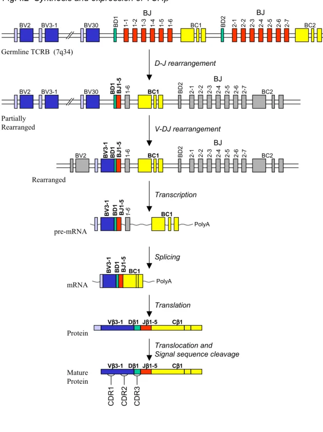
As an example of the gene rearrangement, Figure I.2 depicts the rearrangement of the TCRβ-gene locus. The first rearrangement to occur is the D-to-J joining reaction, followed by V-to-DJ rearrangement. The fully rearranged gene is transcribed into pre-messenger RNA, spliced and translated into polypeptide, which is co-translationally translocated into the endoplasmic reticulum where it folds, pairs with TCRα and is glycosylated. Nearly all of the antigen-binding diversity arises from three loops, named complementarity determining regions, or CDR1, CDR2, and CDR3, on the top of both the TCRα and TCRβ chains, and similarly on the Ig heavy and light chains. CDR1 and CDR2 are contained within, and therefore determined by, Vα, Vβ, VH or VL. CDR3 is
located at the V-(D)-J junction and is therefore the most diverse region, its diversity arising from junctional diversity and N-region diversity as well as from permutational diversity. In all TCR-MHC structures studied so far, CDR1 and CDR3 contact both MHC and MHC-bound peptide, while CDR2 contacts only MHC (Hennecke & Wiley 2001). In antibodies, all three regions can directly contact antigen (Janeway & Travers 2001).
I. C. T cells and the interaction of TCR with MHC-peptide complex I. C.1 Introduction to T cells and APCs
15 TCR molecules on T cells accomplish the critical goal of self/antigen
determination, but they are effective only if the antigen-presentation machinery presents a representative and reliable sampling of peptides. Thus, a large portion of the
responsibility for thorough surveillance falls on the antigen-presenting cells (APCs). APCs have divided this job into two categories, intracellular and extracellular sampling, and have specialized machinery to deal with each type. In nearly all cells of the body, cytoplasmic proteins are sampled by degradation via the proteasome complex, cathepsins or other proteases (reviewed in (Rock & Goldberg 1999)), and are transported into the ER by the TAP membrane channel, loaded onto class I MHC proteins by specialized machinery, and finally presented on their cell surface (see (Pamer & Cresswell 1998) for a review of these processes). The job of extracellular surveillance falls in the hands of a specialized set of APCs, made up primarily of macrophages, B cells, and dendritic cells. These cells circulate throughout the organism and phagocytose, endocytose and
pinocytose proteins, particles, viruses and microbes by a variety of mechanisms (including the PAMP-receptors of the innate immune system and the BCR on B cells, reviewed in (Watts 1997)), degrade proteins in their lysosomes (reviewed in (Villadangos & Ploegh 2000)), load the resultant peptides onto class II MHC proteins in endosomes by a specialized machinery (reviewed in (Busch et al 2000)), and present them at their cell surface for examination by T cells.
The cellular response required to combat intracellular and extracellular infection is quite different, and T cells are divided into two groups in order to respond properly to class I and class II MHC proteins. Antigenic (foreign) peptides bound to class I MHC molecules indicate that the presenting cell contains foreign peptides in its cytoplasm,
16 signaling infection. In most circumstances, the appropriate response is to kill such infected cells. In contrast, antigen peptides bound to class II MHC molecules indicate that an APC has encountered a foreign invader somewhere in the body. These cells should not be killed and, in fact, may be valuable to help the immune system locate the pathogen. As befits this logic, T cells restricted by (reactive with) class I MHC proteins are cytotoxic and bear the cell-surface glycoprotein CD8 (CD8+ T cells) which binds class I MHC proteins, whereas T cells restricted by class II MHC proteins are non-cytotoxic, and bear the cell surface marker CD4 (CD4+ T cells) which binds class II MHC proteins. CD4+ are alternately termed helper T cells. It should be noted that there exist exceptions to the above rules, and that a subset of CD4+ T cells appear to have some cytotoxic activity (Norris et al 2001; Porakishvili et al 2001; Suni et al 2001). The role of these cells is not well understood.
I. C.2 MHC allelic polymorphism
Every individual carries a set of MHC alleles inherited from each parent. In humans, these genes are carried on chromosome 6 and are termed HLA for Human Leukocyte Antigen, and in mice they are termed H-2 for group II histocompatibility antigens. Humans carry HLA-A, HLA-B, and HLA-C class I MHC genes, and HLA-DR, HLA-DP and HLA-DQ class II MHC genes. Mice carry H-2K, H-2D, H-2L, Qa and Tla class I MHC genes, and H-2 IA and H-2 IE class II MHC genes. Furthermore, MHC genes are highly polymorphic within all mammalian species. For example, within HLA-DR (the most highly expressed human class II MHC protein) there approximately 350 different alleles, which can be grouped into sixteen different families. The set of class I
17 and class II MHC alleles carried by an individual is referred to as his/her haplotype. The gene product of HLA-DRA*0101 and HLA-DRB1*0101, commonly termed HLA-DR1 or DR1, is carried by 10-20% of the human population and is the MHC studied in this thesis work.
I. C.3 Peptide binding to MHC molecules and recognition by TCR
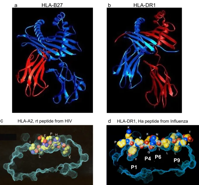
The role of MHC molecules in this immuno-recognition process is to bind a variety of peptides and to present them for examination to T cells. Figure I.3 shows examples of the two classes of MHC molecules, class I and class II. The modular structure of the two classes is similar, although the domain connectivity is different (Fig. I.3a,b). Each protein is composed of side-by-side immunoglobulin-superfamily domains (beta-sheet sandwiches) topped by a beta sheet bearing a pair of alpha helices. The peptide binding groove is formed between the alpha helices with the beta sheet as the bottom.
Class I MHC molecules bind peptides of 8-10 residues primarily based on their N- and C- terminal residues. These residues are buried into binding pockets at either end of the groove, and the intermediate residues are bulged up in the middle of the groove, available for examination by TCR (Fig. I.3c). The peptide binding motif of nearly all human and murine class I alleles is well understood, and protein sequences can be straightforwardly searched for class I binding peptides (Rammensee et al 1995).
In contrast, class II MHC molecules have an open-ended groove with several distinct pockets for side-chains (P1, P4, P6, P9). These pockets determine the peptide-binding motif of class II MHC molecules. In the case of HLA-DR1, P1 is the most
18 important pocket for determining specificity, binding large aliphatic or aromatic residues (Fig. I.3d). Peptide binding prediction algorithms for class II MHC molecules (Hammer et al 1994; Southwood et al 1998; Sturniolo et al 1999) have been less successful than for class I MHC molecules, but can nonetheless be an invaluable guide when attempting to identify antigenic peptides. The peptide residues not buried in these pockets are exposed and therefore available for examination by T cells (Fig. I.3d).
I. C.4 The transmembrane signaling mechanism of T cells
The engagement of cell-surface T cell receptor by cognate MHC-peptide complex on the surface of an adjacent APC triggers activation of the T cell. We have separately reviewed the activation mechanism of T cells (Cochran et al 2001a). I will briefly outline the relevant issues and comment on their importance in this thesis work (Cameron et al 2001c; Reth 2001).
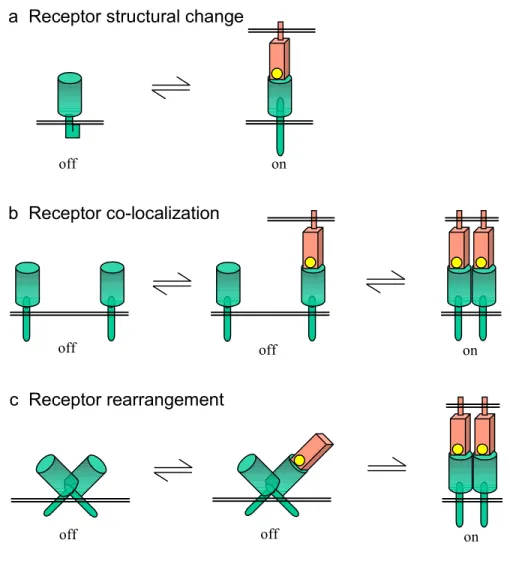
Several transmembrane signaling mechanisms are depicted in Figure I.4. In the first mechanism (Fig. I.4a), typical of seven transmembrane span receptors that activate G-proteins (Wess et al 1997), ligand binding induces a structural change in the receptor that can be sensed by effector proteins within the cell. In another mechanism (Fig. I.4b), binding of a multivalent ligand induces receptor co-localization. This mechanism is typical of receptor tyrosine kinases, in which receptor clustering facilitates
transphosphorylation by the cytoplasmic kinase domains (Hubbard & Till 2000). In a third mechanism (Fig. I.4c), ligand binding induces a rearrangement of a receptor oligomer into a specific activating conformation. One example of this mechanism is the
19 bacterial aspartic acid receptor, in which ligand binding induces a helix reorganization that activates receptor-associated cytoplasmic signaling proteins (Falke et al 1997).
The transmembrane signaling mechanism utilized by the TCR on T cells remains controversial (see Cochran, et al., 2001 for a review). Research on T cells until the 1980s focused on genetic and cellular identification and analysis of T cell immunity. In 1982, Jonathan Lamb and coworkers demonstrated activation of influenza-specific T cell clone (HA1.7) by adding chemically synthesized peptide (Ha) to B cells (Lamb et al 1982), capping decades of work demonstrating the interaction between MHC-peptide complex and TCR. But molecular analysis of T cell activation was hindered by the inability of researchers to produce soluble ligand for TCR. To circumvent this problem, researchers spent the next decade studying T cell activation by using anti-TCR antibodies (Kappler et al 1983; Kaye et al 1983; Yoon et al 1994), and chimeric TCRs carrying unrelated extracellular domains (Irving & Weiss 1991; Letourneur & Klausner 1991; Romeo & Seed 1991). These studies combined to show that intermolecular crosslinking of TCR cytoplasmic domains is sufficient to activate some (but not all) signaling pathways, including phosphorylation of tyrosines on the cytoplasmic tails of the CD3 subunits, recruitment of tyrosine kinases, and Ca++ mobilization.
In the 1990s the technology to produce soluble versions of some MHC and TCR molecules enabled a detailed molecular analysis of this interaction. Biochemical analysis of the revealed the weak (~10-4-10-6 M) and short-lived (t½ ~1-100 sec) nature
TCR/MHC-peptide interactions (Crawford et al 1998; Davis et al 1998). Studies utilizing soluble monomers or oligomers of MHC-peptide complexes to trigger T cells have thus far suggested that CD4+ T cells are activated by receptor dimerization
20 (Cochran et al 2000) or trimerization (Fig. I.4b) (Boniface et al 1998), whereas CD8+ T cells may be activated by MHC-peptide monomers (Delon et al 1998; Sykulev 1996). Although the monomer-activation results appear to suggest receptor structural change as the transmembrane signaling mechanism (Fig. I.4a), CD8 has been suggested to play a key role in the activation by monomers (Delon et al 1998), suggesting a mechanism similar to the receptor co-localization mechanism (Fig. I.4b) but utilizing two different receptors rather than two identical ones. In further support of the receptor co-localization model of T cell activation, structural analysis of a single TCR bound to MHC containing peptides of varying degrees of antigenicity (Ding et al 1999) failed to show any allosteric changes in TCR which might trigger intracellular events. It remains difficult to
conclusively rule out receptor rearrangement as a mechanism of T cell activation, although results from our lab indicating that MHC dimers in different orientations are similarly capable of activating CD4+ T cells (Cochran et al 2001b) argue against this model. We recently proposed a variant of this model in which multi-valent engagement of a pre-formed TCR oligomer disrupts a specific inhibited conformation (Cameron et al 2001c). This model will be discussed further in Chapter V.
Another approach to the study of T cell activation has been the use of confocal microscopy to study the real-time molecular organization of the T cell-APC contact region. Recent studies have shown a large-scale molecular segregation of proteins into distinct central and peripheral regions within ten minutes of initial cellular conjugation (Fig. I.5), termed c-SMACs and p-SMACs by one group (Monks et al 1998), and collectively labeled an immunological synapse by another (Grakoui et al 1999). This phenomenon has lead to the proposal of another type of T cell activation mechanism
21 based on the segregation of regional co-localization of various proteins based on ligand interactions, membrane domain localization, and steric crowding (van der Merwe et al 2000). The role that these large-scale molecular reorganization events play in T cell activation relative to the receptor oligomerization models described above remains controversial (see (Cochran et al 2001a) for a discussion), although recent work suggests that TCR triggering occurs upstream of molecular segregation (Zaru et al 2002).
Despite the remaining confusion regarding the transmembrane signaling mechanism of TCR, one result remains clear: TCR clustering (by either monoclonal antibodies or soluble oligomers of MHC-peptide) can trigger the some degree of activation in either CD4+ or CD8+ T cells (Boniface et al 1998; Cochran et al 2000; Irving & Weiss 1991; Letourneur & Klausner 1991; Yoon et al 1994). This is an important point for the work described in this thesis.
I. D. Placing the work of this thesis into context
The work presented in this thesis hinges on the use of a recently developed
reagent for the investigation of T cells, commonly called MHC tetramers, although in this work most often termed MHC oligomers. The most common use of MHC tetramers is to identify T cells of particular antigen-specificity, often by clinical researchers
investigating particular diseases. We have been among the first to use class II MHC oligomers on CD4+ T cells, and have used them in collaborations with several clinical researchers, as well as for probing activation characteristics of T cells. I will begin by reviewing techniques for the identification of antigen-specific T cells and finish with a brief discussion of the work presented in the following chapters.
22
I. D.1 Techniques of antigen-specific T cell identification
The identification and enumeration of antigen-specific T cells is extremely important in the study of diseased states in order to understand what the immune system is doing right, what it is doing wrong, and, potentially, how we might be able to help it work differently. For example, the difference between HIV-infected patients with progressive and non-progressive pathologies has been proposed to reside in the presence or absence of T cells with particular reactivities (Cohen 2001; Rosenberg et al 1997). Autoimmune diseases are widely thought to be aberrant recognition of self-antigens by the immune system (such as the hypothesized recognition of myelin basic protein in multiple sclerosis (Martin et al 1992)), although so far no such antigens have been incontrovertibly identified.
A variety of methods currently exist for the identification of antigen-specific T cells and are summarized in Table I.2. Several of these methods depend critically on the response of the T cells to activation. They fall into two categories representing the two hallmarks of T cell activation: the secretion of soluble factors, and cell proliferation. Depending on their subset, T cells secrete a variety of soluble proteins called cytokines when activated. These cytokines can be measured in bulk by enzyme-linked immuno-sorbent assay (ELISA). The actively secreting cells themselves can be directly detected by disrupting vesicle traffic with a metabolic inhibitor, fixing, permeabilizing and fluorescently staining the cells with anti-cytokine antibodies (intracellular cytokine staining, (Prussin & Metcalfe 1995)). Alternatively, the cytokines can be captured either on the cell surface (surface-capture cytokine staining, (Manz et al 1995)) or in the local
23 environment (ELISPOT assay, (Czerkinsky et al 1988)) using combinations of anti-cytokine antibodies either fluorescently labeled or enzyme-conjugated.
Bulk assay of lymphocyte proliferation has been routinely measured for several decades by the incorporation of tritiated thymidine in dividing cells (Strong et al 1973). More recently, a method utilizing limiting dilution of T cells followed by expansion and tritiated thymidine incorporation (limiting dilution analysis, (Merkenschlager et al 1988)) was developed to estimate the number of responding clones in the original mixed population. Finally, the ultimate proof of antigen-specific T cells is achieved by
establishing T cell clones by the isolation of single cells followed by massive expansion (Fathman & Fitch 1982), and subsequent demonstration of their functions and specificity directly.
However, none of the above methods can directly identify antigen-specific T cells, independent of their functional capacity. This is critical, as phenomena that might obscure or blunt T cell functions, including T cell tolerance, exhaustion, suppression, and anergy, have become increasingly important aspects of immunological research.
Other methods to detect antigen-specific T cells rely on the skewing of the TCR repertoire by monoclonal or oligoclonal expansions. Large expansions of T cells within TCR Vβ families can be detected using Vβ-specific monoclonal antibodies (Diu et al 1993). Similar data can be derived using semi-quantitative PCR utilizing distinct sets of Vα- or Vβ-specific oligonucleotide primers, combined with fine analysis of CDR3 lengths by high-resolution electrophoresis (spectratyping, (Pannetier et al 1993)). Finally, dominant clones can be visualized by PCR amplification, hybridization with similar but non-identical excess of carrier DNA, and non-denaturing DNA
24 electrophoresis (heteroduplex analysis, (Wack et al 1996)). These methods ignore low-frequency T cells, and identify T cells only by means of statistical comparison with typical distributions, rather than by direct identification based on specificity.
Several years ago a novel technique of antigen-specific T cell detection termed "MHC tetramer analysis" was developed by Altman and colleagues (Altman et al 1996) which has proved extremely popular in the analysis of CD8+ T cells. MHC tetramers are soluble versions of the MHC molecules, biotinylated at the C-terminus of one of the subunits, and subsequently oligomerized using fluorescent streptavidin. Although TCR/MHC-peptide interactions are typically weak (~10-4-10-6 M) with short half-lives (~1-100 sec) (Crawford et al 1998; Davis et al 1998), oligomers of MHC-peptide should have relatively high avidity and slow off-rates. Making these oligomers fluorescent turns them into convenient and sensitive probes of the TCR/MHC-peptide interaction. The technique of MHC tetramer staining of T cells has enabled the direct identification of antigen-specific CD8+ T cells in principle based on TCR specificity, and neither T cell function nor TCR sequence (McMichael & O'Callaghan 1998). The success of this technique (Callan et al 1998; Goulder et al 2000; Kuroda et al 1998; McHeyzer-Williams et al 1996; Moser et al 2001; Murali-Krishna et al 1998; Yee et al 1999) has led to the establishment of a NIH-sponsored facility that produces class I MHC tetramers and provides them free of charge to researchers around the world (see
www.niaid.nih.gov/reposit/tetramer/index.html). Only recently has this technology been extended to the use of class II MHC tetramers to study CD4+ T cells (Cameron et al 2001a; Crawford et al 1998; Kwok et al 2000; Meyer et al 2000; Novak et al 1999; Savage et al 1999).
25
I. D.2 Overview of thesis
This thesis describes the development and use of soluble fluorescent streptavidin-based oligomers of HLA-DR1 to identify and investigate antigen-specific CD4+ T cells.
Chapter II describes the production of soluble fluorescent streptavidin-based oligomers of the class II MHC HLA-DR1 and their use on CD4+ T cell clones. The experimental behavior of these reagents is characterized using fluorescent microscopy, biochemical experiments, and various inhibitors of cellular functions. In this chapter we conclude that the staining of some CD4+ T cells requires a cellular response and is not, therefore, function-independent, as the theory of MHC tetramers suggests. Jennifer R. Cochran contributed to this work, and Yassine-Diab Bader in the lab of Dr. Rafick Pierre-Sekaly provided a valuable short-term T cell line for this work. This chapter was
published in January 2001 as an article in the Cutting Edge section of the Journal of Immunology (Cameron et al 2001a).
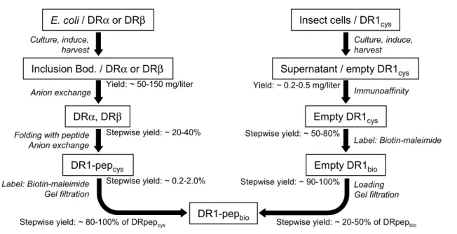
Chapter III describes in greater detail the method for production of biotinylated DR-peptide complex from E. coli and compares it to production from insect cell culture. DR oligomers are used to stain various CD4+ T cell clones and polyclonal lines and the temperature-dependence of staining these lines is analyzed. Finally, the use of these reagents as probes for T cell avidity modulation is discussed. Valuable reagents and advice were contributed to this work by Philip J. Norris and Eric S. Rosenberg at Mass. Gen. Hospital, Alka Patel and Lucy R. Wedderburn at the Institute for Child Health in London, Corinne Moulon at Dictagene in Switzerland, and Elizabeth D. Mellins at Stanford University. This chapter has been submitted to the Journal of Immunological
26 Methods for inclusion in a special issue devoted to MHC tetramers slated for publication in early 2002 (Cameron et al 2001b).
Chapter IV describes the use of MHC oligomers and IFN-γ surface-capture staining to identify CD4+ T cells specific for a peptide derived from influenza in samples of peripheral blood. The repertoire of TCRβ proteins utilized by these cells is found to be extremely broad, and the importance of these findings and directions for future research within the study of the TCR repertoire are discussed. This work was a close collaboration with George B. Cohen at Mass. Gen. Hospital, and has been prepared in the form of a manuscript for submission in the near future.
Chapter V describes other research in this laboratory to which I have contributed and discusses the implication of results for current models of T cell triggering. These studies (Cochran et al 2000; Cochran et al 2001b), performed primarily by Jennifer Cochran, utilized soluble oligomers of MHC-peptide complexes at precise valency to probe the triggering mechanism of CD4+ T cells. TCR dimerization was found to be the critical factor determining T cell triggering. The relative orientation of clustered TCR appeared to be irrelevant to the signal transduced, and TCR activation increased with shortened inter-TCR distances. I discuss the relation between these results and various models of transmembrane signal transduction. In particular, I propose a novel
mechanism of signal transduction in which the inhibitory conformation of an oligomeric receptor is disrupted by multivalent engagement. A portion of this chapter was
published as a Letter in Trends in Immunology (Cameron et al 2001c).
Chapter VI summarizes the conclusions of this thesis and discusses possible future directions of research based on the work presented here.
27 The Appendices include detailed protocols used or developed during my graduate career, tables of content for my research notebooks, and notes on various DNA constructs and cell transfectants.
Figure I.1. Antigen receptors of the adaptive immune response. Schematic diagrams of the subunit composition of B cell Receptor and T cell receptor and their cognate ligands. The precise stoichiometry of subunits in either receptor is unclear. The antigen-binding sites of Ig and TCRαβ are composed of CDR1, CDR2 and CDR3 loops and are highlighted in red at the top of each protein. (a) The B cell receptor is shown engaged with a microbe or virus. B cell receptor is composed of one membrane-anchored immunoglobulin molecule (Ig) responsible for binding antigen associated with Igα-Igβ heterodimer responsible for triggering intracellular signaling cascades. (b) The T cell receptor is shown binding to MHC-peptide complex presented by an adjacent antigen presenting cell (APC). T cell receptor is composed of one TCRαβ heterodimer responsible for antigen-binding associated with ζ-ζ homodimer and CD3δ-ε and/or CD3γ-CD3δ-ε heterodimers, collectively responsible for triggering intracellular signaling cascades. S-S APC T Cell Peptide from Microbe or Virus T cell receptor MHC Activation
b
B Cell Microbe or virus B cell receptor Igα-Igβ Ig Activationa
TCR α β ε γ δ ε ζ−ζ CD3 28Combini- Junctional N-region Somatic
Ig V D J torial Flexibility Diversity Mutation
κ light chain ~ 100 - 5 + + - + λ light chain ~ 100 - 6 + + - + heavy chain 75-250 30 6 + + + + TCRαβ α chain 46 - 50 + + + -β chain 48 2 13 + + + -TCRγδ γ chain 6 5 + + + -δ chain 8 3 4 + + + -# of gene segments
Table I.1. Sources of diversity in the B cell and T cell repertoires
Fig. I.2 Synthesis and expression of TCRβ 1-6 BC1 BD1 BJ1-5 BV 3-1 PolyA BC1 BD1 BJ1-5 BV 3-1 PolyA Cββββ1 Dββββ1 Jββββ1-5 Vββββ3-1 BV2 BV3-1 BV30 BJ BD 1 1-1 1-2 1-3 1-4 1-5 1-6 BJ 2-1 2-2 2-3 2-4 2-5 2-6 BD 2 BC1 2-7 BC2 BV2 BV3-1 BV30 BD1 BJ1-5 1-6 BJ 2-1 2-2 2-3 2-4 2-5 2-6 BD 2 BC1 2-7 BC2 BV2 BV 3-1 BD1 BJ1-5 1-6 BJ 2-1 2-2 2-3 2-4 2-5 2-6 BD 2 BC1 2-7 BC2 D-J rearrangement V-DJ rearrangement Transcription Splicing Translation Translocation and
Signal sequence cleavage
Germline TCRB (7q34) Partially Rearranged pre-mRNA mRNA Protein Mature Protein Rearranged CDR1 CDR2 CDR3 Cββββ1 Dββββ1 Jββββ1-5 Vββββ3-1 30
Figure I.2. Synthesis and expression of TCRβ. The diversity of the antigen-binding sites of Ig and TCRαβ are generated during somatic rearrangements of the Ig and TCR genes during lymphocyte development. TCRβ rearrangement and expression is shown as an example. In the first step, a D segment is joined to a J segment of its neighboring set. In the second step one of the V segments is joined to the DJ pair. In each case the intervening DNA is excised and discarded. The rearranged VDJ gene is transcribed with the adjacent C segment, spliced, translated into protein, translocated into the ER and paired with a surrogate TCRα chain. If during rearrangement there was an inappropriate frame shift or insertion of a stop codon, the gene product is unproductive and so either further rearrangement at this locus or at the locus site on the other TCRβ gene-bearing chromosome occur. If the rearrangements resulted in a productive VDJ combination, then rearrangements commence at the TCRα locus in a similar manner.
a HLA-B27 b HLA-DR1
P1
P4 P6 P9
d HLA-DR1, Ha peptide from Influenza
c HLA-A2, rt peptide from HIV
Figure I.3. Structures of MHC-peptide complexes. Peptide-binding MHCs are divided into class I and class II proteins. Structures determined by x-ray crystallography of examples from each class are shown. (a,b) The human class I MHC protein HLA-B27 (a) and human class II MHC protein HLA-DR1 (b) are shown without peptide looking down the peptide-binding groove. The lower two domains of each MHC are beta-sheet sandwiches and are members of the immunoglobulin-superfamily of protein domains. On top of these domains sits a beta-sheet, topped by an alpha-helix on either side of the peptide binding groove. In native proteins, transmembrane domains would extend from the C-termini of each MHC at the bottom of the proteins as they are shown here. (c,d) These images are 90 degrees rotated around the vertical axis of the images shown in (a,b). Surface topologies of just the upper peptide-binding domain are shown with space-filled representations of bound peptide. In (c) the class I MHC HLA-A2 is shown bound to a peptide derived from the reverse transcriptase protein of HIV. Note that the terminal peptide residues are buried in the MHC while the intervening residues are bulged upwards available for contact by T cell receptor. In (d) HLA-DR1 is shown bound to a peptide derived from the hemagglutinin protein of influenza. In contrast to the class I MHC-peptide complex, in the class II MHC the peptide binding groove is open-ended and the terminal peptide residues appear to make minimal contact with the MHC. Instead, peptide binding is determined by residues in several distinct pockets along the groove, labeled P1, P4, P6 and P9, and resides of the peptide in between these pocket-bound residues are available for contact by T cell receptor. Panels (c,d) are reproduced from Stern & Wiley, Structure, 1994.
a Receptor structural change off on off b Receptor co-localization off on off c Receptor rearrangement on off
Figure I.4. Mechanisms of transmembrane signaling. (a) Ligand-induced conformational change, typified by seven transmembrane span receptors that activate G-proteins. (b) Ligand-induced receptor co-localization, typified by the fibroblast growth factor receptor tyrosine kinase, which is activated by dimerization. (c) Ligand-induced allosteric rearrangement, typified by the bacterial aspartic acid receptor, in which ligand binding induces a helical reorientation.
10 µm 0.5 5 60 Time (minutes) MHC-pep
T-cell
TCR LFA-1 ICAM-1 a bFigure I.5. Large-scale membrane rearrangements during T cell activation. (a) Fluorescent microscopy of interaction between a T cell and a supported bilayer carrying labeled MHC-peptide complexes (green) and ICAM adhesion molecules (red). The view is normal to the membrane interface. Image excerpted from Grakoui, Science, 1999 with permission for
publication in Cochran, TiBS, 2001. Copyright 1999 American Association for the Advancement of Science. (b) Schematic diagram of the process visualized in (a) with appropriate color coding.
Table I.2. Methods to Detect Antigen-specific T cells
Property Technique Principle Notes
Proliferation Bulk Proliferation Assay T Cell Cloning Limiting Dilution Analysis
Cytokine Intra. Cytokine Staining Secretion Cytokine Surface-capture EliSpots TCR TCR Vbeta mAb Diversity Spectratyping Heteroduplex analysis TCR MHC "tetramer" staining Specificity
"Tetramers" bind to cell surface TCR and identify ag-specificity directly.
Depends on cytokine profile, activation state, and in vitro stimulation.
Difficult to determine %-antigen-specific in original sample. Assays all biased for clones which most easily proliferate in vitro .
Indirect; doesn't identify the specificity of the populations examined. Relatively insensitive. Spectratyping and Heteroduplex analysis are technically challenging. Extremely direct and facile assay. However, reagents difficult to obtain. Requires knowledge of antigenic peptides.
Detects, and Quantitates individual cells which secrete particular cytokines in response to APC-ag stimulation.
In vitro stimulation of T cells.
Probe for skewing of the normal TCR repertoire caused by monoclonal or oligoclonal expansion of cells.
36
II.
Detection of Antigen-Specific CD4
+T cells by HLA-DR1 Oligomers
Depends on the T cell Activation State
Class I MHC tetramers have proven to be invaluable tools for following and deciphering the CD8+ T-cell response, but the development of similar reagents based on class II MHC proteins for detection of CD4+ T cells has been more difficult. We evaluated fluorescent streptavidin-based oligomers of HLA-DR1 for use as reagents to analyze antigen-specific human CD4+ T cells. Staining was blocked at low temperatures and by drugs that disrupt microfilament formation and endocytosis. Cell-associated MHC oligomers were resistant to a surface stripping protocol, and were observed by microscopy in intracellular compartments. This behavior indicates that detection of CD4+ T cells using class II MHC oligomers can depend on an active cellular process in which T cells cluster and/or endocytose their antigen receptors. T cells of identical specificity but in different activation states varied greatly in their ability to be detected by class II MHC oligomers.
II. A. Introduction
Tetramers of class I MHC proteins have been shown to bind to antigen-specific CD8+
T cells with high specificity and sensitivity, thus providing a rapid in vitro assay for T-cell detection and enumeration that in principle depends on neither the activation nor
differentiation state of the T cell (McMichael & O'Callaghan 1998). The ability of class I MHC tetramers to detect low frequency antigen-specific T cells in mixed populations has led to increasing use of these reagents in studies of the immune response (McMichael &
37
In recent reports, MHC tetramer technology has been extended to the class II system (Crawford et al 1998; Kotzin et al 2000; Kwok et al 2000; Savage et al 1999). These reports used relatively high concentrations of tetramer (20 µg/ml), extended incubation times (1-3 hours), and elevated temperatures (22°C or 37°C). A requirement for such conditions would be surprising, given the avidity and fast on-rates expected for the oligomeric MHC-TCR
interaction (Davis et al 1998; Eisen et al 1996). This suggests that class II MHC tetramer staining may reflect a more elaborate process than simple association of a soluble reagent with surface receptors.
In the current study, we investigated the utility of oligomers of human class II MHC-peptide complexes in detection of antigen-specific CD4+ T cells. We show by flow cytometry that fluorescent oligomers of DR1 in complex with a peptide from influenza virus specifically stain two DR1-restricted, influenza-specific T cell clones and an antigen-specific polyclonal T cell line, and that the oligomers are internalized efficiently. Treatments that interfere with cytoskeletal rearrangements and endocytosis block class II MHC oligomer staining, showing that an active cellular process is required. Implications of these results for the use of class II MHC oligomers in detection of antigen-specific CD4+ T cells are discussed.
II. B. Materials and Methods II. B.1 Peptides
Ha [306-318] (PKYVKQNTLKLAT), TT [830-844] [QYIKANSKFIGITEL], A2 [103-114] (VGSDWRFLRGYHQYA), and TfR [680-696] (RVEYHFLSPYVSPKESP) were
synthesized using solid-phase Fmoc chemistry, and purified by C18 reverse-phase HPLC. All peptides bind tightly to DR1 with dissociation constants below 100 nM (Zarutskie et al 1999).
38
II. B.2 Preparation of labeled antibodies and streptavidin.
Murine monoclonal antibodies OKT3 or OKT4 (American Type Culture Collection) purified from hybridoma supernatant or streptavidin (Prozyme) were incubated with ten-fold molar excess FITC (Sigma-Aldrich), succinimidyl 6-(biotinamido) hexanoate (NHS-LC-biotin, Pierce), or N-(6-(biotinamido)hexyl)-3'-(2'pyridyldithio) propionamide (Biotin-HPDP, Pierce), at pH 7.5 for 3 hours at room temperature and isolated by gel filtration using Sephadex G-50 (Pharmacia). Streptavidin was pre-incubated with 2-hydroxyazobenzene-4’-carboxylic acid (HABA, Pierce Chemical Co.) before fluorescent labeling. R-phycoerythrin-conjugated streptavidin was purchased from Biosource, Inc.
II. B.3 Preparation of fluorescent class II MHC oligomers.
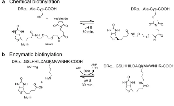
Soluble HLA-DR1 peptide complexes carrying an alpha subunit C-terminal cysteine (Cochran et al 2000) were reacted with excess maleimide-oxyethylene-biotin (PEO-maleimide activated biotin) or pyridyldithio-propionamide-biotin (HPDP-biotin, Pierce Chemical Co.) and isolated by gel filtration in phosphate buffered saline (PBS), pH 7.0. Oligomers were formed by stepwise addition of streptavidin-R-phycoerythrin conjugate (PE, Biosource, Inc.). SA-PE from other sources were tested and found to label cells less brightly. FITC-labeled
tetramers were isolated by gel filtration using sequential SEC-3000 (Phenomenex) and
Superdex 200 (Pharmacia) columns. Streptavidin absorbed to the gel filtration matrix, but the DR-saturated SA-FITC tetramer eluted at its expected molecular weight.
39
T cell clones HA1.7 and Cl-1 were cultured as described (Cochran et al 2000), and rested six or more days before use. A short-term polyclonal CD4+ T cell line was raised by repeated in vitro stimulation of CD8-depleted PBMCs from a DR1-homozygous donor using autologous mitomycin C-treated PBMCs in the presence of 1 µM Ha peptide.
II. B.5 Flow cytometry.
T cells (~107/ml) were mixed with DR1-peptide oligomers for 3-5 hrs. at 37°C, chilled for 5 min., supplemented with secondary antibodies for 30 min., and washed twice with cold wash buffer (PBS, 1% fetal bovine serum, 15 mM sodium azide). Inhibitors (stock solutions in DMSO, ethanol or PBS; final concentration of solvent ≤ 0.5%) were pre-incubated with cells in PBS for 1 hour, after which the cells were stained with oligomer as above. In cell surface stripping experiments, chilled and washed cells were resuspended in wash buffer containing 25 mM 2-mercaptoethanesulfonic acid (βME-SO3-), incubated for fifteen minutes at 37°C, and
then washed twice with cold wash buffer. In inactivation experiments, cells were pretreated overnight in complete medium with peptide or with immobilized OKT3 (5 µg/ml in PBS, 2hrs), and then were stained as above.
II. B.6 Fluorescence microscopy.
Live HA.1.7 T cells were isolated using a Ficoll gradient and incubated with 10 mg/ml FITC-dextran (average MW 10 kDa, Sigma-Aldrich) and 70 µg/ml DR1-Ha SA-PE oligomer, or 35 µg/ml SA-PE alone, for three hours at 37°C, seeded onto cold Cell-Tak (Becton
40
(Electron Microscopy Sciences), and visualized using a DeltaVision digital deconvolution microscope system (Applied Precision Inc.).
II. B.7 Dynamic light scattering.
Measurements were made at 22°C using a Protein Solutions DynaPro-MS/X dynamic light scattering instrument. Protein samples were filtered through 0.2 micron spin filters (Costar) before analysis. All samples were measured at two different protein concentrations and with identical results. Molecular weight equivalents (Da) were estimated from
hydrodynamic radii (RH, nm) using an empirical model for globular proteins, log MW = 2.426
log (1.549xRH), as recommended by the manufacturer.
II. C. Results
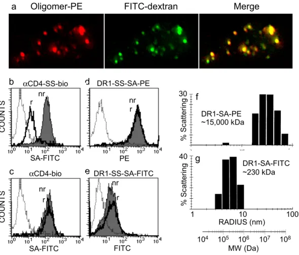
II. C.1 DR1 oligomers detect antigen-specific CD4+ T cells.
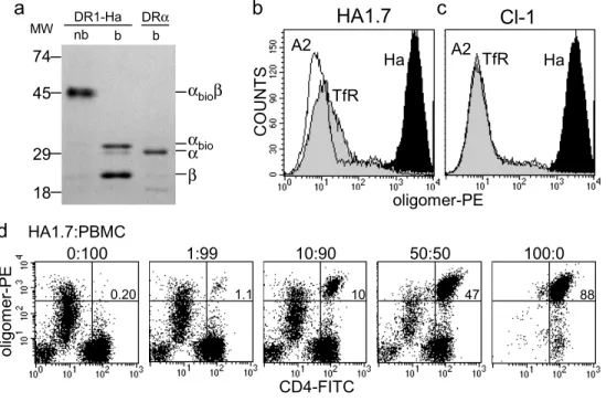
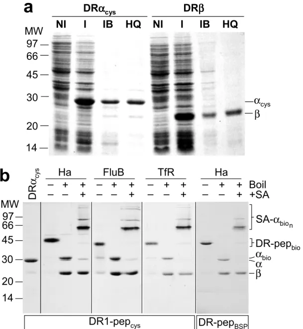
The oligomerization strategy used in this work relies on biotin covalently coupled to a cysteine residue at the C-terminus of the HLA-DR1α subunit (Cochran & Stern 2000), with subsequent oligomerization using streptavidin. Soluble DR1-peptide complexes, folded in vitro from subunits expressed in E. coli (Frayser et al 1999), were biotinylated at the introduced cysteine with >90% efficiency, using a maleimide reagent carrying biotin at the end of a 29 Å hydrophilic linker (Fig. II.1a). The DR1-peptide complexes were resistant to SDS-induced chain dissociation at room temperature, indicating quantitative peptide loading (Fig. II.1a). For routine use, biotinylated DR1-peptide complexes were oligomerized with streptavidin
conjugated to R-phycoerythrin. Such DR1-SA-PE oligomers carrying the Ha peptide exhibited antigen-specific binding to two DR1-restricted, Ha-specific, T cell clones, HA1.7 (Lamb et al
41
1982) and Cl-1 (De Magistris et al 1992) (Fig. II.1b,c). Oligomers carrying unrelated peptides showed no significant binding (Fig. II.1b,c; A2 and TfR traces). To confirm the specificity of oligomer binding we analyzed a series of dilutions of the HA1.7 clone mixed into non-specific peripheral blood mononuclear cells (PBMCs). Monocytes present in the PBMC mixture exhibited non-specific oligomer binding, but could be distinguished by their CD4med phenotype (Fig. II.1d). The fraction of T cells staining with the DR1-SA-PE oligomers correlated closely with the fraction of antigen-specific cells in the mixture, highlighting the specificity of staining (Fig. II.1d).
II. C.2 Oligomer staining is observed only under conditions permissive for internalization.
We examined the experimental conditions necessary for oligomer staining of the T cell clones. Both clones required relatively high oligomer concentration (Fig. II.2a), with Cl-1 exhibiting saturating staining intensity at >300 µg/ml (∼0.6 µM), while for HA1.7, staining did not appear to saturate even at 750 µg/ml (∼2 µM). Staining intensity increased with increasing temperature for both clones (Fig. II.2b). At 4°C, the temperature usually employed for antibody staining, no staining was observed for either clone. The staining signal developed slowly, and continued to increase for at least five hours after addition of oligomer (Fig. II.2c). These concentration, temperature, and time requirements for efficient staining with these reagents are consistent with those described by other researchers using MHC class II oligomers (Crawford et al 1998; Kotzin et al 2000; Kwok et al 2000). Our typical staining protocol employs 20-50 µg/ml oligomer reagent for 3-5 hours at 37°C.
The elevated temperature and extended time required for efficient staining suggested that a metabolic process might be involved. We investigated the staining of T cells by
DR-SA-42
PE oligomers after treatments that interfere with various normal cellular functions (Fig. II.2d). Treatments that inhibited conventional T cell signaling pathways, including genistein (Akiyama et al 1987), staurosporine (Tamaoki et al 1986), PP2 (Hanke et al, 1996), and methyl-
β-cyclodextrin (Scheiffle et al, 1997), weakly inhibited staining or had no effect. Agents that disrupt endosomal proteolysis, such as chloroquine, NH4Cl, and monensin, had little or no
effect. However, staining was substantially blocked by treatments that interfere with endocytosis and cytoskeletal rearrangements, including the microfilament-disrupting drugs latrunculin A (Spector et al 1989) and cytochalasin D (Goddette & Frieden 1986), the phosphatase inhibitor phenylarsine oxide (Lazari et al 1997), sodium azide (80mM), and paraformaldehyde fixation. T cell receptors are efficiently internalized following engagement by cell-surface MHC-peptide complexes on other cells (Itoh et al 1999; Valitutti et al 1995). Since MHC oligomer staining was blocked by treatments which block endocytosis, we
postulated that oligomers might be internalized along with TCR during the staining protocol.
II. C.3 Bound class II MHC oligomers are present in internalized compartments.
To test whether MHC class II oligomers were internalized after binding, we performed fluorescence microscopy on HA1.7 T cells after incubation with DR-SA-PE oligomers.
Oligomer staining was detected in intracellular compartments (Fig. II.3a, red), co-localized with endocytic compartments as visualized by FITC-dextran (Fig. II.3a, green). Cells incubated with SA-PE reagent alone (without MHC) showed normal FITC-dextran internalization but no detectable PE signal (not shown). These results show that MHC class II oligomers are internalized efficiently by T cells.
43
We used a surface stripping protocol to evaluate the relative contributions of
internalized and surface-bound oligomers to the staining signal observed by flow cytometry. A disulfide bond was introduced into MHC tetramers between the DR1 and biotin moiety (DR1-SS-SA-PE), to allow cleavage by the membrane-impermeant reducing agent 2-mercaptoethane sulfonate (βME-SO3-). DR1-SS-SA-PE oligomer staining of HA1.7 T cells was not affected by
the βME-SO3- treatment (Fig. II.3d), indicating that cell-associated oligomers were not present
at the cell surface. To demonstrate the effectiveness of the stripping protocol, we examined HA1.7 T cells that had been surface-stained with an antibody carrying the cleavable linker (αCD4-SS-bio). In this experiment, staining was limited to the cell surface by incubation at 4°C. Fluorescent streptavidin was efficiently stripped from the –SS-bio antibody by the βME-SO3- treatment (Fig. II.3b), but not from a non-cleavable biotinylated antibody (Fig. II.3c). The
insensitivity of the oligomer-binding signal to the βME-SO3- surface-stripping protocol, and the
lack of surface staining observed by fluorescence microscopy, both indicate that essentially all of the oligomers associated with T-cell staining are present in internal compartments.
One possibility for the correlation between MHC oligomer staining and internalization might be a low valency in preparations of DR1-SA-PE. The actual oligomeric state of MHC-SA-PE oligomers has not been reported. Moreover, physical characterization is difficult due to the large size of the PE moiety (~250 kDa) and heterogeneous crosslinking in commercial preparations of SA-PE. To obtain MHC oligomers with a well-defined valency, we prepared oligomers using SA labeled with FITC. Because several preparations of commercially available SA-FITC conjugates exhibited sub-stoichiometric biotin-binding capacity, we developed a strategy to prevent damage to the biotin-binding sites during FITC labeling, by protection of the biotin-binding sites using the weakly binding biotin analog HABA (see
44
Methods). DR1-SA-FITC oligomers prepared with this reagent exhibited an average molecular weight (230 kDa) consistent with one SA (60 kDa) and four DR1 (45 kDa), indicating that the desired tetrameric species had been formed (Fig. II.3g). Gel filtration analysis gave a similar result (265 kDa, data not shown). By contrast, DR1-SA-PE oligomers exhibited a heterogeneous population distributed around MW 15,000 kDa (Fig. II.3f), indicating that they are composed of multiple SA and/or PE moieties; they are either large oligomers or non-covalent aggregates, and properly they should not be referred to as “tetramers.” T cell staining by the defined DR1-SA-FITC tetramers (Fig. II.3e) was considerably less intense than that observed for the DR1-SA-PE oligomers (Fig. II.3d), even after expression of the staining results in terms of numbers of fluorophores bound per cell (data not shown). Nonetheless, the observed staining still required elevated temperature and was insensitive to surface stripping by βME-SO3- (Fig. II.3e). Thus, the observed association of staining and endocytosis was not due
to a reduced valency in the DR1-SA-PE oligomers.
To characterize the internal compartment associated with MHC class II staining, we made
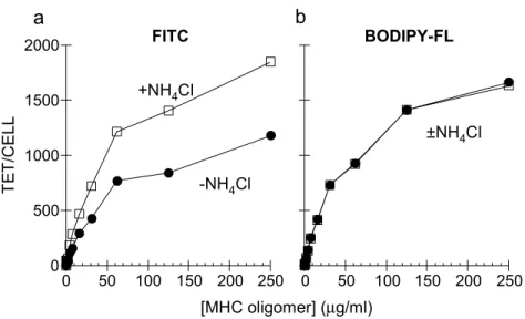
use of the differential pH sensitivity of two fluorescent dyes with similar spectral characteristics, FITC and Bodipy-FL
(4,4’-difluoro-5,7-dimethyl-4-bora-3a,4a-diaza-s-indacene-3-propionic acid). FITC conjugates show reduced staining intensity at lower pH (for example exhibiting 60% reduction from pH 7.2 to pH 6.0) (Yguerabide et al, 1994), whereas Bodipy-FL conjugates are insensitive to pH in the range 2-10 (Karolin et al, 1994). We stained HA1.7 T cells with DR1-Ha tetramers labeled with either FITC or Bodipy-FL, and converted the fluorescence intensities to number of tetramers bound per cell, to account for differences in the intrinsic fluorescent yield of the dyes. The curve describing the binding of the FITC-labeled tetramers (Fig II.4a) was significantly lower than that for the Bodipy-FL tetramers (Fig. II.4b).
45
Addition of NH4Cl increased the FITC signal to a level comparable to the Bodipy-FL signal
(Fig. II.4a, open squares), but had no effect on the Bodipy-FL signal (Fig. II.4b, open squares). Both the reduced intensity of the FITC signal relative to that for Bodipy-FL, and the NH4Cl
sensitivity of the FITC signal, indicate that MHC tetramers are internalized to acidic compartments.
II. C.4 A polyclonal T cell line contains subpopulations with different requirements for staining.
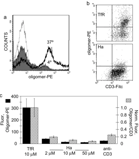
To test the generality of the observation that internalization is required for staining with
MHC class II oligomers, we tested a polyclonal T cell line restricted by HLA-DR1 and specific for the Ha peptide. Oligomer staining experiments were performed in parallel at 37°C and at 4°C (Fig. II.5a). At 37°C, 67% of the total polyclonal cell population exhibited DR1-SA-PE staining (PE +), which was specific for the appropriate peptide. By contrast, only 12% of the cells were PE + when stained at 4°C, and these exhibited ~2-fold reduced intensity. Both the minor PE+ population detected at 4°C and the major population detected at 37°C were
oligoclonal, as shown by TCR Vβ3 and Cβ1 analysis (not shown), and both had characteristics of CD4+ memory T cells (CD3+, CD4+, CD8-, CD25+, CD45RO+ and CD62L-). These results show that most of the polyclonal T cells share with HA.17 and Cl-1 the requirement for an active cellular process to observe MHC oligomer staining, although some cells can stain in the absence of such processes.
46
T cells can enter a non-responsive or anergic state in response to a partial activation stimulus (Schwartz et al, 1996). We tested whether such treatments would effect T cell staining by MHC oligomers. Treatment with high concentrations of antigenic but not control peptides (Lamb et al, 1983), or with immobilized anti-CD3 (Jenkins et al, 1990), each dramatically reduced oligomer staining of the polyclonal T cell line (Fig. II.5b). TCR surface expression was reduced somewhat by these treatments (Fig. II.4c), but the effect was much smaller and accounted for less than 10% of the overall reduction in oligomer staining (Fig. II.5b). These results show that MHC oligomer staining of T cells is dependent on the T cell activation state.
II. D. Discussion
The results presented here show that bound HLA-DR1 oligomers are internalized efficiently by two antigen-specific T clones. Oligomer staining was blocked by low temperature, and by endocytosis inhibitors, suggesting that an active process requiring
cytoskeletal rearrangement was required. Essentially all of the fluorescence of oligomer-stained T cells resulted from internalized oligomers, and cell-surface staining was not detected. For a polyclonal CD4+ T cell line, most cells exhibited the same behavior as the T cell clones, with staining blocked at 4°C. (A sub-population of CD4+ T cells capable of staining at 4°C was
present, consistent with the observations of other investigators, W. Kwok and G. Nepom, personal communication). Staining was greatly reduced by treatments that anergize or inactivate T cells. These results indicate that class II MHC oligomer staining of CD4+ T cells require active T-cell processes, and depends on the T cell activation state.
This behavior is not likely to be limited to the particular MHC-TCR interaction