Saccharomyces cerevisiae
by
Vida Praitis
B.A., Biology. Swarthmore College, 1988
Submitted to the Department of Biology in Partial Fulfillment of the Requirements for the Degree of
Doctor of Science in Biology at the
Massachusetts Institute of Technology
June 1995
©1995 Vida Praitis. All rights reserved.
The author hereby grants to MIT permission to reproduce and to
distribute publicly paper and electronic copies of this thesis
document in whole or in part.
Signature of
Author ...
Department of Biology
Certified
by... ...
... Professor Frank SolomonThesis Supervisor
Accepted
by ... ... ...
Professor Frank Solomon
Chairman, Graduate Thesis Committee
Science
MASSACHIJSETTS INSTITUTE OF TF mF.lil tyV 'JUN 06 1995
Saccharomyces cerevisiae
by
Vida Praitis
Submitted to the Department of Biology on May 5, 1995 in partial fulfillment of the requirements for the Degree of Doctor of Science in Biology
Abstract:
Microtubules, involved in a number of critical, diverse cellular structures and functions, are composed primarily of two related but non-identical, highly conserved subunits, alpha- and beta-tubulin. The precise secondary and tertiary interactions of the tubulin subunits are still poorly understood, likely because native alpha- or beta-tubulin has not been purified in large enough quantities to perform structural or biochemical analysis. Crystal structures, which would provide extensive structural information about these molecules and their interactions, have not been reported. I sought to characterize the alpha- and beta-tubulin polypeptides using two approaches, one genetic and one biochemical.
E. Raff's laboratory characterized a series of mutations in the testes-specific beta-tubulin of D. melanogaster. Strains homozygous for B2t8 exhibited an intriguing phenotype. All microtubule structures were disrupted, with S- or U-shaped, rather than O-shaped microtubule cross-sections. The phenotype suggested a defect in packing within the microtubule polymer. Sequence analysis revealed a glutamic acid to lysine substitution at highly conserved position 288. I generated the same mutation in the sole beta-tubulin of S.
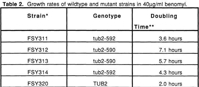
cerevisiae. Phenotypic analysis revealed no defects in growth at several temperatures, mating, sporulation, or germination. The only phenotype was a slight alteration in the sensitivity to the anti-mitotic drug benomyl. These results demonstrate that microtubules are differentially susceptible to in vivo growth conditions or alterations in the beta-tubulin primary sequence.
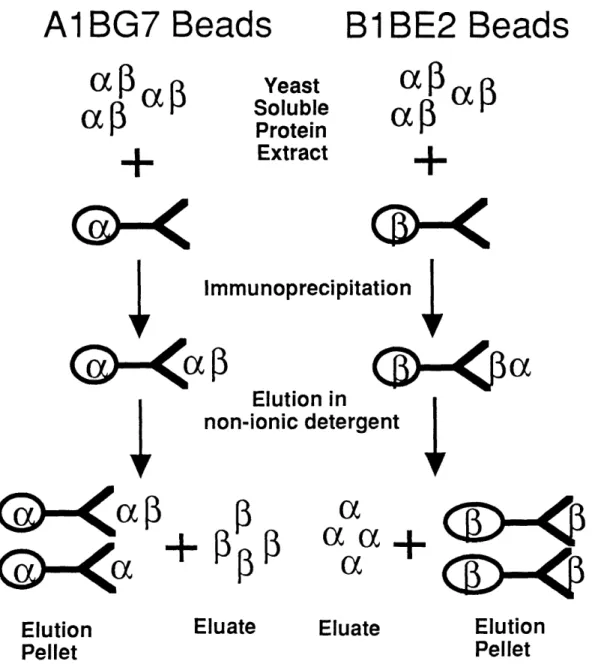
To examine the properties of the individual tubulin subunits biochemically, we developed a procedure to enrich for alpha- or beta-tubulin in the absence of
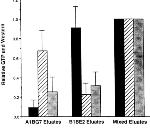
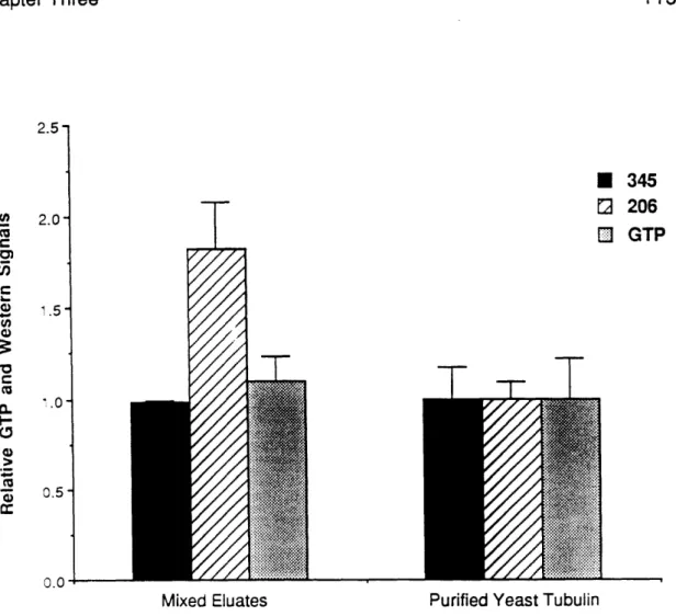
its heterodimeric partner. Co-immunoprecipitated alpha- and beta-tubulin dissociated when exposed to low concentrations of non-ionic detergents. GTP inhibited the detergent-mediated separation of tubulin heterodimer. Relatively pure alpha- and beta-tubulin was tested for its ability to bind GTP, using a photo-cross-linking assay. Native levels of GTP binding could be restored in the eluates when -both tubulin subunits were present in equimolar amounts,
demonstrating both chains contribute to tubulin GTP binding. Thesis supervisor: Dr. Frank Solomon
Education:
1988-1995 1984-1988
Ph.D., Biology. M.I.T. Advisor: Dr. Frank Solomon.
B.A., Biology, with Distinction. Swarthmore College, PA.
Publications:
Vida Praitis, Laurie Connell, Brant Weinstein, and Frank Solomon, 1995. "GTP Binding Involves Both a- and -tubulin Polypeptides" in preparation.
Solomon, F., S. Guenette, D. Kirkpatrick, V. Praitis, B. Weinstein, and J. Archer. 1992. A Genetic Analysis of Microtubule Assembly and Function in Yeast. in Chromosome Segregation and Aneuploidy, Ed. B. Vig. Springer-Verlag, Berlin. 199-210
Solomon, F., L. Connell, V. Praitis, B. Weinstein, and D. Kirkpatrick. 1991. "Methods for Studying the Cytoskeleton in Yeast." The Cytoskeleton. A Practical Approach. Eds. K.L. Carraway and C.A.C. Carraway. IRL Press, Oxford.
Vida Praitis, Wendy Katz, and Frank Solomon. 1991. "A Codon Change in 3-Tubulin Which Drastically Affects Microtubule Structure in Drosophila Fails to Produce A Significant Phenotype in S. Cerevisiae." Molecular and Cellular Biology, 11:4726-4731.
Professional/Academic Societies and Awards:
American Society for Cell Biology, Sigma Xi, and Phi Beta Kappa. National Merit Finalist. Lawrence LaFore Scholar..
Research Experience
1986 Rocky Mountain Biological Laboratory. Gothic, CO.
Field/laboratory work on yellow-bellied marmot diets (Summer). 1985-1987 AT&T Omaha, NE. Chemistry laboratory technician. (Summers).
Office of Minority Affairs . Biochemistry Tutor. M.I.T. (Spring). Developmental/Cell Biology teaching assistant. M.I.T. (Spring). Human Physiology teaching assistant. M.I.T. (Fall).
Teaching Experience
1993 1992
I owe thanks to a number of people without whom this dissertation would have been impossible to accomplish. First, I would like to thank all the current and former members of the Solomon laboratory. Group meetings and informal discussions in the lab and coffee room about research, politics, movies, and life have been wonderful. Thanks also to the members of my MIT class: you made first year, and subsequent years, fun. In particular, thanks to David Litwack and
Karen Hicks for scientific advice and fun conversations.
I would also like to thank the members of my thesis committee, Chris Kaiser, Bob Sauer, and Terry Orr Weaver for their intellectual contributions to my research.
Thanks to my collaborators on various research projects. Wendy Katz taught me everything I needed to know about yeast but was afraid to ask. Brant Weinstein and Laurie Connell made the quagmires associated with tubulin
splitting and GTP binding bearable.
To my bay mate Bettina Winckler: I have tremendously appreciated your sense of humor, intellect, and in particular, your patience. Our discussions of your experiments, (13H9 stains the upper band and 904 stains the lower band; phosphorylation?; and shoot-outs) and my experiments ("This is a beta blot of alpha beads which have beta. This is an alpha blot of beta beads that have alpha") were always confusing initially, but edifying eventually. I remember them with joy. Thank you.
To Charo Gonzalez-Agosti: Charo, you have managed to restore my sanity on more occasions than I care to recall. You have given excellent advice, about both science and life. Thank you.
To Margaret Magendantz: Margaret, thank you for providing me with advice, protocols, reagents, and information over the years. The lab would not run smoothly without you. To Wilma Wasco, thank you for teaching me a number of the "tricks" in molecular biology protocols. Do we really need to boil protein preps a second time? To Suzanne Guenette, thanks for all the wonderful scientific discussions over dinner or on the way to aerobics.
To my advisor Frank Solomon: I admire your intellectual toughness and high scientific standards. You taught me to critically examine the intellectual and the experimental constructs of research and provided me with the tools to pursue a scientific career.
Last but not least, I would like to thank my family -- Aciu labai daug. Their support has provided me with the confidence to pursue my dream.
I dedicate this dissertation to my husband Roger, for his love and friendship. S.B. cubed.
Table of Contents
Characterization of the Alpha- and Beta-tubulin
Polypeptides in Saccharomyces cerevisiae 1
Abstract 3
Biography 5
Acknowledgments 7
Table of Contents 9
Chapter One: Introduction 13
Introduction 15
Microtubule Function and Theory 15
Microtubule Function 15
Models of Microtubule Functional Characteristics 16
Multi-tubulin Hypothesis 17
Dynamic Instability Hypothesis 18
Microtubule-associated Protein Hypothesis 19
Structural and Biochemical Properties of Tubulin 20
The Tubulin Genes 20
Alpha- and Beta-Tubulin Isoforms 20
Mutation analysis 22
Regulation of Tubulin mRNA 23
Tubulin Protein Folding 26
Structure and Biochemistry of Tubulin 28
Tubulin Protein Structure 28
The Tubulin Heterodimer 31
Heterodimer Dissociation 33
Buffer Components 34
Interactions with Small Molecules 38
Post-Translational Modification of Tubulin 43
Microtubule Structure 44
Formation of Microtubules 44
Polymerization and Depolymerization 46
Role of GTP Hydrolysis 47
Chapter Two: A codon change in -tubulin which drastically
affects microtubule structure in Drosophila fails to
produce a significant phenotype in yeast 67
Abstract 67
Introduction 69
Materials and Methods 70
Plasmid constructions 70
Strains and media 70
Genetic Techniques and Transformation 71
Immunofluorescence 71
Protein Blotting 72
Growth Rate Analysis 72
Southern Blots 72
Results 74
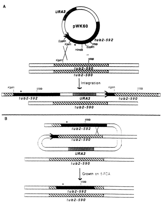
Construction of a mutant TUB2 gene 74
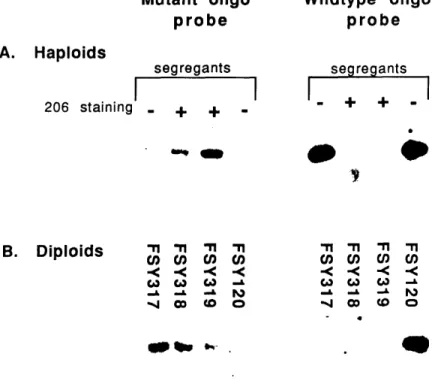
tub2-592 transformants contain the glu to lys change at
position 288 74
Phenotypic Analysis of tub2-592 76
Growth, sporulation, germination, and mating. 76
Temperature and cold sensitivity. 79
Benomyl sensitivity. 79
Discussion 83
Acknowledgments 85
References 86
Chapter Three: GTP Binding to Tubulin Involves both ao- and
13-Tubulin 91
Abstract: 91
Introduction 93
Materials and Methods 94
Monoclonal Antibody Generation 94
Yeast Protein Preparations 95
Immunoprecipitations and Elutions 95
Protein Quantitation, Western Blotting, and Silver staining 96
GTP Binding 97
Results 98
Discussion 115
Acknowledgments: 117
Chapter Four: The Detergent-Mediated Separation of a-and B-tubulin: Experimental Parameters
Introduction
Materials and Methods
Strains, Media, and Culture Conditions Monoclonal Antibody Generation
Preparation of yeast total soluble protein extracts Immunoprecipitation of tubulin and isolation of tubulin
monomers.
Protein Quantitation, Western Blotting, and Silver-staining Results
I mmu noprecipitation
Preparation of Yeast Soluble Protein Extracts Monoclonal Antibody Preparations
Immunoprecipitation Buffer Time of Binding
Saturation of Antibody Binding Sites Elution
Buffer Composition Volume of Eluant
Elution Incubation Time Purity and Quantity
Discussion References
Chapter Five: Conclusions
References
Appendix
Introduction
Materials and Methods
Strains, Media, and Culture Conditions Monoclonal Antibody Generation
Preparation of yeast total soluble protein extracts Immunoprecipitation of tubulin and isolation of tubulin monomers.
SDS and Native Gel Western Blotting GTP cross-linking assays
Co-immunoprecipitations Results
Native gel eletrophoresis
Co-lmmunoprecipitation experiments GTP Binding Discussion References 123 125 126 126 126 127 128 129 130 135 135 135 136 136 137 141 141 147 150 152 166 169 173 180 183 185 186 186 186 186 187 187 188 189 190 190 194 197 203
205
Introduction
Microtubules are involved in a vast number of critical and complex cellular functions. The biochemistry and structure of the tubulin heterodimer and microtubules have been examined for more than thirty years, yet much remains to be explored. This introduction examines a number of hypotheses and experiments from the literature exploring tubulin biochemistry and structure. The intention is to provide a historical and experimental framework for the research described in later chapters of this dissertation.
Microtubule Function and Theory
Microtubule Function
Microtubules are polymeric structures, composed primarily of alpha- and beta-tubulin, present in a number of cellular structures. They are involved in several critical cellular processes which can be broadly characterized in four ways. First, they provide structural rigidity required to specify a cell's characteristic shape, including that of highly specialized cells such as neurons and erythrocytes. Second, microtubules are required for the structural rigidity of cellular structures, such as the mitotic spindle. Third, they provide a structural matrix upon which critical materials are transported within cells. Finally, they produce structures required for cell motility, such as sperm axonemal complexes and flagella.
The vast functional capacity of microtubules requires they have several almost contradictory intrinsic properties. First, within a single microtubule
structure a number of different microtubule-mediated events occur
simultaneously. Microtubules must either be multi-functional as a population, or they must contain specialized microtubules within a population, each capable of performing a single or few tasks. Second, microtubule structures reorganize, often quite rapidly, in response to changes in cellular requirements or as cells differentiate. Yet once microtubules are assembled, they must be structurally
The events in one microtubule-mediated event, mitosis, provide a number of good examples of microtubule functional complexity. Microtubules are the primary structural components of the mitotic spindle, responsible for the accurate and reproducible separation of chromosomes during nuclear division. The transition from interphase to mitosis requires a dramatic reorganization of microtubules, necessitating that the microtubules be capable of radically and rapidly reorganizing in response to cellular signals. During mitosis itself, some microtubules reorganize while others appear fairly stable. Within the mitotic spindle, microtubules provide the rigid framework of the mitotic spindle. Other microtubules interact, directly or indirectly, with chromosomal material to align chromosome pairs at the metaphase plate. Only after the correct pairing of the chromosomes, microtubules mediate the poleward separation of chromosomes at anaphase. Within the mitotic spindle, complex microtubule-mediated events are occurring simultaneously.
There are a number of other excellent examples of microtubule functional diversity within a single specialized microtubule structure. Clearly, microtubules are capable of performing a number of varied and complex roles. How these
polymers perform these complex tasks has been a critical question in cell biology. The question is complicated by the composition of microtubules; they are primarily composed of two subunits, alpha- and beta-tubulin, which interact to form a heterodimer. Alpha- and beta-tubulin proteins are highly homologous to one another within a species and each is quite similar to alpha- and beta-tubulins across highly divergent species (Little and Seehaus, 1988; MacRae and Langdon, 1989; Raff, et al., 1987). In addition, the basic physical requirements for a microtubule polymer are contained within the tubulin heterodimer, since purified alpha- and beta-tubulin heterodimer has the capacity to polymerize (Lee and Timasheff, 1975). A critical question then arises. How can such a seemingly simple system account for the diversity and complexity in microtubule structure and function?
Models of Microtubule Functional Characteristics
Three major models which are not mutually exclusive have emerged to explain the ability of microtubules to participate in a diverse array of structures
Hypothesis, and The Microtubule-associated Protein Hypothesis. I will briefly describe each of these models.
Multi-tubulin Hypothesis
The Multi-tubulin Hypothesis, first described prior to the identification and characterization of tubulin primary sequences, proposed that microtubule functional diversity was possible because of the presence of multiple isoforms of the primary subunits of microtubules, alpha- and beta-tubulin (Fulton and Simpson, 1976; Stephens, 1975). A specialized isoform of alpha- and/or beta-tubulin was required for the establishment of a specific cellular microtubule-structure. As a consequence, isoforms of alpha- and beta-tubulin would be spatially and developmentally restricted.
As research on tubulin isoforms advanced, it became clear that each isoform of alpha- and beta-tubulin expressed in a cell co-localized, and was present in both general and specialized microtubule structures (Lewis, et al., 1987). However, expression of some isoforms was restricted to certain cell types. The Multi-tubulin Hypothesis was modified to address these results (Reviewed In: (Cleveland, 1987; Joshi and Cleveland, 1990; Katz and Solomon, 1989; MacRae and Langdon, 1989; Solomon, 1991)). The hypothesis contended that only specialized isoforms could participate in all cellular microtubule structures, including specialized microtubule structures such as the erythrocyte marginal band. In contrast, abundant isoforms could participate only in general microtubule structures. Implicit in this model was the notion that conserved regions of alpha- and beta-tubulin primary sequence were required for conserved functions while divergent regions were required for divergent microtubule functions.
There are numerous alternatives to the Multi-Tubulin Hypothesis that explain the presence of spatially and temporally expressed multiple isoforms of alpha-and beta-tubulin. One is that alpha- alpha-and beta-tubulin isoforms arose through gene duplication and they diverged slightly in portions of their primary sequence not required for function. Alternatively, isoforms of alpha- and beta-tubulin may be multi-functional and interchangeable, but the genes are not. Specialized 5' untranslated regions may have evolved to spatially or developmentally regulate expression of tubulin isoforms dueto tissue- or
cell-specific requirements (Raff, et al., 1987). Finally, it is possible that alpha- and
beta-tubulin isoforms diverged because some isoforms, expressed in specialized cells, participated only in specialized microtubule-mediated
functions or structures. As a consequence, the tubulin primary sequences required for other, non-specialized microtubule functions diverged because there was no selective pressure to maintain them. The prediction of this model is that the most highly conserved isoforms would be able to participate in most microtubule-mediated functions while the divergent isoforms would be restricted only to roles in the microtubule structures of the cells in which they are normally expressed (Hoyle and Raff, 1990; Little and Seehaus, 1988).
Dynamic Instability Hypothesis
A second major model designed to address questions of microtubule dynamics was the Dynamic Instability Hypothesis. The Dynamic Instability Hypothesis arose because of discrepancies between data examining the kinetics of microtubule polymerization and data from in vivo measurements of microtubule polymerization dynamics (Kirschner and Mitchison, 1986; Mitchison and Kirschner, 1984; Mitchison and Kirschner, 1984). At equilibrium, the rates of polymerization and depolymerization should be dependent upon one another. Equilibrium models using experimentally measured rates of microtubule assembly, which were sufficiently high to account for the rates of microtubule polymerization observed in vivo, predicted disassembly rates that were simply too low to account for the rapid rates of depolymerization observed
in vivo. This led Mitchison and Kirschner to examine the dynamics of individual microtubules in vitro. Tubulin heterodimer was diluted below the predicted critical concentration for microtubule polymerization. Surprisingly, some individual microtubules continued to grow under these conditions, behavior not predicted by polymerization equilibrium models. Mitchison and Kirschner provided a new model to explain these data, the Dynamic Instability Hypothesis
(Mitchison and Kirschner, 1984; Mitchison and Kirschner, 1984).
The Dynamic Instability Hypothesis contended that microtubules existed at steady state rather than equilibrium. Within a population of microtubules, some
individual microtubules grew, while others stochastically underwent catastrophic collapse. Kirschner and Mitchison predicted that the
interconversion between growth and collapse was mediated by the presence of a GTP-tubulin "Cap" at the plus, or faster growing, end of the microtubules, which stabilized the microtubule ends. Loss of the GTP cap through tubulin GTP hydrolysis accounted for the rapid depolymerization of microtubules. They observed nucleator proteins at the minus, or slower growing, end of the microtubule polymer in vitro and predicted that microtubule polymerization from nucleators would be favored in vivo.
Microtubule-associated Protein Hypothesis
The third hypothesis that explains microtubule diversity is the Microtubule-associated Protein Hypothesis. The basic tenet of this hypothesis is that the innate property of tubulin heterodimers to polymerize is regulated and shaped by interactions between tubulin and other proteins. Interactions between these polypeptides and microtubules are responsible for the functional diversity of microtubules. One prediction of this hypothesis is that the expression of
microtubule-associated proteins with specialized functions will be
developmentally and spatially regulated.
This area of research is extremely active and numerous microtubule-associated proteins (MAPs) have been identified (Reviewed in: (Solomon, 1991; Solomon, et al., 1994)). MAPs can be categorized in one of several ways. First, associated proteins act as nucleators or microtubule organizing centers. Second, associated proteins are involved in interactions between microtubules and DNA at kinetechores. Third, associated-proteins known as motor proteins appear to mediate microtubule-based transport by traveling along microtubule polymers. Fourth, associated proteins are involved in the stabilization of microtubule polymers. Additional categories are also possible.
Unfortunately, an extensive discussion about the identification and
characterization of MAPs and their roles in microtubule dynamics is beyond the scope of this dissertation. Since the research described in subsequent chapters has relevance to tubulin genes, biochemistry, and structure, the remainder of this chapter will focus on the literature related to these issues.
Structural and Biochemical Properties of Tubulin
The Tubulin Genes
Alpha- and Beta-Tubulin Isoforms
Microtubules are primarily composed of two basic subunits, alpha- and beta-tubulin. They are highly homologous, with little sequence divergence between species. Extensive sequencing of alpha- and beta-tubulin genes in variety of organisms has revealed that most organisms contain multiple isoforms of alpha-and beta-tubulin. Chickens, for example, contain 5 alpha-tubulin genes alpha-and 5 tubulin genes. In Drosophila melanogaster, there are 4 alpha- and 4 beta-tubulin isoforms (Little and Seehaus, 1988; MacRae and Langdon, 1989; Raff,
et al., 1987).
Isoforms of alpha- and beta-tubulin have been categorized based on their characteristic divergent regions. Tubulin isoforms appear to be spatially and temporally regulated and beta-tubulin isoforms of the same class tend to be expressed in the same tissues of different organisms, lending support to the idea that they have evolved divergent functions. However, alpha-tubulin isoforms are not localized neatly according to class categories, revealing categorization or expression patterns may be complex (MacRae and Langdon, 1989). These data, taken together, appeared to support the Multi-tubulin Hypothesis because they suggested that at least some isoforms evolved because of specific cellular microtubule requirements.
A careful examination of the functional restrictions of tubulin isoforms have contradicted this major tenet of the Multi-Tubulin Hypothesis. First, in S.
cerevisiae, where genetics is readily accessible, deletions in one of two alpha-tubulin isoforms are rescued by increased expression of the other isoform, demonstrating the isoforms are functionally interchangeable (Schatz, et al., 1986). In A. nidulans, divergent beta-tubulin isoforms are also functionally interchangeable (May, 1989). Second, the microtubules of the marginal band, a specialized microtubule structure characteristic of chicken erythrocytes, can be removed by detergent extraction, forming erythrocyte ghosts. The marginal band can be reconstructed in detergent-extracted cells by adding back calf brain microtubule preparations (Swan and Solomon, 1984). This experiment reveals that all the necessary structural information to form the marginal band
must be contained in the erythrocyte ghosts, and conserved in calf brain tubulin. Third, chicken and yeast beta-tubulin chimeras containing a highly divergent carboxy-terminus from yeast beta-tubulin are stably expressed in tissue culture cells. The expressed chimeric beta-tubulin was incorporated into all cellular microtubules (Bond, et al., 1986). These data, taken together, strongly support
the notion that even highly divergent tubulin isoforms are functionally
interchangeable.
Results of experiments in Drosophila melanogaster demonstrated that some tubulin isoforms might have functional restrictions. D. melanogaster has a
beta-tubulin isotype, B2t, expressed exclusively in testes (Kemphues, et al., 1979). The sequence of a second beta-tubulin isoform with developmentally regulated expression, B3, is highly divergent (Rudolph, et al., 1987). To test whether the B3 isoform was functionally equivalent to the B2t isoform, Elizabeth Raff's laboratory produced a construct containing B2t 5' non-coding sequence and B3 coding sequence and introduced it into cells. The recombinant protein accumulated to only 15% of the level of wildtype B2t and exhibited no detectable change in protein half-life over nine hours. In testes cells homozygous for B2t null and the construct, all microtubule structures were affected, although the cytoplasmic microtubules associated with mitochondrial derivatives were nearly normal. Axonemal structures were dramatically altered in strains containing the construct. These data demonstrated that the B2/B3 construct did not produce beta-tubulin capable of sustaining wildtype testicular
microtubule structures (Hoyle and Raff, 1990).
These data did not show, however, that the two isoforms were not functionally interchangeable. The fact that all microtubule structures in B2/B3 strains were altered from wildtype strains showed that the expressed B3 sequence was insufficient to sustain any normal microtubule structure, suggesting the protein was defective in some way. That mitochondrial derivative microtubules were altered only slightly may be more a reflection of the sensitivity of these structures to tubulin alterations, rather than the ability of B3 to substitute for B2. Given that a number of mutations in B2t affect the axonemal complex (Fuller, et al., 1987; Kemphues, et al., 1982; Kemphues, et
al., 1979), this structure may be particularly susceptible to tubulin alterations. That the proteins in B2/B3 strains only accumulated to 15%_of wildtype levels
lends credence to the possibility that the expressed B3 is altered in some way. Negative results in experiments testing whether two isoforms are functionally interchangeable are extremely difficult to interpret because failure to function may occur for a number of reasons.
Mutation analysis
One prediction of the Multi-Tubulin Hypothesis is that conserved residues within tubulin genes are responsible for conserved function while divergent regions are involved specialized functions. The molecular and genetic tools available in S. cerevisiae and D. melanogaster have enabled researchers to examine the functional implications of mutations at specific amino-acids and regions of the alpha- and beta-tubulin genes in great detail.
A test of one element of the Multi-tubulin Hypothesis, that variable domains are necessary for species or cell-specific functions, was tested in S. cerevisiae. Constructs containing insertions of as many as 46 nucleotides (18 amino acids) were introduced into a variable domain near the amino-terminus of alpha-tubulin. Alpha-tubulin generated from these constructs, which provided the only source of alpha-tubulin in these cells, produced no phenotype (Schatz, et al., 1987).
The carboxy-terminus of beta-tubulin represents a second variable domain, used in the classification of beta-tubulins. The beta-tubulin of S. cerevisiae contains twelve amino-acids at the carboxy-terminus not found in other sequenced beta-tubulins. Deletion of this sequence, resulted in no phenotype other than a slight increase in sensitivity to the anti-mitotic drug benomyl (Katz and Solomon, 1988). As the number of amino-acids deleted from the carboxy-terminus of beta-tubulin increased, the phenotypes became increasingly severe (Matsuzaki, et al., 1988), showing that alterations in conserved rather than divergent regions had serious repercussions. In D. melanogaster, beta-tubulin generated from genes in which early stop codons at 276 or 397 were introduced, did not accumulate. Beta-tubulin containing a deletion of 15 amino-acids at the carboxy-terminus produced less stable protein than the wildtype gene, but this beta-tubulin supported nearly normal microtubule structures. The exceptions was the axonemal complex, which is likely particularly susceptible to altered protein stability (Fackenthal, et al., 1993; Kemphues, et al., 1982).
Expression of a chicken:yeast beta-tubulin chimera in tissue culture cells produced similar results. Beta-tubulin produced from constructs containing 25% yeast sequence at the carboxy-terminus assembled into all microtubules. As the proportion of yeast sequence increased, beta-tubulin was less stable and assembled at lower efficiency than wildtype protein (Fridovich-Keil, et al., 1987). These data show that cells were less sensitive to changes in the variable carboxy-terminal domain of beta-tubulin than to changes in conserved domains, contradicting one tenet of the Multi-tubulin Hypothesis.
To examine whether small regions in the primary sequences of alpha- and beta-tubulin were responsible for specific microtubule functions, a number of mutations were generated in the tubulin genes of S. cerevisiae, D.
melanogaster, and other organisms (Kemphues, et al., 1982; Reijo, et al., 1994;
Schatz, et al., 1988; Sullivan and Huffaker, 1992; Thomas, et al., 1985). A number of these mutations, such as those affecting GTP hydrolysis, will be discussed below.
One mutation, a glutamic acid to lysine alteration at position 288 of beta-tubulin exhibited a particularly intriguing structural phenotype. Microscopic examination of cross-sections of microtubules from Bt28 mutant strains were
S-or U-shaped rather than O-shaped. Since the mutation occurred in an almost absolutely conserved residue, these data suggested a specific defect in heterodimer interaction within the microtubule polymer. In Chapter Three of this dissertation, I describe the consequences of introducing an identical mutation
into the sole beta-tubulin of S. cerevisiae.
Regulation of Tubulin mRNA
One of the predictions of the Dynamic Instability Hypothesis is that microtubule structures are sensitive to changes in the availability of the soluble tubulin heterodimer. While there are numerous means by which a cell might regulate the availability of tubulin heterodimer, such as sequestration of tubulin heterodimers or modification of heterodimers to assembly-incompetent forms, one mechanism, the regulation of alpha- and beta-tubulin protein levels by the regulation of tubulin messenger RNA levels, was examined extensively in a series of experiments.
The identification of tubulin mRNA (Cleveland, et al., 1978) enabled researchers to examine the sensitivity of tubulin message and protein in response to microtubule-depolymerizing and stabilizing drugs. 3 5S-Methionine protein pulse label experiments and quantitative northern blotting analysis revealed that tubulin message levels and the rate of tubulin synthesis decreased after the addition of colchicine and nocodazole to cellular growth media, drugs that decreased the level of tubulin in polymeric form. Addition of taxol or vinblastine, drugs which increased the level of polymeric tubulin or decreased soluble tubulin, did not cause decreased tubulin message levels or protein synthesis. These authors concluded that tubulin message levels were sensitive to changes in the pool of tubulin heterodimer (Ben-Ze'ev, et al., 1979; Cleveland, et al., 1981).
Tubulin microinjected into cells also induced "autoregulation" of tubulin message and new tubulin synthesis. Oddly, lower concentrations of purified tubulin, of which only a fraction was assembly competent, decreased mRNA levels more efficiently than thrice-cycled tubulin preparations or colchicine-bound tubulin (Cleveland, 1983). These data suggested that the regulation of tubulin message might be a response to partially degraded or denatured tubulin
rather than to changes in the tubulin soluble pool.
Tubulin autoregulation appeared to occur in the cytoplasm, because
enucleated cells treated with colchicine or colcemid, a microtubule
depolymerizing drug similar to colchicine, regulated tubulin message levels (Caron, et al., 1985b; Pittenger and Cleveland, 1985). Colchicine-induced autoregulation occurred in the presence of cycloheximide, a drug which inhibits translation, but does not inhibit translation initiation. Cells treated with puromycin and pactamycin, which cause premature release of transcripts or inhibit translation initiation, respectively, did not regulate tubulin message levels (Pachter, et al., 1987). Taken together, these data point strongly to a mechanism of me-ssage regulation and degradation in the cytoplasm that requires translational initiation.
The first four amino acids of the beta-tubulin sequence, MR(E/D)I, were
sufficient for colchicine-induced autoregulation to occur, but constructs
containing the tetra-peptide sequence did not autoregulate unless they were sufficiently long to code for at least 41 additional amino-acids. _(Gay, et al., 1987;
Yen, et al., 1988a; Yen, et al., 1988b). Since the fourth amino-acid is not conserved between alpha- and beta-tubulin, the requirement for isoleucine was carefully examined. Constructs containing isoleucine exhibited message level autoregulation, while polar residues at position four were only mildly autoregulatory. Cysteine, located at position four of alpha-tubulin, did not confer autoregulatory properties to the constructs tested. In fact, of several transcripts containing alpha-tubulin sequence introduced into cells, including one which contained the MREI sequence, none was autoregulated (Bachurski,
et al., 1994). These data implied that alpha- and beta-tubulin mRNA were not
regulated in the same manner. It seems rather remarkable that two separate mechanisms evolved for regulating the same phenomena for interacting proteins.
There are a number of additional concerns about tubulin message autoregulation. First, tubulin protein half-life is extremely long -- 50 hours in some cases (Caron, et al., 1985b). It is unlikely that a mechanism would evolve to regulate the level of a long-lived protein by regulating the level of messenger
RNA for that protein. A more likely mechanism, alterations in protein stability, has been demonstrated to occur in vivo (Gong and Brandhorst, 1988). Newly synthesized tubulin may be particularly susceptible to degradation. In cultured neuronal cells treated with LiCI, newly synthesized tubulin was preferentially degraded (Bennett, et al., 1991). Second, cells plated on low concentrations of laminin decreased microtubule length and increased the soluble tubulin pool. New tubulin synthesis decreased in these cells, consistent with the tubulin autoregulatory hypothesis. However, the tubulin half-life increased, consistent
with thermodynamic models that suggested higher concentrations of
heterodimer were required to sustain microtubules in round cells (Mooney, et
al., 1994). Third, researchers were unable to detect a physical interaction between tubulin heterodimer and the amino-terminal peptide required for tubulin message level regulation (Theodorakis and Cleveland, 1992). Fourth, the precise consequences of microtubule-depolymerizing drugs were unknown, making it difficult to ascertain the specific event triggering the autoregulatory response. Fifth, abrupt alterations in polymer stability were unlikely to occur in normal cells, making it a concern that the autoregulatory response may not be a physiologically relevant event. Sixth, S. cerevisiae cells with 2 copies of the
major alpha-tubulin gene to one copy of beta-tubulin decreased the level of alpha-tubulin message and protein to the match that of beta-tubulin, to near 50% of wildtype levels (Katz, et al., 1990). These cells were phenotypically normal, and there were no adverse affects to dramatic alterations in tubulin protein levels. These results suggest it is unlikely that tubulin message autoregulation is the primary mechanism involved in regulating tubulin heterodimer availability, an important component of the Dynamic Instability Hypothesis.
Tubulin Protein Folding
The correct three-dimensional structure of alpha- and beta-tubulin proteins are critical for the function of these polypeptides. While some proteins appear to contain sufficient information in their primary sequences to specify correct protein folding and secondary structure, for tubulin folding is mediated by chaperonins. Chaperonins are a family of proteins, often toroidal-shaped complexes made up of several subunits. Discovery of the role chaperonins play
in correctly folding alpha- and beta-tubulin occurred in experiments
characterizing the protein products of the in vitro translation of tubulin genes. Attempts to produce alpha- and beta-tubulin in E. coli in order to examine the biochemical and physical properties of these proteins were unsuccessful because the proteins aggregated (Gao, et al., 1993; Zabala and Cowan, 1992). When tubulin message was translated in vitro in rabbit reticulocyte lysate preparations, 67% of the alpha-tubulin but only 13% of the beta-tubulin was competent to assemble into microtubules (Cleveland, et al., 1978). Subsequent experiments have characterized the protein products of in vitro translation systems more carefully.
Alpha-tubulin eluted as a single peak from anion exchange columns and appeared to be monomeric based on its mobility. Co-immunoprecipitation
experiments failed to identify interactions with beta-tubulin from the rabbit reticulocyte lysate preparations. The alpha-tubulin produced in vitro interacted and cycled with purified brain microtubules (Yaffe, et al., 1988b). However, examination of alpha-tubulin mobility during native polyacrylamide gel electrophoresis revealed the protein was part of a 900 kDa complex (Zabala and Cowan, 1992).
Three forms of beta-tubulin eluted from anion exchange columns, as 55 kDa (Bll), 110 kDa (Bill), and 900 kDa (BI) proteins (Yaffe, et al., 1988a), with consistent results in native gel electrophoresis assays (Zabala and Cowan, 1992). Nearly all of the Bill dimeric fraction, composed of both rabbit alpha-tubulin and chicken beta-alpha-tubulin, cycled with purified brain microtubules, but only 20-35% of the Bll monomeric fraction cycled with exogenous microtubules. A portion of the tubulin from the BI and Bill fractions ran as 110 kDa protein after exposure to exogenous microtubules, and contained alpha-tubulin, as assayed by co-immunoprecipitation with anti-alpha-tubulin antibodies (Yaffe, et al., 1988a). Addition of purified brain microtubules and GTP produced more beta-tubulin in the dimeric fraction (Yaffe, et al., 1992; Zabala and Cowan, 1992). Pulse chase analysis revealed that labeled tubulin moved from BI to Bll in the absence of exogenous tubulin and to Bill with exogenous microtubules. These data suggested the BI form is an intermediary between newly synthesized beta-tubulin and beta-tubulin heterodimer (Yaffe, et al., 1992).
One of the proteins present in the tubulin 900 kDa complex was TCP1 (Frydman, et al., 1992), a chaperonin-like protein with some sequence homology to GroEL, but structurally and biochemically unique (Frydman, et al., 1992). TCP1 formed a complex with seven or eight other proteins from rabbit
reticulocyte lysates and bovine testes, as assayed by denaturing gel
electrophoresis (Rommelaere, et al., 1993).
The interaction between tubulin and the chaperonin protein TCP1 occurred in a 900 kDa complex in vivo in CHO cells, with labeled tubulin moving from the BI to Bill fractions in pulse chase experiments (Sternlicht, et al., 1993). In vitro folding assays with purified TCP1 protein complex proteins from bovine testes
demonstrated chaperonin-mediated folding of tubulin into functional
heterodimers in the presence of GTP and exogenous microtubules (Frydman, et
al., 1992; Rommelaere, et al., 1993). Actin and gamma-tubulin also interacted
with TCP1 complex homologs (Gao, et al., 1992; Melki and Cowan, 1994; Melki,
et al., 1993).
Characterization of proteins in rabbit reticulocyte lysate preparations
identified two protein cofactors, A and B, required, along with exogenous microtubules, to release alpha- and beta-tubulin from chaperonin complexes, in vitro. The resultant alpha- and beta-tubulin polypeptides were competent to
co-assemble with microtubules. Cofactor A alone released monomeric beta-tubulin, but it was not assembly-competent (Gao, et al., 1993). Cofactor A was purified based on its ability to release beta-tubulin. The protein was 28 kDa, expressed abundantly in testis, and had no similarity to known proteins (Gao, et
al., 1994).
A S. cerevisiae TCP1 homolog was identified in a screen for genes with homology to mouse and Drosophila TCP1. The gene was essential. Temperature sensitive alleles produced excess unbudded anucleated or multinucleated cells. The strains were sensitive to the anti-mitotic drug benomyl and exhibited altered alpha-tubulin immunofluorescence staining (Ursic and Culbertson, 1991). A second member of the TCP1 family, TCPlbeta, was identified based on its homology to TCPlalpha. This gene was also essential. Additional proteins co-immunoprecipitated with antibodies to TCPlalpha under non-denaturing conditions, suggesting the yeast TCP1 homologs resided in a protein complex (Miklos, et al., 1994). TCPlbeta (named BIN3) and a third member of the chaperonin/TCP1 family (BIN2) were identified as supressors of strains with excess binucleated cells. Mutations in BIN2 and BIN3 exhibited defective microtubule and actin phenotypes. Double mutants were inviable (Chen, et al., 1994). ANC2, a fourth member of the TCP1 family, was identified as a suppressor of defects that enhance actin mutations. It was an essential gene. Temperature sensitive mutants exhibited cellular phenotypes similar to those produced with actin mutants; they had delocalized actin structures. ANC2 mutant strains also exhibited a slight microtubule defect and were sensitive to benomyl (Vinh and Drubin, 1994). The phenotypic consequences of chaperonin homolog mutants in yeast demonstrate a critical role for these proteins, and suggest a direct interaction between TCP1 and tubulin and/or actin. However, the microtubule- or actin-related phenotypes could be due to indirect or downstream consequences of defects in these genes.
Structure and Biochemistry of Tubulin
Tubulin Protein Structure
The primary sequences of alpha- and beta-tubulin have been extensively characterized, but the secondary structure of the alpha- and beta-tubulin
polypeptides is still poorly understood. Research in this area has been limited by the technical difficulties in producing native alpha- or beta-tubulin polypeptides in sufficient quantities for biochemical and structural studies. As described above, in vitro translation of tubulin genes produced only small
quantities of monomeric alpha- and beta-tubulin and the ability of these proteins to assemble was uncertain. Attempts to separate purified tubulin heterodimer into functional monomeric alpha- and beta-tubulin have also been unsuccessful. Alpha- and beta-tubulin polypeptides from heterodimer pools were separable by gel electrophoresis techniques (Bryan and Wilson, 1971) and by HPLC (Stephens, 1988), but these techniques did not preserve native structure. Crystal structures of alpha- or beta-tubulin, which would provide extensive structural information about these polypeptides, have not been reported.
In the absence of techniques to produce large quantities of native monomeric alpha- and tubulin, structural studies of the alpha- and beta-tubulin polypeptides have chiefly utilized pure sources of heterodimeric protein. Experiments examining tubulin monomeric and heterodimeric structure fall into several general classes: proteolysis accessibility, antibody epitope accessibility and obstruction; biophysical chemistry studies; and microscopy experiments. These experiments have provided information about the general structure of the tubulin heterodimer, although much remains to be elucidated.
Early experiments with proteases such as thermolysin, trypsin, and chymotrypsin demonstrated that unpolymerized alpha- and beta-tubulin were more accessible to proteases than polymerized alpha- and beta-tubulin (Brown and Erickson, 1983). These data suggested that proteolytic sites were differentially accessible due to the structural conformations of tubulin in polymerized and unpolymerized forms. Antibodies generated against
gel-purified full-length alpha- or beta-tubulin made it possible to identify
characteristic alpha- or beta-tubulin proteolytic fragments by size (Mandelkow,
et al., 1985). Proteolysis of beta-tubulin, followed by covalent cross-linking, and
separation by gel electrophoresis identified an interaction between the small 20 kDa beta-tubulin domain and a 55 kDa protein, presumably alpha-tubulin. The larger 30 kDa beta-tubulin domain did not cross-link. Similar experiments with
presumably beta-tubulin, and the large N-terminal domain of alpha-tubulin. These data led to a model of heterodimer interactions. Tubulin heterodimer is composed of four domains, two in alpha-tubulin and two in beta-tubulin. The small alpha-tubulin domain interacts with the large beta-tubulin domain, and the small beta-tubulin domain interacts with the large alpha-tubulin domain (Kirchner and Mandelkow, 1985).
Antibodies generated against unique peptides of alpha- and beta-tubulin, enabled researchers to extensively characterize tubulin cleavage patterns. Data from proteolysis experiments showed a cluster of proteolytic cleavages around 115-165, 260-300, and the carboxy-terminus of each tubulin chain, suggesting three tubulin domains, separated by protease accessible sections (de la Viia, et al., 1988). X-ray studies of microtubule polymers at 18A resolution produced images of tubulin polymers with a repeated motif pattern in the polymer also suggestive of three domain repeats in the tubulin heterodimer. Unfortunately, the relative positions of alpha- and beta-tubulin could not be determined from this analysis (Beese, et al., 1987).
A second method used to characterize tubulin heterodimer structure relied on the hypothesis that antibodies would be able to stain microtubules only if the epitopes were present at the microtubule surface, allowing identification of these regions. Antibodies with epitopes at the carboxy-terminus of alpha- or beta-tubulin strongly stained microtubules, suggesting these domains resided at the microtubule surface (Breitling and Little, 1986; Draber, et al., 1989). In contrast, antibodies generated against amino-terminal domains of tubulin
stained microtubules poorly, suggesting these domains reside within the microtubule polymer (Draber, et al., 1989).
Subsequent research has focused extensively on the carboxy-terminal domains of alpha- and beta-tubulin, revealing potential interaction sites
between tubulin and the microtubule-associated proteins MAP2 and tau. In overlay experiments, iodinated MAP2 and tau associated with proteolytic fragments located in the carboxy-terminal regions of alpha- and beta-tubulin (Littauer, et al., 1986). Peptides generated from the alpha- and beta-tubulin sequences responsive to MAPs inhibited binding of tau and MAP2 to microtubule polymers (Maccioni, et al., 1988). Antibodies generated against the
polymerization, although these microtubules polymerized under conditions where MAPs were not essential (Rivas, et al., 1988; Vera, et al., 1988).
Classical biophysical chemistry tools, such as fluorescence spectra, circular dichroism, Raman spectroscopy and sedimentation equilibrium have also been used to examine properties of tubulin secondary structure. These techniques have been particularly useful in identifying the structural consequences of a number of buffer conditions, including pH, altered temperatures, incubation time, and the presence or absence of GTP/GDP, colchicine, proteases, etc. (see for examples, (Audenaert, et al., 1989; Kanazawa and Timasheff, 1989; Mozo-Villarias, et al., 1991; Vera, et ai., 1989). Unfortunately, the resolution of these techniques in characterizing alpha- and beta-tubulin secondary structure is
imprecise.
In Chapter 3 of this dissertation, I will describe a procedure for isolating tubulin protein preparations greatly enriched for alpha- and beta-tubulin
monomer. This procedure, which produces alpha- or beta-tubulin functional by at least one criteria, GTP binding, may be useful for tubulin structural studies.
The Tubulin Heterodimer
Microtubules, originally purified based on their colchicine binding activity, were found to be composed of an active subunit of -120 kDa, which could be converted into a -60 kDa unit under denaturing conditions (Weisenberg, et al., 1968). The -60 kDa component could be separated electrophoretically after exposure to reducing agents into two protein bands, with distinguishable
amino-acid compositions. These data showed that microtubules were
composed of heteropolymers. The authors hypothesized that the 1 to 1 stoichiometry was consistent with the presence of heterodimer subunits, although homo-dimers could not be ruled out (Bryan and Wilson, 1971). On native gels, purified microtubule protein exhibited a complex pattern, with protein present at -55 kDa, -110 kDa, -220 kDa, -330 kDa, etc. Denaturation by exposure to 8M urea reduced the pattern to two protein bands, each sized -55 kDa (Lee, et al., 1973). These data suggested that microtubules were composed of two, non-identical proteins, present in -1 to 1 stoichiometry, that formed a -110 kDa subunit. These experiments could not distinguish whether
the basic subunits of microtubules were composed of equal quantities of alpha/alpha and beta/beta subunits, or exclusively alpha/beta subunits.
The evidence that microtubules were composed of tubulin heterodimer was derived from several indirect experiments. First, Luduena and colleagues (Luduena, et al., 1977) cross-linked tubulin protein and examined the products under gel electrophoresis to distinguish alpha-alpha, beta-beta, and alpha-beta dimeric forms. The alpha-beta forms were as abundant as alpha-alpha forms, and slightly more abundant than beta-beta forms. The researchers hypothesized that if the interactions between alpha-alpha and beta-beta were
non-specific, their frequency should increase under conditions where
aggregation increased, such as long incubation times. The opposite result
should occur if alpha-beta interactions were non-specific. They found
increased beta-beta forms as incubation time increased. When microtubules were incubated with vinblastine and colchicine, conditions where denaturation and aggregation would be inhibited, the dominant cross-linked species was alpha/beta, comprising greater than 90% of the cross-linked species when both drugs were present. Unfortunately, cross-linking efficiency in these experiments was poor; only 10% of the protein was cross-linked, of which 7.6% was dimeric, with the remaining 2.4% aggregated (Luduena, et al., 1977). As a result, only a small fraction of tubulin protein was actually characterized. Protein preparations highly enriched for or beta-tubulin show formation of alpha-alpha or beta homodimers, based on migration and alpha-alpha- and beta-tubulin localization on native gels western blots (this thesis, see Appendix One), demonstrating these forms occur under some conditions.
A second line of evidence that the basic microtubule subunit is composed of alpha- and beta-tubulin heterodimers is based on results from antibody experiments. In microtubule structures, alpha- and beta-tubulin consistently co-localized, based on indirect immunofluorescence experiments using antibodies
specific for alpha- or beta-tubulin. Alpha- and beta-tubulin
co-immunoprecipitated with antibodies generated against either tubulin chain, demonstrating these proteins directly interacted with one another (For example, Chapter Three, Figure 3.1).
Third, the results of genetic experiments in S. cerevisiae have provided evidence for a direct interaction between alpha- and beta-tubulin. Screens for
suppressors of mutations in alpha-tubulin identified beta-tubulin and screens for suppressors of mutations in beta-tubulin identified alpha-tubulin (Stearns and Botstein, 1988). These data showed that alpha- and beta-tubulin participated in the same cellular structure, and suggested a direct interaction.
Heterodimer Dissociation
There have been a series of biochemical experiments measuring the strength of the association between alpha- and beta-tubulin. The experiments fall into three general categories: sedimentation equilibrium experiments, change in proteolysis susceptibility, and the change in the fluorescence pattern of labeled tubulin. These techniques have produced a range of heterodimer dissociation constants, between KD = 1 x 10⁻⁷ M and 1 x 10⁻⁶ M. While some of the variability in the tubulin heterodimer dissociation constant values was certainly due to differences in experimental approach, some was also due to differences in temperature, pH, and buffer conditions at which the experiments were performed.
The earliest measurements of tubulin heterodimer dissociation used
sedimentation coefficient equilibrium techniques. Within a sedimentation pattern, the tubulin heterodimer was present in a molecular weight distribution between 80,000 to 110,000 kDa, with the average dependent upon the starting concentration of tubulin. The molecular weights represented a continuum of rapidly equilibrating monomer and dimer. From these data, the authors extrapolated a dissociation constant for tubulin dimer at KD = 8 x 10- 7 M at 4°C in Pipes buffer (pH = 6.9, with EGTA, MgSO4, GTP, and DTE). Unfortunately,
this technique required 15 to 30 hours. Although 80% of the tubulin maintained at 4 C in this buffer retained the ability to polymerize, it is difficult to know the effects of prolonged incubation on the dissociation constant (Detrich III and Jr., 1978). Shorter columns and faster equillibration times yielded a slightly stronger association, KD = 2 x 10⁻⁷ M at 4 °C in MES buffer (pH = 6.9, with MgC 2 , EGTA, and GTP) (Sackett and Lippoldt, 1991). In the absence of
guanine nucleotides, the association between alpha- and beta tubulin was weakened slightly, with KD = 1 x 10⁻⁶ M at 10 °C in sodium phosphate buffer
(with EGTA), as measured by sedimentation equilibrium (Shearwin, et al., 1994a).
A second method to measure the dissociation constant of tubulin heterodimer took advantage of the different susceptibilities of tubulin monomer and dimer to subtilisin protease activity. By treating decreasing concentrations of tubulin heterodimer with subtilis n and measuring the resultant proteolytic activity, the authors could estimate a dissociation concentration. In these experiments, the measured tubulin heterodimer dissociation constant was KD = 1.7 x 10- 7 M in MES buffer, (with MgCI2 , EGTA, and GTP). It was difficult,
however, to ascertain whether the dissociation reached equilibrium in the 30 to 45 minute experiments (Sackett and Wolff, 1986; Sackett, et al., 1989).
The third method to measure tubulin heterodimer dissociation constants relied on the change in fluorescence pattern for labeled tubulin heterodimer. The change of fluorescence of Nile Red, a probe for non-polar surfaces of proteins, was used as a probe of tubulin heterodimer dissociation, yielding a KD = 1 x 10⁻⁶ M in MES buffer, (MgCI2 , EGTA, and GTP) (Sackett, et al., 1990). Assembly-competent tubulin labeled with DTAF or FITC was examined at different concentrations to measure the anisotropy of fluorescence. These experiments yielded a dissociation constant of 8.4 x 10- 7 M at 20 °C in Pipes buffer (with EGTA and MgSO4) (Mejillano and Himes, 1989) or 7.2 x 10- 7 M in
Pipes buffer (EGTA, MgSO4, and GTP) (Panda, et al., 1992). Increased temperature, from 20°C to 360C, increased the measured KD two-fold and decreased temperature, to 10°C, decreased it two-fold (Mejillano and Himes, 1989).
Buffer Components
The strength and stability of the association between alpha- and beta-tubulin and the tubulin polymerization reaction are sensitive to buffer conditions.
Numerous buffer components have been examined to determine their effects on the tubulin heterodimer. While some of these components affect the dissociation constant of the tubulin heterodimer, others primarily affect
microtubule assembly or disassembly.
Glycerol and Sucrose
Glycerol and sucrose promote both the stability of the tubulin heterodimer and microtubule assembly. The earliest purification of tubulin heterodimer, based on its interaction with colchicine, included sucrose in the purification
buffer (Weisenberg, et al., 1968). Glycerol and sucrose dramatically increased the half-life of colchicine binding activity, in a concentration dependent manner, from 5 hours to 220 hours (Frigon and Lee, 1972; Solomon, et al., 1973). Glycerol protected purified brain tubulin against denaturation (Arai and Kaziro,
1977), possibly by stabilizing the tubulin heterodimer. Sedimentation equilibrium experiments demonstrated increased stability of the tubulin
heterodimer in the presence of glycerol (Detrich ii, et al., 1982). The presence of glycerol in the assembly buffer improved the efficiency of microtubule assembly (Lee and Timasheff, 1975; Lee and Timasheff, 1977; Shelanski, et al.,
1973). The enhancement of assembly was likely due to a non-specific interaction between tubulin and glycerol that thermodynamically favored tubulin self-association (Na and Timasheff, 1981).
Detergent
The physical interactions between tubulin and a variety of detergents, such as Triton X-100, octylglucoside, and deoxycholate, have been extensively studied. Deoxycholate and octylglucoside had moderate effects on tubulin colchicine binding activity while Triton X-100 had very little effect, retaining 88% of the binding activity of tubulin without detergent. Nearly total binding activity was restored upon removal of the octylglucoside and deoxycholate detergents (Andreu, 1982) showing that short term exposure to detergents did not have
permanent effects on tubulin colchicine binding. Deoxycholate and
octylglucoside produced moderate changes in the UV circular dichroism and fluorescence patterns of tubulin, qualitatively different from changes induced by sodium dodecylsulfate(SDS) denaturation. The patterns suggested slight unfolding of the tubulin heterodimer (Andreu, 1982; Andreu, 1986a; Andreu and Munoz, 1986b; Andreu, et al., 1986c). Tubulin with or without deoxycholate was analyzed by X-ray scattering using synchroton radiation. The results demonstrated a detergent-mediated swelling on the protein (Andreu, et al., 1989). Tubulin was more susceptible to proteases in detergent buffers (Andreu and Munoz, 1986b), consistent with the idea that detergents alter protein structure.
The temperature dependence of heterodimer dissociation constants
measured by sedimentation equilibrium suggested heterodimers are held together by hydrophobic forces (Sackett and Lippoldt, 1991), as did
measurements using a second method, alterations in Nile Red fluorescence (Sackett, et al., 1990). Decay of tubulin colchicine binding activity was accompanied by an increase in binding sites for Bis(8-anilinonaphtalene-1-sulfonate), indicating the appearance of hydrophobic regions as tubulin denatured (Prasad, et al., 1986). Detergents may interact with the hydrophobic alpha/beta interface, destabilizing the heterodimer and making the protein incompetent to assemble. Octylglucoside and deoxycholate inhibited the self-assembly of tubulin into microtubules, but the effect was reversible (Andreu, 1982). A milder detergent, Triton X-100, did not interfere with tubulin self-assembly (Friden, et al., 1987), demonstrating it had slight effects on tubulin hydrophobic interactions.
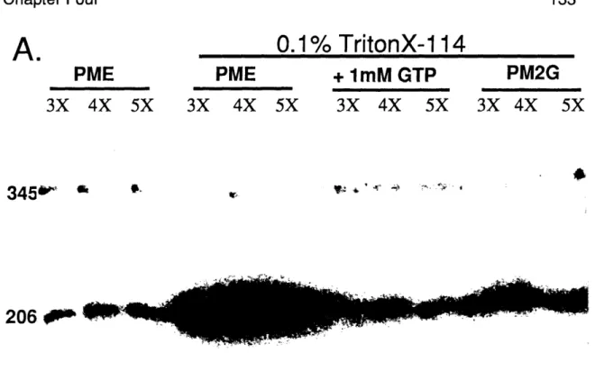
In chapter three of this dissertation, I will discuss the in vitro separation of alpha- and beta-tubulin from heterodimer using Triton X-114 or Nonidet P-40. While it is not clear why the detergent-mediate separation of alpha- and beta-tubulin occurs, work from beta-tubulin heterodimer dissociation and detergent interaction experiments suggest the detergent interferes with heterodimer interactions, favorably altering the dissociation constant.
Other buffer components and conditions
A number of other buffer components and conditions have been examined, including drugs which affect the microtubule heterodimer or polymer state, buffer type, buffer pH, buffer temperature, and salt concentration. An extensive review of all the literature related to these topics is not possible, so I will briefly describe a few experiments examining these parameters.
The polymerization of purified microtubules was not sensitive to buffer type. Studies demonstrated no significant change in tubulin heterodimer folding properties, as assayed by circular dichroism, or tubulin polymerization in a number of commercially available buffers, such as Tris, MES, or Pipes (Lee and Timasheff, 1977). Altering buffer pH from 6.0 to 7.0, however, resulted in increased polymerization (Lee and Timasheff, 1977). Buffer pH greater than 9.0 resulted in irreversible tubulin denaturation. Tubulin in pH 8.0 buffer was competent to assemble only if the buffer contained either 2 M glycerol or 1 mM GTP (Brown-Croom, et al., 1986). High salt concentrations, > 0.7 M NaCI caused irreversible aggregation of tubulin heterodimer (Brown-Croom, et al., 1986).
A number of interactions between microtubules and anti-mitotic drugs, such as colchicine, benomyl, and taxol, have been experimentally examined. The best characterized is colchicine, which depolymerizes microtubules. The interaction is direct, as tubulin co-purifies with colchicine in treated cells with one mole of colchicine per mole of heterodimer (Weisenberg, et al., 1968). Colchicine stabilizes the tubulin heterodimer, since it increased the association constant for tubulin heterodimer (Detrich ill, et al., 1982; Sackett and Lippoldt, 1991). Colchicine may preferentially interact with beta-tubulin. In cross-linking experiments, colchicine was cross-linked to beta-tubulin (Wolff, et al., 1991) and beta-tubulin, but not alpha-tubulin, was more sensitive to proteases when colchicine was present (Sackett and Varma, 1993).
Taxol, isolated as an anti-tumor agent purified from the Pacific Yew tree, had the unique property of stabilizing microtubule polymers, making them resistant to temperature- or calcium-induced depolymerization (Schiff, et al., 1979). Purified tubulin assembled without GTP or MAPs in the presence of taxol (Collins and Valee, 1987; Diaz and Andreu, 1993; Schiff and Horwitz, 1981) and taxol-treated cells exhibited bundles of microtubules, were cold-resistant, and did not migrate properly (Schiff and Horwitz, 1980). 0.6 mol of labeled taxol bound to 1 mol of tubulin in vitro, with a Kapp of 8.1 x 10⁻⁷'M for taxol to tubulin monomer (Parness and Horwitz, 1981). Although the precise action of taxol is unknown, taxol likely binds along the length of the microtubules, halting microtubule disassembly when the taxol molecule is encountered (Caplow, et
al., 1994).
Benomyl is an anti-mitotic drug used in a number of organisms, including S.
cerevisiae. Methyl Benzimidazol-2-YL Carbamate (MBC), the active breakdown
product of benomyl, directly bound to tubulin. The binding was competitively inhibited by colchicine and vinblastine, suggesting the drug may act at the same site as those microtubule dissassembly drugs (Davidse and Flach, 1977). In S.
cerevisiae, treatment with MBC produced large-budded cells and spindle-pole
duplication but not separation (Quinlan, et al., 1980). The precise action of the drug is uncertain.