HAL Id: hal-02415505
https://hal.archives-ouvertes.fr/hal-02415505
Submitted on 17 Dec 2019
HAL is a multi-disciplinary open access
archive for the deposit and dissemination of
sci-entific research documents, whether they are
pub-lished or not. The documents may come from
teaching and research institutions in France or
abroad, or from public or private research centers.
L’archive ouverte pluridisciplinaire HAL, est
destinée au dépôt et à la diffusion de documents
scientifiques de niveau recherche, publiés ou non,
émanant des établissements d’enseignement et de
recherche français ou étrangers, des laboratoires
publics ou privés.
P. Fichet, S. Leblond, R. Laumonier, P. Sardini, S. Billon
To cite this version:
P. Fichet, S. Leblond, R. Laumonier, P. Sardini, S. Billon. MAUD Project - Characterization by
Autoradiography Tech-nique of facilities under dismantlement. DEM 2018 - Dismantling Challenges:
Industrial Reality, Prospects and Feedback Experience, Oct 2018, Avignon, France. �hal-02415505�
MAUD PROJECT- Characterization by Autoradiography Technique
of facilities under dismantlement
Pascal Fichet1, Sylvain Leblond1, Rémi Laumonier2, Paul Sardini 3, Sophie Billon3*
1Den – Service d’Etudes Analytiques et de Réactivité des Surfaces (SEARS), CEA, Université Paris-Saclay,
F-91191, Gif sur Yvette, France
2ARL (Ateliers Laumonier), 11 Rue de Chenival, 95690 Nesles-la-Vallée, France 3IC2MP-HydrASA/UMR 7285 CNRS, 86073 Poitiers cedex 9, France
* Main Author: Pascal Fichet, pascal.fichet@cea.fr
Abstract
In the framework of nuclear facilities dismantling, there is a strong need to obtain accurate analytical results on remaining radioactivity on different solid materials (concrete, metal, …). On the basis of various commercial developments dedicated to biological researches, the Dig-ital Autoradiography method has already been used for different real characterizations of nu-clear wastes. Different developments have proved the efficiency of the Autoradiography Tech-nique but drawbacks have been identified and the industrial research project MAUD has pro-posed several innovative solutions to improve the use of the autoradiography technique for characterization.
KEYWORDS:nuclear characterization, dismantling, tritium, alpha, radioactivity images.
Introduction
During the dismantling of nuclear facilities, and after extensive historical researches, stakeholders are always facing a high demand of characterizations (destructive and nondestructive) to ensure the safety and security of workers and to guide nuclear wastes up to the appropriate repository. Detection of gamma emitters in situ with nondestructive techniques appears generally feasible but alpha and beta characterizations on solid is far less obvious. In particular, to provide new techniques coming from other domains of research (1), the autoradiography has been tested to provide efficient way to characterize alpha and beta emitters on solid matrices. Researches on the Autoradiography technique are still on-going at CEA for decommissioning (2). Initially developed for biological application where H-3 and C-14 are mainly used as tracers, the autoradiography has shown high potentialities as a nondestructive tech-nique for decommissioning. Various applications can be highlighted to address this particular issue: - to provide an image of radioactivity and thus improving the sampling processes, - to obtain surface map-ping of remaining radioactivity and - to obtain information on contamination in depth. The Research conducted at CEA aims at showing real examples of all the potentialities of the Autoradiography tech-niques.
The Autoradiography Technique
The Autoradiography, a nondestructive technique providing nuclear measurements, has been devel-oped for several years by the Laboratory of Analysis and Operators Support at the French Atomic En-ergy Commission. Autoradiography refers to a radiation detection technique where the radiosensitive material is exposed to a radiation (, , , X, neutrons) of an unknown source of radioactivity to analyze, in order to evaluate its activity, and to locate it. It exists different devices to provide Autoradiography images (1), by gas detector, by specific camera detectors, but for this work at the very beginning of dismantling facilities investigations, the autoradiography using phosphor screens (also known as imag-ing plates or films) has been developed (Figure 1). The radiosensitive material (Figure 1) is deposited on a two-dimensional flexible screen (size 12.5 cm * 25.2 cm), whose blue or white (Figure 1) surfaces exposed to ionizing particle are risen to an electronically metastable state. The blue or white covers are composed with the same radio sensitive material BaF(Br,I):Eu2+. The difference between these two
screens is a plastic cover which protects the white screen. The blue screen is dedicated particularly to tritium, the radiosensitive material (blue color) is deposited directly on the surface of a solid waste to investigate.
Figure 1: Phosphor screens used for autoradiography measurements
The phosphor screen technique (Figure 1) was commercialized in the 90s but for biological studies. The main steps of the use of these screens are depicted in Figure 2. Screens can be reused several times if not contaminated. The screen is initialized by a strong bright light during a couple of minute. Then the screen is deposited on a surface to investigate the potential radioactivity. The exposure time is difficult to set because it depends on the activity and the nature of the radionuclide investigated. For dismantling application, this exposure time can vary from several minutes to several days. The screen is kept as much as possible in light tight storage. The raw data obtained by autoradiography technique correspond to the visible image of the surface potentially contaminated by radionuclides.
Figure 2: Methodology to use and reuse phosphor screens for activity measurements.
time, the surface of the screen is swept by a laser. The laser (continuous red laser) is focused on small areas of the screens and the mechanism of the scanner allows to investigate the whole screen surface of the screen with a resolution of several tenth of microns (typically 40 µm). The description of the mechanism induced on Europium, composing the screen, by the radioactivity is explained in Figure 3B. The screen in contact with radioactivity keeps the Europium in a metastable state. The laser excites this state and a visible photon from an unstable state is immediately emitted. In each surface analyzed by the laser a PMT tube records the photons due to the radioactivity that was in contact with the screen surface.
(A) (B)
Figure 3: Laser scanner (A) used to extract information after screens deposition and to obtain an image of radioactivity. (B) Physical mechanism of the measurement involving Eu.
The laser stimulation produces very rapidly (around 2 minutes, for a spatial resolution of 169 µm and approximately 106 pixels) a high resolution image (Figure 4) showing the location of the radioactivity that
was in contact with the screen.
(A) (B)
Figure 4: Images corresponding to the radioactivity measurement (A) compared to a typical background (B)
In Figure 4 (A) a large spot of radioactivity can be easily observed (here in color but it could be repre-sented also in grey gradient) whereas in Figure 4 (B) the lack of radioactivity observed by the screen can be validated by a homogeneous grey color. The color scale displayed in Figure 4A is linearly pro-portional with the radioactivity value (4).
Improvement of sampling processes by Characterization with Autoradiography
The scaling factors technique is widely used to characterize nuclear wastes. This technique allows to declare, to the authority in charge of waste management, a measurement of contamination by nuclides difficult to measure using those (mainly gammas) that can be remotely measured. The determination of these scaling factors can be very hard. To address this issue, destructive analysis in dedicated labora-tories are required, but collecting about 1 g of representative wastes on a whole waste drum as an example can be a real problem (example in Figure 5).
Figure 5: Difficult Sample collection of nuclear wastes
Autoradiography can be used to assess this problem because it provides an image of radioactivity on surface materials (an example in Figure 6). The radioactivity observed here (Figure 6) by the technique of imaging plates used for Autoradiography, appears in black whereas the lack of radioactivity appears in white. For the effectiveness of a destructive analysis requiring only 1 g, it is therefore interesting and very easy to use the autoradiography technique to improve the sampling efficiency. One must pay at-tention in the result presented in Figure 6, because due to the software of the laser scanner, the auto-radiography image is flipped. Moreover for this types of tries, because the screen can be reused thou-sands of times, the autoradiography produces very little waste. The only waste produced is the screen.
Figure 6: Real image of typical plastic waste (left) – analysis by autoradiography (right)
Contamination mapping
To obtain an evaluation of the residual of radioactivity on a concrete floor, it is possible to use batches of imaging plates (Figure 7) to reconstruct an image of various contaminated locations of a facility being dismantled. In Figure 7, screens are not deposited side by side but in a large grid in order to allow a large area investigation and a time saving. The idea was also to use this type of grid to allow the potential recognition of spot of radioactivity with a certain size: exciding around 20 cm2. By using efficient software
that implements geostatistical calculations (3), the autoradiography technique can provide contamina-tion mapping of different radionuclides and even tritium (Figure 8).
Figure 7: Batch of imaging plates (black rectangles) deposited on potentially contaminated concrete sur-face of a shutdown nuclear facility.
(A) (B) Figure 8: mappng obtained after mathematical treatment of around thousand autoradiography images
ob-tained on the floor of a 250 m2 laboratory area. Spots of radioactivity are due in this case to tritium.
(A) Real measurements (B) Same image after kriging process obtained with KartotrakTM software. The KartotrakTM software is able to calculate, by using a geostatistical method, with raw data a variogram
function that represents the mean effect of all the neighbors to calculate the radioactivity value of an individual point on the concrete surface. The kriging process is based on assumptions and particularly that all radioactivity spots can be clearly observed by autoradiography measurements in this case.
Contamination in depth
Regarding potential contamination that can be found in nuclear facilities during their dismantling, it is very difficult to find techniques for assessing the depth of contamination. This is highly problematic par-ticularly for alpha and beta emitters. Following the realization of a core sample, autoradiography tech-nique can also provide an efficient way of characterizing potential contamination in depth. As an exam-ple, Figure 9 shows an example of a result obtained for a cut concrete block. The color appearing in the left part of the block corresponds to the contamination in depth of Cl-36. This application highlights the potentiality of the technique to measure very quickly the contamination in depth of some radionuclides. That can be a very efficient technique to test the efficiency of different barriers used for nuclear wastes repository.
Figure 9: autoradiography image obtained on a concrete surface contaminated in depth by Cl-36 radionu-clide.
Image of Cl-36 contamination in
depth
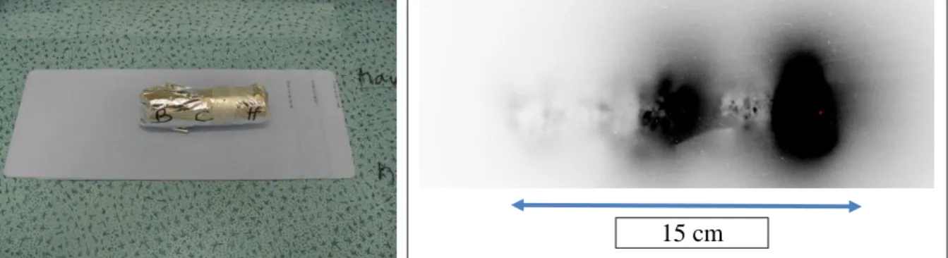
Another interesting application of measurement by autoradiography is displayed in Figure 10. A core sample containing irradiating radionuclides (typically Cs-137) is deposited during few minutes on a screen. The core was wrapped by an aluminum foil to prevent any potential contamination. In this case the result obtained (Figure 10B) clearly shows two spots. This very rapid investigation of core sample could be a very efficient way to collect effective samples for destructive analysis.
Figure 10: Contamination in depth obtained after investigation by autoradiography technique on a con-crete core.
Conclusion
Autoradiography characterization is being developed to evaluate its potentialities as an in situ and non-destructive method to measure alpha and beta radionuclides that must be characterized in dismantling facilities. Great care is needed in these developments because autoradiography is also sensitive to all types of radioactive emitters and in particular gamma.
Digital autoradiography producing images is a quantitative, non-destructive, and inexpensive method for the localization of radioactive contamination on different surfaces of facilities being dismantled. It is compliant with most requirements in such a context (very few constraints, minimal exposure of opera-tors, neither production of wastes nor transport of radioactive materials). While traditional wipe tests for beta emitters characterization can only detect removable contamination, digital autoradiography detects both fixed and removable contamination.
Autoradiography has high potential to improve sampling processes.
In addition, it provides 2D images enabling accurate localization of the potential contamination even in facilities with large areas. However, for a large area investigated, the number of images is huge, requir-ing a mapprequir-ing method to compare results and also to extrapolate data without actual measurement. The use of a Geographic Information System (GIS) with a geostatistical method is a very efficient way to locate and compare the radioactive spots observed by the Digital Autoradiography Technique. The image obtained by the technique can allow the evaluation of the contamination in depth.
The imaging plate technique to obtain autoradiographs has proven to be very efficient, but some im-provements has to be provided, that are currently studied with the MAUD industrial project considering other type of detector for autoradiography.
Acknowledgment
Work performed within the Investments for the future program of the French Government and operated by the French National Radioactive Waste Management Agency (Andra).
References
1) “Handbook of radioactivity analysis”, MF L’Annunziata, chap 19, Third edition 2012.
2) MAUD industrial national project, PIA 2016-2019, https://www.andra.fr/download/site-principal/document/inno-vation/fiche-maud.pdf
3) Kartotrak User's guide. All-in-one software for contaminated site characterization. Geovariances, Fon-tainebleau, 532 pp, 2017
4) P. Fichet, F. Bresson, A Leskinen, F Goutelard, J Ikinen, M Siitari-Kaupi, « Tritium analysis in building disman-tling process using digital autoradiography”, J Radioanal Nucl Chem, 291, 869-875, (2012)