Aneuploidy reveals insights into control of protein complex
stoichiometry
by Christopher M. Brennan B.S. Biology Providence College, 2013SUBMITTED TO THE DEPARTMENT OF BIOLOGY
IN PARTIAL FULFILLMENT OF THE REQUIREMENTS FOR THE DEGREE OF DOCTOR OF PHILOSOPHY IN BIOLOGY
AT THE
MASSACHUSETTS INSTITUTE OF TECHNOLOGY SEPTEMBER 2019
© 2019 Christopher M. Brennan. All rights reserved.
The author hereby grants to MIT permission to reproduce and to distribute publicly paper and electronic copies of this thesis document in whole or in part in any medium now known or
hereafter created.
Signature of the Author: __________________________________________________________ Department of Biology July 15, 2019 Certified by: ___________________________________________________________________
Angelika Amon Kathleen and Curtis Marble Professor of Cancer Research Investigator, Howard Hughes Medical Institute Thesis Supervisor Accepted by: __________________________________________________________________
Stephen Bell Uncas and Helen Whitaker Professor of Biology Investigator, Howard Hughes Medical Institute
Aneuploidy reveals insights into control of protein complex
stoichiometry
By
Christopher M. Brennan
Submitted to the Department of Biology on July 15, 2019 in Partial Fulfillment of the Requirements for the Degree of
Doctor of Philosophy in Biology Abstract
Aneuploidy, or an incorrect number of chromosomes, is caused by errors in chromosome segregation during cell division. Because genes are expressed in accordance with their copy number, aneuploidy simultaneously alters the gene dosage of hundreds to thousands of genes. The outcome is an imbalanced proteome, which has a negative impact on cellular physiology and places intense demand on the protein quality control system of the cell to effectively fold and/or degrade proteins. Aneuploidy further represents an ideal model for studying how cells cope with imbalances in their proteome as it allows for interrogation of the fate of hundreds to thousands of imbalanced proteins simultaneously.
Here, we identify protein complex stoichiometry imbalances as a major cause of protein aggregation in aneuploid cells. Subunits of protein complexes encoded on excess chromosomes aggregate in aneuploid cells, which is suppressed when expression of other subunits is
coordinately altered. We further show that excess subunits are either degraded or aggregate, a fate that is largely mutually exclusive for individual subunits. We also demonstrate that protein aggregation is nearly as effective as protein degradation at lowering levels of excess proteins. Our study explains why proteotoxic stress is a universal feature of the aneuploid state and reveals protein aggregation as a form of dosage compensation to cope with disproportionate expression of protein complex subunits. This work informs both our comprehension of aneuploid cell physiology, and also provides a more complete understanding of how aneuploid and euploid cells cope with stoichiometric imbalances, namely that protein aggregation can function as protein quality control mechanism in this regard.
Thesis supervisor: Angelika Amon
Title: Kathleen and Curtis Marble Professor of Cancer Research; Investigator, Howard Hughes Medical Institute
Acknowledgements
First, I need to thank my advisor, Angelika, for her unparalleled enthusiasm. Thank you for always encouraging me and pushing me to be my best. I am so fortunate to have had a mentor that cares so deeply about my development as a scientist. It was an absolute honor and privilege to be able to learn from such an outstanding scientist and mentor. I also need to thank my undergraduate research advisor, Brett, whose passion for science is the reason why I decided to pursue a PhD.
The Amon lab has been a fantastic place to spend these last 5 years. To past and present members of the lab, thank you for creating a wonderful environment to do science in. Thank you for all of your time and energy spent thinking about my work. I need to highlight Ana, my rotation mentor who was instrumental in getting this project started and spent her last summer in the lab teaching me. Also, my two bay mates, Luke and Summer, both of whom I have learned so much from. Your friendship made coming to lab every day much more enjoyable.
I would also like to acknowledge my thesis committee members, Steve Bell and Gene-Wei Li. Your input throughout my grad career was invaluable for guiding the direction of my project and career. Also, thank you to Dan Finley for graciously agreeing to serve as the outside member of my thesis committee.
I have been fortunate to have great friends both in and out of the biology department. Thank you to my Biograd2013 classmates for always keeping things fun and light-hearted. To my non-MIT friends, you have been the most welcomed distraction from work. Thank you to Rushmore, the North End trivia crew, Pin and Tonic, and ENDR for drinking beers with me, bowling, playing video games, and generally just having fun. Thank you to my roommates Corey, Eric, and Erik who always at least tried to understand what happened in my day. You made living in Boston a great time in my life. I need to thank my oldest friend, Brianna, for putting up with me since we were born. I can’t believe that 28 years later, our dogs are best friends.
I consider myself extremely fortunate to have a family that are also my best friends and greatest supporters. Thank you to my grandparents Mamie, Pa, Nana, and Pop for your constant love. Family gatherings with my aunts, uncles, and cousins, both Brennan’s and Roberts’ have been a consistent source of happiness throughout my life. And thank you to the Fletcher’s for welcoming me into their family. I’ve greatly enjoyed all of my trips to Washington and your visits to Boston.
I could not have done this without the support of my Mom and Dad. Mom, thank you for fostering my love of science. Dad, thank you for encouraging me to find what I love to do, then figure out how to make a career out of it. To my brother and best friend JR, thank you for
reminding me not to take things (especially myself) too seriously. To my dog Bradley, thank you for attacking me with same affection and enthusiasm when I come home regardless of whether my experiment failed or succeeded. Lastly, thank you to my wife and life partner, Marissa, for being my greatest source of support and inspiration since we met on the first day of graduate school. Meeting you was the best thing that ever happened to me.
Table of Contents
Abstract ... 3
Acknowledgements ... 6
Chapter 1: Introduction ... 11
ANEUPLOIDY DECREASES FITNESS ... 12
Causes of aneuploidy ... 12
Models of aneuploidy used in this thesis ... 17
Effects of aneuploidy on organismal physiology ... 19
Aneuploid fitness defects are caused by changes in gene expression ... 20
Cancer and the aneuploidy paradox ... 23
MAINTENANCE OF PROTEIN HOMEOSTASIS ... 24
Molecular chaperones assist protein folding ... 24
Protein complex formation is the final step in protein folding ... 27
The ubiquitin proteasome system degrades misfolded proteins ... 29
Protein aggregation as a strategy for protein quality control ... 31
EFFECTS OF AN ANEUPLOID PROTEOME ... 38
Chromosome gain and loss cause protein aggregation ... 38
Protein quality control defects underlie the fitness disadvantage of aneuploid cells ... 40
Control of gene dosage by protein degradation ... 41
Concluding remarks ... 42
References ... 44
Chapter 2: Protein aggregation mediates stoichiometry of protein complexes in aneuploid cells 56 ABSTRACT ... 57
INTRODUCTION ... 58
RESULTS ... 59
Identification of proteins that aggregate in aneuploid yeast cells ... 59
Proteins that aggregate in multiple disomes also aggregate in cells with compromised protein quality control ... 72
Duplicated proteins are highly enriched in aneuploid aggregates ... 74
Increasing gene copy number by one causes protein aggregation in human cells ... 75
Stoichiometric imbalance of protein complexes can cause protein aggregation ... 78
Protein complex subunits that aggregate when in excess have lower turnover rates than
degraded subunits ... 88
Dosage compensation by protein aggregation ... 93
DISCUSSION ... 99
Which proteins aggregate in aneuploid cells? ... 100
Cellular response to excess subunits of protein complexes ... 101
Dosage compensation by protein aggregation ... 102
MATERIALS AND METHODS ... 103
Supplemental Table S1. Yeast strains used in this study ... 117
Supplemental Table S2. Primers used in this study ... 120
Supplemental Table S3. Plasmids used in this study ... 121
Acknowledgments ... 121
References ... 122
Chapter 3: Conclusions and Future Directions ... 127
Stoichiometric imbalance causes protein aggregation ... 128
The fate of excess protein: to aggregate or to degrade ... 129
Subunits that are neither aggregated nor degraded ... 131
Protein aggregation mediates protein complex stoichiometry ... 133
Is aggregation protective in aneuploid cells? ... 134
How do cancer cells cope with proteotoxic stress? ... 135
Does aggregation commonly control protein stoichiometry in euploid cells? ... 137
Concluding Remarks ... 140
Aneuploidy, or an incorrect number of chromosomes, is caused by errors in chromosome segregation during cell division. Every time a cell divides, it must accurately segregate equal numbers of chromosomes between the mother and daughter cells. While mechanisms exist to ensure faithful chromosome segregation, errors occur at a rate of 5x10⁻⁴ in yeast and 10-4 to 10-5 in mammals (Hartwell and Smith, 1985; Rosenstraus and Chasin, 1978). Because genes are expressed in accordance with their copy number, aneuploidy simultaneously alters the gene dosage of hundreds to thousands of genes. The result is an imbalanced proteome, which has a negative impact on cellular physiology and places intense demand on the protein quality control system of the cell to effectively fold and/or degrade proteins.
In this introduction, I will first discuss the effects of aneuploidy on cellular and
organismal fitness. I will then provide an overview of how cells utilize protein quality control mechanisms to maintain a healthy proteome, including the role of protein aggregation in protein homeostasis. Finally, I will discuss the effects of aneuploidy on the proteome and our current understanding of how cells cope with the problem of an imbalanced proteome.
ANEUPLOIDY DECREASES FITNESS Causes of aneuploidy
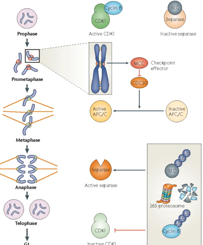
Aneuploidy is caused by errors in chromosome segregation during cell division. In a normal mitotic division, replicated sister chromatids are linked by the protein cohesin during S phase (Nasmyth and Haering, 2009). In prophase, these sisters are then attached to opposite poles of the mitotic spindle by the interaction between spindle microtubules and the kinetochore, a large protein assembly that forms at the centromeres of chromosomes. Attached chromosomes align during metaphase and are bioriented, i.e. with sister kinetochores facing in opposite
directions. At this point, the spindle assembly checkpoint (SAC) ensures that sister chromatids are properly attached to the spindle and bioriented such that chromosome segregation will result in partitioning of one sister chromatid into each daughter (Fig. 1, Musacchio and Salmon, 2007). If sister chromatids are correctly attached and bioriented, the pulling force in opposite directions generated by the spindle results in tension across the sister kinetochores. This tension satisfies the checkpoint, resulting in the activation of the ubiquitin ligase known as the anaphase-promoting complex or cyclosome (APC/C) and its activating subunit Cdc20. APC/C bound to Cdc20 leads to cleavage of cohesin by degrading Securin, the inhibitor of the protease called Separase. Cleavage of cohesin by Separase begins anaphase and results in the separation of sister chromatids to opposite poles.
Figure 1. The spindle assembly checkpoint (SAC) ensures faithful chromosome segregation (adapted from (Musacchio and Salmon, 2007))
Unattached kinetochores and sister chromatids that lack tension lead to the generation of the mitotic checkpoint complex (MCC). The MCC keeps the APC/C inactive by inhibiting its
activating subunit Cdc20. Once all chromosomes have been correctly attached the spindle and tension is generated, the SAC is inactivated relieving the inhibition of Cdc20. APC/C-Cdc20 mediates ubiquitination and proteolysis of Securin, the inhibitor of Separase. Once activated, Separase cleaves cohesin molecules that hold the bioriented sister chromatids together causing them to be pulled to opposite poles. APC/C-Cdc20 also facilitates the degradation of mitotic cyclins, thus lowering CDK activity and preparing cells to exit from mitosis.
One source of chromosome mis-segregation is the premature loss of sister-chromatid cohesion. This can be caused by incorrect establishment of cohesion, hyperactivation of
Separase, or by aberrantly low Securin activity. In these cases, sister chromatids are segregated as soon as they attach to the mitotic spindle, so any incorrect attachments result in aneuploid daughter cells (Nasmyth and Haering, 2009).
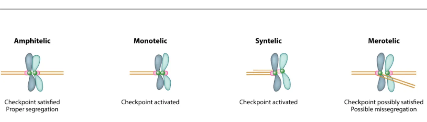
Incorrect attachments of chromosomes to the spindle and faulty inactivation of the SAC are another source of chromosome-segregation errors. Amphitelic attachments describe properly attached sister chromatids, with each sister attached to microtubules emanating from opposite polls. Monotelic (only one kinetochore is attached to the spindle) and syntelic (both sister kinetochores are attached to the same pole) do not generate tension and thus activate the SAC (Fig. 2, (Knouse et al., 2017)). If the SAC fails to arrest cells in metaphase due to mutations in checkpoint components, these types of attachments lead to chromosome mis-segregation and aneuploidy. Merotelic attachments occur when one kinetochore is attached to both poles and are thought to be poorly sensed by the SAC since merotely can still result in tension across the sister kinetochores. In these cases, chromosomes lag behind the rest of the migrating chromosomes and
can be mis-segregated or incorporated into micronuclei (Cimini et al., 2001; Thompson and Compton, 2011).
Figure 2. Types of kinetochore-microtubule attachments (adapted from Knouse et al., 2017)
Kinetochore-microtubule attachments that result in properly bioriented sister chromatids attached to opposite poles of the spindle are termed amphitelic. Amphitelic attachments satisfy the spindle assembly checkpoint (SAC) and result in equal segregation of chromosomes. Monotelic
attachments occur when only one kinetochore attaches to one pole, while syntelic attachments occur when both kinetochores are attached to the same pole. Both cause activation of the SAC and, under normal circumstances, delay anaphase until the an amphitelic attachment is achieved. Merotelic attachments occur when one kinetochore is attached to both poles. Depending on the strength of the attachments to each pole, the SAC can be satisfied by merotelic attachments and anaphase proceeds. The chromatid that is attached to both poles lags behind the rest of the migrating chromosomes resulting in mis-segregation or the formation of a micronucleus. A micronucleus forms when the lagging chromosome is surrounded by membrane that is separate from the main nucleus causing the isolation of the lagging chromosome.
If chromosome mis-segregation occurs in a mitotic division early in development, this can result in a large percentage of cells in the organism being aneuploid. Chromosome
segregation errors can also occur during either division of meiosis, producing aneuploid gametes that give rise to whole organisms with constitutional aneuploidy.
Models of aneuploidy used in this thesis
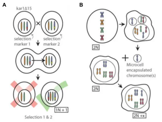
In this thesis, I have employed two models of aneuploidy in budding yeast and one model in human cells to study the molecular nature of protein aggregates in aneuploid cells. In general, aneuploid cells can be generated such that they have specific, known karyotypes or random karyotypes. Defined aneuploidies have the advantage of enabling assessment of chromosome-specific effects of aneuploidy. Cells with randomly generated karyotypes are useful for studying general effects of aneuploidy and are also capable of resulting in more complex combinations of chromosome gains and losses. However, they are often karyotypically unstable. The primary model used in this work are haploid budding yeast containing a single extra copy of one of their 16 chromosomes (n+1) called disomes (Fig. 3A). Each copy of the duplicated chromosome is marked with a unique selectable marker, allowing for stable maintenance of the aneuploid karyotype. Disomes were constructed by marking the same locus with different markers in separate haploid strains. One of the parental strains contains a mutation in the karyogamy gene, KAR1, which when mutated in one mating partner prevents nuclear fusion from occurring (Conde and Fink, 1976). These strains were then crossed to one another, resulting in an abortive mating. Occasionally entire chromosomes are transferred from one nucleus to the other, resulting in aneuploid progeny (Torres et al., 2007). Chromosome transfer events can then be selected for by growing progeny in conditions that require the selectable marker on both chromosomes.
To study higher levels of chromosomal imbalance, I have utilized a strain harboring a temperature sensitive allele of the kinetochore component NDC10 (Goh and Kilmartin, 1993).
When grown at the semi-permissive temperature, ndc10-1 mutants mis-segregate chromosomes at a very high rate, resulting in a heterogeneous population of aneuploid cells with a range of karyotypes (Oromendia et al., 2012).
To assess whether principals uncovered in yeast are generalizable to higher eukaryotes, I have also utilized a series of human cell lines with stable, defined karyotypes. Retinal pigmented epithelial (RPE-1) cells immortalized by the expression of telomerase reverse transcriptase are nearly diploid, allowing for an adequate euploid control in experiments. These cells were made aneuploid by microcell mediated chromosome transfer (Fournier and Ruddle, 1977; Stingele et al., 2012). By this method, micronuclei containing whole chromosomes are generated by treating cells with a microtubule poison, then purified and fused with acceptor cells, resulting in whole chromosome gains (Fig. 3B).
(A) Haploid yeast containing a single extra copy of one chromosome (disomes) were created using an abortive mating scheme. One parental strain harbored a mutation in the KAR1 gene to prevent nuclear fusion upon mating. The two parental strains harbored distinct selectable
markers at the same locus allowing for selection of progeny that had inherited both copies of the chromosome due to transfer of the whole chromosome.
(B) Human cells with trisomies were generated by microcell mediated chromosome transfer. Micronuclei containing whole chromosomes can be created by treating cells with a microtubule poison. Chromosome-containing micronuclei can be purified and fused with a cell, resulting in the incorporation of the extra chromosome into nucleus of the acceptor cell.
Effects of aneuploidy on organismal physiology
Over 100 years ago, Marcella O’Grady and Theodor Boveri noted the detrimental effects that aneuploidy had on organismal fitness by observing the failed development of sea urchin embryos that had undergone chromosome mis-segregation (Boveri, 1902). Today we understand that aneuploidy that arises during gamete formation is the leading cause of miscarriage in
humans because, with rare exceptions, aneuploid human embryos fail to survive to term
(Alberman and Creasy, 1977; Hassold and Jacobs, 1984). Humans with trisomy of chromosome 13 (Patau syndrome) and trisomy 18 (Edwards syndrome) can survive to birth, but only ~10% of these individuals live to be one year of age (Rasmussen et al., 2003). Trisomy of chromosome 21 causes Down syndrome, which is characterized by developmental defects including impaired growth and brain development (Siegel and Amon, 2012). In all other organisms examined, aneuploid progeny either fail to survive to term or have developmental defects reminiscent of those observed in humans.
Aneuploidy can also arise spontaneously in adult organisms due to mitotic chromosome mis-segregation. In most adult organisms, aneuploidy is quite rare, occurring in only ~2-5% of cells (Knouse et al., 2014). Mosaic variegated aneuploidy (MVA) is caused by mutations in the mitotic machinery or SAC components that ensure chromosomes are properly attached to the mitotic spindle, resulting in individuals with upwards of 25% of their cells being aneuploid (Hanks et al., 2004; Snape et al., 2011). Like individuals with constitutional aneuploidies, humans with MVA exhibit growth and developmental deficiencies. Mice harboring a
hypomorphic mutation in the SAC were found to have higher numbers of aneuploid cells in non-regenerating adult tissues such as the brain, while highly proliferative tissues like the intestine were almost entirely euploid, indicating that aneuploid cells may be selected against in vivo (Pfau et al., 2016). This could in part be due to euploid cells outcompeting slow growing aneuploid cells, but recent evidence suggests that aneuploid cells can be recognized by the immune system (Andriani et al., 2016; Santaguida et al., 2017; Sullivan et al., 2016). At least in vitro, aneuploid cells that have arrested in the cell cycle with complex karyotypes secrete pro-inflammatory signals that facilitates their clearance by natural killer (NK) cells (Santaguida et al., 2017). Combined with cell autonomous methods to ensure faithful chromosome segregation, immune-mediated clearance of aneuploid cells may also protect organisms against the harmful effects of aneuploidy.
Aneuploid fitness defects are caused by changes in gene expression
To understand why aneuploidy is universally detrimental to organismal development, it is important to explore how an imbalanced karyotype affects individual cells. The most important question to address is whether the extra chromosome is expressed. Using disomic yeast described
above, it was established that chromosome gain leads to a corresponding increase in RNA and protein levels for most genes on the gained chromosome (Torres et al., 2007; 2010). Consistent with developmental defects observed in aneuploid multicellular organisms, disomic yeast have impaired proliferation. This phenotype is caused by the expression of the extra chromosome, as euploid yeast containing a yeast artificial chromosome of up to 1.6 Mb (13% of the genome) encoding no yeast genes have no proliferation delay (Torres et al., 2007).
Fundamentally, aneuploidy is a problem of gene dosage. Phenotypes could be caused by changing the dosage of just one to a few genes, affecting the related cellular process, or they could be caused by mass action of simultaneously altering the dosage of many genes. There is evidence that aneuploid phenotypes are caused by both of these mechanisms.
Single gene effects of aneuploidy could be caused by haploinsufficiency (chromosome loss in a diploid) and sensitivity to increased copy number (chromosome gain).
Haploinsufficiency describes when a phenotype in a diploid organism is caused by a
heterozygous loss of function mutation. In yeast grown in rich medium, about 3% of the genome is haploinsufficient, and most of these genes are involved in protein translation, particularly ribosomal subunits (Deutschbauer et al., 2005). With the exception of chromosome I, all yeast chromosomes contain at least one ribosomal subunit, which could in part explain the fitness defects caused by chromosome loss.
Increasing gene expression of a single gene by just two-fold can also have fitness
consequences. Perhaps the most dramatic example comes from studies of tubulin in yeast. Just a 1.4-fold overexpression of β-tubulin causes a decrease in cell viability (Katz et al., 1990). Other instances of changes in fitness due to a single extra copy of a gene have been harder to discover. In a study of dosage sensitive genes (defined as causing fitness penalties when encoded in
greater than five copies) only one gene was found to have a proliferation delay when expressed at one extra copy (Bonney et al., 2015). A recent study using a pooled growth competition of strains harboring barcoded genes on single copy plasmids found that 15% of genes significantly delayed proliferation (Morrill and Amon, 2019). Thus, particularly for chromosome gain, it is uncommon for single genes to account for the entirety of proliferation defects observed in aneuploid cells. Karyotype specific aneuploid phenotypes are often observed, arguing that a two-fold increase in expression of one or a small group of genes can have specific phenotypic
consequences. For example, disome XVI has defects in protein transport not observed in other disomes as evidenced by its unique sensitivity to gene deletions in protein trafficking
components and drugs targeting protein trafficking (Dodgson et al., 2016a). What gene or group of genes on chromosome XVI causes this effect is still elusive.
General phenotypic consequences of aneuploidy have been studied more readily. These phenotypes have been observed in aneuploid cells with distinct karyotypes even across species. Apart from decreased proliferation, aneuploidy commonly causes metabolic stress and genomic instability (Blank et al., 2015; Hwang et al., 2017; Passerini et al., 2016; Sheltzer et al., 2011; Tang et al., 2017; Torres et al., 2007; Williams et al., 2008). These various stresses along with slow growth of aneuploid cells also causes the activation of a transcriptional response known as the environmental stress response (ESR) (Sheltzer et al., 2012). The ESR was found to
downregulate genes involved in macromolecule biosynthesis and up regulate stress response genes to help cells cope with multiple different environmental stresses (Gasch et al., 2000). Recently it was discovered that multiple distinct karyotypes in yeast and mice cause an increase in variability, or “noise”, in biological pathways (Beach et al., 2017). Finally, as a direct result of altering the proteome, aneuploid cells have defects in protein folding and turnover, a condition
known as proteotoxic stress. The causes and consequences of proteotoxic stress in aneuploid cells will be discussed in depth later in this introduction and in Chapter 2.
Cancer and the aneuploidy paradox
Despite the numerous penalties of aneuploidy described above, aneuploidy is observed in 77% of tumors in The Cancer Genome Atlas (Knouse et al., 2017). This striking feature of cancer cells prompted Theodor Boveri to propose that aneuploidy causes cancer (Boveri, 1914). This presents a paradox: in most contexts, aneuploidy impairs cellular proliferation, yet it is frequently associated with cancer, a disease defined by cells with increased proliferation. This begs the question of whether aneuploidy is a cause or simply a consequence of transformation. Theoretically, aneuploidy could promote tumorigenesis by altering the copy number of tumor suppressors and oncogenes. It is also possible that dysregulation of the cell cycle observed in cancer leads to an increase in chromosome segregation errors, and aneuploidy is an unwanted side effect of rapid proliferation. Evidence for both of these scenarios exist, and the complexity of the role of aneuploidy in cancer is perhaps best exemplified by the fact that people with Down syndrome have a decreased risk of forming solid tumors and an increased risk of developing leukemia (Hasle et al., 2000; Yang et al., 2002).
Analysis of many cancer genomes revealed that gains of oncogenes and loss of tumor suppressors do impact the karyotype of tumors (Davoli et al., 2013). Direct evidence that aneuploidy can drive tumorigenesis comes from studies of chromosomal instability (CIN), a condition which causes aneuploidy due to increased rates of chromosome mis-segregation. In one study, CIN was able to accelerate tumor progression by promoting loss of heterozygosity of key tumor suppressors such as p53 (Baker et al., 2009). Indeed, individuals with mosaic
variegated aneuploidy are at an increased risk of developing childhood cancers (Hanks et al., 2004).
While genomic or chromosomal instability may be able to facilitate tumorigenesis, much evidence suggests that aneuploidy per se does not cause cancer. Aneuploid cells of multiple karyotypes transformed with oncogenes do not proliferate faster than euploid cells transformed with the same oncogene (Sheltzer et al., 2017). Additionally, levels of aneuploidy tend to be lower in early lesions, and higher in more developed tumors suggesting that aneuploidy arises later in oncogenesis (Ried et al., 1996; Ross-Innes et al., 2015). While specific chromosomal alterations are associated with some cancers, the lack of a common “pan-cancer” karyotype and the variability of karyotypes even among individuals with same type of cancer strongly argues that aneuploidy is infrequently a cancer driver (Knouse et al., 2017). Though it is rarely the cause of cancer onset, it is possible that aneuploidy plays a role in metastasis and resistance to
treatment by providing genetic variability within the tumor population, thus allowing for adaptation. This is supported by the fact that high levels of aneuploidy are associated with poor patient prognosis (Byrd et al., 2002; Emdin et al., 1987). Establishing the role of aneuploidy in later stages of cancer development will be important in fully understanding the impact of karyotype on disease progression.
MAINTENANCE OF PROTEIN HOMEOSTASIS Molecular chaperones assist protein folding
From bacteria to humans, the final step needed for expression of a gene to result in a functional protein is the acquisition of its correct three-dimensional structure. Proteins can adapt many different energetically favorable conformations, but these misfolded states do not result in
a functional protein (Balchin et al., 2016) and negatively impact the fitness of the cell (Geiler-Samerotte et al., 2011). Although the information needed for a protein to fold correctly is encoded in its amino acid sequence (Anfinsen, 1973), most proteins require assistance from molecular chaperones to achieve a properly folded state. The major cellular chaperones are HSP70s, HSP90s, and chaperonins.
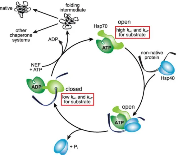
HSP70 binds short stretches of hydrophobic amino acids on nascent peptides and newly synthesized proteins (Rüdiger et al., 1997). Through ATP hydrolysis cycles, HSP70 binds and releases substrates, giving them an opportunity to fold (Fig. 4). ATP-bound HSP70 has low affinity for substrates, allowing substrates to associate and dissociate freely (Mayer, 2010). HSP40, a co-chaperone for HSP70, can deliver substrates to HSP70 and stimulates its ATPase activity, causing HSP70 to transition into a high affinity, ADP-bound state (Kampinga and Craig, 2010). Nucleotide exchange factors then facilitate the exchange of ADP for ATP, causing the substrate to be released into the cytoplasm. If the substrate is able to fold, the hydrophobic residues become buried and are no longer recognized by HSP70 (Hartl et al., 2011).
Figure 4. HSP70 conformation cycles are driven by ATP hydrolysis (adapted from (Balchin et al., 2016))
HSP70 in its ATP bound state has fast on and off rates for substrate binding and release (open conformation). The HSP70 co-chaperone, HSP40, delivers non-native proteins to ATP bound HSP70. HSP40 also stimulates the ATPase activity of HSP70, causing it to hydrolyze ATP. ADP bound HSP70 (closed conformation) tightly binds the substrate that was delivered by HSP40. HSP70 nucleotide exchange factors (NEFs) then facilitate exchange of ADP for ATP, causing HSP70 to transition back to its open conformation, releasing its substrate. This substrate can either be re-captured by HSP70 if it did not fold, be delivered to another chaperone system, or reach its fully native form with no further assistance.
HSP90 functions downstream of HSP70, assisting in the folding of a large number of client proteins that include protein kinases and steroid receptors (Taipale et al., 2010). Like HSP70, HSP90 works via an ATP hydrolysis cycle, however it functions as a dimer and features many more co-chaperones that facilitate client selection and modulate ATP hydrolysis (Shiau et al., 2006).
Chaperonins are barrel shaped protein complexes that encapsulate a single substrate protein allowing it to fold in isolation thereby preventing inappropriate interactions with other proteins. The chaperonin of the eukaryotic cytoplasm, known as TRiC (or CCT), is composed of two sets of 8 subunits that form back-to-back rings (Muñoz et al., 2011). It is most notably responsible for aiding in the folding of actin and tubulin. HSP70 can deliver substrates to TRiC, which enter the barrel and are entrapped by a lid domain which opens and closes in an ATP-dependent manner (Douglas et al., 2011). The inner walls of chaperonins are composed of
hydrophilic, negatively-charged residues that encourage the enclosed substrate to fold by burying hydrophobic residues (Horwich and Fenton, 2009). Upon completion of the ATP hydrolysis cycle, the lid domain opens and the substrate is released. If one round of ATP hydrolysis was not enough time for the substrate to attain its folded conformation, the substrate can reenter the barrel for subsequent rounds of entrapment and release (Hartl et al., 2011).
Protein complex formation is the final step in protein folding
In addition to achieving a correct three-dimensional structure, many proteins rely on stable interactions with other proteins to function. Homomeric protein complexes are composed of multiple subunits of the same protein while heteromeric protein complexes are composed of at least two unique subunits. In principal, this final step in protein folding involves two or more
proteins diffusing through a crowded cytoplasm then colliding with one another in an orientation that results in their proper association (Ellis, 2001; Williams and Dichtl, 2018). This presents a large problem, as protein-protein interfaces are “sticky” and have the potential to form
inappropriate interactions, especially with unfolded nascent polypeptides, when they are not buried within the structure of the mature protein complex (Levy et al., 2012; Pechmann et al., 2009). In some cases, the cell copes with this problem by employing specific chaperones to aid in a multi-step, ordered assembly process, e.g. the 20S proteasome and yeast vacuolar ATPase (Le Tallec et al., 2007; Smardon et al., 2002).
In theory, this problem could also be solved by assembling complexes as the translated proteins emerge from the ribosome. For homomeric complexes, this can happen because a single mRNA is typically translated by multiple ribosomes, resulting in spatially proximal subunits (Kiho and Rich, 1964; Zipser and Perrin, 1963). In prokaryotes, where subunits of the same complex are often encoded by the same polycistronic message, even heteromeric complexes can be assembled co-translationally (Shieh et al., 2015). In fact, subunit-encoding genes are ordered within operons to optimize complex assembly (Wells et al., 2016). Operons are rare in
eukaryotes, so co-translational complex assembly would require co-localization of mRNAs or localization of one mature subunit to the site of translation of another subunit. Indeed, mRNAs encoding separate subunits of the actin nucleation complex, Arp2/3, are co-localized to the leading edge protrusions of migrating fibroblasts (Mingle et al., 2005). In fission yeast, it was estimated that ~40% of proteins (without RNA binding domains) associated with mRNAs of binding partners (Duncan and Mata, 2011). More recent evidence confirms that co-translational assembly of complexes is common in budding yeast. Shiber and colleagues performed
nascent polypeptides of their binding partners using ribosome profiling (Shiber et al., 2018). This was the case for nine out of twelve tested complexes. The three that did not show evidence of co-translational assembly have dedicated chaperones to assist in assembly as described above. Interestingly, for six of the nine complexes, assembly is directed, i.e. fully translated subunit A interacts with nascent subunit B, but not vice versa (Shiber et al., 2018). Using a similar
approach, co-translational assembly of three nuclear complexes was also observed in mammalian cells (Kamenova et al., 2019). Thus, co-translational assembly of heteromeric complexes appears to be the norm and not the exception. However, it is still unclear how one subunit is targeted to the translation site of other subunits.
The ubiquitin proteasome system degrades misfolded proteins
When proteins fail to achieve their native conformation or are damaged, the ubiquitin-proteasome system (UPS) can degrade them. Misfolded or damaged proteins are recognized by ubiquitin ligases, which aid in the covalent attachment of a charged ubiquitin molecule from a conjugating enzyme to a lysine residue on the substrate by binding both the ubiquitin-conjugating enzyme and the substrate (Finley et al., 2012). Thus, ubiquitin ligases confer
substrate selectivity to the ubiquitination reaction. The two classes of ubiquitin ligases are RING domain and HECT domain ubiquitin ligases that differ in the mechanism by which they facilitate the ubiquitination reaction. RING domain ubiquitin ligases position the ubiquitin-conjugating enzyme to effectively transfer the ubiquitin molecule directly to the substrate (Deshaies and Joazeiro, 2009). For HECT domain ubiquitin ligases, the ubiquitin-conjugating enzyme first transfers the ubiquitin molecule to a cysteine residue on the ubiquitin ligase before it is covalently bound to the substrate (Scheffner et al., 1995).
There are numerous ubiquitin ligases that control many cellular processes, including a set that are dedicated to recognizing misfolded and damaged proteins in various cellular
compartments. Some recognize misfolded proteins by binding to exposed hydrophobic residues (Fredrickson et al., 2011), whereas others bind to chaperones in order to find misfolded proteins (Heck et al., 2010). Multiple rounds of ubiquitin conjugation on a single substrate results in the creation of a polyubiquitin chain. Ubiquitin itself contains seven lysines, all of which can be ubiquitinated (Komander and Rape, 2012). All types of chains except lysine 63 (K63) linkages are competent to signal for degradation by the proteasome with K48 linkages being most common (Xu et al., 2009). A chain of at least four ubiquitin molecules is required for efficient recognition by the proteasome (Thrower et al., 2000). Polyubiquitinated substrates can be delivered to the proteasome by proteins that act as shuttles. These shuttle proteins contain a ubiquitin-like (UBL) domain that is bound by the proteasome and a ubiquitin binding (UBA) domain that binds the polyubiquitin chain on the substrate thereby physically linking the ubiquitinated protein to the proteasome (Elsasser and Finley, 2005).
The proteasome is a holoenzyme composed of two multi-subunit particles: the 19S regulatory particle recognizes substrates and the 20S core particle degrades them. The common cytosolic form of the proteasome is the 26S proteasome, which is composed of the barrel-shaped 20S core particle and two 19S regulatory particles situated on opposite ends of the barrel that serve as lids (Finley, 2009). Ubiquitin chains conjugated to substrates are recognized by two lid subunits that bind ubiquitin. Along with deubiquitinating enzymes, the lid then removes
ubiquitin chains from proteins prior to their degradation so that ubiquitin can be recycled
(Leggett et al., 2002; Verma et al., 2000; 2002; Yao and Cohen, 2002). The lid also performs the important function of unfolding proteins and threading them into the core particle by ATP
hydrolysis (Sauer and Baker, 2011). The barrel-shaped core particle is composed of four stacked rings, with the two inner rings harboring subunits that have trypsin-like, chymotrypsin-like, and caspase-like protease activity (Groll et al., 1997). Once inside the core particle, peptide bonds of the substrate are cleaved, resulting in short polypeptides that are typically between 3 and 22 amino acids long (Kisselev et al., 1999).
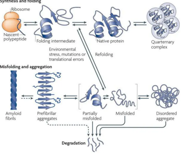
Protein aggregation as a strategy for protein quality control
When proteins do not fold properly and fail to be degraded, aggregation can occur as a result of inappropriate interactions between exposed hydrophobic residues that would be buried within the folded structure of the protein (Fig. 5, (Tyedmers et al., 2010)). Types of protein aggregates were historically defined by electron microscopy as either amyloid or amorphous. Amyloid aggregates are highly ordered, characterized by cross-β sheet secondary structure instead of the typical folded state of the protein (Dobson, 2003; Fändrich and Dobson, 2002). The amyloid form of some proteins can perform functions separate from the soluble form of the protein such as translational control in yeast meiosis and long-term memory persistence in Drosophila (Berchowitz et al., 2015; Majumdar et al., 2012). However, amyloid proteins are perhaps most known for their role in disease phenotypes including Alzheimer’s, Parkinson’s, and prion diseases (Knowles et al., 2014).
Amorphous aggregates have no defined structure and it is becoming apparent that they vary widely in their nature. The types of aggregates formed varies based on both the identity of the aggregating proteins and the conditions that cause aggregation (Stathopulos et al., 2003; Wang et al., 2010). Whether amorphous aggregates are toxic to cells will be discussed below.
Figure 5. Protein folding and aggregation (adapted from (Tyedmers et al., 2010)) Protein folding begins at the ribosome as nascent chains adopt three dimensional structures, resulting in the formation of native proteins. Nascent chains, partially folded intermediates, and orphan complex subunits are particularly vulnerable to misfolding and aggregation because they contain hydrophobic stretches that become buried in the core of the fully folded protein or protein complex. Molecular chaperones aid in the process of protein folding and prevent aggregation by binding these exposed hydrophobic residues. When aggregation does occur,
proteins can form cross-β sheets resulting in the formation of amyloids or disordered aggregates that lack any defining structure. Proteins in aggregates can then be disaggregated and either refolded or degraded. Entire aggregates can also be degraded by selective autophagy.
Once an aggregate has formed, cells generally have two mechanisms for removing them: disaggregation and autophagy. In yeast, disaggregation is carried out by Hsp70 (Ssa1), Hsp40 (Ydj1), and the AAA+ ATPase Hsp104 (Glover and Lindquist, 1998). Hsp104 functions as a homohexameric ring, with each subunit capable of hydrolyzing ATP. Hsp70 and Hsp40 transfer proteins from the aggregates to the central pore of the Hsp104 ring (Haslberger et al., 2007; 2008). Hsp104 then threads the substrate through its central pore by hydrolyzing ATP, thus disentangling it from the aggregate and allowing for refolding or degradation (Mogk et al., 2018). Metazoans notably lack Hsp104, however they still effectively eliminate protein aggregates (Pinto et al., 1991). Instead, separate classes of metazoan HSP40s form transient complexes that are capable of conferring disaggregation activity to HSP70 (Nillegoda et al., 2015). Precisely how this system is capable of removing proteins from aggregates is still unknown.
Rather than individually disaggregate proteins, cells can also degrade aggregates entirely utilizing the cellular recycling system known as autophagy. Autophagy involves the formation of a double membrane structure within the cytoplasm of the cell around the substrate that is to be degraded. Eventually, this autophagosome fuses with the lysosome (or vacuole in yeast) where the contents are deposited for degradation (Reggiori and Klionsky, 2013). This process can be used to turn over bulk cytoplasm or specific cargo such as entire organelles. In mammals, p62 functions as the cargo receptor for protein aggregates, tagging them for turnover by autophagy.
p62 interacts with ubiquitinated protein aggregates via its ubiquitin binding (UBA) domain and with the autophagosome membrane bound receptor, LC3, through its LC3-interacting region (Bjørkøy et al., 2005; Pankiv et al., 2007). In yeast, Cue5 has been identified as the link between ubiquitination and autophagy. Like, p62, Cue5 contains a ubiquitin-binding domain and was demonstrated to bind Atg8, the yeast homolog of LC3 (Lu et al., 2014). Cue5 is unrelated to p62, as it binds ubiquitin via its CUE domain rather than a UBA domain. Interestingly, the
identification of Cue5 in yeast led to the discovery of CUE domain containing autophagy
adaptors in mammals, suggesting that these proteins may be the ancestral autophagy adaptors for ubiquitin conjugated proteins (Lu et al., 2014). In yeast, disaggregation and refolding of
aggregated proteins appears to be favored over clearance either by disaggregation followed by proteolysis or aggregate autophagy (Wallace et al., 2015). Since disaggregation is a highly ATP demanding process, it is possible that under starvation conditions, autophagy may become the dominant pathway for aggregate clearance (Miller et al., 2015b).
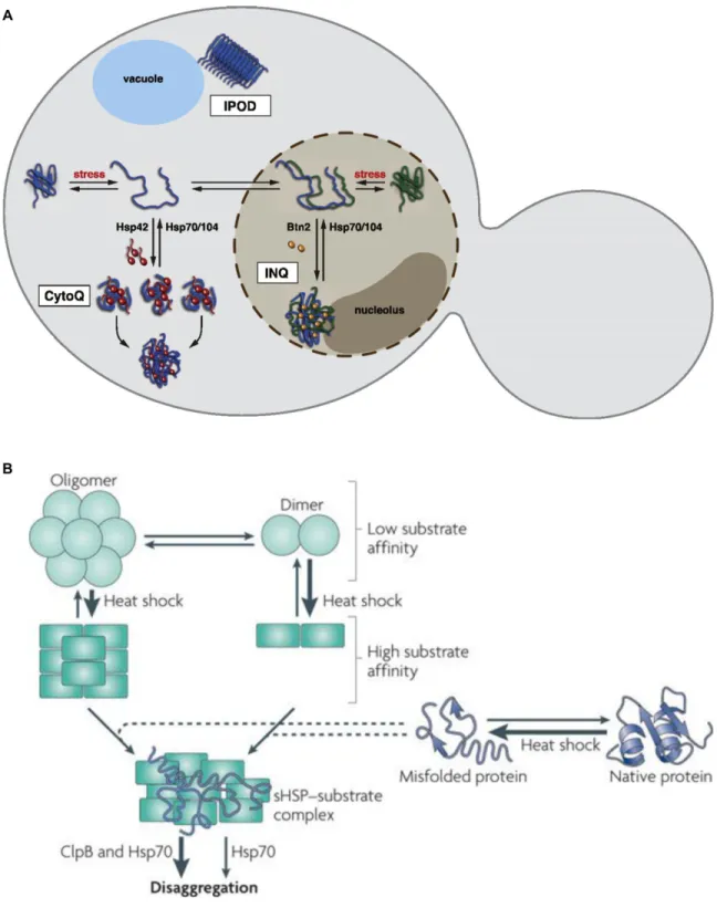
Increasing evidence suggests that rather than being an uncontrolled cellular catastrophe, protein aggregation may be an additional branch of protein quality control (Chen et al., 2011). Aggregating misfolded proteins instead of degrading or attempting to fold them may relieve components of the protein quality control system. Further, if misfolded proteins can be
sequestered, their potential toxicity caused by their ability to form inappropriate interactions with native or nascent proteins could be mitigated (Mogk et al., 2018). Indeed, yeast and mammals contain specific aggregate deposition sites. In yeast, the main sites for protein aggregation are called IPOD (insoluble protein deposit), INQ (intra nuclear quality control), and CytoQ (cytosolic quality control compartment) (Fig. 6A, (Kaganovich et al., 2008; Miller et al.,
the yeast prions (Kaganovich et al., 2008; Kumar et al., 2016). The INQ forms inside the
nucleus, adjacent to the nucleolus and contains misfolded nuclear and cytosolic proteins (Miller et al., 2015a). The CytoQ describes the cytoplasmic aggregation site, which begins as many distinct aggregates upon stress and eventually coalesces into a few or even just one aggregate (Specht et al., 2011). Interestingly, the formation of the INQ and CytoQ depend on the proteins Btn2, and Hsp42, respectively (Malinovska et al., 2012; Specht et al., 2011). Btn2 contains a nuclear localization sequence and upon induction by heat, colocalizes with the INQ. Hsp42 is a constitutively expressed, cytosolic small heat shock protein (sHSP) (Haslbeck et al., 2004). sHSPs are ATP-independent chaperones that function as “holdases” in that they bind partially unfolded substrates, rendering them competent for refolding upon alleviation of the stress (Fig. 6B, (Jakob et al., 1993; Ungelenk et al., 2016)). sHSPs frequently exist as dimers but can form oligomers of 48 or greater, facilitating their ability to bind many misfolded proteins
simultaneously (Basha et al., 2012). Because of their capacity to directly promote aggregation as well as fuse many small aggregates into larger aggregates, Hsp42 and other sHSPs have been called “aggregases” (Mogk et al., 2018). Mammalian cells contain a specialized aggregation site near their microtubule organizing center (MTOC) that forms upon stress called the aggresome (Johnston et al., 1998). Misfolded proteins are marked with K63 ubiquitin chains by the ubiquitin ligase, Parkin (Olzmann et al., 2007). Histone deacetylase 6 (HDAC6) simultaneously binds ubiquitin chains and the microtubule motor dynein, thereby allowing transport of the misfolded protein to the MTOC (Kawaguchi et al., 2003).
Figure 6. Small heat shock proteins (sHSPs) facilitate aggregation at deposition sites (adapted from (Miller et al., 2015a) (A) and (Tyedmers et al., 2010) (B))
(A) Yeast contain three identified aggregate deposition sites. The insoluble protein deposit (IPOD) resides near the vacuole which contains mainly amyloid proteins such as yeast prions.
The intra nuclear quality control (INQ) compartment contains misfolded proteins in the nucleus and requires Btn2 for its formation. The cytosolic quality control compartment (CytoQ) contains cytoplasmic misfolded proteins and requires the sHSP Hsp42. This compartmentalization has been proposed to help cells by allowing the concentration of dissagregation, refolding, and degradation factors at only a few sites of protein aggregation.
(B) sHSPs are ATP-independent molecular chaperones that hold misfolded proteins during stress. They can exist as dimers or high molecular weight oligomers. Some sHSPs undergo a conformational change at increased temperatures that make them become high affinity binders of misfolded proteins. sHSPs are thought to interact with proteins before they become fully
unfolded, thereby keeping them in a folding competent conformation for refolding after dissagregation by Hsp104 or Hsp70.
The existence of such cellular mechanisms to promote aggregation argues in favor of the hypothesis that aggregation is cytoprotective. In addition to response to protein-folding stress, increasing evidence suggests that the toxic form of amyloid proteins are actually smaller, soluble oligomers, and that the insoluble amyloid form of the protein is in fact cytoprotective (Caughey and Lansbury, 2003). Furthermore, amyloid forming proteins were found to only be toxic when present in the cytoplasm, but were harmless when targeted to the nucleus (Woerner et al., 2016). In C. elegans, it has been demonstrated that an increase in protein aggregation mediated by sHSPs correlates with longevity, indicating that sHSP induced protein aggregation can aid in protein quality control within aged cells (Walther et al., 2015). The emerging picture is that, although protein misfolding is undesirable, controlled aggregation can be an effective mechanism to cope with the problem.
EFFECTS OF AN ANEUPLOID PROTEOME Chromosome gain and loss cause protein aggregation
Aneuploidy presents a unique challenge to the protein quality control machinery. In the case of a gain of a single chromosome in a diploid cell, the cell must cope with a 1.5-fold
increase in the amount of gene products for hundreds to thousands of otherwise normal proteins. This is a rather different scenario than contexts in which protein homeostasis has previously been studied, namely where heat or chemical stress causes misfolding of the majority of the proteome or expression of a single toxic protein that misfolds. Aneuploid cells must fold or degrade this excess protein or else they are at risk of containing large amounts of potentially harmful
misfolded protein. Disomic yeast harbor twice as many protein aggregates compared to euploid cells when visualized by Hsp104 foci, regardless of which chromosome is amplified (Oromendia et al., 2012). In disomes, it appears that aggregation occurs as a result of the chaperone systems and the UPS becoming overwhelmed, as disomic cells have reduced capacity to fold a model Hsp90 substrate and are sensitive to chemical and genetic inhibition of chaperones and the UPS (Dodgson et al., 2016b; Oromendia et al., 2012; Torres et al., 2007). Likewise, mammalian cells trisomic or tetrasomic for single chromosomes are deficient in protein folding, increase
autophagy in an attempt to cope with misfolded protein, and are sensitive to drugs that inhibit chaperones and autophagy (Donnelly et al., 2014; Tang et al., 2011).
Although it is clear that increasing the amount of protein in the cell by gaining a chromosome causes proteotoxic stress, it is less intuitive that having less protein caused by chromosome loss would have the same effect. Heterogeneous populations of aneuploid cells generated by random chromosome mis-segregation show increased protein aggregation and lysosomal stress caused by altered autophagic flux respectively (Oromendia et al., 2012;
Santaguida et al., 2015; Stingele et al., 2013; Tang et al., 2011). The most compelling piece of evidence that chromosome loss causes proteotoxic stress comes from the generation of yeast monosomic for specific chromosomes (Beach et al., 2017). Like disomic and trisomic yeast, monosomic yeast form protein aggregates immediately following chromosome mis-segregation. In this system, the degree of protein aggregation is well correlated with the number of protein coding genes on the aneuploid chromosome(s) for chromosome gain and loss (Beach, 2016). This suggests that the source of proteotoxic stress in aneuploid cells originates from an
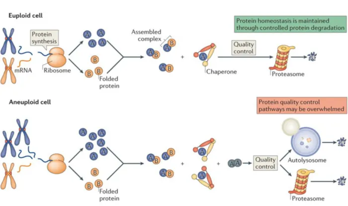
imbalance in the proteome, not simply excess protein. It has been proposed that this is caused by stoichiometric imbalance of protein complexes, where proteins that are expressed at levels higher than their binding partners due to aneuploidy are prone to misfold (Fig. 3, (Torres et al., 2008)). This idea will be explored in detail in Chapter 2.
In euploid cells (top) synthesis of protein complex subunits is proportional to their stoichiometry in the complex. Any slight deviations in synthesis results in excess subunits that can be dealt with by cellular protein quality control (PQC) mechanisms. In aneuploid cells (bottom), proteins are synthesized in accordance with their genetic copy number, resulting in stoichiometric
imbalance. Any protein complex with a subunit encoded on the aneuploid chromosome(s) then has excess subunits that need to be handled by PQC machinery. This has been proposed to overwhelm the PQC system, leading to the observed proteotoxic stress that is a universal consequence of aneuploidy.
Protein quality control defects underlie the fitness disadvantage of aneuploid cells Although aneuploid cells exhibit multiple different stresses, evidence suggests that coping with an imbalanced proteome underlies the reduced proliferation that is common among aneuploid cells. First, as described above, aneuploid cells are exquisitely sensitive to additional perturbations to the protein quality control network. Second, in disomic yeast, the magnitude of each strains proliferation disadvantage is well correlated with the number of protein coding genes on that chromosome (Torres et al., 2007). Additionally, yeast artificial chromosomes that contain no yeast protein-coding genes do not slow proliferation. Finally, increasing the cells capacity to fold and/or degrade proteins is one of the few mechanisms shown to rescue the proliferation defects of multiple aneuploid lines with distinct karyotypes. In a screen of disomic yeast for mutations that confer growth advantages to aneuploid cells, a loss of function mutation in the deubiquitinase, UBP6, was one of just three genetic alterations to rescue the proliferation of more than one disomic strain (Torres et al., 2010). Ubp6 functions at the proteasome to recycle ubiquitin from degraded substrates, however this activity has the potential to allow
substrates to escape degradation effectively antagonizing the UPS (Hanna et al., 2006; Leggett et al., 2002). As such, deletion of UBP6 causes increased degradation of some proteins. In disomic cells harboring a deletion in ubp6, it was shown that many proteins encoded by the extra
chromosome were more effectively degraded, thereby attenuating the imbalance caused by aneuploidy at the protein level (Torres et al., 2010). In human cells, increased expression of HSF1, the master transcription factor of the heat shock response, rescues sensitivity to autophagy inhibition by increasing expression of HSP90 and restoring the folding capacity of cells
(Donnelly et al., 2014). This corresponds with an increase in the proliferative capacity for trisomic cells (Donnelly et al., 2014). Thus, proteotoxic stress can account for at least part of the proliferative defects that are characteristic of aneuploidy.
Control of gene dosage by protein degradation
Because imbalance in the proteome is at the heart of many deleterious aneuploid phenotypes, aneuploid cells would benefit by decreasing expression of proteins from excess chromosomes, or increasing expression of proteins on lost chromosomes. Some mechanism of dosage compensation would thus restore the balance of the proteome to a euploid state. As discussed above, most genes are expressed in proportion to their genetic copy number at both the RNA and protein level, indicating that widespread dosage compensation on autosomes does not exist. However, it was initially reported that most genes encoded in extra copy fail to accumulate more protein, because only a few proteins were examined and most of them were subunits of protein complexes (Torres et al., 2007). Subsequent analyses utilizing mass spectrometry (ms) demonstrated that it is more common for protein levels to reflect genetic copy number (Pavelka et al., 2010; Torres et al., 2010). Later work generated a comprehensive picture of the proteome for individual gains of most yeast chromosomes utilizing mass spec, and identified many
proteins that fail to accumulate in aneuploid cells despite having twice as much mRNA
(Dephoure et al., 2014). These attenuated proteins are downregulated primarily by degradation because inhibition of proteasomal and autophagic degradation causes them to accumulate to levels predicted by their copy number. Further, disomic yeast do not utilize translational control as a dosage compensation strategy (Taggart and Li, 2018). These proteins that are degraded when encoded in excess are highly enriched for subunits of protein complexes, indicating that aneuploid cells utilize protein degradation to control stoichiometry of imbalanced complexes (Dephoure et al., 2014). The finding that aneuploid cells rely on protein degradation to perform dosage compensation is likely one reason why they are so sensitive to perturbations in the UPS.
Concluding remarks
Aneuploidy results in an imbalanced proteome that has numerous fitness penalties to both cells and organisms. These phenotypes occasionally derive from change in dosage of one or a few genes, but more commonly are caused by the simultaneous change in dosage of many genes. This large-scale change in the proteome of the cell results in proteotoxic stress, a universal feature of aneuploid cells that is at the heart of their proliferation defects. The molecular basis for aneuploid-associated proteotoxic stress is unknown, though it has been proposed that
stoichiometric imbalance of protein complexes could account for the increased burden on the protein quality control system. Additionally, aneuploidy represents an ideal model to study how cells cope with stoichiometric imbalance. Since eukaryotes rarely employ operons to ensure that stoichiometric ratios of complex subunits are properly maintained, how eukaryotic cells deal with imbalances in their proteome is a fundamental question.
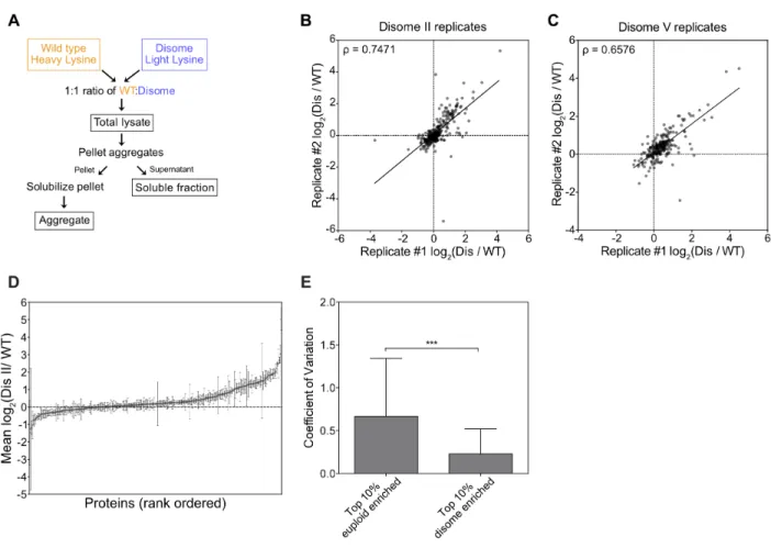
In this thesis, I have developed an assay to purify protein aggregates from aneuploid yeast and human cells to identify and quantify proteins within those aggregates using mass spectrometry. Uncovering the identity of proteins in aggregates revealed that stoichiometric imbalance of protein complexes causes protein aggregation in both aneuploid and euploid cells. Further, by combining this dataset with previously published data, I was able to track the fate of unassembled subunits of protein complexes genome-wide, revealing protein degradation and aggregation as mutually exclusive mechanisms to neutralize proteins that lack their binding partners. Remarkably, this work identified protein aggregation as a mechanism of dosage compensation, capable of eliminating excess protein by rendering it insoluble.
In light of recent findings that aggregation functions as an additional branch of protein quality control rather than a source of cellular toxicity, I propose that dosage compensation by protein aggregation may function as a cytoprotective mechanism in aneuploid and euploid cells. This is may be of particular importance to cancer cells. Cancer cells are frequently highly aneuploid, yet exhibit high proliferative potential relative to untransformed cells. How cancer cells avoid the negative fitness penalties caused by aneuploidy-induced stoichiometric imbalance could reveal therapeutic strategies. It will be important to study whether cancer cells employ aggregation or other dosage compensation mechanisms to maintain a balanced proteome.
References
Alberman, E.D., and Creasy, M.R. (1977). Frequency of chromosomal abnormalities in miscarriages and perinatal deaths. Journal of Medical Genetics 14, 313–315.
Andriani, G.A., Almeida, V.P., Faggioli, F., Mauro, M., Tsai, W.L., Santambrogio, L., Maslov, A., Gadina, M., Campisi, J., Vijg, J., et al. (2016). Whole Chromosome Instability induces senescence and promotes SASP. Nature Publishing Group 6, 35218.
Anfinsen, C.B. (1973). Principles that govern the folding of protein chains. Science 181, 223– 230.
Baker, D.J., Jin, F., Jeganathan, K.B., and van Deursen, J.M. (2009). Whole Chromosome
Instability Caused by Bub1 Insufficiency Drives Tumorigenesis through Tumor Suppressor Gene Loss of Heterozygosity. Cancer Cell 16, 475–486.
Balchin, D., Hayer-Hartl, M., and Hartl, F.U. (2016). In vivo aspects of protein folding and quality control. Science 353, aac4354.
Basha, E., O’Neill, H., and Vierling, E. (2012). Small heat shock proteins and α-crystallins: dynamic proteins with flexible functions. Trends Biochem. Sci. 37, 106–117.
Beach, R.R. (2016). Insights into the consequences of chromosome gains and losses in S. cerevisiae. Massachusetts Institute of Technology.
Beach, R.R., Ricci-Tam, C., Brennan, C.M., Moomau, C.A., Hsu, P.-H., Hua, B., Silberman, R.E., Springer, M., and Amon, A. (2017). Aneuploidy Causes Non-genetic Individuality. Cell 169, 229–242.e21.
Berchowitz, L.E., Kabachinski, G., Walker, M.R., Carlile, T.M., Gilbert, W.V., Schwartz, T.U., and Amon, A. (2015). Regulated Formation of an Amyloid-like Translational Repressor Governs Gametogenesis. Cell 163, 406–418.
Bjørkøy, G., Lamark, T., Brech, A., Outzen, H., Perander, M., Øvervatn, A., Stenmark, H., and Johansen, T. (2005). p62/SQSTM1 forms protein aggregates degraded by autophagy and has a protective effect on huntingtin-induced cell death. J. Cell Biol. 171, 603–614.
Blank, H.M., Sheltzer, J.M., Meehl, C.M., and Amon, A. (2015). Mitotic entry in the presence of DNA damage is a widespread property of aneuploidy in yeast. Mol. Biol. Cell 26, 1440–1451. Bonney, M.E., Moriya, H., and Amon, A. (2015). Aneuploid proliferation defects in yeast are not driven by copy number changes of a few dosage-sensitive genes. Genes & Development 29, 898–903.
Boveri, T. (1902). Uber mehrpolige Mitosen als Mittle zur Analyse des Zellkerns. Verhandl Phys-Med Ges Wulzburg NF 35, 67–90.
Boveri, T. (1914). Zur Frage der Entstehung maligner Tumoren (Jena, Germany: Gustav Fischer Verlag).
Byrd, J.C., Mrózek, K., Dodge, R.K., Carroll, A.J., Edwards, C.G., Arthur, D.C., Pettenati, M.J., Patil, S.R., Rao, K.W., Watson, M.S., et al. (2002). Pretreatment cytogenetic abnormalities are predictive of induction success, cumulative incidence of relapse, and overall survival in adult patients with de novo acute myeloid leukemia: results from Cancer and Leukemia Group B (CALGB 8461). Blood 100, 4325–4336.
Caughey, B., and Lansbury, P.T. (2003). Protofibrils, pores, fibrils, and neurodegeneration: separating the responsible protein aggregates from the innocent bystanders. Annu. Rev. Neurosci. 26, 267–298.
Chen, B., Retzlaff, M., Roos, T., and Frydman, J. (2011). Cellular strategies of protein quality control. Cold Spring Harb Perspect Biol 3, a004374.
Cimini, D., Howell, B., Maddox, P., Khodjakov, A., Degrassi, F., and Salmon, E.D. (2001). Merotelic kinetochore orientation is a major mechanism of aneuploidy in mitotic mammalian tissue cells. J. Cell Biol. 153, 517–527.
Conde, J., and Fink, G.R. (1976). A mutant of Saccharomyces cerevisiae defective for nuclear fusion. Proc Natl Acad Sci USA 73, 3651–3655.
Davoli, T., Xu, A.W., Mengwasser, K.E., Sack, L.M., Yoon, J.C., Park, P.J., and Elledge, S.J. (2013). Cumulative haploinsufficiency and triplosensitivity drive aneuploidy patterns and shape the cancer genome. Cell 155, 948–962.
Dephoure, N., Hwang, S., O'Sullivan, C., Dodgson, S.E., Gygi, S.P., Amon, A., and Torres, E.M. (2014). Quantitative proteomic analysis reveals posttranslational responses to aneuploidy in yeast. Elife 3, e03023.
Deshaies, R.J., and Joazeiro, C.A.P. (2009). RING domain E3 ubiquitin ligases. Annu. Rev. Biochem. 78, 399–434.
Deutschbauer, A.M., Jaramillo, D.F., Proctor, M., Kumm, J., Hillenmeyer, M.E., Davis, R.W., Nislow, C., and Giaever, G. (2005). Mechanisms of haploinsufficiency revealed by genome-wide profiling in yeast. Genetics 169, 1915–1925.
Dobson, C.M. (2003). Protein folding and misfolding. Nature 426, 884–890.
Dodgson, S.E., Kim, S., Costanzo, M., Baryshnikova, A., Morse, D.L., Kaiser, C.A., Boone, C., and Amon, A. (2016a). Chromosome-Specific and Global Effects of Aneuploidy in
Saccharomyces cerevisiae. Genetics 202, 1395–1409.
Dodgson, S.E., Santaguida, S., Kim, S., Sheltzer, J., and Amon, A. (2016b). The pleiotropic deubiquitinase Ubp3 confers aneuploidy tolerance. Genes & Development 30, 2259–2271.
Donnelly, N., Passerini, V., Dürrbaum, M., Stingele, S., and Storchová, Z. (2014). HSF1 deficiency and impaired HSP90-dependent protein folding are hallmarks of aneuploid human cells. The EMBO Journal 33, 2374–2387.
Douglas, N.R., Reissmann, S., Zhang, J., Chen, B., Jakana, J., Kumar, R., Chiu, W., and Frydman, J. (2011). Dual action of ATP hydrolysis couples lid closure to substrate release into the group II chaperonin chamber. Cell 144, 240–252.
Duncan, C.D.S., and Mata, J. (2011). Widespread cotranslational formation of protein complexes. PLoS Genet 7, e1002398.
Ellis, R.J. (2001). Macromolecular crowding: obvious but underappreciated. Trends Biochem. Sci. 26, 597–604.
Elsasser, S., and Finley, D. (2005). Delivery of ubiquitinated substrates to protein-unfolding machines. Nat Cell Biol 7, 742–749.
Emdin, S.O., Stenling, R., and Roos, G. (1987). Prognostic value of DNA content in colorectal carcinoma. A flow cytometric study with some methodologic aspects. Cancer 60, 1282–1287. Fändrich, M., and Dobson, C.M. (2002). The behaviour of polyamino acids reveals an inverse side chain effect in amyloid structure formation. The EMBO Journal 21, 5682–5690.
Finley, D. (2009). Recognition and processing of ubiquitin-protein conjugates by the proteasome. Annu. Rev. Biochem. 78, 477–513.
Finley, D., Ulrich, H.D., Sommer, T., and Kaiser, P. (2012). The ubiquitin-proteasome system of Saccharomyces cerevisiae. Genetics 192, 319–360.
Fournier, R.E., and Ruddle, F.H. (1977). Microcell-mediated transfer of murine chromosomes into mouse, Chinese hamster, and human somatic cells. Proc Natl Acad Sci USA 74, 319–323. Fredrickson, E.K., Rosenbaum, J.C., Locke, M.N., Milac, T.I., and Gardner, R.G. (2011). Exposed hydrophobicity is a key determinant of nuclear quality control degradation. Mol. Biol. Cell 22, 2384–2395.
Gasch, A.P., Spellman, P.T., Kao, C.M., Carmel-Harel, O., Eisen, M.B., Storz, G., Botstein, D., and Brown, P.O. (2000). Genomic Expression Programs in the Response of Yeast Cells to Environmental Changes. Mol. Biol. Cell 11, 4241–4257.
Geiler-Samerotte, K.A., Dion, M.F., Budnik, B.A., Wang, S.M., Hartl, D.L., and Drummond, D.A. (2011). Misfolded proteins impose a dosage-dependent fitness cost and trigger a cytosolic unfolded protein response in yeast. Proc. Natl. Acad. Sci. U.S.a. 108, 680–685.
Glover, J.R., and Lindquist, S. (1998). Hsp104, Hsp70, and Hsp40: a novel chaperone system that rescues previously aggregated proteins. Cell 94, 73–82.
Goh, P.Y., and Kilmartin, J.V. (1993). NDC10: a gene involved in chromosome segregation in Saccharomyces cerevisiae. J. Cell Biol. 121, 503–512.
Groll, M., Ditzel, L., Löwe, J., Stock, D., Bochtler, M., Bartunik, H.D., and Huber, R. (1997). Structure of 20S proteasome from yeast at 2.4 A resolution. Nature 386, 463–471.
Hanks, S., Coleman, K., Reid, S., Plaja, A., Firth, H., FitzPatrick, D., Kidd, A., Méhes, K., Nash, R., Robin, N., et al. (2004). Constitutional aneuploidy and cancer predisposition caused by biallelic mutations in BUB1B. Nat. Genet. 36, 1159–1161.
Hanna, J., Hathaway, N.A., Tone, Y., Crosas, B., Elsasser, S., Kirkpatrick, D.S., Leggett, D.S., Gygi, S.P., King, R.W., and Finley, D. (2006). Deubiquitinating enzyme Ubp6 functions noncatalytically to delay proteasomal degradation. Cell 127, 99–111.
Hartl, F.U., Bracher, A., and Hayer-Hartl, M. (2011). Molecular chaperones in protein folding and proteostasis. Nature 475, 324–332.
Hartwell, L.H., and Smith, D. (1985). Altered fidelity of mitotic chromosome transmission in cell cycle mutants of S. cerevisiae. Genetics 110, 381–395.
Haslbeck, M., Braun, N., Stromer, T., Richter, B., Model, N., Weinkauf, S., and Buchner, J. (2004). Hsp42 is the general small heat shock protein in the cytosol of Saccharomyces cerevisiae. The EMBO Journal 23, 638–649.
Haslberger, T., Weibezahn, J., Zahn, R., Lee, S., Tsai, F.T.F., Bukau, B., and Mogk, A. (2007). M domains couple the ClpB threading motor with the DnaK chaperone activity. Molecular Cell 25, 247–260.
Haslberger, T., Zdanowicz, A., Brand, I., Kirstein, J., Turgay, K., Mogk, A., and Bukau, B. (2008). Protein disaggregation by the AAA+ chaperone ClpB involves partial threading of looped polypeptide segments. Nat. Struct. Mol. Biol. 15, 641–650.
Hasle, H., Clemmensen, I.H., and Mikkelsen, M. (2000). Risks of leukaemia and solid tumours in individuals with Down's syndrome. The Lancet 355, 165–169.
Hassold, T.J., and Jacobs, P.A. (1984). Trisomy in man. Annu. Rev. Genet. 18, 69–97. Heck, J.W., Cheung, S.K., and Hampton, R.Y. (2010). Cytoplasmic protein quality control degradation mediated by parallel actions of the E3 ubiquitin ligases Ubr1 and San1. Proc. Natl. Acad. Sci. U.S.a. 107, 1106–1111.
Horwich, A.L., and Fenton, W.A. (2009). Chaperonin-mediated protein folding: using a central cavity to kinetically assist polypeptide chain folding. Q. Rev. Biophys. 42, 83–116.
Hwang, S., Gustafsson, H.T., O'Sullivan, C., Bisceglia, G., Huang, X., Klose, C., Schevchenko, A., Dickson, R.C., Cavaliere, P., Dephoure, N., et al. (2017). Serine-Dependent Sphingolipid Synthesis Is a Metabolic Liability of Aneuploid Cells. Cell Rep 21, 3807–3818.