HAL Id: inserm-01980579
https://www.hal.inserm.fr/inserm-01980579
Submitted on 14 Jan 2019
HAL is a multi-disciplinary open access
archive for the deposit and dissemination of
sci-entific research documents, whether they are
pub-lished or not. The documents may come from
teaching and research institutions in France or
abroad, or from public or private research centers.
L’archive ouverte pluridisciplinaire HAL, est
destinée au dépôt et à la diffusion de documents
scientifiques de niveau recherche, publiés ou non,
émanant des établissements d’enseignement et de
recherche français ou étrangers, des laboratoires
publics ou privés.
Emergence of Wesselsbron virus among black rat and
humans in Eastern Senegal in 2013
Moussa Diagne, Martin Faye, Oumar Faye, Abdourahmane Sow, Fanny
Balique, Mbacké Sembène, Laurent Granjon, Pascal Handschumacher,
Ousmane Faye, Mawlouth Diallo, et al.
To cite this version:
Moussa Diagne, Martin Faye, Oumar Faye, Abdourahmane Sow, Fanny Balique, et al.. Emergence
of Wesselsbron virus among black rat and humans in Eastern Senegal in 2013. One Health, Elsevier,
2017, 3, pp.23-28. �10.1016/j.onehlt.2017.02.001�. �inserm-01980579�
Emergence of Wesselsbron virus among black rat and humans in Eastern
Senegal in 2013
Moussa M. Diagne
a,b, Martin Faye
a,b, Oumar Faye
a, Abdourahmane Sow
a, Fanny Balique
a,
Mbacké Sembène
b,c, Laurent Granjon
c, Pascal Handschumacher
d, Ousmane Faye
a,
Mawlouth Diallo
a, Amadou A. Sall
a,⁎
aInstitut Pasteur de Dakar, Dakar, Sénégal
bUniversité Cheikh Anta Diop de Dakar, Dakar, Sénégal c
IRD CBGP, CS 30016, 34988 Montferrier-sur-Lez cedex, France
d
IRD, UMR 912 SE4S, IRD-INSERM-U2, France
a b s t r a c t
a r t i c l e i n f o
Article history:
Received 7 November 2016
Received in revised form 23 January 2017 Accepted 7 February 2017
Available online 9 February 2017
Wesselsbron disease is a neglected mosquito transmitted Flavivirus infection that causes abortions and has tera-togenic effects on sheep and cattle in Africa. Human can also be infected. The detection of human or animal cases is complicated by the non-specific symptoms close to Rift Valley Fever (RVF) in domestic livestock species or Dengue like syndrome in humans. Then, these detections are usually made during RVF investigations in sheep. These domestic animals should take a role in the life cycle of the virus but some evidences of Wesselsbron virus (WSLV) presence in wild animals suggest that the latter may be involved in the virus maintenance in nature. However, the reservoir status of wild vertebrate in general and rodents particularly for WSLV is only based on an isolation from a Cape short-eared gerbil in southern Africa. Most of WSLV isolations are from southern parts of Africa even if it has been found in western and central Africa or Madagascar. In Senegal, there are serological evidences of WSLV circulation in human since the 1970s and some isolations, the last one of which dates back in 1992. Despite the detection of the virus on mosquitoes until the 2000s in different parts of the country, no new human case has been noted. In this paper, we report the WSLV re-emergence in eastern Senegal in 2013 with 2 human cases and itsfirst isolation from a black rat Rattus rattus. Sequencing analyses show the circulation of the same strain between these humans and the commensal rodent. The putative impact on WSLV transmission to human populations could be more important if the reservoir status of the black rat is confirmed. Focused sur-vey in human populations, specific entomological and mammalogical investigations would permit a better un-derstanding of the life cycle of the virus and its impact on public health.
© 2017 Published by Elsevier B.V. This is an open access article under the CC BY-NC-ND license (http://creativecommons.org/licenses/by-nc-nd/4.0/). Keywords: Wesselsbron virus Rodents Black rat Eastern Senegal 1. Introduction
Wesselsbron disease (WSL) is a mosquito borne infection of sheep and cattle in Africa which can also infect human in whom fever and my-algia are the most common symptoms. It is caused by Wesselsbron virus (WSLV) which belongs to the Flavivirus genus. WSLV wasfirst isolated in blood of a febrile man and dead lamb during an outbreak in 1955 in Wesselsbron in the Free State Province, South Africa[17,34].
The disease in sheep is clinically similar to Rift Valley fever (RVF) with abortions and 20% mortality in pregnant ewes. Hydrops amnii and teratogenic effects such as arthrogryposis, hydrocephaly or neuro-genic muscular atrophy are also observed in lambs[4]whereas infection
causes less severe fever in goat, cattle, and pig[5]. WSL has been de-scribed as a cause of neurological disease in two horses of South Africa (Venter et al., 2008)[38]. WSL outbreaks can be unnoticed as they are often concomitant with RVF epidemics in South Africa[35]and its inci-dence can be underestimated. In humans, the infection causes arthral-gia, myalgia and fever during a short and mild acute phase[33].
WSLV has a wide geographic distribution in Africa[34]and viral iso-lations from mosquitoes have been reported in South Africa, Botswana, Zimbabwe, Uganda, Mozambique, Uganda, Cameroon, Central African Republic, Mauritania, Senegal, Nigeria, DR Congo and Madagascar[36]. Aedes mosquitoes are generally associated with virus detection[18].
WSLV was also isolated from several domestic livestock species in-cluding camels, cattle, pigs, donkeys and horses, and serological evi-dence of its circulation was found in wild animals including South African zebras[2]and wild ruminants from Chad[2,34]which raised the question of its reservoir. The reservoir status of wild vertebrate for
⁎ Corresponding author.
E-mail address:asall@pasteur.sn(A.A. Sall).
http://dx.doi.org/10.1016/j.onehlt.2017.02.001
2352-7714/© 2017 Published by Elsevier B.V. This is an open access article under the CC BY-NC-ND license (http://creativecommons.org/licenses/by-nc-nd/4.0/).
Contents lists available atScienceDirect
One Health
WSLV is also suspected since an isolation from Cape short-eared gerbil Desmodillus auricularis in southern Africa (Kokernot et al., 1960)[39].
Between 1955 and 2011, 35 WSLV isolates were obtained from humans, mostly from Central African Republic (CAR) and South Africa
[34,37], Jupp and Kemp, 1998[40],[13,39]. In western Africa, most human isolates are from Senegal (4 strains in 1965, 1974, 1982 and 1992).
In Senegal, WSLV was serologically detected in most of the children from Upper Casamance and Eastern Senegal between 1972 and 1975
[29]. Swanepoel[34]reports a laboratory-acquired infection in 1965 and another one in 1974. A human infected isolation was also obtained in 1992 in the capital city Dakar (Annual report of Institut Pasteur de Dakar (IPD), 1992) after a previous one 10 years before.
Even if Monlun et al.[26]found that WSLV did not seem to represent a major public health concern in southeastern Senegal, entomological investigations between 1998 and 1999 in some parts of Senegal and Mauritania, spanning the Senegal River basin permitted to detect 51 WSLV strains from Aedes vexans[11]. Already,N40 strains were found from 1974 to 1999 in Kedougou, Barkedji and Yonofere, a village in Saint Louis region (Annual report of IPD, 1999).
Moreover, recent emergences of viruses within populations initially naive showed the necessity of anticipation of risk factors. In this pur-pose, in the Kedougou region (southeastern Senegal), a surveillance of arboviruses has been undertaken on mosquitoes and human populations.
In this paper, we report on the re-emergence WSLV in Eastern Sen-egal in 2013 with 2 human cases and itsfirst isolation from the rodent Rattus rattus (the black rat).
2. Material and methods 2.1. Study sites
In the Kedougou region (Fig. 1), a surveillance of acute febrile illness (AFI) on human populations has been led since 2009[31]. Patients pre-senting AFI were recruited from seven healthcare facilities of the Kedougou region, including Ninefesha rural hospital, Kedougou and
Saraya health centres, Bandafassi and Khossanto health posts, the Kedougou military health post, and the Catholic Mission mobile team, which targets populations in remote areas.
2.2. Samples collection
Samples were collected from human presenting acute febrile ill-nesses and from rodent trapped in Kedougou. Serum or blood speci-mens of patients with AFI, as well as brain, serum and blood collected from rodents, are then submitted to the Arboviruses and Hemorrhagic Fever viruses Unit of IPD for a diagnostic of arboviruses.
Trapping sessions targeting rodents and shrews have been under-taken in a number of localities of Eastern Senegal to study the putative circulation of arboviruses in these small mammal populations. Briefly, traps (wire-mesh locally made and folding aluminium Sherman© traps) were set inside buildings for trapping sessions of one to six con-secutive days with peanut butter as bait[6]. One of each type of traps was set per room and inspected for captures each morning. Each trapped specimen was identified to the species level based on morpho-logical or geographical knowledge[14,16]or, in case of ambiguity, by further molecular or chromosomal analyses (see details in[12,15,21]). Trapped individuals were euthanatized by cervical dislocation as rec-ommended by Mills et al.[25]. The brain, the tissues and the serum of each individual were separated and put in dry ice for transportation to IPD. Permission to work within the different villages was obtained from appropriate authorities, and animals were correctly treated fol-lowing Sikes et al.[32].
2.3. Viral isolation and identification
Each sample was inoculated into 2- to 3-days-old suckling mice for isolation of live virus (Shope and Sather, 1979)[41]according to a pro-tocol approved by the National Health Laboratory Service Animal Ethics Committee (reference number, 107/06).
Suckling mice derived viruses were identified by reverse transcrip-tion PCR targeting a segment at the partial NS5 gene using the generic
Flavivirus primers pair FD3/FU1[20]and Sanger sequencing performed out-of-house by GENEWIZ, Inc. (Essex, UK).
2.4. RNA extraction and RT-PCR amplification
RNA was extracted from suckling mice derived viruses stocks using the QIAamp RNA Viral Kit (Qiagen, Hilden, Germany) according to the manufacturer's recommendations. RNA was eluted in 50μl of AVE buffer and stored at−80 °C until use. For cDNA synthesis, 10 μl of viral RNA was mixed with 1μl of the reverse primer (2 pmol) and the mixture was heated at 95 °C for 2 min. Reverse transcription was performed in 20μl mixture containing mixed of 2.5 U RNasin (Promega, Madison, USA), 1 μl of deoxynucleotide triphosphate (dNTP) (10 mM each DNTP), 5 U of AMV reverse transcriptase (Promega, Madison, USA) and incubated at 42 °C for 60 min. PCR products were generated using the primers FD3/FU1 to amplify partial NS5 region. Five microliters of cDNA were mixed with 10× buffer, 5μl of each primer, 5 μl of dNTPs 10 mM, 3μl of MgCl2, and 0.5μl of Taq polymerase (Promega, Madison, USA).
2.5. Genome sequencing and analysis
The genomic polyprotein of each confirmed WSLV strain was obtain-ed using overlapping sets of primers (Table 1) designed with the com-plete genome of the WSLV reference strain from South Africa (GenBank accession number: gi| 389595538|gb|JN226796.1|;https://
www.ncbi.nlm.nih.gov/nuccore/389595538/, consulted on January
07th 2017). Sequences obtained were merged using EMBOSS Merger software andfinal results were analyzed using the Basic Local Align-ment Tool (BLAST,www.ncbi.nlm.nih.gov/).
Nucleotide sequences alignment were generated using the MUSCLE algorithm implemented in Mega version 6 (Tamura, Stecher, Peterson, Filipski, and Kumar 2013)[42].
3. Results
3.1. Isolation and identification of WSLV in black rat sample
Between May 2012 and December 2013, 6 trapping sessions yielded 1313 small mammal captures. After inoculation in suckling mice, WSLV was isolated from the brain of a male black rat trapped in November 2013 in Kedougou. Day 4-brain derived virus identity was done by RT-PCR targeting the Flavivirus partial NS5. The RT-RT-PCR amplicon obtained showed a sequence which was highly conserved after comparison to the reference strain (GenBank accession number: gi | 389595538 | gb | -JN226796.1|), with 99% of homology.
3.2. Isolation and identification of WSLV in human sera
Two human WSLV cases from the region of Kedougou were con-firmed from patients showing AFI. The first case was a 4-year-old girl from Thiabedji (Fig. 1), a village located about 35 km from Kedougou, presenting in local healthcare facility in July 2013 with a fever over 39 °C, headache and diffuse pains associated with malaria. The second one was a 30-year-old woman from Bantaco (Fig. 1) presenting jaun-dice and fever in February 2014 in Kedougou and who was found Hep-atitis E positive. Both samples were tested by RT-PCR. Amplification of polymerase region (NS5) showed a similitude of 100% and 99% respec-tively for thefirst and second case compared to the strain found in black rats. An additional test of complementfixation test targeting WSLV was performed as described by Casey[3]on the suckling mice derived virus obtained from the Thiabedji case sample to check the consistency with sequencing results (data not shown).
These two co-infected WSLV human cases recovered after few days without sequelae.
Table 1
Designed primers used for genome sequencing.
Primers pair Primers name Orientation Primers position Sequence (5′ → 3′)
1 WSL1F F 4nt–23nt ATATTCTGCGTGCTAATCGT WSL1R R 1467nt–1486nt CCATAGCCTGTAAAAGCAAC 2 E-FW-WSL F 1387nt–1406nt CCACTCAGGAGCAAAGAAGG E-REV-WSL R 2362nt–2381nt TGAAGCCCATTGACATTGAA 3 WSL3F F 2392nt–2411nt GAGCCTTACTGCTAGTGCTG WSL3R R 3829nt–3848nt GATCTCCTAATGCAAGTTGG 4 WSL4F F 3559nt–3578nt GATGAAGAGGTTCTCCATGA WSL4R R 4996nt–5015nt CATAAAGGCCAATGACATCT 5 WSL5F F 4741nt–4760nt TGCAGGAAAAAGAATGACTC WSL5R R 6180nt–6199nt AACTGGCATGTCAAGTCTCT 6 WSL6F F 6107nt–6126nt ACCAAGACAACAACAAGTCC WSL6R R 7571nt–7590nt CCATTGTAAGCAAGTCCAAT 7 WSL7F F 7303nt–7322nt TCCAGTAGTTGATGGGAATC WSL7R R 8368nt–8387nt ACACGTCTCCTTCTATGACG 8 WSL8F F 8297nt–8316nt AACATCACTCACATGGTCAA WSL8R R 9332nt–9351nt ACCACCTTGTTTTTGTATGC 9 NS5-FW-WSL F 9084nt–9104nt TGGGATTCYTAAATGAAGACC NS5-REV-WSL R 10,081nt–10,101nt GTCTGATGTGGATTGTCTTCT 10 WSL10F F 9556nt–9575nt GAGATGTTGGCTTGACAGAT WSL10R R 10,790nt–10,811nt CACTAGTTGGTTCTCAACTTCC
11 WSL Rat Gap F1 F 1041nt–1060nt GCAGCTGCGTGACTTTGATA
WSL Rat Gap R1 R 1575nt–1594nt GTCGTGAACCCATTGCTTGT
12 WSL Rat Gap F2 F 2156nt–2175nt ACAATGAAAGGAGCCCAACG
WSL Rat Gap R2 R 2873nt–2892nt ACCCGTATTGAGTTCCACAC
13 WSL Rat Gap F3 F 3356nt–3373nt CCAGAATGGTGCTGTCGC
WSL Rat Gap R3 R 4262nt–4281nt AGTAGCACTCCACCAACAGC
14 WSL Rat Gap F4 F 4669nt–4688nt AGTCGGAGTGGTGAAGGATG
WSL Rat Gap R4 R 5372nt–5391nt GTCGGCTCTAACATGCGATG
15 WSL Rat Gap F5 F 5508nt–5527nt TATTCATGTCAGCCACCCCT
WSL Rat Gap R5 R 6360nt–6379nt ACTGCATACCCTGGTGTCAA
16 WSL Rat Gap F6 F 6780nt–6799nt TAATACCAGAACCGGGCACA
WSL Rat Gap R6 R 7479nt–7499nt TGCCTTCAATAAGTGGTCCCA
17 WSL human Gap F1 F 5790nt–5809nt TGGCCACTGACATAGCTGAA
3.3. Phylogeny of WSLV isolates
Using 17 designed overlapping primers pairs (Table 1), sequencing of the coding regions of the virus genome permitted to obtain 10,643 bp and 10,648 bp for the human cases from Thiabedji (isolate WSLV-IP248525/SEN/2013; access number: KY056256) and Bantaco (isolate IP262451/SEN/2014; access number: KY056257), respectively, and 10,645 bp for the black rats' one (isolate IP259570/SEN/2013; ac-cess number: KY056258).
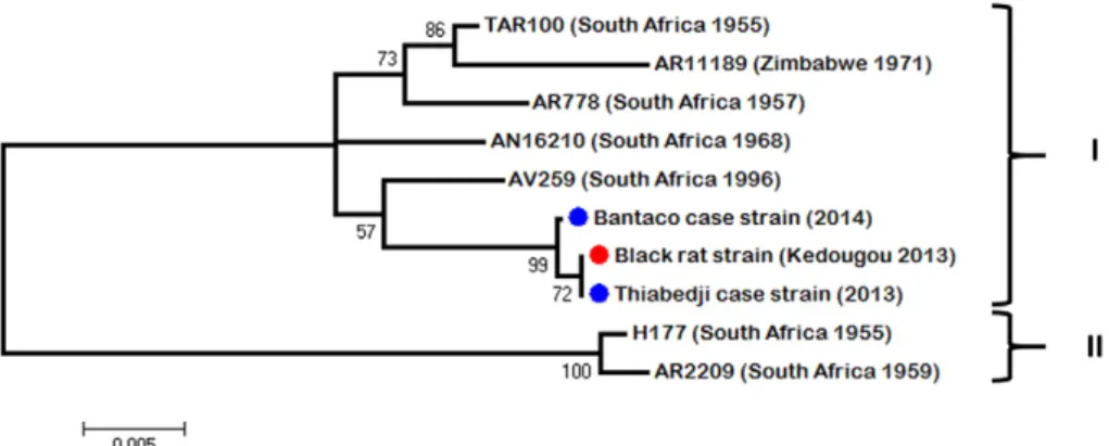
Pairwise nucleotide comparison of the three genomes yielded an av-erage of 1% nucleotide diversity across the black rat strain (ref. in GenBank) and the two human cases from Thiabedji (ref. in GenBank) and Bantaco (ref. in GenBank). The sequences of partial polymerase (NS5) and envelope (E) genes of the WSLV strains isolated in this study were aligned with the other available sequences obtained from isolates of South Africa, Kenya and Zimbabwe (from 1955 to 2011). Analysis of both regions showed two clades. The Senegalese strains iso-lated from eastern Senegal were included in afirst clade (I) containing the largest number of sequences from South Africa, Zimbabwe and Kenya (Fig. 2). The other one (II) (Fig. 3) contained some South African strains isolated before 1970s. Intra-clade diversity was at most 1%. Inter-clade diversity was of 6% in sequenced region of polymerase (NS5) and 8% in envelope (E). Despite this diversity, no geographical or spatial cor-relation could be observed as previously argued by Weyer et al.[36]. 4. Discussion
The Kedougou region is an area prone to infectious diseases[7,9,10]. The surveillance of AFI on human populations is then very important for the anticipation of possible emergence. In this study, thefirst human case of WSL disease was a co-infection of a malaria positive young girl from Bantaco. In Kedougou, malaria remains highly prevalent with a cir-culation of at least two Plasmodium species[28]. Sow et al.[32]showed that concurrent infection malaria/arboviruses occurred among 48.7% of patients infected with arboviruses in southeastern Senegal from July 2009 to March 2013. According to this previous study where co-infected
patients were significantly younger than non-co-infected ones, the WSL case from Bantaco is a 4-year-old girl. If mosquito biting infection seems obvious, this co-infection could be the consequence of consecutive bites from two different species. In fact, only 10 strains of WSLV have been isolated in Anopheles vectors from Senegal, Central African Republic and Cameroon from 1962 to 1999 (Annual report of Arboviruses and Hemorrhagic Fever Viruses Unit of the Institute Pasteur, Dakar, 1999). However we should note the possibility of an asymptomatic carriage of Plasmodium, which often occurs in Senegal as showed by Zwetyenga et al.[37]or Males et al.[22]for examples.
The second case from Thiabedji is another co-infection during a con-comitant hepatitis E virus (HEV) outbreak (personal communication). Thiabedji is a village located near to some little mining areas where breeding sites have proliferated promoting vector/human contacts in unhealthy environment.
An interesting fact in this study is the WSLV isolation in a black rat specimen trapped in Kedougou. To our knowledge, it is the second iso-lation of the virus from a rodent after D. auricularis in South Africa (Kokernot et al., 1960)[39]. The analysis showed that the same WSLV strain has circulated among the patients and the black rat. From this, it appears that this commensal rodent may have been involved in WSLV circulation in dwelling areas.
Previous studies on WSLV transmissibility on mice model showed various results. If Muspratt et al.[27]showed that about 90% of arti ficial-ly infected Ae. circumluteolus mosquitoes were able to transmit WSLV to mice while successful transmission rate was very low for most of other studies including Ae. caballus s.l.[18], Culex theileri[24], Aedes aegypti and Cx. quinquefasciatus[30]. Therefore, inoculation of WSLV by mos-quitoes seems to be exceptional in rodents and it can explain the small number of isolates obtained from them. The recent environmental changes in Kedougou with the substantial increase of mosquito breed-ing sites near of human habitats and gold minbreed-ing areas could foster the transmission on a black rat specimen. However, entomological stud-ies in this area at this same time did not permit any WSLV detection in the mosquito pools trapped (data not shown). As for many arboviruses, vertical transmission appears to not be sufficient for explaining virus
Fig. 2. Unrooted maximum likelihood tree of 948-bp of the NS5 region of WSLV strains isolated in this study (blue circle for human strain and red circle for black rat strain) and in previous ones from southern Africa countries.
maintenance in nature and secondary cycles involving vertebrates is an-other track study[8]. Except the Namaqua gerbil D. auricularis in which viremia induced by WSLV (7.2 LD50/ml; 1–3 days) could induce
infec-tion in mosquito[1,24], rodents appeared to be refractory to WSLV as showed by McIntosh [23] with Mastomys natalensis or Aethomys chrysophylus for example. However, the black rat was not involved in these studies. The Namaqua gerbil is a desert species most often meet in the dry Kalahari area, between Botswana, Namibia and western South Africa. The black rat is an urban species close to human habitats and also one of the most invasive in the world. The putative impact on human populations concerning WSLV transmission could be then more important if the reservoir status of this rat is confirmed. It would certainly be too anticipate to link these two last human cases with the recent introduction of black rat specimens in southeastern Senegal at the beginning of the 21st century[19]but investigations on the implica-tion of this species in the life cycle of WSLV should be undertaken.
Basic phylogenetic analysis of the genome of the three isolates showed that the same WSLV strain has probably circulated among humans and the black rat specimen in Kedougou region between 2013 and 2014. The current results let envisage the circulation of two lineages, afirst one (clade I) found in some countries of southern Africa, in Kenya and Senegal and another one (clade II) detected in South Africa between 1955 and 1960. More isolates and sequences from other parts of Africa could help for a better understanding of the phylogenetic situ-ation of WSLV at least in Africa.
WSLV impact on public health, although no fatal human case has been identified, may be underestimated given the lack of monitoring of the virus. In fact, most of the cases have been noted in a context of other investigations. The described WSLV human cases were found to be co-infection.
Further investigations are needed in human populations, insects and mammals to understand the life cycle and the impact on public health of the virus. Conferring to the hypothesis of rodents' implication in gener-al, the most abundant order among mammals, in the life cycle of the virus, combined with the Aedini populations' extension worldwide, WSLV could have a larger distribution than expected.
Financial support
This study is a part supported by the French National Research Agen-cy (ANR) CEP&S 2011 under the references ANR-11-CEPL-0010 and ANR-11-JSV7-0006 and by the Institut Pasteur (IPD24_01) de Dakar funds.
Disclosures regarding real or perceived conflicts of interest No competing interests in this scientific work.
Acknowledgements
We thank the CBGP teams of IRD (Institut de Recherche pour le Développement) of Montpellier, France and Dakar, Senegal for their outstanding work for the rodent trappings.
References
[1] Arbovirus Research Unit, National Institute for Virology, Sandringham, unpublished laboratory records, 1953 to 1985.
[2] B.J. Barnard, Antibodies against some viruses of domestic animals in southern Afri-can wild animals, Onderstepoort J. Vet. Res. 64 (2) (1997 Jun) 95–110.
[3] H.L. Casey, II. Adaptation of LCBF-method microtechnique, Standardized Diagnostic Complement Fixation and Adaptation to Microtest. U.S. Public Health Monograph No. 74, U.S. Public Health Service, Washington, D.C 1965, pp. 31–34.
[4] J.A.W. Coetzer, B.J.H. Barnard, Hydrops amnii in sheep associated with hydranencephaly and arthrogryposis with Wesselsbron disease and Rift Valley fever viruses as ethological agents, Onderstepoort J. Vet. Res. 44 (1977) 119–126.
[5] J.A. Coetzer, A. Theodoridis, Clinical and pathological studies in adult sheep and goats experimentally infected with Wesselsbron disease virus, Onderstepoort J Vet Res. 49 (1) (1982 March) 19–22.
[6] A. Dalecky, K. Ba, S. Piry, C. Lippens, C.A. Diagne, M. Kane, A. Sow, M. Diallo, Y. Niang, A. Konecny, N. Sarr, E. Artige, N. Charbonnel, L. Granjon, J.M. Duplantier, C. Brouat, Range expansion of the invasive house mouse Mus musculus domesticus in Senegal, West Africa: a synthesis of trapping data over three decades, 1983–2014, Mammal Rev. 45 (3) (2015) 176–190.
[7] Diawo Diallo, Amadou A. Sall, Michaela Buenemann, Rubing Chen, Oumar Faye, Cheikh T. Diagne, Ousmane Faye, Yamar Ba, Ibrahima Dia, Douglas Watts, Scott C. Weaver, Kathryn A. Hanley, Mawlouth Diallo, Landscape ecology of sylvatic chikungunya virus and mosquito vectors in Southeastern Senegal, PLoS Negl. Trop. Dis. 6 (6) (June 2012), e1649. Published online 2012 June 1210.1371/journa. pntd.0001649.
[8] D. Diallo, R. Chen, C.T. Diagne, Y. Ba, I. Dia, A.A. Sall, S.C. Weaver, M. Diallo, Bloodfeeding patterns of sylvatic arbovirus vectors in southeastern Senegal, Trans. R. Soc. Trop. Med. Hyg. 107 (3) (2013 Mar) 200–203,http://dx.doi.org/10.1093/ trstmh/trs095.
[9] Diawo Diallo, Amadou A. Sall, Cheikh T. Diagne, Oumar Faye, Kathryn A. Hanley, Michaela Buenemann, Yamar Ba, Ousmane Faye, Scott C. Weaver, Mawlouth Diallo, Patterns of a sylvatic yellow fever virus amplification in Southeastern Sene-gal, 2010, Am.J.Trop. Med. Hyg. 90 (6) (2014) 1003–1013 June 410.4269/ajtmh. 13-0404.
[10] Diawo Diallo, Amadou A. Sall, Cheikh T. Diagne, Oumar Faye, Ousmane Faye, Yamar Ba, Kathryn A. Hanley, Michaela Buenemann, Scott C. Weaver, Mawlouth Diallo, Zika Virus Emergence in Mosquitoes in Southeastern Senegal, 2011, PLoS One 9 (10) (2014), e109442. Published online 2014 October 1310.1371/journal.pone.0109442. [11]M. Diallo, P. Nabeth, K. Ba, A.A. Sall, Y. Ba, M. Mondo, L. Girault, M.O. Abdalahi, C. Mathiot, Mosquito vectors of the 1998–1999 outbreak of Rift Valley Fever and other arboviruses (Bagaza, Sanar, Wesselsbron and West Nile) in Mauritania and Senegal, Med. Vet. Entomol. 19 (2) (Jun 2005) 119–126.
[12] G. Dobigny, C. Tatard, M. Kane, P. Gauthier, C. Brouat, K. Bâ, J.-M. Duplantier, A cyto-taxonomic and DNA-based survey of rodents from Northern Cameroon and West-ern Chad, Mamm. Biol. 76 (2011) 417–427.
[13] J.-P. Durand, X. de Camaret, H. Zeller, A. Sall, F. Tock, E. Czarnecki, R. Régis, S. Guesdon, H. Tolou, First isolation of Wesselsbron virus in Chad, Virologie 5 (4) (2001) 305–306.
[14] L. Granjon, J.M. Duplantier, Les Rongeurs de l'AfriqueSahélo-soudanienne, IRD Edi-tions/Publications scientifiques du Muséum, Marseille, France, 2009.
[15] L. Granjon, G. Dobigny, The importance of cytotaxonomy in understanding the bio-geography of African rodents: Lake Chad murids as an example, Mammal Rev. 33 (2003) 77–91.
[16]M. Happold, D.C.D. Happold, Mammals of Africa, Vol. IV, Hedgehogs, Shrews and Bats, Bloomsbury Publishing, London, UK, 2013.
Fig. 3. Unrooted maximum likelihood tree of 845-bp of the E region of WSLV strains isolated in this study (blue circle for human strain and red circle for black rat strain) and in previous ones from southern Africa countries.
[17] N. Karabatsos (Ed.), International Catalogue of Arboviruses, Including Certain Other Viruses of Vertebrates, third ed., San Antonio, Texas: Am. Soc. Trop. Med. Hyg, The 1986–1995 Supplements to the International Catalogue, CDC Div. Vector-Borne In-fect. Dis., Ft. Collins, 1985.
[18] R.H. Kokernot, H.E. Paterson, B. De Meillon, Studies on the transmission of Wesselsbron virus by Aedes. (Ochlerotatus) caballus (Theo.), S. Afr. Med. J. 32 (1958) 546–548.
[19] A. Konečnỷ, Consequences of Anthropogenic Changes on Rodent Communities and Populations: Study Cases on Native and Introduced Species in Eastern Senegal, Université de Montpellier 2, 2009.
[20] G. Kuno, G.J.J. Chang, K.R. Tsuchiya, N. Karabatsos, C.B. Cropp, Phylogeny of the genus flavivirus, J. Virol. 10 (1) (1998) 73–83.
[21] E. Lecompte, C. Brouat, J.M. Duplantier, M. Galan, L. Granjon, A. Loiseau, K. Mouline, J.F. Cosson, Molecular identification of four cryptic species of Mastomys (Rodentia, Murinae), Biochem. Syst. Ecol. 33 (2005) 681–689.
[22] S. Males, O. Gaye, A. Garcia, Long-term asymptomatic carriage of Plasmodium falciparum protects from malaria attacks: a prospective study among Senegalese children, Clin. Infect. Dis. 46 (4) (Feb 15 2008) 516–522,http://dx.doi.org/10. 1086/526529.
[23] B.M. McIntosh, Susceptibility of some African wild rodents to infection with various arthropod borne viruses, Trans. R. Soc. Trop. Med. Hyg. 55 (1961) 63.
[24] B.M. McIntosh, The Epidemiology of Arthropod-borne Viruses in Southern Africa(D.Sc.thesis) University of Pretoria, Pretoria, 1980.
[25] J.N. Mills, J.E. Childs, T.G. Ksiazek, C.J. Peters, W.M. Velleca, Methods for Trapping and Sampling Small Mammals for Virologic Testing, U.S. Department of Health & Human Services, CDC, Atlanta, Georgia, USA, 1995.
[26] E. Monlun, H. Zeller, B. Le Guenno, M. Traoré-Lamizana, J.P. Hervy, F. Adam, L. Ferrara, D. Fontenille, R. Sylla, M. Mondo, et al., Surveillance of the circulation of ar-bovirus of medical interest in the region of eastern Senegal, Bull. Soc. Pathol. Exot. 86 (1) (1993) 21–28.
[27]J. Muspratt, K.C. Smithburn, H.E. Paterson, R.H. Kokernot, Studies on arthropod-borne viruses of Tongaland. X. The laboratory transmission of Wesselsbron virus by the bite of Aedes (Banksinella) circumluteolus Theo, S. Afr. J. Med. Sci. 22 (2– 3) (Oct 1957) 121–126.
[28] M. Niang, L.G. Thiam, A. Sow, C. Loucoubar, N.S. Bob, F. Diop, B. Diouf, O. Niass, A. Mansourou, M.L. Varela, R. Perraut, A.A. Sall, A. Toure-Balde, A molecular survey of acute febrile illnesses reveals Plasmodium vivax infections in Kedougou, southeast-ern Senegal, Malar. J. 14 (Jul 19 2015) 281, http://dx.doi.org/10.1186/s12936-015-0808-y.
[29] J. Renaudet, C. Jan, J. Ridet, C. Adam, Y. Robin, A serological survey of arboviruses in the human population of Senegal, Bull. Soc. Pathol. Exot. Filiales 71 (2) (Mar-Apr 1978) 131–140.
[30]P. Simasathien, L.C. Olson, Factors influencing the vector potential of Aedes aegypti and Culex quinquefasciatus for Wesselsbron virus, J. Med. Entomol. 10 (6) (Dec 30 1973) 587–590.
[31] A. Sow, C. Loucoubar, D. Diallo, O. Faye, Y. Ndiaye, C.S. Senghor, A.T. Dia, O. Faye, S.C. Weaver, M. Diallo, D. Malvy, A.A. Sall, Concurrent malaria and arbovirus infections in Kedougou, southeastern Senegal, Malar. J. 15 (2016) 47.
[32] Sikes RS, Gannon WI, the Animal Care and Use Committee of the American Society of Mammalogists (2011).
[33] R. Swanepoel, Wesselsbron virus disease, in: T.P. Monath (Ed.), The Arboviruses Ep-idemiology and Ecology, vol. 5, CRC Press, Inc., Boca Raton, FL, 1989.
[34] K.E. Weiss, D.A. Haig, R.A. Alexander, Wesselsbron virus—a virus not previously de-scribed, associated with abortion in domestic animals, Onderstepoort Vet. J. 27 (1956) 183–195.
[35] K.E. Weiss, Rift Valley fever—a review, Bull. Epizoot. Dis. Afr. 5 (1957) 431–458.
[36] J. Weyer, J. Thomas, P.A. Leman, A.A. Grobbelaar, A. Kemp, J.T. Paweska, Human cases of Wesselsbron disease, South Africa 2010–2011, Vector Borne Zoonotic Dis. 13 (5) (2013 May) 330–336,http://dx.doi.org/10.1089/vbz.2012.1181(Epub 2013 Mar 8). [37] J. Zwetyenga, C. Rogier, A. Spiegel, D. Fontenille, J.F. Trape, O. Mercereau-Puijalon, A cohort study of Plasmodium falciparum diversity during the dry season in Ndiop, a Senegalese village with seasonal, mesoendemic malaria, Trans. R. Soc. Trop. Med. Hyg. 93 (4) (Jul–Aug 1999) 375–380.
[38] M. Venter, S. Human, G.H. Gerdes, J. Williams, P.A. Leman, A. Kemp, et al., Investiga-tion of the pathogenesis of West Nile virus and other zoonoticflavi and alpha virus-es in humans and horsvirus-es in South Africa, Proceedings of the South African Society for Veterinary Epidemiology and Preventive Medicine 8th Annual Conference, 20– 22 August, Gauteng, South Africa, 2008.
[39]R.H. Kokernot, K.C. Smithburn, H.E. Paterson, B. de Meillon, Further isolations of Wesselsbron virus from mosquitos, S. Afr. Med. J. 34 (1960) 871–874.
[40]P.G. Jupp, A. Kemp, Studies on an outbreak of Wesselsbron virus in the Free State Province, South Africa. J. Am. Mosquito. Contr. 14 (1998) 40–45.
[41]E. Shope, G.E. Sather, Arboviruses, in: E.H. Lenette, N.J. Schmidt (Eds.), Diagnostic procedures for viral, rickettsial and chlamydial infections, American Public Health Association, Washington DC 1979, pp. 767–814.
[42] Koichiro Tamura, Glen Stecher, Daniel Peterson, Alan Filipski, Sudhir Kumar, MEGA6: Molecular Evolutionary Genetics Analysis version 6.0, Mol. Biol. Evol. 30 (2013) 2725–2729.