HAL Id: hal-01840327
https://hal.umontpellier.fr/hal-01840327
Submitted on 4 Dec 2020HAL is a multi-disciplinary open access archive for the deposit and dissemination of sci-entific research documents, whether they are pub-lished or not. The documents may come from teaching and research institutions in France or abroad, or from public or private research centers.
L’archive ouverte pluridisciplinaire HAL, est destinée au dépôt et à la diffusion de documents scientifiques de niveau recherche, publiés ou non, émanant des établissements d’enseignement et de recherche français ou étrangers, des laboratoires publics ou privés.
saladensis (Monogenea: Ancyrocephalidae) from Mugil
liza (Teleostei: Mugilidae) in southern Brazil
Natalia Marchiori, Antoine Pariselle, Joaber Pereira, Jean-François Agnèse,
Jean-Dominique Durand, Maarten P.M. Vanhove
To cite this version:
Natalia Marchiori, Antoine Pariselle, Joaber Pereira, Jean-François Agnèse, Jean-Dominique Durand, et al.. A comparative study of Ligophorus uruguayense and L. saladensis (Monogenea: Ancyrocephal-idae) from Mugil liza (Teleostei: MugilAncyrocephal-idae) in southern Brazil. Folia Parasitologica, Institute of Parasitology Czechoslovak Academy of, 2015, 62, �10.14411/fp.2015.024�. �hal-01840327�
http://folia.paru.cas.cz
This is an Open Access article distributed under the terms of the Creative Commons Attribution License (http://creativecommons.org/licenses/by/4.0), which permits unrestricted use, distribution, and reproduction in any medium, provided the original work is properly cited.
Research Article
Address for correspondence: N.C. Marchiori, Epagri, Rua Joaquim Garcia, s/nº, Centro, CEP 88340-000 Camboriú, SC, Brazil. Phone: +55 47 33651319; E-mail: namarchiori@gmail.com
© Institute of Parasitology, Biology Centre CAS Folia Parasitologica 2015, 62: 024 doi: 10.14411/fp.2015.024
A comparative study of
Ligophorus uruguayense and
L. saladensis (Monogenea: Ancyrocephalidae)
from Mugil liza (Teleostei: Mugilidae) in southern Brazil
Natalia C. Marchiori1, Antoine Pariselle2,3, Joaber Pereira Jr.4, Jean-François Agnèse2, Jean-Dominique Durand5
and Maarten P.M. Vanhove6,7,8,9
1 Empresa de Pesquisa Agropecuária e Extensão Rural de Santa Catarina (Epagri), Campo Experimental de Piscicultura de Camboriú,
Camboriú, Santa Catarina, Brasil;
2 Institut des Sciences de l’Évolution, IRD-CNRS-Université Montpellier 2, Montpellier, France; 3 Present address: Institut de Recherche pour le Développement (IRD), Yaoundé, Cameroon;
4 Laboratory of Parasite Biology of Aquatic Organisms, Oceanographic Institute, Federal University of Rio Grande Foundation, Rio
Grande, Rio Grande do Sul, Brazil;
5Institut de Recherche pour le Développement (IRD), University of Montpellier 2, Montpellier, France;
6 Laboratory of Biodiversity and Evolutionary Genomics, Department of Biology, University of Leuven, Leuven, Belgium;
7 Department of Botany and Zoology, Faculty of Science, Masaryk University, Brno, Czech Republic; 8 Invertebrates Unit, Biology Department, Royal Museum for Central Africa, Tervuren, Belgium;
9 Institute of Marine Biological Resources and Inland Waters, Hellenic Centre for Marine Research, Anavyssos, Greece
Abstract: Representatives of Ligophorus Euzet et Suriano, 1977 were found on the gills of Mugil liza Valenciennes caught in southern
Brazil. They were identified as Ligophorus uruguayense Failla Siquier et Ostrowski de Núñez, 2009 and Ligophorus saladensis Mar-cotegui et Martorelli, 2009, even though specific identification proved to be difficult due to inconsistencies in some diagnostic features reported for these two species. Therefore, a combined morphological and molecular approach was used to critically review the validity of these species, by means of phase contrast and confocal fluorescence microscopical examination of sclerotised hard parts, and assess-ing the genetic divergence between L. saladensis, L. uruguayense and their congeners usassess-ing rDNA sequences. The main morphological differences between the two species relate to the shape of the accessory piece of the penis and the median process of the ventral bar. The accessory piece in L. uruguayense is shorter than in L. saladensis, has a cylindrical, convex upper lobe and straight lower lobe (vs with the distal tip of the lower lobe turning away from the upper lobe in the latter species). The ventral bar has a V-shaped anterior median part in L. uruguayense (vs U-shaped in L. saladensis). The two species are suggested to be part of a species complex together with L. mediterraneus Sarabeev, Balbuena et Euzet, 2005. We recommend to generalise such comparative assessment of species of Ligophorus for a reliable picture of the diversity and diversification mechanisms within the genus, and to make full use of its potential as an additional marker for mullet taxonomy and systematics.
Keywords: morphology, molecular systematics, mullet, parasite, taxonomy
The identification of species belonging to Ligophorus Euzet et Suriano, 1977, monogenean gill parasites of mul-lets (Mugilidae), mainly relies on the morphology and size of the sclerotised parts of the haptor and of the male copu-latory complex (Blasco-Costa et al. 2012). However, dis-tinguishing between closely-related species may be very difficult, not only because of the small size of these struc-tures, but also because of their close resemblance under optical microscopy. Common flaws in describing species of Ligophorus include inadequate flattening of the haptoral
parts of fixed specimens and considering the differences between dorsal and ventral views of the ventral bar as in-traspecific or interspecific character variation (Dmitrieva et al. 2009a and references therein). Overlooking functional aspects of the structures used for morphological iden-tification has also led to errors (Dmitrieva et al. 2009b). Moreover, the relationship between morphology-based and molecular taxonomy is not yet clear, although congruence between the two approaches was reported to be high by Sarabeev and Desdevises (2014).
To date, six species of Ligophorus were described from
Mugil liza Valenciennes (syn. Mugil platanus Günther –
see Fraga et al. 2007) in South America: Ligophorus
uru-guayense Failla Siquier et Ostrowski de Núñez, 2009 from Laguna de Rocha, Uruguay, Ligophorus saladensis Marco-tegui et Martorelli, 2009 from Samborombón Bay, Argen-tina and four other species from the Guandu River, state of Rio de Janeiro, Brazil, namely L. brasiliensis, L.
guan-duensis, L. lizae and L. tainhae, all described by Abdallah
et al. (2009), i.e. Abdallah, Azevedo et Luque, 2009. Specimens of M. liza from southern Brazil were sam-pled for investigation of their monogenean fauna. Numer-ous individuals of Ligophorus were found on the gills of this fish host. They were identified as L. uruguayense and
L. saladensis, even though specific identification proved to
be difficult mostly because some of the diagnostic morpho-logical characters reported for these species (e.g. presence/ absence of transverse annulations at the distal end of the vaginal tube as well as the presence of a thick process at the distal end of the inner root of ventral anchors) seemed to vary continuously in the sample. In addition, the male copulatory complex of these two closely-related species may be very similar, depending on the position in which this structure is observed. Therefore, the validity of both species needed to be critically reviewed. In a recent revi-sion, Sarabeev et al. (2013) indicated problems in the orig-inal descriptions of these species, such as a surprisingly wide range in haptoral morphometrics for L. uruguayense and recommended to revise the morphology of the repro-ductive organs for both species.
The present study critically reviews the validity of
L. uruguayense and L. saladensis using an integrated
ap-proach combining both morphological and molecular anal-yses.
MATERIALS AND METHODS
In October 2012, thirty fingerlings of Mugil liza (total length of 3.0 ± 0.4 cm; weight of 0.3 ± 0.9 g) were collected from a stream that flows into the Atlantic Ocean at Querência, Cassino Beach, Rio Grande do Sul, Brazil (32°11'S; 52°10'W). For each fish specimen, gills were excised, fixed in 95% ethanol and kept separately from the rest of the body, which was then also fixed in 95% ethanol and kept in another glass jar.
Monogeneans were collected from the fish gills and some of them were mounted in Hoyer’s medium (Humason 1979) on a slide to study the morphology of the sclerotised hard parts using a Reichert-Jung Polyvar compound microscope at a magnifica-tion of ×1 000 using interference phase contrast.
Specimens found in this host were identified as Ligophorus uruguayense and L. saladensis, both originally described from M. liza in Uruguay and Argentina, respectively (Failla Siquier and Ostrowski de Núñez 2009, Marcotegui and Martorelli 2009). Both species are very similar to each other, but could be most reli-ably distinguished on the basis of the shape of the accessory piece of the male copulatory complex (see Failla Siquier and Ostrowski de Núñez 2009, Marcotegui and Martorelli 2009). The specimens that could not be identified to the species level reliably were ex-cluded from the morphometrical study. To confirm the validity of
both species, specimens that could be unquestionably assigned to one of the species were used for molecular study.
Measurements of the sclerotised structures in 18 specimens of L. uruguayense and 10 of L. saladensis (except for vagina length, which was measured in one and three specimens, respectively) were taken as defined by Failla Siquier and Ostrowski de Núñez (2009) using a Leica DM2500 microscope and LAS 6.3 software. They are given in micrometres (µm), with the range followed by the mean in parentheses. The following abbreviations for the characters are used throughout the text: DAA – dorsal anchor to-tal length; DAB – dorsal anchor main part length; DAC – dorsal anchor outer root length; DAD – dorsal anchor inner root length; DDIOR – distance between inner and outer root of dorsal anchors; LWL – penis accessory piece lower lobe length; PAPL – penis accessory piece total length; PL – total length of penis; VAC – ventral anchor outer root length; VAD – ventral anchor inner root length; VBAP – distance between membranous anterior process-es of ventral bar; VBL – ventral bar length; VDIOR – distance be-tween inner and outer root of ventral anchors. Voucher specimens were deposited in the Helminthological Collection of the Instituto Oswaldo Cruz (CHIOC), Rio de Janeiro, Brazil (CHIOC 37951a, 37951b – L. uruguayense and 37952a, 37952b – L. saladensis).
For molecular identification, other specimens were mounted in Malmberg’s medium (ammonium picrate-glycerine – Malm-berg 1957) and analysed under a phase contrast microscope. Only specimens with a well-characterised male copulatory complex (MCC) (clearly a usable character based on the original descrip-tions) were considered as identified to the species level: this se-lection took into consideration the observation of the shape of the upper and lower lobes of this structure, which was greatly facilitated by the use of Malmberg’s medium. Specimens were then photographed and later each individual was dismounted and rinsed over a 24-hour period with 95% ethanol. After that, they were transferred, under a stereomicroscope, into a 2 ml Eppen-dorf® tube filled with 15 µl of sterilised nuclease-free distilled
water, and grinded with the aid of a disposable micropipette tip (used for P20–200 micro-pipettor).
DNA suspended in water with lipids, proteins and other components was directly used as template for amplification. DNA concentration, measured with a Qubit (Life Technolo-gies, Carlsbad, CA, USA) fluorometer with a Qubit (Life Tech-nologies) dsDNA BR assay Kit, varied between 0.066 µg/µl and 0.120 µg/µl. Primer combinations followed Blasco-Costa et al. (2012). A portion (D1–D2) of 28S rDNA was amplified us-ing U178 (5'-GCACCCGCTGAAYTTAAG-3') (Lockyer et al. 2003) and LSU1200R (5'-GCATAGTTCACCATCTTTCGG-3') (Littlewood et al. 2000). For the ITS-1 rDNA region, Lig18endF (5'-GTCTTGCGGTTCACGCTGCT-3') and Lig5.8R (5'-GA-TACTCGAGCCGAGTGATCC-3') (Blasco-Costa et al. 2012) were used. The amplification protocol consisted of 40 cycles be-ginning with 2 min at 93 °C for initial denaturation followed by cycles of 30 s at 93 °C, 30 s at 56 °C for annealing, 1 min 30 s at 72 °C for extension, with a final 5 min extension step at 72 °C. The different reagents’ final concentrations were as followed: GoTaq Flexibuffer (Promega) 1×, MgCl2 2.5 mM, PCR nucleotide mix, 0.2 nM of each DNTP, forward and reverse primers 1 µM each, 2 U GoTaq (Promega) DNA polymerase, template DNA 0.2 µg (between 1.6 to 3 µl depending on the DNA extract concentra-tion), and nuclease-free water to a total volume of 20 µl.
doi: 10.14411/fp.2015.024 Marchiori et al.: Validity of two species of Ligophorus
Folia Parasitologica 2015, 62: 024 Page 3 of 10
Sequences were aligned using Clustal W (Thompson et al. 1994) implemented in MEGA v.6 (Tamura et al. 2013). The latter software was also used to calculate the genetic distances and for model selection and phylogenetic tree building. With this phylog-eny reconstruction, we do not aim to redo or repeat the work by Blasco-Costa et al. (2012) or Sarabeev and Desdevises (2014); it is intended merely to situate L. saladensis and L. uruguayense among their congeners that have been genetically characterised. Following Blasco-Costa et al. (2012), both fragments are ana-lysed separately. Based on the Bayesian Information Criterion, the optimal model for molecular evolution was the Kimura two parameter (Kimura 1980) + Г model, with a gamma shape param-eter of 0.15 for 28S and 0.34 for ITS-1. Pairwise genetic distances were calculated according to the optimised model, using the pair-wise deletion option of gap-handling. A neighbour-joining (NJ) analysis was performed using the selected parameters and with the complete deletion option of gap-handling, assessing nodal support through 1 000 bootstrap samples. Using 1 000 replicates of Tree Bisection and Reconnection branch swapping, a maxi-mum parsimony (MP) search was carried out making use of all sites.
Because of too little overlap, the ITS and 28S sequences of Ligophorus leporinus (Zhang et Ji, 1981) (EF152321 and DQ537380 – Wu et al. 2007) and the 28S fragment ascribed to L. mugilinus (Hargis, 1955) (AF131710 – Mollaret et al. 2000; but see Blasco-Costa et al. 2012) were omitted from the analyses. The same applies for the 28S sequence of L. vanbenedenii (Pa-rona et Perugia, 1890) published by Wu et al. (2006) (DQ157655) as it was suggested to stem from another genus than Ligophorus (see Blasco-Costa et al. 2012, Sarabeev and Desdevises 2014). Hence, the analysis was chiefly based on the sequence dataset of Blasco-Costa et al. (2012) (GenBank accession Nos. JN996801– JN996869), stemming from the Mediterranean and Black Sea
representatives of Ligophorus, and our own data from Brazil. Trees were rooted using the lineages of Ligophorus that appeared as basal in the trees of Blasco-Costa et al. (2012).
In addition, other specimens (6 of L. saladensis and 4 of L. uruguayense) were stained with Gomori’s trichrome, mounted in Histochoice (Amresco, Solon, OH, USA) and imaged using a Carl Zeiss LSM780 confocal fluorescence microscope and a PL APO 63× 1.4 oil immersion lens. Three-dimensional (3D) stacks were acquired with a typical voxel size of 100 × 100 × 500 nm (XYZ). The sample was excited using the 488 nm line of an Argon Laser. Spectral emission was analysed using the internal GaAsP multianode detector and a 490 nm to 694 nm window (8.9 nm subchannel window size). Spectral separation was used and the stain was separated from the general autofluorescence of the sam-ple using the Linear Unmixing algorithm (weighted unmixing). After offline dye separation from autofluorescence, 3D stacks were further visualised using Imaris 7.4.2 software (Bitplane) and segmented with the isosurface option (with preliminary lo-cal background estimation). Finally, for the purpose of clarity, in some acquisitions, unwanted signal (remains of unseparated autofluorescence) was cut-off using the cut function of Imaris. The cut surface has no 3D texture.
RESULTS
Seventeen out of thirty (57%) fingerlings of Mugil liza were infected with both Ligophorus uruguayense and
L. saladensis. The overall mean abundance was 6.7
speci-mens of both species per individual fish.
Phase contrast microscopy
It was possible to distinguish two morphological groups identified as Ligophorus saladensis, based on the descrip-tion provided by Marcotegui and Martorelli (2009), and
Fig. 1. Photomicrographs of hard parts of Ligophorus saladensis Marcotegui et Martorelli, 2009 (A, C) and Ligophorus uruguayense
Failla Siquier et Ostrowski de Núñez, 2009 (B, D), both from Mugil liza Valenciennes in southern Brazil. A, B – haptor; C, D – male
compulatory complex. Abbreviations: MPVB – median process of the ventral bar; PAP – penis accessory piece.
A
B
C
L. uruguayense, which was described by Failla Siquier and
Ostrowski de Núñez (2009), based on the shape of the me-dian process of the ventral bar and the accessory piece of the MCC (Fig. 1).
However, the specimens studied revealed variation in other diagnostic features ascribed either to L. saladensis or L. uruguayense, such as the presence of transverse an-nulations at the distal end of the vaginal tube and a thick process at the distal end of the inner root of the ventral an-chors (Fig. 2). Specimen 1 (Fig. 2A,C,E) possesses a ven-tral bar with a wide median-squared process characteristic for L. saladensis, but also an annulated vagina (a feature assigned to L. uruguayense). Moreover, the accessory piece displays an intermediate shape between the two spe-cies. Such condition was clearly caused by the position in which the structure was observed on the slide. Specimen 2 (Fig. 2B,D,F) exhibits the characteristic shape of the ven-tral bar, accessory piece and vagina for L. uruguayense, but lacks the remarkable thick process at the distal end of the inner root of the ventral anchor, which is allegedly charac-teristic to this species.
Molecular characterisation
A fragment spanning partial 18S rDNA, the entire ITS-1 rDNA spacer and the beginning of the 5.8S rRNA gene
was sequenced from a specimen of L. uruguayense (722 bp; deposited in GenBank under accession No. KF442626) and from four specimens of L. saladensis (714 bp in view of indels as compared to L. uruguayense; KF442627). Their uncorrected pairwise genetic distance was 3% (without indels and calculated over ITS-1 only, the other sequence portions being identical). From the 28S rRNA gene, 971 bp were sequenced for L. saladensis (six specimens, yielding two haplotypes: KF442628, KF442629). For the two speci-mens belonging to L. uruguayense sequenced, the same 28S rDNA fragment (KF442630) amounted to 970 bp in view of an indel. The two species differed in 9 or 10 substitutions (equaling around 1% of uncorrected genetic distance, not counting the indel).
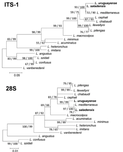
In the ITS-1 MP tree, L. saladensis formed a clade to-gether with L. mediterraneus Sarabeev, Balbuena et Euzet, 2005, with a bootstrap support of 86 %. In the 28S MP tree, L. mediterraneus was nested within L. saladensis and clustered with one of its haplotypes with a bootstrap sup-port of 68% (not shown in Fig. 3). Based on ITS-1 as well as on 28S rDNA sequences, the two species under study clustered firmly with L. mediterraneus (Fig. 3). Although relationships within this clade were poorly resolved,
L. saladensis seems a sister species to L. mediterraneus in
the 28S tree. Only one or two substitutions (i.e. maximum
Fig. 2. Light micrographs of hard parts of specimen 1 (A, C, E) and specimen 2 (B, D, F) collected from Mugil liza Valenciennes in
southern Brazil. A, B – ventral bar; C, D – sclerotised vagina; E, F – male copulatory complex. Abbreviations: AV – annulated vagina;
MPVB – median process of the ventral bar; PAP – penis accessory piece.
A
B
E
F
doi: 10.14411/fp.2015.024 Marchiori et al.: Validity of two species of Ligophorus
Folia Parasitologica 2015, 62: 024 Page 5 of 10
Fig. 3. Neighbour-joining phylogram constructed for the ITS-1 (above, 734 bp) and 28S (below, 18 unique haplotypes, 929 bp) rDNA
sequences of Ligophorus Euzet et Suriano, 1977. The scale bar indicates the number of substitutions per site. The focal taxa of the pres-ent study are shown in bold. Statistical support for each node is shown: neighbour-joining bootstrap/maximum parsimony bootstrap. Nodes that did not receive a bootstrap support of at least 65 in at least one of both analyses are collapsed, creating a polytomy. A clade not recovered in a particular analysis is indicated by ‘-’.
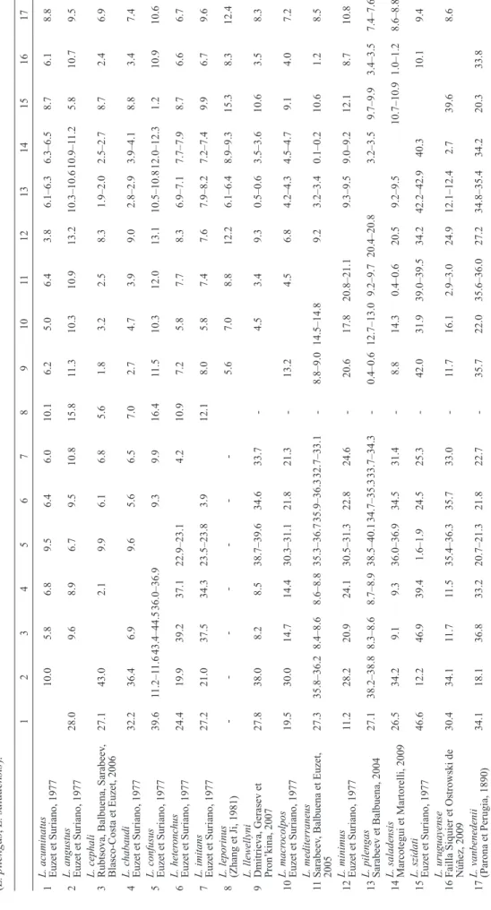
0.2% corrected pairwise genetic distance) were observed between the partial 28S rRNA gene sequences for these two species (Table 1). Their ITS-1 sequences only differed by 0.4–0.6%. This distance is smaller than that between either of them and L. uruguayense (Table 1).
Confocal fluorescence microscopy
Imaging of species of Ligophorus under a confocal fluo-rescence microscope is depicted in Fig. 4. This provided accurate data on the morphology of the ventral bar and pe-nis accessory piece for the two species.
The ventral bar of L. saladensis (Fig. 4C) is charac-terised by one median-squared process with an anteriorly extended membraneous protuberance on both sides. The protuberances and the median process are united to form a U-shaped anterior median part of the ventral bar. In con-trast, the ventral bar of L. uruguayense (Fig. 4D) possesses one ventral, median V-shaped process with an enlarged membraneous protuberance on both sides. The protuber-ances with the median process form a V-shaped anterior median part of the ventral bar.
As for the penis accessory piece, the upper lobe in
L. saladensis is cylindrical, with two slightly spine-like
protrusions where the accessory piece curves; these may
serve in guiding the penis (Fig. 4E). The lower lobe is smaller than the upper lobe, with the distal tip turned away from the upper lobe. In L. uruguayense, this structure was shown to be quite similar to its original description (Fig. 4F); its upper lobe is cylindrical, convex and longer than the lower lobe, which, in turn, is straight and proxi-mally united to the upper lobe.
Morphometrics
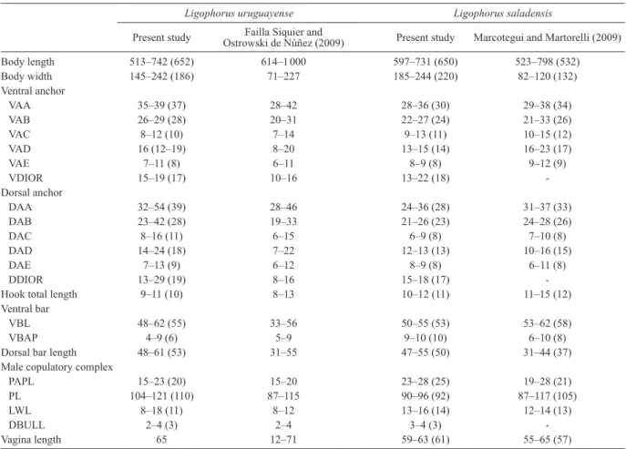
Comparative morphometric data obtained in the present study for L. uruguayense and L. saladensis and from their original descriptions are shown in Table 2. Similarly as ob-served by Marcotegui and Martorelli (2009), L. saladensis also presented greater VBAP, PAPL and LWL than L.
uru-guayense in the present study. The upper ranges for some
characters of L. uruguayense reported in its original de-scription (e.g. body width, VDIOR, DAA, DAB, DDIOR, VBL, dorsal bar length, PL and LWL) were lower than those found in the present study. Additionally, the ranges for some characters of L. saladensis found in the present study were outside the upper and lower ranges reported in its original description (e.g. body width, dorsal bar length and VAD).
Table 1. Gamma-corrected pairwise distanc es (in %) between ITS-1 (below diagonal) and 28S (above diagonal) sequences of species of Ligophorus Euzet et Suriano, 1977. For species with reported multiple haplotypes, intraspecific distances for ITS-1 range from 0.1 % (L. mediterraneus , L. pilengas ) to 0.3 % (L. confusus ). Over the 28S fragment, these amount to 0.1% (L. pilengas , L. saladensis ). 1 2 3 4 5 6 7 8 9 10 11 12 13 14 15 16 17 1
L. acuminatus Euzet et Suriano, 1977
10.0 5.8 6.8 9.5 6.4 6.0 10.1 6.2 5.0 6.4 3.8 6.1–6.3 6.3–6.5 8.7 6.1 8.8 2
L. angustus Euzet et Suriano, 1977
28.0 9.6 8.9 6.7 9.5 10.8 15.8 11.3 10.3 10.9 13.2 10.3–10.6 10.9–1 1.2 5.8 10.7 9.5 3
L. cephali Rubtsova, Balbuena, Sarabeev
, Blasco-Costa et Euzet, 2006 27.1 43.0 2.1 9.9 6.1 6.8 5.6 1.8 3.2 2.5 8.3 1.9–2.0 2.5–2.7 8.7 2.4 6.9 4
L. chabaudi Euzet et Suriano, 1977
32.2 36.4 6.9 9.6 5.6 6.5 7.0 2.7 4.7 3.9 9.0 2.8–2.9 3.9–4.1 8.8 3.4 7.4 5
L. confusus Euzet et Suriano, 1977
39.6 11.2–1 1.6 43.4–44.5 36.0–36.9 9.3 9.9 16.4 11.5 10.3 12.0 13.1 10.5–10.8 12.0–12.3 1.2 10.9 10.6 6 L. heter onchus Euzet et Suriano, 1977 24.4 19.9 39.2 37.1 22.9–23.1 4.2 10.9 7.2 5.8 7.7 8.3 6.9–7.1 7.7–7.9 8.7 6.6 6.7 7
L. imitans Euzet et Suriano, 1977
27.2 21.0 37.5 34.3 23.5–23.8 3.9 12.1 8.0 5.8 7.4 7.6 7.9–8.2 7.2–7.4 9.9 6.7 9.6 8
L. leporinus (Zhang et Ji, 1981)
-5.6 7.0 8.8 12.2 6.1–6.4 8.9–9.3 15.3 8.3 12.4 9
L. llewellyni Dmitrieva, Gerasev et Pron’kina, 2007
27.8 38.0 8.2 8.5 38.7–39.6 34.6 33.7 -4.5 3.4 9.3 0.5–0.6 3.5–3.6 10.6 3.5 8.3 10 L. macr ocolpos Euzet et Suriano, 1977 19.5 30.0 14.7 14.4 30.3–31.1 21.8 21.3 -13.2 4.5 6.8 4.2–4.3 4.5–4.7 9.1 4.0 7.2 11 L. mediterraneus Sarabeev , Balbuena et Euzet, 2005 27.3 35.8–36.2 8.4–8.6 8.6–8.8 35.3–36.7 35.9–36.3 32.7–33.1 -8.8–9.0 14.5–14.8 9.2 3.2–3.4 0.1–0.2 10.6 1.2 8.5 12
L. minimus Euzet et Suriano, 1977
11.2 28.2 20.9 24.1 30.5–31.3 22.8 24.6 -20.6 17.8 20.8–21.1 9.3–9.5 9.0–9.2 12.1 8.7 10.8 13
L. pilengas Sarabeev et Balbuena, 2004
27.1 38.2–38.8 8.3–8.6 8.7–8.9 38.5–40.1 34.7–35.3 33.7–34.3 -0.4–0.6 12.7–13.0 9.2–9.7 20.4–20.8 3.2–3.5 9.7–9.9 3.4–3.5 7.4–7.6 14
L. saladensis Marcotegui et Martorelli, 2009
26.5 34.2 9.1 9.3 36.0–36.9 34.5 31.4 -8.8 14.3 0.4–0.6 20.5 9.2–9.5 10.7–10.9 1.0–1.2 8.6–8.8 15
L. szidati Euzet et Suriano, 1977
46.6 12.2 46.9 39.4 1.6–1.9 24.5 25.3 -42.0 31.9 39.0–39.5 34.2 42.2–42.9 40.3 10.1 9.4 16
L. uruguayense Failla Siquier et Ostrowski de Núñez, 2009
30.4 34.1 11.7 11.5 35.4–36.3 35.7 33.0 -11.7 16.1 2.9–3.0 24.9 12.1–12.4 2.7 39.6 8.6 17
L. vanbenedenii (Parona et Perugia, 1890)
34.1 18.1 36.8 33.2 20.7–21.3 21.8 22.7 -35.7 22.0 35.6–36.0 27.2 34.8–35.4 34.2 20.3 33.8
doi: 10.14411/fp.2015.024 Marchiori et al.: Validity of two species of Ligophorus
Folia Parasitologica 2015, 62: 024 Page 7 of 10
DISCUSSION
Since the erection of Ligophorus by Euzet and Suriano (1977), the number of species in the genus has increased to approximately 50. Nevertheless, according to Blasco-Costa et al. (2012), this number can augment greatly as new hosts and localities are studied and, thus, the develop-ment of a reliable taxonomic framework based on morpho-logical and molecular grounds is becoming increasingly needed (this was partially done by Sarabeev et al. 2013 and Sarabeev and Desdevises 2014). In the present study, we observed that scrutiny of the published diagnostic fea-tures under phase contrast microscopy does not succeed in unambiguously discriminating between L. saladensis and
L. uruguayense.
Ligophorus uruguayense was described from M. liza
(= M. platanus) in Uruguay by Failla Siquier and Os-trowski de Núñez in 2009. It was distinguished from its congeners on the basis of the shape of the ventral bar and anchors, the accessory piece of the penis and the vaginal aperture, the host species and the geographical distribution. Later that same year, L. saladensis was described from the same fish host in Argentina by Marcotegui and Martorelli (2009). The authors differentiated their new species from other species of Ligophorus by the shape of the ventral bar and accessory piece and by morphometrics. Concerning its closely related species L. uruguayense, it was distin-guished from the latter on the basis of the morphology of the accessory piece, the absence of transverse annulations at the distal end of the vaginal tube, as well as by the thick process at the distal end of the inner root of the ventral an-chors. Additionally, L. saladensis was distinguished from
L. uruguayense by having a greater distance between the
membraneous anterior processes (protuberances) of the ventral bar, and a greater total length and lower lobe length of the accessory piece.
The accessory pieces of the MCC (Fig. 4E,F) of
L. saladensis and L. uruguayense can be sometimes very
similar to each other, depending on the position in which this structure is examined, because of its delicate form and small size. Besides, its total length cannot be easily stud-ied due to large amounts of vitelline follicles covering it. Nevertheless, each species presents a unique shape for this structure: whereas in L. uruguayense it has a smaller total length, with a cylindrical, convex upper lobe and straight lower lobe (because of that, it can easily remind of a ‘box glove’), in L. saladensis it is longer, with the distal tip of the lower lobe turning away from the upper lobe.
Sarabeev et al. (2013) attributed a different morphology to the penis accessory piece of L. saladensis: according to them, it has a main lobe and a secondary one, the latter possessing a trident format, with three unequal branches. While the upper one extends beyond the distal end of the main lobe and is two times longer than the lower one, the medial branch has the smallest size. However, we did not identify such configuration in our analysed specimens. In addition, examination of one of the paratypes of L.
salad-ensis (MLP-He 5935) deposited in the Collection of
In-vertebrates of the Museo de la Plata in Argentina revealed a similar morphology of the median process of the ventral bar and the accessory piece of the penis when compared to the presently studied specimens. Other characteristics
Fig. 4. Three-dimensional Imaris reconstruction of hard parts of Ligophorus saladensis Marcotegui et Martorelli, 2009 (A, C, E) and
Ligophorus uruguayense Failla Siquier et Ostrowski de Núñez, 2009 (B, D, F) both collected from Mugil liza Valenciennes in southern Brazil. A, B – transverse bar, dorsal; C, D – transverse bar, ventral; E, F – penis accessory piece.
A
B
E
F
Table 2. Comparison of morphometric characters (in micrometers, expressed as range with mean in parentheses) of Ligophorus
uru-guayense Failla Siquier et Ostroswki de Núñez, 2009 and Ligophorus saladensis Marcotegui et Martorelli, 2009 from present and previous studies.
Ligophorus uruguayense Ligophorus saladensis
Present study Ostrowski de Núñez (2009)Failla Siquier and Present study Marcotegui and Martorelli (2009)
Body length 513–742 (652) 614–1 000 597–731 (650) 523–798 (532) Body width 145–242 (186) 71–227 185–244 (220) 82–120 (132) Ventral anchor VAA 35–39 (37) 28–42 28–36 (30) 29–38 (34) VAB 26–29 (28) 20–31 22–27 (24) 21–33 (26) VAC 8–12 (10) 7–14 9–13 (11) 10–15 (12) VAD 16 (12–19) 8–20 13–15 (14) 16–23 (17) VAE 7–11 (8) 6–11 8–9 (8) 9–12 (9) VDIOR 15–19 (17) 10–16 13–22 (18) -Dorsal anchor DAA 32–54 (39) 28–46 24–36 (28) 31–37 (33) DAB 23–42 (28) 19–33 21–26 (23) 24–28 (26) DAC 8–16 (11) 6–15 6–9 (8) 7–10 (8) DAD 14–24 (18) 7–22 12–13 (13) 10–16 (15) DAE 7–13 (9) 6–12 8–9 (8) 6–11 (8) DDIOR 13–29 (19) 8–16 15–18 (17)
-Hook total length 9–11 (10) 8–13 10–12 (11) 11–15 (12)
Ventral bar
VBL 48–62 (55) 33–56 50–55 (53) 53–62 (58)
VBAP 4–9 (6) 5–9 9–10 (10) 6–10 (8)
Dorsal bar length 48–61 (53) 31–55 47–55 (50) 31–44 (37)
Male copulatory complex
PAPL 15–23 (20) 15–20 23–28 (25) 19–28 (21)
PL 104–121 (110) 87–115 90–96 (92) 87–117 (105)
LWL 8–18 (11) 8–12 13–16 (14) 12–14 (13)
DBULL 2–4 (3) 2–4 3–4 (3)
-Vagina length 65 12–71 59–63 (61) 55–65 (57)
DAA – dorsal anchor total length; DAB – dorsal anchor main part length; DAC – dorsal anchor outer root length; DAD – dorsal anchor inner root length; DAE – dorsal anchor point length; DBULL – distance between upper and lower lobe of penis accessory piece; DDIOR – distance between in-ner and outer root of dorsal anchors; LWL – penis accessory piece lower lobe length; PAPL – penis accessory piece total length; PL – total length of penis; VAA – ventral anchor total length; VAB – ventral anchor main part length; VAC – ventral anchor outer root length; VAD – ventral anchor inner root length; VAE – ventral anchor point length; VBAP – distance between membranous anterior processes of ventral bar; VBL – ventral bar length; VDIOR – distance between inner and outer root of ventral anchors.
pointed out to be diagnostic ones by the original describers could not be observed.
Besides the shape of the accessory piece, Marcotegui and Martorelli (2009) also differentiated L. saladensis from L. uruguayense based on the absence of transverse annulations at the distal end of the vaginal tube and a thick process at the distal end of the inner root of the ventral anchors. However, in the presently studied specimens, nei-ther the first nor the second character was found to be reli-able for distinguishing one species from the other. Trans-verse annulations in the vagina were always present in both species, whilst the presence of a process in the inner root of the ventral anchors was highly variable for both of them (sometimes also present in L. saladensis). Furthermore, Sarabeev et al. (2013) also commented on the presence of transverse annulations at the distal end of the vaginal tube reported for L. uruguayense and concluded that this feature corresponds, to a muscular or fibrous sheath surrounding the sclerotised vaginal tube, being similar to that of other
Ligophorus species.
With regard to the surprisingly wider ranges in some haptoral morphometrics of L. uruguayense signaled by
Sarabeev et al. (2013) (e.g. VAC, VAD, DAC and DAD) (but we found narrower ranges for these characters), they were still wider than those found for other Ligophorus spe-cies, including L. saladensis (see Table 2). The metrical dif-ferences found in the ranges between our observations and the original descriptions for both species should be consid-ered as intraspecific ones or even a result of the different procedure applied (in the present study the measurements were taken from specimens fixed in 95% ethanol whereas they were taken from both fixed and live specimens and from heat-fixed specimens preserved in 10% formalin, re-spectively, in the original descriptions of L. uruguayense and L. saladensis).
Whereas the genetic divergence between L. saladensis and L. uruguayense is low as compared to most distances observed between representatives of Ligophorus (Table 1), comparable genetic distances between closely related species of Ligophorus exist (see also Blasco-Costa et al. 2012). Examples include L. llewellyni Dmitrieva, Gerasev et Pronkina, 2007 vs L. pilengas Sarabeev et Balbuena, 2004, and L. confusus Euzet et Suriano, 1977 vs L. szidati Euzet et Suriano, 1977. For example, despite their limited
doi: 10.14411/fp.2015.024 Marchiori et al.: Validity of two species of Ligophorus
Folia Parasitologica 2015, 62: 024 Page 9 of 10
divergence, which may suggest L. llewellyni and L.
pilen-gas are conspecific, Blasco-Costa et al. (2012) argued that
the unambiguous morphological distinction between these species confirms species delineation. Similarly, considering the observed genetic and phenotypic differences between
L. saladensis and L. uruguayense, our results support the
validity of these species.
However, inter- and intraspecific distances seem to con-verge for the 28S rDNA fragment for L. saladensis and
L. mediterraneus, with interspecific phylogenetic
relation-ships poorly resolved. Despite their morphological similari-ty (both species have a bilobed accessory piece of the MCC, with the lower lobe smaller than the upper, and a ventral bar with a median process; see also Dmitrieva et al. 2009a), the fact that they do not share haplotypes leads us to the suggestion that L. saladensis, L. mediterraneus and L.
uru-guayense belong to a complex of closely related species.
This situation is comparable for the host fishes: Mugil
cephalus Linnaeus, the host of L. mediterraneus,
prob-ably belongs to the same species complex as M. liza. Both species are, despite their low genetic divergence, consid-ered valid (Durand et al. 2012, Whitfield et al. 2012). The presence of separate but closely related parasite species on these closely related hosts corroborates the suggestion of El Hafidi et al. (2013) that species of Ligophorus can be used as a marker for taxonomy and evolution of mullet species.
To date, records for both L. uruguayense and L.
salad-ensis are limited to the southwestern Atlantic. Whereas the
former species was reported in Laguna de Rocha, Uruguay, the latter was later reported in Samborombón Bay, between Punta Piedras and Punta Rasa (Failla Siquier and Ostrowski de Núñez 2009, Marcotegui and Martorelli 2009).
Interestingly, although Samborombón Bay is very close to the Uruguayan coast, the authors reported only the pres-ence of L. saladensis and no specimen of L. uruguayense was found. Furthermore, they suggested that the occurence of parasites could be related to host body size. However, this idea is not supported by the present study since our
fish sample consisted of specimens with a body size of no more than 4 centimetres, which were parasitised by both parasite species.
It seems that because of the great latitudinal range of
M. liza along the Atlantic coast, this host fish may face
a wide spectrum of abiotic conditions. We can assume that speciation of species of Ligophorus is a result. This hy-pothesis was also supported by Pahor-Filho et al. (2012). Furthermore, we believe that future integrative studies similar to the present one should be carried out for other species of Ligophorus reported for M. liza in order to better comprehend the diversity and diversification of these fish parasites.
Blasco-Costa et al. (2012) suggest that both within-host duplication and host-switching contributed to the diversi-fication of Ligophorus, whereas Sarabeev and Desdevises (2014) stress the importance of host-switching. The ob-served limited divergence between two species of
Ligo-phorus, that share the same host raises the question as to
which parasite speciation modes have occurred in this ge-nus. To formally reconstruct a diversification scenario for the representatives of Ligophorus and to check for poten-tial co-speciation events, co-phylogenetic analyses of this parasite and its mullet hosts are recommended.
Acknowledgments. The authors thank Coordination for
Im-provement of Higher Education Personnel (CAPES) for fi-nancial support to N.C. Marchiori and to J. Pereira Jr. (Project No. 23038.005284/2011-60) and the National Council for Sci-entific and Technological Development (CNPq; project No. 300753/2012-8). Obtaining sequence newly produced during this work was financed by the Intitut de Recherche pour le Dévelop-pement and obtained through the technical facilities of the “Cen-tre Méditerraéen de l’Environnement et de la Biodiversité” (Ce-MEB). This is publication ISEM 2015-016 S. M.P.M. Vanhove was supported by a PhD fellowship of the Research Foundation – Flanders (FWO-Vlaanderen) and currently by the Czech Sci-ence Foundation (project no. P505/12/G112 – European Centre of Ichthyoparasitology).
REFERENCES
Abdallah V.D., Azevedo R.K., Luque J.L. 2009: Four new species of Ligophorus (Monogenea: Dactylogyridae) parasitic on Mugil liza (Actinopterygii: Mugilidae) from Guandu River, southeastern Brazil. J. Parasitol. 95: 855–864.
Blasco-Costa I., Míguez-Lozano R., Sarabeev V., Bal-buena J.A. 2012: Molecular phylogeny of species of Ligopho-rus (Monogenea: Dactylogyridae) and their affinities within the Dactylogyridae. Parasitol. Int. 61: 619–627.
Dmitrieva E.V., Gerasev P.I., Merella P., Pugachev O.N. 2009a: Redescription of Ligophorus mediterraneus Sarabeev, Balbuena and Euzet, 2005 (Monogenea: Ancyrocephalidae) with some methodological notes. Syst. Parasitol. 73: 95–105. Dmitrieva E.V., Gerasev P.I., Merella P., Pugachev O.N.
2009b: Redescription of Ligophorus cephali Rubtsova, Bal-buena, Sarabeev, Blasco-Costa and Euzet, 2006 and L. chabaudi Euzet and Suriano, 1977 (Monogenea: Ancyrocephalidae), with notes on the functional morphology of the copulatory organ. Syst. Parasitol. 73: 175–191.
Durand J.D., Shen K.N., Chen W.J., Jamandre B.W., Blel H., Diop K., Nirchio M., Garcia de León F.K., Whitfield A.K., Chang C.W., Borsa P. 2012: Systematics of the grey
mullets (Teleostei: Mugiliformes: Mugilidae): molecular phylo-genetic evidence challenges two centuries of morphology-based taxonomy. Mol. Phylogenet. Evol. 64: 73–92.
El Hafidi F., Berrada Rkhami O., de Buron I., Durand J.D., Pariselle A. 2013: Ligophorus species (Monogenea: Ancyro-cephalidae) from Mugil cephalus (Teleostei: Mugilidae) off Mo-rocco with the description of a new species and remarks about the use of Ligophorus spp. as biological markers of host popula-tions. Folia Parasitol. 60: 433–440.
Euzet L., Suriano D.M. 1977: Ligophorus n. g. (Monogenea, An-cyrocephalidae) parasite des Mugilidae (Téléostéens) en Medi-terranée. Bull. Mus. Natl. Hist. Nat. Zool. 472: 799–821. Failla Siquier G.F., Ostrowski de Núñez M. 2009:
Ligopho-rus uruguayense sp. nov. (Monogenea, Ancyrocephalidae), a gill parasite from Mugil platanus (Mugiliformes, Mugilidae) in Uru-guay. Acta Parasitol. 54: 95–102.
Fraga E., Schneider H., Nirchio M., Santa-Brigida E., Ro-drigues-Filho L.F., Sampaio I. 2007: Molecular phylogenetic analyses of mullets (Mugilidae, Mugiliformes) based on two mi-tochondrial genes. J. Appl. Ichthyol. 23: 598–604.
Received 31 October 2013 Accepted 1 February 2015 Published online 1 May 2015
Cite this article as: Marchiori N.C., Pariselle A., Pereira Jr. J., Agnèse J.-F., Durand J.-D., Vanhove M.P.M. 2015: A comparative
study of Ligophorus uruguayense and L. saladensis (Monogenea: Ancyrocephalidae) from Mugil liza (Teleostei: Mugilidae) in southern Brazil. Folia Parasitol. 62: 024.
Humason, G.L. 1979: Animal Tissue Techniques. Fourth Edition. W.H. Freeman and Company, San Francisco, 661 pp.
Kimura M. 1980: A simple method for estimating evolutionary rate of base substitutions through comparative studies of nucle-otide sequences. J. Mol. Evol. 16: 111–120.
Lockyer A.E., Olson P.D., Littlewood D.T.J. 2003: Utility of complete large and small subunit rRNA genes in resolving the phylogeny of the Neodermata (Platyhelminthes): implica-tions and a review of the cercomer theory. Biol. J. Linn. Soc. 78: 155–171.
Littlewood D.T.J., Curini-Galletti M., Herniou E.A. 2000: The interrelationships of Proseriata (Platyhelminthes: Seriata) tested with molecules and morphology. Mol. Phylogenet. Evol. 16: 449–466
Malmberg G. 1957: [On the occurrence of Gyrodactylus in Swed-ish fSwed-ishes.] Skrifter utgivna av Södra Sveriges Fiskeriföreningen 1956: 19–76. (In Swedish.)
Marcotegui P.S., Martorelli S.R. 2009: Ligophorus saladen-sis n. sp. (Monogenea: Ancyrocephalidae) from Mugil platanus Günther in Samborombón Bay, Argentina. Syst. Parasitol. 74: 41–47.
Mollaret I., Jamieson B.G.M., Justine J.L. 2000: Phylog-eny of the Monopisthocotylea and Polyopisthocotylea (Platy-helminthes) inferred from 28S rDNA sequences. Int. J. Parasitol. 30: 171–185.
Pahor-Filho E., Miranda-Filho K.C., Pereira J. Jr. 2012: Parasitology of juvenile mullet (Mugil liza) and effect of formal-dehyde on parasites and host. Aquaculture 354–355: 111–116.
Sarabeev V., Desdevises Y. 2014: Phylogeny of the Atlantic and Pacific species of Ligophorus (Monogenea: Dactylogyridae): morphology vs. molecules. Parasitol. Int. 63: 9–20.
Sarabeev V., Rubtsova N., Yang T., Balbuena J.A. 2013: Tax-onomic revision of the Atlantic and Pacific species of Ligopho-rus Euzet and Suriano, 1977 (Monogenea: Dactylogyridae) from mullets (Teleostei: Mugilidae) with proposal of a new genus and description of four new species. Vestn. Zool. 2013, Suppl. 28: 1–112.
Tamura K., Stecher G., Peterson D., Filipski A., Kumar S. 2013: MEGA6: Molecular Evolutionary Genetics Analysis Ver-sion 6.0. Mol. Biol. Evol. 30: 2725–2729.
Thompson J.D., Higgins D.G., Gibson T.J. 1994: CLUSTAL W: improving the sensitivity of progressive multiple sequence align-ment through sequence weighting, position-specific gap penal-ties and weight matrix choice. Nucl. Acids Res. 22: 4673–4680. Whitfield A.K., Panfili J., Durand J.D. 2012: A global review
of the cosmopolitan flathead mullet Mugil cephalus Linnaeus, 1758 (Teleostei: Mugilidae), with emphasis on the biology, ge-netics, ecology and fisheries aspects of this apparent species complex. Rev. Fish Biol. Fisher. 22: 641–681.
Wu X.Y., Zhu X.Q., Xie M.Q., Li A.X. 2006: The radiation of Haliotrema (Monogenea: Dactylogyridae: Ancyrocephalinae): molecular evidence and explanation inferred from LSU rDNA sequences. Parasitology 132: 659–668.
Wu X.Y., Zhu X.Q., Xie M.Q., Li A.X. 2007: The evaluation for generic-level monophyly of Ancyrocephalinae (Monogenea, Dactylogyridae) using ribosomal DNA sequence data. Mol. Phy-logenet. Evol. 44: 530–544.