Acoel regeneration mechanisms indicate an
ancient role for muscle in regenerative patterning
The MIT Faculty has made this article openly available.
Please share
how this access benefits you. Your story matters.
Citation
Raz, Amelie A. et al. “Acoel Regeneration Mechanisms Indicate
an Ancient Role for Muscle in Regenerative Patterning.” Nature
Communications 8, 1 (October 2017): 1260 © 2017 The Author(s)
As Published
http://dx.doi.org/10.1038/S41467-017-01148-5
Publisher
Nature Publishing Group
Version
Final published version
Citable link
http://hdl.handle.net/1721.1/114708
Terms of Use
Creative Commons Attribution 4.0 International License
Acoel regeneration mechanisms indicate an
ancient role for muscle in regenerative patterning
Amelie A. Raz
1
, Mansi Srivastava
2
, Ranja Salvamoser
1,3
& Peter W. Reddien
1
Positional information is required for animal regeneration, yet how it is harbored in adult
tissues is poorly understood. In planarians, positional control genes (PCGs) control
regen-eration outcomes and are regionally expressed predominately in the musculature. Acoels are
early diverging bilaterally symmetric animals, having separated from other bilaterians
> 550
million years ago. Here, we
find that PCGs in the acoel Hofstenia miamia are expressed
together and speci
fically in a primary differentiated cell type: muscle. The vast majority of
Hofstenia muscle cells in regions tested express PCGs, suggesting positional information is a
major feature of muscle. PCG expression domains are dynamic in muscle after injury,
consistent with known PCG roles in guiding regeneration. These data demonstrate an
instructive positional role for Hofstenia muscle and this similarity with planarians suggests
mesodermal muscle originated at the base of the Bilateria not only for contraction, but also as
the source of positional information guiding regeneration.
DOI: 10.1038/s41467-017-01148-5
OPEN
1Howard Hughes Medical Institute, MIT Biology, Whitehead Institute for Biomedical Research, 9 Cambridge Center, Cambridge, MA 02142, USA. 2Department of Organismic and Evolutionary Biology, Harvard University, Cambridge, MA 02138, USA.3Present address: Walter and Eliza Hall Institute of
T
he ability to regenerate is widespread in the animal
king-dom
1. However, whether similar or distinct underlying
mechanisms explain regeneration across diverse animals
remains unclear. Positional information refers to the molecular
processes by which the regional identity of cells and tissues can be
specified
2. Positional information is necessary for regulating
regeneration, yet the identity of adult cell types across animals
that are the source of positional information involved in
regen-eration is poorly understood
3,4. In one classical model system for
studying regeneration, planarian
flatworms, muscle cells have
been identified as the primary site of expression of patterning
genes
5. This specific expression of patterning molecules indicates
that muscle is a major source of
flatworm adult positional
information
5. Muscle was subsequently found to specifically
express regionally restricted Wnts in the regenerative
flatworm
parasite Echinococcus multilocularis
6. Mesoderm, and specifically
mesoderm-derived muscle, arose at the base of the Bilateria over
550 million years ago
7–13. We sought to test whether muscle
originated in evolution not only for generating contractile force,
but also as the tissue responsible for the control of regenerative
patterning. This would suggest a broad role for muscle in
con-trolling regeneration across bilaterians.
Acoels, an enigmatic taxon of marine worms, are highly
regenerative and have recently been highlighted as a model for
understanding the evolution of regeneration mechanisms
14–17.
Recent phylogenetic work using transcriptome-level sequences
supports a position for the acoels, together with
nemertoderma-tids and Xenoturbella, as a sister clade to all other bilaterians
(the bilaterally symmetric animals) (Fig.
1
a)
17–22. This
phyloge-netic placement of acoels suggests that they diverged from other
bilaterians over 550 million years ago
7. This position is significant
for experimental approaches to animal biology because
compar-ison of biology in acoels, serving as an outgroup to other
bila-terians, can point to attributes likely present in the last common
ancestor of the Bilateria. By assessing the role of muscle in
regenerative patterning in an acoel, we can seek to determine the
ancestral and broadly conserved functions of muscle.
Hofstenia miamia is an acoel capable of whole body
regeneration
17,23–25. We recently developed Hofstenia as a new
and powerful model for studying regeneration
17. Hofstenia
express molecules constitutively in adult tissues that are required
for regional tissue identity in regeneration
17. Similar genes in
planarians have been referred to as positional control genes
(PCGs), which are genes that display constitutive regionalized
expression and either a patterning abnormal RNAi phenotype or
encode a protein predicted to act in the pathway of such a
patterning molecule
5. In Hofstenia, genes encoding several Wnt
pathway components, including Wnt signaling ligands, Frizzled
d
b
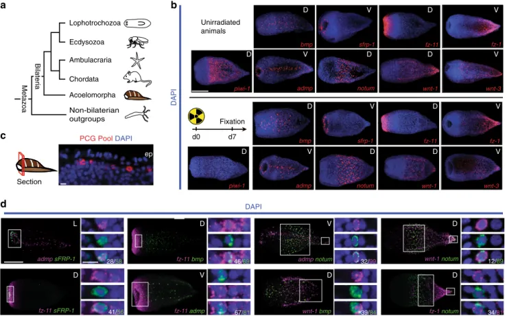
a
PCG Pool DAPIc
Section Ecdysozoa Ambulacraria Chordata Non-bilaterian outgroups Metazoa Bilateria DAPI DAPI d0 D D D D D V V V D V D V V D D D D ep D D L D D V V V V d7 Fixation sfrp-1 admp fz-11 bmp wnt-3 wnt-1 fz-1 piwi-1 piwi-1 notum Acoelomorpha Lophotrochozoa admp notum 32/99 fz-1 notum 34/81 39/84 fz-11 admp 67/81 admp sFRP-1 28/58 fz-11 bmp 46/69 wnt-1 notum 12/89 fz-11 sFRP-1 41/56 Unirradiated animals sfrp-1 admp fz-11 bmp wnt-3 wnt-1 fz-1 notum wnt-1 bmpFig. 1 PCGs are expressed in a common differentiated cell type. a A phylogenetic tree representing the relative position of acoels within the Metazoa. Phylogenetic analyses support the position of acoels as the sister group to other bilaterian lineages, diverging shortly after the origin of the Bilateria> 550 million years ago17,18,20.b As a marker for cycling cells, piwi-1 expression is sensitive to irradiation and is ablated by 7 days post-irradiation with 10,000 rads17. PCG expression domains17were maintained, suggesting that they are primarily expressed in non-cycling, differentiated cells.c Fluorescent image of a transverse optical section in the anterior. ep epidermis. PCG pool comprised of simultaneously hybridized sFRP-1, frizzled-11, bmp, admp, notum, wnt-1, and frizzled-1 RNA probes. d Eighteen RNA probe pairs were used in double-FISH experiments. The denominator for each quantification is the total number of cells in the channel indicated (i.e., magenta or green); the numerator is the total number of double-positive cells for that pair. Quantification was performed in area indicated by the white box, and inset panels below are higher magnification images of cells for this region. See Supplementary Fig.1b for remaining pairs. Scale bars:b 100μm; c 10 μm; d 100 μm, overview image; 10 μm, cell inset. D dorsal view, V ventral view, L lateral view. Overview images are representative of 5/5 animals examined
receptors, and secreted antagonists (e.g., secreted frizzled-related
protein, sFRP, or the secreted Wnt deacylase Notum
26,27) are
regionally expressed along the Hofstenia anterior–posterior (AP)
axis
17. Perturbation of several Wnt pathway components resulted
in abnormal AP patterning in regeneration
17. For instance, RNAi
of the posteriorly expressed wnt-1 gene resulted in regeneration of
a head in place of a tail, generating two-headed acoels, whereas
RNAi of the Wnt-antagonist notum resulted in two-tailed acoels.
Genes encoding several Bmp pathway components, including
Bmp-family signaling ligands, are expressed in different patterns
on the dorsal–ventral (DV) and medial–lateral (ML) axes
17.
RNAi of the dorsally expressed bmp gene or the ventrally
expressed admp gene resulted in abnormal dorsal–ventral
regenerative patterning
17. These Wnt and Bmp signaling
path-ways are, therefore, hypothesized to instruct the regeneration of
correct axial structures after amputation and involve regional and
constitutive expression of signaling ligands, receptors, and
secreted pathway inhibitors
17.
Here, we show that Hofstenia PCGs are co-expressed with one
another in a common differentiated, subepidermal cell type,
consistent with a single primary source of adult positional
information. Analysis by in situ hybridization and single-cell
quantitative real-time PCR (qRT-PCR) demonstrated that all
known Hofstenia PCGs are specifically expressed in muscle cells
during both homeostatic tissue maintenance and regeneration. In
tested regions, Hofstenia muscle cells expressed one or more
PCGs, suggesting that most Hofstenia muscle cells express
patterning molecules as well as control body contraction. The
expression domains of PCGs shifted in muscle after amputation,
consistent with muscle guiding regenerative patterning choices.
These data demonstrate that despite being separated by over
550 million years of evolution, acoels, like planarians, use muscle
as a physical positional map to guide tissue turnover and
regenerative patterning. This is consistent with a model in which
mesodermal muscle was the primary source of regenerative
positional information in the last common ancestor of the
fz-11 V 194/213 fz-1 D 235/264 sFRP-1 D 91/95*
wnt-3 D 126/135 V bmp 228/240 admp 177/186 wnt-1 213/229 notum 282/294 D D Da
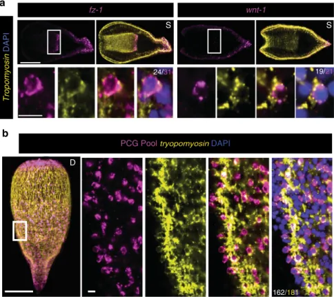
Tropomyosin DAPIb
tropomyosin sFRP-1 frizzled-1 1 notum frizzled-1 wnt-1 wnt-3 admp bmp PC2 synapsin Genes Cellsc
PC2 4/415 synapsin 18/488 1/512 hm.98018390 PCG Pool 0 – Δ Ct –5 –10 –15 D D S DAPIFig. 2 Muscle specifically harbors positional information in Hofstenia. a PCG( + ) cells also expressed tropomyosin. Both muscle fibers and nuclei are marked by tropomyosin mRNA; PCG mRNA is localized primarily around the nucleus. Denominator for each quantification is the total number of PCG-positive cells; the numerator is the total number of PCG/tropomyosin double-positive cells. Asterisk denotes fiber expression of PCG. b PCGs were not significantly expressed in PC2- or synapsin-positive neuron populations, nor in an internal differentiated cell population marked by expression of hm.98018390. The PCG RNA probe pool was the same as in Fig.1c.c Single-cell cDNA libraries were screened by qRT-PCR with primers specific to PCGs, tissue-type markers (tropomyosin, PC2, and synapsin), and a housekeeping gene GAPDH. Any cell without expression of GAPDH was excluded from the analysis. Shown are all remaining cells positive for at least one PCG and/or positive for at least one neural marker.−ΔCt = −[Ct(gene of interest)−Ct(GAPDH)]. All Ct values greater than 32 or undetected were analyzed as Ct= 32. 29/30 PCG-positive cells also expressed tropomyosin. 0/30 PCG-positive cells expressed the neural markers PC2 or synapsin. tropomyosin expression was never found in single-cell libraries with expression of either neural marker, indicating that our libraries were indeed amplified from single cells. Scale bars: a, b 100 μm, overview image; 10 μm, cell inset. D dorsal view, V ventral view, L lateral view, S coronal section. Overview images are representative of 5/5 animals examined
Bilateria and has broad instructive roles in regeneration in extant
animals.
Results
Hofstenia PCGs are expressed in a common differentiated cell
type. We found that all Hofstenia PCGs examined were primarily
expressed in a peripheral, subepidermal cell layer of non-cycling
differentiated cells (Fig.
1
b, c, Supplementary Fig.
1
a,
Supple-mentary Table
2
). Eighteen Hofstenia PCG pairs with overlapping
expression domains were examined for co-expression by double
fluorescence in situ hybridization (FISH) (Fig.
1
d and
Supple-mentary Fig.
1
b). In most instances where the expression
domains of PCG pairs overlapped, the tested genes were
expressed together in the same cells to a substantial degree
(Fig.
1
d and Supplementary Fig.
1
b). This is consistent with the
possibility that a primary single cell type, such as muscle, might
harbor Hofstenia positional information.
Hofstenia muscle produces positional information. To test
whether Hofstenia PCG expression was in muscle, we developed a
FISH protocol that allowed detection of the nuclei connected to
muscle
fibers, involving visualizing tropomyosin messenger RNA
(mRNA) transcripts within individual cells (Fig.
2
a and
Supple-mentary Fig.
2
a–c). Hofstenia muscle cells are organized into long
fibers; dissociation of muscle fibers reveals that individual muscle
cells appear to be mononucleate (Supplementary Fig.
2
d). These
muscle
fibers are comprised of several subtypes, including
body-wall longitudinal and circular
fibers, as well as fibers that radiate
from or encircle the pharynx; all of these subtypes are marked by
tropomyosin expression
17,28(Supplementary Fig.
2
a).
Strikingly, every PCG analyzed was co-expressed with
tropomyosin to a high degree (89%–96% of PCG-positive cells
also expressed tropomyosin) (Fig.
2
a). PCG transcripts were
predominantly perinuclearly localized in these cells. Because
PCGs largely encode secreted or transmembrane proteins,
this localization likely reflects perinuclear association with
ribosomes on the endoplasmic reticulum. We could not
confidently identify every muscle nucleus with this method
because of relatively weak perinuclear tropomyosin(
+ ) signal
with FISH, indicating our quantification of the percentage of PCG
+ cells that expressed tropomyosin was an underestimate of the
degree of overlap, and PCG expression may be almost exclusive to
muscle cells.
We also identified an additional Hofstenia muscle marker,
titin-like, with mRNA localized primarily along muscle
fibers
(Supplementary Fig.
2
e). Co-expression analyses with this
alternative marker also showed restriction of PCG expression to
muscle (Supplementary Fig.
2
e). The PCG pairs that were not
robustly co-expressed together (e.g., bmp and notum) were
nonetheless individually each expressed in tropomyosin(
+ ) cells,
indicating that some PCGs are expressed in different subsets of
muscle cells (Supplementary Fig.
1
b, Fig.
2
a). We conclude that a
major site of PCG expression in Hofstenia is muscle.
To test the specificity of PCG expression to muscle cells, we
assessed by FISH whether there was any co-expression of PCGs
with other cell types. PCGs were not significantly expressed in
neurons (synapsin
+ or PC2 + cells) or an internal
differen-tiated cell type marked by hm.98018390 expression (Fig.
2
b).
The cellular source of regeneration in Hofstenia is a cycling,
mesenchymal piwi-1 + cell population
17. PCG expression was
primarily non-overlapping with piwi-1
+ cells (Supplementary
Tropomyosin DAPI
a
fz-1 wnt-1 24/31 19/21 S S 162//181PCG Pool tryopomyosin DAPI
D
b
Fig. 3 PCG expression is a major attribute of Hofstenia muscle. a The posterior PCGs frizzled-1 and wnt-1 were expressed both in the body wall and interiorly. The interior expression was specific to tropomyosin-positive pharyngeal muscle fibers. b Co-expression of PCGs and tropomyosin, detected with hybridization of seven pooled PCG probes as in Figs.1c and2b. Quantification was specific for region shown. Denominator is total number of tropomyosin-positive nuclei; numerator is the total number of double-tropomyosin-positive.a, b 100μm, overview image; 10 μm, cell inset. D dorsal view, S coronal section. Overview images are representative of 5/5 animals examined
Fig.
2
f). One PCG, frizzled-1, was expressed in some piwi-1 +
cells, as well as in piwi-1(−) cells (Supplementary Fig.
2
f). In
principle, frizzled-1 could encode a Wnt receptor that participates
in both Wnt signaling within differentiated cells and in
transducing signals from differentiated cells to responsive
piwi-1(
+ ) cycling cells.
We next utilized single-cell qRT-PCR to assess the identity of
cells expressing PCGs (Fig.
2
c). This approach allowed
quanti-fication of the fraction of PCG + cells that were positive for
muscle markers and further assessment of the specificity of PCG
expression in muscle. Animals were dissociated and cells were
individually sorted into wells of a 96-well plate using
fluorescence
activated cell sorting (FACS) (Supplementary Fig.
2
g).
Comple-mentary DNA (cDNA) libraries were generated from the single
cells and assayed by qRT-PCR for expression of PCGs,
tropomyosin, and the neural markers PC2 and synapsin. The
overwhelming majority (29/30) of PCG + single cells were muscle
(tropomyosin + ) (Fig.
2
c). Furthermore, no PCG-positive cell
(0/30) expressed either neural marker (Fig.
2
c).
Although most PCGs were specifically expressed in cells near
the body wall, some were expressed internally, including wnt-1
and frizzled-1. The internal expression of both of these PCGs was
specifically in pharyngeal muscle cells (Fig.
3
a). Altogether, these
data suggest that muscle is the major tissue responsible for
expression of all known patterning genes in adult Hofstenia.
To determine the degree to which muscle cells express one or
more PCGs, RNA probes against seven different PCGs were
pooled and simultaneously utilized for hybridization with
endogenous transcripts (Fig.
3
b). In a central body region
analyzed, where multiple known PCG expression zones exist, the
vast majority of muscle cells identified (89%) also expressed at
least one PCG (Fig.
3
b). This observation suggests that, in
addition to contraction, a major function of Hofstenia muscle
cells is maintenance of adult tissue positional identity (Fig.
3
b).
Muscle cells dynamically re-express PCGs during regeneration.
If muscle-specific PCG expression is responsible for patterning
new tissues during regeneration, PCGs must be rapidly
re-expressed in muscle cells after amputation of their expression
zones. To test this prediction, we assessed re-expression of PCGs
with restricted expression along the AP axis following
amputa-tion. Some PCGs were significantly re-expressed as early as 12 h
post-amputation (hpa), and all PCGs examined were re-expressed
in domains reflecting their normal axial regions by 24 hpa
(Fig.
4
). At both of these time points, the vast majority of these
newly PCG-positive cells were identifiably muscle cells, consistent
with the hypothesis that muscle expression of PCGs is responsible
for early patterning decisions in regeneration (Fig.
4
).
Single-cell qRT-PCR was also performed on cells isolated from
24 hpa regenerating Hofstenia (Supplementary Fig.
3
a). Eight out
wnt-1 fz-1 px 0/0 D 135/150 D Tropomyosin DAPI 24 hpa 0 hpa 12 hpa 146/154 V 17/19 V 164/166 V 78/86 D sFRP-1 fz-1 1 0/0 D 90/106 D 92/97 L 0/0 D 22/30 L 46/47 D
Fig. 4 PCGs are re-expressed in muscle during regeneration. Animals were amputated to remove expression zones of anteriorly or posteriorly restricted genes. In the schematic, the black outline represents the regenerating piece assayed; grey outline represents the piece removed. Successful removal of body wall expression region was verified by observation of PCG expression at the time of amputation (0 h post-amputation [hpa]). The fraction of PCG-positive cells that expressed tropomyosin was assayed at 12 and 24 hpa. Intact interior pharynx (px) expression was excluded from analysis. The denominator for each quantification is the total number of PCG-positive cells; the numerator is the total number of PCG/tropomyosin double-positive cells. 100μm, overview image; 10 μm, cell inset. D dorsal view, V ventral view, L lateral view. Overview images are representative of 5/5 animals examined
of ten PCG( + ) cells coexpressed tropomyosin by this assay, and
none expressed the neural markers PC2 or synapsin. The
remaining two tropomyosin(−), PCG( + ) cells expressed the
cycling cell marker piwi-1. These two cells could represent a
muscle-progenitor class that is enriched during regeneration.
Similar PCG expression in rare cycling neoblast cells can be
observed in planarians
29.
Following this early phase of regeneration (0–24 hpa),
amputated Hofstenia form regenerative outgrowths, termed
blastemas, at wound sites
17. New tissues are formed and
patterned in these blastemas. We assessed several PCGs with
broader expression across the AP axis to determine if there is a
transient state of non-muscle PCG expression during blastema
formation. PCG( + ) cells at the wound site or in the blastemas at
1–3 days post-amputation (dpa) were almost exclusively muscle
cells (Fig.
5
a), suggesting that muscle acts as the major patterning
tissue throughout all stages of Hofstenia tissue maintenance and
regeneration.
The rapid re-expression of PCGs after amputation could
involve production of new muscle cells with a specified positional
program, and/or re-expression of PCGs in existing muscle cells,
which would suggest that muscle cells are capable of dynamic
changes in their positional identity. To explore the latter
possibility, we used irradiation (10,000 rads) to ablate all cycling
cells
17(Supplementary Fig.
3
b). Post-irradiation, animals were
amputated to remove specific PCG expression zones, and then
assayed at 12 and 24 hpa for re-expression of those PCGs. Several
PCGs assayed were not re-expressed after amputation in this
assay, suggesting that re-expression of some PCGs requires new
tissue production from cycling cells, or some other influence of
the cycling cells. (Supplementary Fig.
3
c). However, the
poster-iorly expressed PCG frizzled-1 clearly displayed
irradiation-insensitive re-expression within muscle of amputated head
fragments, consistent with the possibility that existing muscle
cells are capable of dynamically changing their gene expression
program to reflect their new positional identity after amputation
(Fig.
5
b). This is consistent with a role for muscle in guiding
patterning outcomes during regeneration.
Discussion
The phylogenetic position of Hofstenia as part of a sister clade to
other bilaterians allows for a powerful point of comparison with
other organisms to identify processes likely present in the last
ancestor of all bilaterians (Fig.
6
a, b)
14. Such processes are
potential candidates to be broadly important in the biology of
extant bilaterians. The development of mesoderm, and of
meso-dermal muscle, is a major evolutionary innovation that appeared
in and is specific to the bilaterians
8–13. Mesodermal muscle might
be one of the earliest mesodermal tissue innovations
30, 31. Our
a
66/75 49/51 18/21 45/46 54/58 33/34 61/64 36/39 24/24 39/42 46/49 29/35 Tropomyosin DAPI admp bmp notum 2 dpa0 hpa 1 dpa 3 dpa
D V D D V V V V D D D D
b
d0 d7 Tropomyosin DAPI 150/162 0/0 V 168/174 V D fz-1 24 hpa 0 hpa 12 hpaFig. 5 Muscle cells dynamically re-express PCGs during regeneration. a Animals underwent decapitation and the fraction of PCG-positive cells that expressed tropomyosin was assayed during blastema formation at 0, 1, 2, and 3 days post-amputation (dpa). The denominator for each quantification is the total number of PCG-positive cells; the numerator is the total number of PCG/tropomyosin double-positive cells. Quantification was performed in area indicated by the white box, and inset panels below are higher magnification images of cells for this region. b Animals were lethally irradiated with 10,000 rads and amputated after ablation of piwi-1 expression (7 days post-irradiation). PCG expression was assayed 12 and 24 hpa. The denominator for each quantification is the total number of PCG-positive cells; the numerator is the total number of PCG/tropomyosin double-positive cells. Animals were from the same cohort as Supplementary Fig.3b, c. Scale bars:a, b 100μm, overview image; 10 μm, cell inset. D dorsal view, V ventral view. Overview images are representative of 5/5 animals examined
data demonstrating an instructive positional role for muscle in
Hofstenia raises the possibility that muscle originated at the base
of the Bilateria not only as a contractile tissue, but also for
expressing a physical positional map responsible for guiding
regenerative patterning (Fig.
6
a, b). Although independent
evo-lution of muscle-driven regenerative patterning in acoels and
flatworms is possible, the similarities in the details of this system
are more parsimonious with a model of homology. The individual
genes and the pathways active in regenerative patterning of acoels
and
flatworms are similar, exhibit similar expression patterns and
functions, and the tissue responsible for their production is also
the same. This model can be further investigated in diverse other
clades. Our
findings demonstrate the use of muscle in
regen-erative patterning in organisms separated by
> 550 million years
of evolution, and predict that regenerative positional information
might be widely active in muscle, or other similar mesodermal
derivatives, in diverse extant bilaterians.
Methods
Cloning. titin-like (forward primer 5′GTTGGCTTGGCGGAAGTA-3′; reverse primer 5-′ATCTCAAGCGGGACCACA-3′) and ho.98018390 (forward primer 5′CGCTGTTGTCCAGACGATT-3′; reverse primer 5′TCTTGGTCTGCCCCA ACT3′) were cloned from cDNA into the pGEM vector (Promega). titin-like was annotated by best BLAST hit.
Fixation andin situ Hybridizations. Animals were relaxed in 1% MgCl2in
sea-water, followed byfixation in 4% paraformaldehyde in 1% phosphate-buffered saline with 0.1% Triton-X (PBST) for one hour, and stored in methanol at−20 °C for up to several months. Digoxigenin (DIG)-,fluorescein isothiocynate (FITC)-, and dinitrophenol (DNP)-labeled riboprobes were synthesized32. Animals were treated with 2μg/mL proteinase K in PBST with 0.1% SDS for 10 min, then with 4% formaldehyde in PBST for 10 min. Animals were rinsed in PBST twice, and washed for 10 min in 1:1 PBST:PreHybe (PreHybe prepared as described17), then incubated in PreHybe for two hours at 56 °C, then hybridized with RNA probes diluted 1:800 in Hybe (Hybe prepared as described17) for at least 16 h at 56 °C. Animals were blocked for 1 h prior to incubating overnight at 4 °C with anti-DIG-POD (1:1500, Roche), anti-FITC-anti-DIG-POD (1:2000, Roche), or anti-DNP-HRP (1:150, Perkin-Elmer) in blocking solutions of PBST with 10% western blocking reagent (Roche) for anti-DIG-POD or with 5% western blocking reagent (Roche) and 5% casein solution (Sigma) for both anti-FITC-POD and anti-DNP-HRP. Fluorescent tyramide development and amplification was performed33,34byfirst placing the animals for 5 min in borate buffer (0.1 M boric acid, 2 M NaCl, pH 8.5), followed by 10 min in borate buffer with rhodamine (1:1000) orfluorescein (1:1500) tyr-amide, and 0.0003% hydrogen peroxide. After development, peroxidase activation was performed in a 1% sodium azide solution for at least 1 h, followed by antibody labeling for the second probe. Animals were stained in 1μg/mL DAPI (Sigma).
Maceration for cellin situ hybridization. Animals were gently macerated in 45 μL calcium- and magnesium-free artificial sea water (CMF-ASW: 449 mM NaCl, 9 mM KCl, 33 mM Na2SO4, 2.14 mM NaHCO310 mM Tris-Cl, 2.5 mM EGTA) and
5μL TH Liberase (Roche) at 35° for 2 h. They were diluted with 100 μL CMF-ASW and 15 uL of the cell suspension was added to a poly-D-Lysine coverslip (BioCoat),
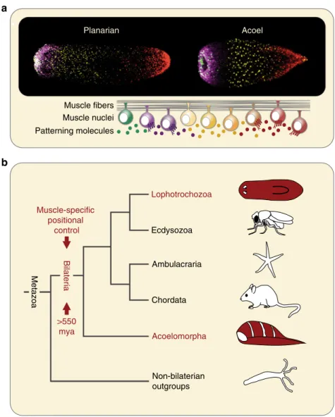
Ecdysozoa Ambulacraria Chordata Non-bilaterian outgroups Bilateria Lophotrochozoa Acoelomorpha Muscle-specific positional control
a
b
Planarian Acoel Muscle fibers Muscle nuclei Patterning molecules >550 mya MetazoaFig. 6 Model for evolution of muscle-specific positional control. a Both planarians and Hofstenia demonstrate muscle-specific expression of positional control genes, despite divergence of over 550 million years.b Muscle-specific expression of PCGs in both lophotrochozoans and acoels suggests that it arose as a mechanism of regenerative patterning at the base of the Bilateria, over 550 million years ago
fixed with 4% PFA in CMF-ASW, and then incubated in PreHybe. Staining pro-tocol was the same as in whole animals.
Image acquisition and quantification. Fluorescence image acquisition performed using a Zeiss LSM 700 confocal microscope. Image J software (Fiji) was used for processing and quantifying images. Images were quantified manually by first counting the number of (denominator)-positive cells while blinded to the other channel, then by counting the number of second-channel positive cells while blinded to thefirst channel. These were then overlayed to determine co-expression. See Supplementary Fig.2c.
Irradiation. Animals were irradiated using a dual Gammacell-40137cesium source
to deliver 10,000 rads.
Fluorescence-activated cell sorting. Animals were macerated in calcium- and magnesium-free artificial sea water (CMF-ASW: 449 mM NaCl, 9 mM KCl, 33 mM Na2SO4, 2.14 mM NaHCO310 mM Tris-Cl, 2.5 mM EGTA) with 1%
heat-inactivated horse serum by vigorously pipetting for 1–2 min. Cell suspensions were passed through a 40μm cell strainer (Falcon) and pelleted at 300 g for 5 min at 4 °C, and resuspended in CMF-ASW. After resuspension, cells were once again passed through a 40 um strainer. In all, 10 mg/mL Hoechst solution (Invitrogen) was added to the cell suspension at a 1:400 dilution and incubated in the dark for 45 min. In all, 5 mg/mL propidium iodide was added to allow for detection of permeabilized cells. Cells were stored on ice for no longer than 30 min after addition of propidium iodide before sorting. Cells were sorted on a MoFlo (Beckman-Coulter) with gating inclusive of all Hoechst-positive, propidium iodide-negative events to exclude dead cells and cellular debris. Cells were sorted into 96-well plates pre-loaded with 5 uL Buffer TCL (Qiagen) with 1% 2-mercaptoethanol. Plates were immediately stored at−80 °C.
Single-cell cDNA library construction. Libraries were prepared using the SmartSeq2 method35,36. In all, 11 uL of Ampure XP beads (Agencourt) were added to each well of a 96-well plate, incubated for 10 min, and put on a 96-well magnet plate (Dynamag 96-side magnet; Life Technologies) for 5 min. Supernatant was removed and beads were washed twice with 80% ethanol. Ethanol was removed and beads were eluted in a mix of 1 uL reverse-transcription primer (5 ′-AAG-CAGTGGTATCAACGCAGAGTACT(30)VN-3′, IDT DNA), 1 uL of dNTP mix (10 mM), 0.1 uL of SUPERase RNase-inhibitor (40 U/uL; Life Technologies), and 1.9 uL of H2O. The plate was incubated at 72 °C for 3 min and placed immediately
on ice. A mix of 1.65 uL H2O, 2 uL of 5× Maxima reverse-transcription buffer
(Thermo-Fischer), 0.9 uL MgCl2(100 mM Sigma-Aldrich; M1028), 2 uL of Betaine
(5 M; Sigma-Aldrich; B0300-5VL), 0.25 uL of SUPERase RNase-inhibitor (40 U/ uL), 0.1 uL of Maxima RNase H- RT (200 U/μL; Thermo-Fischer, EP0753), and 0.1 template switching-oligo (Exiqon; 100 uM; AAGCAGTGGTATCAACGCA-GAGTACrGrG + G; r and“ + ’’ denote RNA and LNA bases, respectively) was added to each well. Plate was incubated at 42 °C for 90 min, followed by 10 cycles of (50 °C for 2 min, 42 °C for 2 min), followed by 70 °C for 15 min. Following reverse-transcription a pre-amplification mix of 14 uL was added to each well [1 uL of H2O;
0.5 uL of PCR primer (10 uM; 5′-AAGCAGTGGTATCAACGCAGAGT-3′), and 12.5 uL of KAPA HiFi HotStart ReadyMix (Kapa Biosystems; KK2601)]. The cDNA was amplified using the following program: 98 °C for 3 min; 20 cycles of (98 °C for 15 s, 67 °C for 20 s, 72 °C for 6 min); 72 °C for 5 min; hold at 4 °C. Following pre-amplification PCR products were purified using × 0.8 Ampure XP beads, and eluted in 20 uL of H2O. Amplified cDNA concentrations were measured using
Qubit HS-DNA reagents (Life Technologies). All samples were diluted and nor-malized to 0.2 ng/uL.
Quantitative real-time PCR. All qRT-PCR primers were tested to ensure effi-ciency between 90%–110% and a standard curve R2> 0.98. A 20 uL mix was made
in MicroAmp Fast Optical 96-Well Reaction Plate (Applied Biosystems) of 50% EvaGreen (BioRad), 5% forward primer, 5% backwards primer, 2% cDNA, and 38% H2O for each single-cell cDNA library. All libraries were initially screened for
expression of GAPDH (forward: 5′-TTCCGTGTACCAACACCAGA-3′, backward 5′-GGAGATTGACTGGCATCCTT-3′). Any library without expression of GAPDH (~ 20% of libraries) was excluded from further analysis. Expression was then analyzed for genes of interest. Primers used for qRT-PCR are listed in Sup-plementary Table1. All qRT-PCR was performed on a 7500 Fast Real-Time PCR System machine (Applied Biosystems) and analyzed on 7500 Software v2.0.5. All Ct values> 32 or undetected were thresholded to Ct = 32. The reported measure -ΔCt equals -[Ct(gene of interest)−Ct(GAPDH)].
Data availability. The authors declare that all data supporting thefindings of this study are available within the article and its supplementary informationfiles or from the corresponding author upon reasonable request. Nucleotide sequences for hm.98018390 and titin-like have been deposited in the GenBank under accession codes: MF568700 (hm.98018390) and MF568701 (titin-like).
Received: 30 June 2017 Accepted: 18 August 2017
References
1. Bely, A. E. & Nyberg, K. G. Evolution of animal regeneration: re-emergence of a field. Trends Ecol. Evol. 25, 161–170 (2010).
2. Wolpert, L. Positional information and the spatial pattern of cellular differentiation. J. Theor. Biol. 25, 1–47 (1969).
3. Poss, K. D. Advances in understanding tissue regenerative capacity and mechanisms in animals. Nat. Rev. Genet. 11, 710–722 (2010).
4. French, V., Bryant, P. J. & Bryant, S. V. Pattern regulation in epimorphicfields. Science 193, 969–981 (1976).
5. Witchley, J. N., Mayer, M., Wagner, D. E., Owen, J. H. & Reddien, P. W. Muscle cells provide instructions for planarian regeneration. Cell Rep. 4, 633–641 (2013).
6. Koziol, U., Jarero, F., Olson, P. D. & Brehm, K. Comparative analysis of Wnt expression identifies a highly conserved developmental transition in flatworms. BMC Biol. 14, 10 (2016).
7. Peterson, K. J. et al. Estimating metazoan divergence times with a molecular clock. Proc. Natl Acad. Sci. USA 101, 6536–6541 (2004).
8. Steinmetz, P. R. H. et al. Independent evolution of striated muscles in cnidarians and bilaterians. Nature 487, 231–234 (2012).
9. Ryan, J. F. et al. The genome of the ctenophore Mnemiopsis leidyi and its implications for cell type evolution. Science 342, 1242592 (2013). 10. Martindale, M. Q., Pang, K. & Finnerty, J. R. Investigating the origins of
triploblasty:‘mesodermal’ gene expression in a diploblastic animal, the sea anemone Nematostella vectensis (phylum, Cnidaria; class, Anthozoa). Development 131, 2463–2474 (2004).
11. Moroz, L. L. et al. The ctenophore genome and the evolutionary origins of neural systems. Nature 510, 109–114 (2014).
12. Seipel, K. & Schmid, V. Evolution of striated muscle: jellyfish and the origin of triploblasty. Dev. Biol. 282, 14–26 (2005).
13. Dayraud, C. et al. Independent specialisation of myosin II paralogues in muscle vs. non-muscle functions during early animal evolution: a ctenophore perspective. BMC Evol. Biol. 12, 107 (2012).
14. Hejnol, A. & Pang, K. Xenacoelomorpha’s significance for understanding bilaterian evolution. Curr. Opin. Genet. Dev. 39, 48–54 (2016). 15. Bely, A. E. & Sikes, J. M. Acoel and platyhelminth models for stem-cell
research. J. Biol. 9, 1–4 (2010).
16. Gehrke, A. R. & Srivastava, M. Neoblasts and the evolution of whole-body regeneration. Curr. Opin. Genet. Dev. 40, 131–137 (2016).
17. Srivastava, M., Mazza-Curll, K. L., Van Wolfswinkel, J. C. & Reddien, P. W. Whole-body acoel regeneration is controlled by Wnt and Bmp-Admp signaling. Curr. Biol. 24, 1107–1113 (2014).
18. Rouse, G. W., Wilson, N. G., Carvajal, J. I. & Vrijenhoek, R. C. New deep-sea species of Xenoturbella and the position of Xenacoelomorpha. Nature 530, 94–97 (2016).
19. Hejnol, A. et al. Assessing the root of bilaterian animals with scalable phylogenomic methods. Proc. R. Soc. B 276, 4261–4270 (2009).
20. Cannon, J. T. et al. Xenacoelomorpha is the sister group to Nephrozoa. Nature 530, 89–93 (2016).
21. Arroyo, A. S., López-Escardó, D., de Vargas, C. & Ruiz-Trillo, I. Hidden diversity of Acoelomorpha revealed through metabarcoding. Biol. Lett. 12, 20160674 (2016).
22. Ruiz-Trillo, I. Acoelflatworms: earliest extant bilaterian metazoans, not members of platyhelminthes. Science 283, 1919–1923 (1999).
23. Hooge, M. et al. A revision of the systematics of panther worms (Hofstenia spp., Acoela), with notes on color variation and genetic variation within the genus. Hydrobiologia 592, 439–454 (2007).
24. Jondelius, U., Wallberg, A., Hooge, M. & Raikova, O. I. How the worm got its pharynx: Phylogeny, classification and bayesian assessment of character evolution in acoela. Syst. Biol. 60, 845–871 (2011).
25. Steinböck, O. Regenerationsversuche mit Hofstenia giselae Steinb. (Turbellaria Acoela). Roux Arch. Entwickl. 158, 394–458 (1967).
26. Kakugawa, S. et al. Notum deacylates Wnt proteins to suppress signalling activity. Nature 519, 187–192 (2015).
27. Zhang, X. et al. Notum is required for neural and head induction via wnt deacylation, oxidation, and inactivation. Dev. Cell 32, 719–730 (2015). 28. Hooge, M. D., Haye, P. A., Tyler, S., Litvaitis, M. K. & Kornfield, I. Molecular
systematics of the Acoela (Acoelomorpha, Platyhelminthes) and its concordance with morphology. Mol. Phylogenet. Evol. 24, 333–342 (2002). 29. Scimone, M. L., Kravarik, K. M., Lapan, S. W. & Reddien, P. W. Neoblast specialization in regeneration of the planarian Schmidtea mediterranea. Stem Cell Rep. 3, 339–352 (2014).
30. Rieger, R. M. & Ladurner, P. The significance of muscle cells for the origin of mesoderm in bilateria. Integr. Comp. Biol. 43, 47–54 (2003).
31. Hejnol, A. & Martindale, M. Q. Acoel development supports a simple planula-like urbilaterian. Phil. Trans. R. Soc. B 363, 1483–1501 (2008).
32. Pearson, B. J. et al. A formaldehyde-based whole mount in situ hybridization method for planarians. Dev. Dyn. 238, 443–450 (2009).
33. King, R. S. & Newmark, P. A. In situ hybridization protocol for enhanced detection of gene expression in the planarian Schmidtea mediterranea. BMC Dev. Biol. 13, 8 (2013).
34. Scimone, M. L., Cote, L. E., Rogers, T. & Reddien, P. W. Two FGFRL-Wnt circuits organize the planarian anteroposterior axis. eLife 5, e12845 (2016). 35. Picelli, S. et al. Smart-seq2 for sensitive full-length transcriptome profiling in
single cells. Nat. Methods 10, 1096–1098 (2013).
36. Wurtzel, O. et al. A generic and cell-type-specific wound response precedes regeneration in planarians. Dev. Cell 35, 632–645 (2015).
Acknowledgements
We acknowledge Isaac Oderberg for illustration assistance, Olivia Johnson and Catherine McQuestion for animal husbandry, and members of the Reddien lab for critical input. P. W.R. is an Investigator of the Howard Hughes Medical Institute and an associate member of the Broad Institute of Harvard and MIT.
Author contributions
A.A.R. and P.W.R. conceived of and designed experiments. A.A.R., R.S. and M.S. optimized protocols. A.A.R. and R.S. performed the experiments. A.A.R. and P.W.R. analyzed the data. A.A.R. and P.W.R. wrote the paper.
Additional information
Supplementary Informationaccompanies this paper at doi:10.1038/s41467-017-01148-5. Competing interests:The authors declare no competingfinancial interests.
Reprints and permissioninformation is available online athttp://npg.nature.com/ reprintsandpermissions/
Publisher's note:Springer Nature remains neutral with regard to jurisdictional claims in published maps and institutional affiliations.
Open Access This article is licensed under a Creative Commons Attribution 4.0 International License, which permits use, sharing, adaptation, distribution and reproduction in any medium or format, as long as you give appropriate credit to the original author(s) and the source, provide a link to the Creative Commons license, and indicate if changes were made. The images or other third party material in this article are included in the article’s Creative Commons license, unless indicated otherwise in a credit line to the material. If material is not included in the article’s Creative Commons license and your intended use is not permitted by statutory regulation or exceeds the permitted use, you will need to obtain permission directly from the copyright holder. To view a copy of this license, visithttp://creativecommons.org/ licenses/by/4.0/.