HAL Id: hal-01330190
https://hal.sorbonne-universite.fr/hal-01330190
Submitted on 10 Jun 2016
HAL is a multi-disciplinary open access
archive for the deposit and dissemination of
sci-entific research documents, whether they are
pub-lished or not. The documents may come from
teaching and research institutions in France or
abroad, or from public or private research centers.
L’archive ouverte pluridisciplinaire HAL, est
destinée au dépôt et à la diffusion de documents
scientifiques de niveau recherche, publiés ou non,
émanant des établissements d’enseignement et de
recherche français ou étrangers, des laboratoires
publics ou privés.
Distributed under a Creative Commons Attribution| 4.0 International License
Serine/Threonine Protein Phosphatase-Mediated
Control of the Peptidoglycan Cross-Linking
L,D-Transpeptidase Pathway in Enterococcus faecium
Emmanuelle Sacco, Mélanie Cortes, Nathalie Josseaume, Louis B Rice,
Jean-Luc Mainardi, Michel Arthur
To cite this version:
Emmanuelle Sacco, Mélanie Cortes, Nathalie Josseaume, Louis B Rice, Jean-Luc Mainardi, et al..
Serine/Threonine Protein Phosphatase-Mediated Control of the Peptidoglycan Cross-Linking
L,D-Transpeptidase Pathway in Enterococcus faecium. mBio, American Society for Microbiology, 2014, 5
(4), pp.e01446-14. �10.1128/mBio.01446-14�. �hal-01330190�
Serine/Threonine Protein Phosphatase-Mediated Control of the
Peptidoglycan Cross-Linking
L
,
D
-Transpeptidase Pathway in
Enterococcus faecium
Emmanuelle Sacco,a,b,cMélanie Cortes,a,b,cNathalie Josseaume,a,b,cLouis B. Rice,dJean-Luc Mainardi,a,b,c,eMichel Arthura,b,c
INSERM, U1138, LRMA, Equipe 12 du Centre de Recherche des Cordeliers, Paris, Francea; Université Pierre et Marie Curie, UMR S 1138, Paris, Franceb; Université Paris
Descartes, Sorbonne Paris Cité, UMR S 1138, Paris, Francec; Rhode Island Hospital, Brown University, Providence, Rhode Island, USAd; Assistance Publique-Hôpitaux de
Paris, Service de Microbiologie, Hôpital Européen Georges Pompidou, Paris, Francee
E.S. and M.C. contributed equally to this article.
ABSTRACT The last step of peptidoglycan polymerization involves two families of unrelated transpeptidases that are the essential
targets of-lactam antibiotics.D,D-transpeptidases of the penicillin-binding protein (PBP) family are active-site serine enzymes that use pentapeptide precursors and are the main or exclusive cross-linking enzymes in nearly all bacteria. However, pepti-doglycan cross-linking is performed mainly by active-site cysteineL,D-transpeptidases that use tetrapeptides in Mycobacterium
tuberculosis, Clostridium difficile, and-lactam-resistant mutants of Enterococcus faecium. We have investigated
reprogram-ming of the E. faecium peptidoglycan assembly pathway by a switch from pentapeptide to tetrapeptide precursors and bypass of PBPs byL,D-transpeptidase Ldtfm. Mutational alterations of two signal transduction systems were necessary and sufficient for
activation of theL,D-transpeptidation pathway, which is essentially cryptic in wild-type strains. The first one is a classical two-component regulatory system, DdcRS, that controls the activity of Ldtfmat the substrate level. As previously described, loss of
DdcS phosphatase activity leads to production of theD,D-carboxypeptidase DdcY and conversion of the pentapeptide into the tetrapeptide substrate of Ldtfm. Here we show that full bypass of PBPs by Ldtfmalso requires increased Ser/Thr protein
phos-phorylation resulting from impaired activity of phosphoprotein phosphatase StpA. This enzyme negatively controlled the level of protein phosphorylation both by direct dephosphorylation of target proteins and by dephosphorylation of its cognate kinase Stk. In combination with production of DdcY, increased protein phosphorylation by this eukaryotic-enzyme-like Ser/Thr pro-tein kinase was sufficient for activation of theL,D-transpeptidation pathway in the absence of mutational alteration of pepti-doglycan synthesis enzymes.
IMPORTANCEThe mechanism of acquisition of high-level ampicillin resistance involving bypass of the penicillin-binding
pro-teins (PBPs) byL,D-transpeptidase Ldtfmwas incompletely understood, as production of tetrapeptide precursors following
tran-scriptional activation of the ddc locus by the DdcRS two-component regulatory system was necessary but not sufficient for full activation of theL,D-transpeptidation pathway. Here, we identified the release of a negative control of Ser/Thr protein phosphor-ylation mediated by phosphatase StpA as the additional factor essential for ampicillin resistance. Thus, bypass of PBPs by Ldtfm
requires the modification of signal transduction regulatory systems without any gain of function by mutational alteration of peptidoglycan biosynthetic enzymes. In contrast, previously characterized mechanisms of antibiotic resistance involve horizon-tal gene transfer and mutational alteration of drug targets. Activation of theL,D-transpeptidation pathway reported in this study is an unprecedented mechanism of emergence of a new metabolic pathway since it involved the recruitment of preexisting func-tions following modificafunc-tions of regulatory circuits.
Received 12 June 2014 Accepted 16 June 2014 Published 8 July 2014
Citation Sacco E, Cortes M, Josseaume N, Rice LB, Mainardi J-L, Arthur M. 2014. Serine/threonine protein phosphatase-mediated control of the peptidoglycan cross-linking
L,D-transpeptidase pathway in Enterococcus faecium. mBio 5(4):e01446-14. doi:10.1128/mBio.01446-14.
Invited Editor Juan Ayala, Consejo Superior Investigaciones Cientificas Editor Fernando Baquero, Ramón y Cajal University Hospital
Copyright © 2014 Sacco et al. This is an open-access article distributed under the terms of theCreative Commons Attribution-Noncommercial-ShareAlike 3.0 Unported license, which permits unrestricted noncommercial use, distribution, and reproduction in any medium, provided the original author and source are credited. Address correspondence to Michel Arthur, michel.arthur@crc.jussieu.fr.
E
nterococcus faecium and Enterococcus faecalis are commensal bacteria but also important nosocomial pathogens (1). These bacteria have an unusually high capacity for resistance to antibi-otics since they combine several mechanisms of intrinsic resis-tance with the acquisition of resisresis-tance determinants by horizon-tal gene transfer (2–4). Intrinsic-lactam resistance is mediated by species-specific low-affinity penicillin-binding proteins(PBPs), such as PBP5, that are thought to be sufficient for pepti-doglycan cross-linking under conditions in which the D,D
-transpeptidase catalytic domain of all other PBPs is inactivated by -lactams (5–7). Although -lactam resistance is conveyed by a single PBP in the enterococci, expression of resistance depends upon additional host factors (8, 9). In E. faecalis, factors essential for PBP5-mediated resistance to-lactams of the cephalosporin
mbio.asm.org
on June 10, 2016 - Published by
class have been detected by genetic analyses but their functions remain largely unknown. These factors include the CroRS two-component regulatory system, which is thought to regulate genes essential for PBP5 activity, although none of the targets of CroRS-mediated transcriptional regulation identified so far has a demon-strated role in peptidoglycan synthesis or cephalosporin resistance (10–13). A serine/threonine protein kinase (IreK) was identified as essential for ceftriaxone resistance in E. faecalis (14). Subse-quent characterization of the associated protein phosphatase, IreP, showed that the phosphorylation of one or several IreK sub-strates is essential for cephalosporin resistance (15). Recently, a substrate of IreK and IreP, designated IreB, has been identified and shown to negatively control cephalosporin resistance (16). Additional proteins essential for ceftriaxone resistance include bi-functional transglycosylase-transpeptidase (class A) PBPs for elongation of glycan chains, either PonA or PBPF (17, 18), one of the two E. faecalis MurA synthetases (UDP-N-acetylglucosamine 1-carboxyvinyl transferase) performing the first committed stem of peptidoglycan synthesis (19), and alanine transferase of the Fem family for synthesis of theL-Ala–L-Ala side chain of pepti-doglycan precursors (20). Genome-wide identification of ampi-cillin resistance determinants in E. faecium identified a different set of proteins contributing to intrinsic-lactam resistance, in-cluding the L,D-transpeptidase Ldtfm, the D,D-carboxypeptidase DdcP, and the glycosyltransferase Pgt (21).
In previous studies, we have identified a PBP5-independent -lactam resistance mechanism in E. faecium based on in vitro selection of mutants resistant to ampicillin (a-lactam of the penam class) (22). We started from E. faecium D344S, a derivative of E. faecium D344R that had spontaneously lost the pbp5 gene (22). Parental strain D344S is hypersusceptible to ampicillin be-cause of the absence of low-affinity PBP5 (22). In five consecutive selection steps with increasing concentrations of ampicillin, we obtained five mutants, M1, M2, M3, M4, and M512, that gradually acquired high-level ampicillin resistance (22, 23). In mutant M512, 4¡3 cross-links formed by PBPs were replaced by 3¡3 cross-links generated by an ampicillin-insensitive L,D
-transpeptidase, Ldtfm(22, 24). This enzyme cleaves theL-Lys3–D -Ala4peptide bond of the acyl donor and links the carbonyl group ofL-Lys3to the acyl acceptor (24). Ldtfmand PBPs were found to be structurally unrelated and to harbor different active-site resi-dues for nucleophilic attack of the carbonyl group of the acyl do-nor (Cys versus Ser, respectively) (24–26). Ldts and PBPs also differ in their spectra of inactivation by-lactams (27). PBPs are potentially inactivated by all-lactams, including members of the penam (ampicillin), cephalosporin (ceftriaxone), carbapenem (imipenem), and monobactam (aztreonam) classes, depending upon the specific-lactam and PBP under consideration (28). In contrast,L,D-transpeptidases are efficiently inactivated by a single
class of-lactams, the carbapenems (27, 29).
The proportion of 3¡3 cross-links generated by Ldtfmis very low in wild-type strains of E. faecium (22). Ldtfmis constitutively produced by parental strain D344S (23, 30), but the participation of theL,D-transpeptidase to peptidoglycan cross-linking is mar-ginal since this enzyme requires a tetrapeptide stem in the acyl donor substrate (24) instead of the pentapeptide stem assembled in the cytoplasm (31). Thus, activation of theL,D-transpeptidation
pathway requires pentapeptide-to-tetrapeptide conversion (23). The corresponding enzyme, DdcY, was identified as a
metallo-D,D-carboxypeptidase that cleaves the C-terminal residue (D-Ala5)
of a UDP-MurNAc pentapeptide to generate the precursor of the
L,D-transpeptidation pathway (30). DdcY is encoded by a cryptic locus, ddc, that is sporadically disseminated in E. faecium strains and encodes a two-component regulatory system (DdcRS) in ad-dition to DdcY (30). Activation of the ddc locus in M512 results from a mutation in the ddcS sensor kinase gene that impairs the phosphatase activity of the enzyme and leads to constitutive ex-pression of the locus (30). In this investigation, we identified a second locus involved in the activation of theL,D-transpeptidation
pathway, which encodes a eukaryotic-enzyme-like serine/threo-nine protein kinase (Stk) and its cognate phosphatase (StpA). We show that decreased protein dephosphorylation by StpA and the previously characterized activation of the ddc locus are both suf-ficient and necessary for high-level-lactam resistance mediated byL,D-transpeptidation in E. faecium.
RESULTS
Identification of mutations necessary and sufficient for high-level ampicillin resistance. We have previously shown that
acqui-sition of high-level resistance to ampicillin by mutant M512 re-quires activation of the ddc locus, which encodes the D,D
-carboxypeptidase DdcY, for formation of the tetrapeptide-containing substrate of the L,D-transpeptidase (30). In mutant
M512, activation of the locus results from a T161A substitution in the sensor kinase DdcS that impairs the phosphatase activity of the protein (30). Here, we show that acquisition of ampicillin resis-tance involves the mutational alteration of a second locus encod-ing a putative serine/threonine kinase (Stk) and a phosphatase (StpA) (Fig. 1A). The mutation was detected in mutant M1 (first selection step) and leads to a Thr-to-Arg substitution at position 101 of StpA, which is invariant in related phosphatases (Fig. 1B). Acquisition of the stpA mutation by strain D344S was associ-ated with a moderate increase in the MIC of ampicillin (from 0.06 to 0.5g/ml) (Fig. 1C). In order to evaluate the contributions of DdcY and StpA to resistance, the ddcY gene was cloned under the control of constitutive promoter P2 of expression vector pJEH11 and the resulting plasmid (pJEH11⍀ddcY) was introduced into D344S. Production of theD,D-carboxypeptidase in this host re-sulted in a moderate level of resistance to ampicillin (MIC, 0.5g/ ml). In contrast, plasmid pJEH11⍀ddcY conferred high-level re-sistance on mutant M1 (MIC,⬎2,000g/ml). Thus, the T101R substitution in StpA and production of DdcY were both necessary and sufficient for high-level ampicillin resistance.
Selection of high-level ampicillin resistance in a D,D -carboxypeptidase-producing background. Spontaneous
mu-tants of D344S/pJEH11⍀ddcY were obtained on agar plates con-taining ampicillin (4 or 8g/ml) at a frequency of ca. 5 ⫻ 10⫺7. The characterization of a total of five independent mutants in each case revealed the expression of high-level ampicillin resistance (MIC,⬎2,000g/ml) and the presence of a mutation in stpA (mutants Ma to Me, Fig. 1B and C). The mutations led to amino acid substitutions in four mutants and to a frameshift mutation in the remaining mutant. Three of the four substitutions affected invariant residues, and the remaining substitution (S192P) was lo-cated within a highly conserved motif (Fig. 1B). The frameshift mutation resulted from a base deletion in codon 236 located close to the 3= end of the stpA gene (246 codons), which encodes a conserved region of the protein. These results indicate that muta-tions in stpA are reproducibly selected by ampicillin in aD,D
-carboxypeptidase-producing background.
Sacco et al.
mbio.asm.org
on June 10, 2016 - Published by
Modulation of the ampicillin resistance level by the balance between kinase and phosphatase activities. Overproduction of
Stk in D344S following the introduction of plasmid pJEH11⍀stk increased the MIC of ampicillin from 0.06 to 0.5g/ml (Fig. 1C). Thus, the resistance phenotypes resulting from overproduction of Stk (D344S/pJEH11⍀stk) and impaired StpA phosphatase activity (mutant M1) were similar. This observation implies that the level of ampicillin resistance increases with the level of serine and thre-onine phosphorylation. In agreement, expression of a functional
copy of stpA impaired the expression of ampicillin resistance in mutant M512 (data not shown). Thus, the level of ampicillin re-sistance is modulated by a balance between kinase and phospha-tase activities.
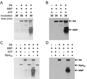
In vitro activities of purified Stk and StpA. A soluble fragment
of the kinase Stk (residues 1 to 338) was produced in Esche-richia coli, purified, and analyzed by Western blotting. Recombi-nant Stk was purified as a phosphoprotein, as shown by the detec-tion of phosphorylated proteins (Fig. 2A) or phosphothreonine
FIG 1 Roles of ddcY and stpA in ampicillin resistance. (A) Map of the E. faecium locus encoding the protein phosphatase StpA and the serine/threonine kinase
Stk. (B) Sequence alignment of protein phosphatases. Identical and conserved amino acids are highlighted in black and gray, respectively. The positions of amino acid substitutions detected in the mutants are indicated below the alignment. In mutant Ma, a frameshift was detected in codon 236 of stpA. (C) MICs of ampicillin for E. faecium D344S and resistant mutants.
mbio.asm.org
on June 10, 2016 - Published by
residues (Fig. 2B). Mass spectrometry analyses revealed several forms of the proteins containing one to five phosphate groups (see Fig. S1A and B in the supplemental material). StpA fully dephos-phorylated Stk, as shown by the complete disappearance of the phosphorylated forms of the kinase by Western blotting (Fig. 2C and D) and mass spectrometry (see Fig. S1 in the supplemental material). Upon incubation with ATP, purified Stk phosphory-lated myelin basic protein (MBP) (Fig. 2A and B). The phospho-protein bands corresponding to phospho-MBP disappeared upon the addition of the phosphatase StpA. These results indicate that StpA and Stk display the protein phosphatase and kinase activities inferred from sequence similarity. These results also indicate that kinase Stk is a substrate of StpA.
Autophosphorylation of purified Stk in vitro. Since
recombi-nant Stk was purified as a phosphoprotein from E. coli extracts, Stk was dephosphorylated in vitro by the addition of StpA in a 1:10 phosphatase-to-kinase ratio (see Fig. S2 in the supplemental ma-terial). The phosphatase StpA was removed by size exclusion chro-matography, and the absence of phosphate groups from purified Stk was confirmed by Western blotting and mass spectrometry
analyses. Incubation of dephosphorylated Stk with ATP and Mn2⫹resulted in the phosphorylation of Stk. The reaction led to a monophosphorylation of Stk, whereas the protein recovered from E. coli extracts contained multiple phosphate groups (compare Fig. S1 and S2 in the supplemental material). These results indi-cate that Stk catalyzes the phosphorylation of at least one of its threonine residues. The difference between the phosphorylation patterns observed in vitro and in vivo indicates that E. coli might produce an unknown kinase responsible for Stk phosphorylation, although this host does not produce any Stk homologue (32).
Impact of stpA mutations on the in vitro activity of the phos-phatase. Recombinant forms of StpA detected in
ampicillin-resistant mutants M1, Ma, Mb, Mc, Md, and Me were produced in E. coli. The truncated form of StpA (mutant Ma) and StpA con-taining the S192P substitution (mutant Mc) were not soluble and could not be functionally characterized. Purification of the four remaining proteins afforded soluble enzymes with reduced phos-phatase activity (M1, Mb, and Me) or no detectable activity (Md) (Table 1). The amino acid substitutions detected in the former mutants mainly led to lower kcatvalues with marginal impacts on the Kmof para-nitrophenylphosphate (pNPP). The values of these catalytic constants were determined in the presence of a high con-centration of Mn2⫹(2 mM). Further analysis performed with the wild-type enzyme showed that Mn2⫹is essential for activity and that this cation cannot be replaced by Mg2⫹, Ca2⫹, or Zn2⫹(data not shown). The phosphatase activity increased with the Mn2⫹ concentration, and a relatively high concentration of the cation (ⱖ2 mM) was required for maximal activity (see Fig. S3 in the supplemental material). Reduction of the Mn2⫹concentration from 2 mM to 50M led to a 100-fold decrease in the catalytic efficiency of wild-type StpA (Table 1). Reduction of the Mn2⫹ concentration also reduced the activity of StpA from mutants Mb and Me. Together, these results indicated that the mutations de-tected in the stpA gene from ampicillin-resistant mutants im-paired the phosphatase activity of the protein, as suggested by the positions of the mutations. Of note, sufficient Mn2⫹is expected to be present in vivo for metalation of StpA since brain heart infusion (BHI) broth contains 20M Mn2⫹(33) and E. faecium accumu-lates Mn2⫹because of active uptake (34, 35).
StpA and Stk do not control expression of the ddc locus or
ldtfmL,D-transpeptidase gene. We have previously shown that the
ddc locus encoding DdcY (D,D-carboxypeptidase) and DdcRS
(two-component regulatory system) is cryptic in parental strain D344S (30). Activation of the locus in mutant M512 is due to the T161A substitution in DdcS, which impairs the phosphatase
activ-FIG 2 In vitro activities of Stk (A and B) and StpA (C and D).
Phosphopro-teins were revealed with a commercial kit (Pro-Q; Invitrogen) (A and C) or with phosphothreonine-specific antibodies (B and D).
TABLE 1 Catalytic constants for pNPP hydrolysis by StpA from parental strain D344S and ampicillin-resistant mutants
Substitution in StpA (strain)
Catalytic constantain presence of Mn2⫹at:
2 mM 50M Km (mM) kcat (min⫺1) kcat/Km (min⫺1mM⫺1) Km (mM) kcat (min⫺1) kcat/Km (min⫺1mM⫺1) None (D344S) 0.66⫾ 0.09 1,200⫾ 100 1,800⫾ 290 0.69⫾ 0.17 12⫾ 1 17⫾ 4.5 T101R (M1) 1.2⫾ 0.2 0.83⫾ 0.04 0.69⫾ 0.12 NDb ND ND D136Y (Mb) 1.8⫾ 0.5 140⫾ 15 80⫾ 23 1.5⫾ 0.4 0.09⫾ 0.01 0.06⫾ 0.02 D18Y (Md) ND ND ⬍0.003 ND ND ND S120R (Me) 1.3⫾ 0.1 28⫾ 0.1 22⫾ 1.6 2.6⫾ 0.7 0.23⫾ 0.02 0.09⫾ 03
aRegression values⫾ standard errors were obtained by fitting experimental data to the Michaelis-Menten equation V ⫽ k
catES/(Km⫹ S), where V is the initial velocity and E and S
are the initial enzyme and substrate concentrations, respectively.
bND, not determined.
Sacco et al.
mbio.asm.org
on June 10, 2016 - Published by
ity of the sensor kinase. Here we show that expression of the ddc locus is not under StpA control since DdcY and DdcS were not detected in extracts of D344S and M1, whereas the two proteins were produced at similar high levels by M512 and M512/ pJEH11⍀stpA (Fig. 3). Likewise, overproduction of Stk in D344S did not lead to any increase in the levels of DdcY and DdcS pro-duction (data not shown). Propro-duction of theL,D-transpeptidase Ldtfmwas not affected by impaired StpA and DdcS phosphatase activities (Fig. 3) or by Stk overproduction (data not shown). These results show that the contribution of impaired StpA phos-phatase activity or increased Stk activity to ampicillin resistance did not depend upon increased production of the L,D
-transpeptidase Ldtfmor activation of the ddc locus for production of the tetrapeptide substrate of this cross-linking enzyme.
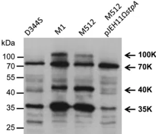
In vivo activity of StpA. Western blot analysis of E. faecium
protein extracts was performed to determine whether the in vivo production of StpA affects the level of threonine phosphorylation. The intensity of three protein bands detected by antiphospho-threonine antibodies (100, 40, and 35 kDa) was increased in mu-tant M1 (StpA T101R) in comparison to that in parental strain D344S (StpA) (Fig. 4). As expected, the phosphoprotein patterns were similar in M1 and M512, whereas production of StpA by M512/pJEH11⍀stpA led to a decrease in the intensity of the same set of phosphoprotein bands. These results indicate that StpA neg-atively controls the level of threonine phosphorylation in vivo.
In vitro dephosphorylation of phosphoproteins by purified
StpA. Comparison of the level of protein phosphorylation in
crude E. faecium extracts (Fig. 4) does not allow determination of whether the negative control mediated by StpA relies on dephos-phorylation of phosphoproteins or an indirect effect involving negative control of the kinase activity of Stk. In order to assay directly for the phosphoprotein phosphatase activity of StpA, E. faecium extracts were incubated with purified StpA and ana-lyzed by Western blot assay (Fig. 5A). The assay was performed with extracts from D344S, M1, M512, and D344S/pJEH11⍀stk.
The latter strain was included to maximize the phosphorylation of putative StpA substrates because of high-level production of the Ser/Thr kinase. Moderate overproduction of Stk by D344S/ pJEH11⍀stk was experimentally established by the increased in-tensity of a 100-kDa protein band detected with anti-Stk antibod-ies (Fig. 5B) and detection of recombinant Stk with antipolyhistidine antibodies (Fig. 5C). The 100-kDa phosphopro-tein band (Fig. 5A) had the same electrophoretic mobility as Stk (Fig. 5B) and recombinant Stk containing a 6⫻His tag (Fig. 5C). Purification of Stk from E. faecium D344S/pJEH11⍀stk (see Fig. S4 in the supplemental material) indicated that the 100-kDa phosphoprotein band corresponds to Stk.
Incubation of the extracts with purified StpA resulted in a de-crease in the intensity of the Stk 100-kDa phosphoprotein band (Fig. 5A). This decrease resulted from dephosphorylation of phospho-Stk by StpA, as previously shown for purified StpA and Stk (Fig. 2). Dephosphorylation of the phosphoprotein bands at 40 and 35 kDa by StpA was not detected (Fig. 5A). Thus, modu-lation of the phosphorymodu-lation levels of these proteins by StpA is indirect and may involve a negative control of the kinase activity of Stk. Upon incubation with StpA, the intensity of the 70-kDa phoprotein band decreased in all four extracts. Thus, the phos-phoprotein band at 70 kDa contains a substrate of StpA. These results suggest that a subset of the proteins phosphorylated by Stk can be dephosphorylated by StpA.
DISCUSSION
The last step of peptidoglycan polymerization involves two fami-lies of unrelated transpeptidases ofDD andLD specificities that
catalyze the formation of 4¡3 and 3¡3 cross-links (36). The relative contributions of the two enzyme types greatly vary among bacterial species and may also vary along the cell cycle. In E. coli, twoL,D-transpeptidases catalyze the formation of a minority (ca.
5%) of the cross-links during the exponential growth phase but the contribution ofL,D-transpeptidases to peptidoglycan
cross-linking increases in the stationary phase (13%) (37, 38). In
Myco-FIG 3 Level of production of proteins encoded by the stpA-stk, ddc, and ldtfm
loci in E. faecium strains. Crude E. faecium extracts were analyzed by Western blotting with polyclonal antibodies raised against the protein phosphatase StpA, the serine/threonine kinase Stk, theD,D-carboxypeptidase DdcY, the sensor kinase DdcS, and theL,D-transpeptidase Ldtfm.
FIG 4 Western blot analysis of phosphoproteins in crude E. faecium extracts.
Phosphoproteins were detected with antiphosphothreonine polyclonal anti-bodies. Arrows indicate the positions of the main phosphoproteins (100, 70, 40, and 35 kDa), which were named according to their relative electrophoretic mobility.
mbio.asm.org
on June 10, 2016 - Published by
bacterium tuberculosis (39, 40) and M. abscessus (41), L,D -transpeptidation is the predominant mode of peptidoglycan cross-linking in both growth phases (70 to 80%), althoughL,D -transpeptidase paralogs appear to be differently regulated during the cell cycle (42, 43). In wild-type E. faecium, the L,D -transpeptidase Ldtfmis constitutively produced but the enzyme makes a marginal contribution to peptidoglycan cross-linking (3%) (23, 30). The activity of Ldtfmis controlled at the substrate level by the production of theD,D-carboxypeptidase DdcY, which cleaves the C-terminalD-Ala of the stem pentapeptide to form the
essential tetrapeptide donor of theL,D-transpeptidation reaction (23, 30). In contrast to the ldtfmgene, which is present in all of the E. faecium isolates that have been examined (24), the ddcY and upstream ddcRS genes are present in only a minority of the isolates belonging to this species (30). The ddc locus is cryptic, and its activation results from the loss of the phosphatase activity of the DdcS sensor kinase in response to selection for ampicillin resis-tance (30). Production of DdcY is not sufficient for the complete bypass of PBPs by Ldtfmsince the production of DdcY was asso-ciated with an only moderate (8-fold) increase in the ampicillin MIC (Fig. 1C). In this study, we show that the full bypass of PBPs also requires an increase in Ser/Thr protein phosphorylation re-sulting from impaired StpA phosphatase activity. Association of a mutation in stpA with expression of ddcY was necessary and suf-ficient for high-level ampicillin resistance (Fig. 1C). In support of
this conclusion, we constructed strains that sequentially acquired a mutation in stpA and a plasmid encoding DdcY in both orders (Fig. 1C). The stpA mutations led to impaired phosphatase activ-ity, as inferred from their nature and positions (Fig. 1B) and a direct assay of StpA phosphatase activity (Table 1). In agreement, homologous residues of the phosphatase Stp from Streptococcus agalactiae were located in the enzyme-active site (44) (see Fig. S5 in the supplemental material). Amino acid substitutions in StpA from E. faecium mutants led to⬎200-fold reductions in kcatfor hydrolysis of pNPP in the presence of 50M MnCl2. The impact of the D136Y and S120R substitutions was less (43-fold and 7-fold reductions, respectively) at the nonphysiological Mn2⫹ concen-tration of 2 mM. This observation suggests that substitutions D136Y and S120R impaired Mn2⫹binding. Together, these results indicate that amino acid substitutions in StpA affected the chem-ical step of the dephosphorylation reaction rather than substrate recognition. Thus, decreased protein dephosphorylation was es-sential for acquisition of ampicillin resistance.
Several assays were used to study the protein kinase and phos-phatase activities of purified Stk and StpA. First, we showed that Stk catalyzes its own phosphorylation on Thr residues and subse-quently transfers the phosphate group to a model protein (MBP) (Fig. 2; see Fig. S2 in the supplemental material). Phospho-Stk and phospho-MBP were dephosphorylated upon the addition of StpA (Fig. 2). This first assay established that Stk and StpA display pro-tein kinase and phosphatase activities, respectively, as expected from sequence similarity to previously characterized enzymes (45). In a second assay, we compared the level of protein phos-phorylation in crude cell extracts from E. faecium strains harbor-ing different alleles of stpA (Fig. 4). This analysis revealed that impaired StpA activity increased the phosphorylation of several phosphoprotein bands (Fig. 4), including Stk (Fig. 5), indicating that StpA negatively modulates the level of protein phosphoryla-tion in vivo. A third assay was used to determine whether StpA directly dephosphorylates proteins. In this assay, crude cell ex-tracts were prepared from an E. faecium strain harboring various alleles of stpA and from an additional Stk-overproducing strain to maximize protein phosphorylation (Fig. 5). Incubation of the ex-tracts resulted in the decreased phosphorylation of three phos-phoprotein bands (100, 70, and 35 kDa). The extent of phosphor-ylation of a 40-kDa protein was greater in mutants with impaired phosphatase activity, but the intensity of the phosphoprotein band did not decrease upon incubation of the extract with purified StpA in vitro. For this protein band, modulation of the level of phosphorylation appears to depend upon the negative control of Stk kinase activity by StpA. Other phosphoproteins were directly dephosphorylated by StpA. For these phosphoproteins, negative control of the level of phosphorylation mediated by StpA may involve both protein dephosphorylation and negative control of the kinase Stk.
Together, our results show that the mutational alteration of two signal transduction systems is necessary and sufficient for reprogramming of the peptidoglycan assembly pathway by the production of tetrapeptide-containing precursors. The first signal transduction system is a classical two-component regulatory sys-tem, DdcRS, that controls the production of DdcY for conversion of pentapeptide into tetrapeptide precursors as previously de-scribed (30). The second system, StpA-Stk, controls the level of phosphorylation of several proteins. Several lines of evidence in-dicate that increased protein phosphorylation resulting from the
FIG 5 Dephosphorylation of E. faecium phosphoproteins by purified StpA.
Proteins from crude cell extracts (10g) were incubated with (⫹) or without (⫺) purified phosphatase StpA (3g) for 30 min at room temperature. Pro-teins were separated by SDS-PAGE and transferred onto PVDF membranes. Phosphoproteins were detected with antiphosphothreonine (A), anti-Stk (B), and antipolyhistidine (C) antibodies. Arrows indicate the positions of the main phosphoproteins (100, 70, 40, and 35 kDa).
Sacco et al.
mbio.asm.org
on June 10, 2016 - Published by
mutation of stpA was required for resistance although this did not affect the level of DdcY production. First, expression of ddcY un-der the control of a heterologous promoter in plasmid pJEH11 was sufficient for high-level ampicillin resistance (MIC,⬎2,000 g/ ml) in mutant M1, which produced a derivative of StpA with reduced phosphatase activity, but not in the parental strain D344S, which harbors a wild-type copy of stpA (Fig. 1C). Thus, production of DdcY and impaired phosphatase activity were both required for ampicillin resistance. Second, Western blot analysis indicated that DdcY was produced at the same level by derivatives of D344S and M1 harboring pJEH11⍀ddcY, indicating, as might have been expected, that the heterologous promoter of vector pJEH11 is functional in both hosts independently of the stpA allele (see Fig. S6 in the supplemental material). Conversely, DdcY was not produced by D344S and M1, indicating that the stpA mutation present in M1 did not lead to transcriptional activation of the chromosomal copy of ddcY. Thus, the essential role of the stpA mutation did not depend upon DdcY production. In agreement, derivatives of D344S/pJEH11⍀ddcY selected on ampicillin (mu-tants Ma to Me) harbored mutations in stpA (Fig. 1C) that im-paired the phosphatase activity of StpA (Fig. 1B and Table 1) and did not affect the level of DdcY production (see Fig. S6 in the supplemental material). Third, acquisition of a mutation in stpA by M1 and mutants Ma to Me did not result in the production of DdcS (Fig. 3; see Fig. S6 in the supplemental material). Thus, the chromosomal locus ddc was not regulated in response to modula-tion of protein phosphorylamodula-tion by StpA. Together, these results establish that alterations of DdcRS and StpA-Stk are both required for resistance and that these signal transduction systems control the activity of distinct sets of proteins.
Production of DdcY resulting from the introduction of plas-mid pJEH11⍀ddcY into M1 or loss of DdcS phosphatase activity by M512 resulted in similar resistance phenotypes. Thus, DdcY is the only target of DdcRS-mediated regulation that is essential for ampicillin resistance. In contrast, StpA may affect the level of phosphorylation of multiple proteins potentially involved in re-sistance, as shown by modulation of the level of phosphorylation of the 100-, 70-, 40-, and 35-kDa protein bands (Fig. 4 and 5). Furthermore, additional fainter phosphoprotein bands were de-tected by anti-phospho-Thr antibodies upon prolonged exposure of Western blots (data not shown) and Ser phosphorylation was not investigated. In agreement, phosphoproteome analyses of Fir-micutes suggest that the number of Ser/Thr phosphoproteins may be on the order of 100 to 200 in E. faecium (46, 47). The role of protein phosphorylation in peptidoglycan synthesis and ampicil-lin resistance remains to be determined.
It is striking that activation of theL,D-transpeptidation
path-way in E. faecium involves modifications of representatives of the two main signal transduction pathways of prokaryotes, the two-component regulatory system relying on His-to-Asp phospho-transfer (48) and the one-component eukaryotic-enzyme-like Ser/Thr phosphorylation system (45), in the absence of modifica-tion of cell wall biosynthesis proteins. In contrast, previously char-acterized mechanisms of acquisition of antibiotic resistance in-volve horizontal gene transfer, for example, van clusters for vancomycin resistance (49), or mutational alteration of an antibi-otic target, for example, PBP5, in E. faecium (8, 9). Acquisition of high-level ampicillin resistance resulting from bypass of the PBPs by Ldtfmwas found here to depend upon the alteration of regula-tory circuits without any gain of function through gene transfer or
modification biosynthetic enzymes. This is an unprecedented ex-ample of the emergence of a new metabolic pathway through the recruitment of functions via the modification of regulatory cir-cuits.
MATERIALS AND METHODS
Bacterial strains, selection of mutants, and antibiotic susceptibility as-say. All cultures were performed at 37°C in BHI agar or broth (Difco
Laboratories). Mutants M1 and M512 were derived from E. faecium D344S by serial selection on medium containing increasing concentra-tions of ampicillin (22, 23). Derivatives of pJEH11 were introduced into strains of E. faecium by conjugation (17) and electroporation (18) as pre-viously described, except that a field of 1 kV (2-mm gap) and a shunt resistance of 1,000⍀ were used for the latter technique. Transformants and transconjugants were selected on gentamicin (128g/ml). Mutants Ma to Me were obtained by plating D344S/pJEH11⍀ddcY on agar con-taining ampicillin at 4 or 8g/ml. Colonies appeared after 5 days of incubation at a frequency of about 5⫻ 10⫺7. MICs of ampicillin (Bristol-Myers, Paris, France) were determined by the agar dilution method after 48 h of incubation (23).
Plasmid construction. For a description of the construction of
recom-binant plasmids encoding the E. faeciumD,D-carboxypeptidase DdcY, ser-ine/threonine kinase Stk, serser-ine/threonine phosphatase StpA, see Text S1 in the supplemental material.
Production and purification of kinase Stk and phosphatase StpA.
Recombinant Stk and StpA containing a C-terminal 6⫻His tag were pro-duced in E. coli BL21(DE3). Bacteria were grown in BHI broth containing ampicillin (100g/ml) at 37°C to an optical density at 600 nm of 0.9. Protein production was induced with 0.5 mM IPTG (isopropyl--D -thiogalactopyranoside) for 19 h at 16°C. Cells were disrupted by sonica-tion in 40 ml of 50 mM Tris-HCl (pH 8.0) containing 300 mM NaCl, and cells debris was removed by centrifugation. Proteins were purified by af-finity chromatography on Ni2⫹-nitrilotriacetate (Sigma). Elution was performed with 500 mM imidazole in 50 mM Tris buffer (pH 8.0) con-taining 300 mM NaCl. Proteins were further purified by size exclusion chromatography on a Superdex 75 HL26/60 column (GE Healthcare) equilibrated with 50 mM Tris-HCl (pH 8.0) containing 150 mM NaCl. Stk and StpA eluted as single peaks corresponding to monomers. Proteins were stored at⫺20°C in the same buffer supplemented with 50% glycerol.
Western blot analysis. Polyclonal anti-DdcY and anti-DdcS rabbit
antisera were previously described (30). Anti-Ldtfm, Stk, and
anti-StpA antibodies were obtained by three subcutaneous injections at 2-week intervals of 500g of purified protein (24) into rats for Ldtfmor into
rabbits for Stk and Stp. Antiphosphothreonine rabbit antibodies were purchased from Invitrogen. Bacteria were lysed in 100 mM Tris-HCl (pH 7.0) with 0.18-m glass beads (6 ⫻ 30 s; FastPrep; QBIOgene, Illkirch, France). Proteins were separated by SDS-PAGE, electrotrans-ferred to a nitrocellulose membrane (Hybond, Amersham Biosciences, Little Chalfont, United Kingdom), and incubated with antisera at dilu-tions of 1/1,000 (Anti-DdcY), 1/200 (anti-DdcS and anti-Ldtfm), 1/2,000
(anti-Stk), 1/1,000 (anti-StpA), and 1/2,500 (antipolyhistidine from Sigma). Antiphosphothreonine antibodies were used at dilutions of 1/250 and 1/1,000 for analyses of crude cell extracts and purified proteins, re-spectively. Western blot assays were incubated in Tris-buffered saline (TBS)-Tween (10 mM Tris [pH 7.5], 150 mM NaCl, 0,025% Tween 20, 2.5% nonfat dry milk) for all antibodies except for antiphosphothreonine antibodies (20 mM Tris [pH 7.5], 137 mM NaCl, 0.1% Tween 20, 3% bovine serum albumin). Goat anti-rabbit or anti-rat IgGs coupled to per-oxidase (SouthernBiotech, Birmingham, AL) were used as secondary an-tibodies, and proteins were detected by chemiluminescence (ECL kit; Pierce, Amersham Biosciences).
Kinase and phosphatase assays. Autophosphorylation of Stk and
phosphorylation of MBP (Sigma) were tested in kinase buffer (50 mM Tris-HCl [pH 7.5], 25 mM NaCl, 1 mM MnCl2, 1 mM MgCl2, 1 mM
dithiothreitol, 0.1 mM EDTA) in the presence or absence of ATP (2 mM)
mbio.asm.org
on June 10, 2016 - Published by
at room temperature. Dephosphorylation of Stk and MBP (Sigma) by StpA was tested by adding purified StpA (4M) to the kinase assay mix-ture. For dephosphorylation of phosphoproteins from E. faecium crude cell extract, purified StpA (3g) was incubated with E. faecium crude cell extract (10g) in 20 l of 100 mM phosphatase buffer (pH 6.4) contain-ing 2 mM MnCl2for 30 min at room temperature. Reactions were quenched with Laemmli SDS sample buffer and boiled for 5 min. Proteins were separated by 12% SDS-PAGE and transferred to polyvinylidene di-fluoride (PVDF) transfer membrane (Immobilon-FL; Millipore). Phos-phorylated proteins were detected with a Pro-Q Diamond phosphopro-tein blot stain kit (Invitrogen) according to the manufacturer’s instructions. Blots were washed in TBS-Tween for serial detection of phosphoproteins with the Pro-Q kit and antiphosphothreonine antibod-ies.
Hydrolysis of pNPP (Sigma) by StpA was determined at 37°C in 50 mM Tris-HCl (pH 8.0) containing various concentrations of MnCl2. Reactions were initiated by the addition of MnCl2, and absorbance at 405 nm ( ⫽ 12,500 M⫺1cm⫺1) was monitored with a Cary 100 Bio
spectrophotometer (Varian).
SUPPLEMENTAL MATERIAL
Supplemental material for this article may be found athttp://mbio.asm.org/ lookup/suppl/doi:10.1128/mBio.01446-14/-/DCSupplemental.
Figure S1, TIF file, 7.8 MB. Figure S2, TIF file, 21.1 MB. Figure S3, TIF file, 1.7 MB. Figure S4, TIF file, 1.9 MB. Figure S5, TIF file, 12.6 MB. Figure S6, TIF file, 5.3 MB. Text S1, DOCX file, 0 MB.
ACKNOWLEDGMENT
This work was supported by National Institute of Allergy and Infectious Diseases grant RO1 AI046626.
REFERENCES
1. Arias CA, Murray BE. 2012 The rise of the Enterococcus: beyond vanco-mycin resistance. Nat. Rev. Microbiol. 10:266 –278.http://dx.doi.org/ 10.1038/nrmicro2761.
2. Hollenbeck BL, Rice LB. 2012. Intrinsic and acquired resistance mecha-nisms in Enterococcus. Virulence 3:421– 433.http://dx.doi.org/10.4161/ viru.21282.
3. Paulsen IT, Banerjei L, Myers GS, Nelson KE, Seshadri R, Read TD,
Fouts DE, Eisen JA, Gill SR, Heidelberg JF, Tettelin H, Dodson RJ, Umayam L, Brinkac L, Beanan M, Daugherty S, DeBoy RT, Durkin S, Kolonay J, Madupu R, Nelson W, Vamathevan J, Tran B, Upton J, Hansen T, Shetty J, Khouri H, Utterback T, Radune D, Ketchum KA, Dougherty BA, Fraser CM. 2003. Role of mobile DNA in the evolution of
vancomycin-resistant Enterococcus faecalis. Science 299:2071–2074.
http://dx.doi.org/10.1126/science.1080613.
4. Willems RJ, Hanage WP, Bessen DE, Feil EJ. 2011. Population biology of Gram-positive pathogens: high-risk clones for dissemination of antibi-otic resistance. FEMS Microbiol. Rev. 35:872–900.http://dx.doi.org/ 10.1111/j.1574-6976.2011.00284.x.
5. Fontana R, Grossato A, Rossi L, Cheng YR, Satta G. 1985. Transition from resistance to hypersusceptibility to beta-lactam antibiotics associ-ated with loss of a low-affinity penicillin-binding protein in a Streptococcus faecium mutant highly resistant to penicillin. Antimicrob. Agents Che-mother. 28:678 – 683.http://dx.doi.org/10.1128/AAC.28.5.678. 6. Leimanis S, Hoyez N, Hubert S, Laschet M, Sauvage E, Brasseur R,
Coyette J. 2006. PBP5 complementation of a PBP3 deficiency in
Entero-coccus hirae. J. Bacteriol. 188:6298 – 6307.http://dx.doi.org/10.1128/ JB.00334-06.
7. Canepari P, Lleo MM, Cornaglia G, Fontana R, Satta G. 1986. In Streptococcus faecium penicillin-binding protein 5 alone is sufficient for growth at sub-maximal but not at maximal rate. J. Gen. Microbiol. 132: 625– 631.
8. Rice LB, Bellais S, Carias LL, Hutton-Thomas R, Bonomo RA, Caspers
P, Page MG, Gutmann L. 2004. Impact of specific pbp5 mutations on
expression of beta-lactam resistance in Enterococcus faecium. Antimicrob. Agents Chemother. 48:3028 –3032. http://dx.doi.org/10.1128/ AAC.48.8.3028-3032.2004.
9. Sifaoui F, Arthur M, Rice L, Gutmann L. 2001. Role of penicillin-binding protein 5 in expression of ampicillin resistance and peptidoglycan structure in Enterococcus faecium. Antimicrob. Agents Chemother. 45: 2594 –2597.http://dx.doi.org/10.1128/AAC.45.9.2594-2597.2001. 10. Comenge Y, Quintiliani R, Jr, Li L, Dubost L, Brouard JP, Hugonnet JE,
Arthur M. 2003. The CroRS two-component regulatory system is
re-quired for intrinsic beta-lactam resistance in Enterococcus faecalis. J. Bac-teriol. 185:7184 –7192. http://dx.doi.org/10.1128/JB.185.24.7184 -7192.2003.
11. Le Breton Y, Muller C, Auffray Y, Rincé A. 2007. New insights into the Enterococcus faecalis CroRS two-component system obtained using a differential-display random arbitrarily primed PCR approach. Appl. En-viron. Microbiol. 73:3738 –3741.http://dx.doi.org/10.1128/AEM.00390 -07.
12. Muller C, Le Breton Y, Morin T, Benachour A, Auffray Y, Rincé A. 2006. The response regulator CroR modulates expression of the secreted stress-induced SalB protein in Enterococcus faecalis. J. Bacteriol. 188: 2636 –2645.http://dx.doi.org/10.1128/JB.188.7.2636-2645.2006. 13. Snyder H, Kellogg SL, Skarda LM, Little JL, Kristich CJ. 2014.
Nutri-tional control of antibiotic resistance via an interface between the phos-photransferase system and a two-component signaling system. Antimi-crob. Agents Chemother. 58:957–965.http://dx.doi.org/10.1128/ AAC.01919-13.
14. Kristich CJ, Wells CL, Dunny GM. 2007. A eukaryotic-type Ser/Thr kinase in Enterococcus faecalis mediates antimicrobial resistance and intes-tinal persistence. Proc. Natl. Acad. Sci. U. S. A. 104:3508 –3513.http:// dx.doi.org/10.1073/pnas.0608742104.
15. Kristich CJ, Little JL, Hall CL, Hoff JS. 2011. Reciprocal regulation of cephalosporin resistance in Enterococcus faecalis. mBio 2(6): e00199 – 00111.http://dx.doi.org/10.1128/mBio.00199-11.
16. Hall CL, Tschannen M, Worthey EA, Kristich CJ. 2013. IreB, a Ser/Thr kinase substrate, influences antimicrobial resistance in Enterococcus faeca-lis. Antimicrob. Agents Chemother. 57:6179 – 6186.http://dx.doi.org/ 10.1128/AAC.01472-13.
17. Arbeloa A, Segal H, Hugonnet JE, Josseaume N, Dubost L, Brouard JP,
Gutmann L, Mengin-Lecreulx D, Arthur M. 2004. Role of class A
penicillin-binding proteins in PBP5-mediated beta-lactam resistance in Enterococcus faecalis. J. Bacteriol. 186:1221–1228.http://dx.doi.org/ 10.1128/JB.186.5.1221-1228.2004.
18. Rice LB, Carias LL, Rudin S, Hutton R, Marshall S, Hassan M,
Jos-seaume N, Dubost L, Marie A, Arthur M. 2009. Role of class A
penicillin-binding proteins in the expression of beta-lactam resistance in Enterococ-cus faecium. J. Bacteriol. 191:3649 –3656.http://dx.doi.org/10.1128/ JB.01834-08.
19. Vesic´ D, Kristich CJ. 2012. MurAA is required for intrinsic cephalosporin resistance of Enterococcus faecalis. Antimicrob. Agents Chemother. 56: 2443–2451.http://dx.doi.org/10.1128/AAC.05984-11.
20. Bouhss A, Josseaume N, Severin A, Tabei K, Hugonnet JE, Shlaes D,
Mengin-Lecreulx D, van Heijenoort J, Arthur M. 2002. Synthesis of the
L-alanyl-L-alanine cross-bridge of Enterococcus faecalis peptidoglycan. J. B i o l . C h e m . 2 7 7 : 4 5 9 3 5 – 4 5 9 4 1 . h t t p : / / d x . d o i . o r g / 1 0 . 1 0 7 4 / jbc.M207449200.
21. Zhang X, Paganelli FL, Bierschenk D, Kuipers A, Bonten MJ, Willems
RJ, van Schaik W. 2012. Genome-wide identification of ampicillin
resis-tance determinants in Enterococcus faecium. PLoS Genet. 8(6):e1002804.
http://dx.doi.org/10.1371/journal.pgen.1002804.
22. Mainardi JL, Legrand R, Arthur M, Schoot B, van Heijenoort J,
Gut-mann L. 2000. Novel mechanism of beta-lactam resistance due to bypass
ofDD-transpeptidation in Enterococcus faecium. J. Biol. Chem. 275: 16490 –16496.http://dx.doi.org/10.1074/jbc.M909877199.
23. Mainardi JL, Morel V, Fourgeaud M, Cremniter J, Blanot D, Legrand R,
Frehel C, Arthur M, Van Heijenoort J, Gutmann L. 2002. Balance
between two transpeptidation mechanisms determines the expression of beta-lactam resistance in Enterococcus faecium. J. Biol. Chem. 277: 35801–35807.http://dx.doi.org/10.1074/jbc.M204319200.
24. Mainardi JL, Fourgeaud M, Hugonnet JE, Dubost L, Brouard JP,
Ouazzani J, Rice LB, Gutmann L, Arthur M. 2005. A novel
peptidogly-can cross-linking enzyme for a beta-lactam-resistant transpeptidation pathway. J. Biol. Chem. 280:38146 –38152.http://dx.doi.org/10.1074/ jbc.M507384200.
Sacco et al.
mbio.asm.org
on June 10, 2016 - Published by
25. Biarrotte-Sorin S, Hugonnet JE, Delfosse V, Mainardi JL, Gutmann L,
Arthur M, Mayer C. 2006. Crystal structure of a novel
beta-lactam-insensitive peptidoglycan transpeptidase. J. Mol. Biol. 359:533–538.
http://dx.doi.org/10.1016/j.jmb.2006.03.014.
26. Lecoq L, Dubée V, Triboulet S, Bougault C, Hugonnet JE, Arthur M,
Simorre JP. 2013. Structure of Enterococcus faeciumL,D-transpeptidase acylated by ertapenem provides insight into the inactivation mechanism. ACS Chem. Biol. 8:1140 –1146.http://dx.doi.org/10.1021/cb4001603. 27. Mainardi JL, Hugonnet JE, Rusconi F, Fourgeaud M, Dubost L, Moumi
AN, Delfosse V, Mayer C, Gutmann L, Rice LB, Arthur M. 2007.
Unexpected inhibition of peptidoglycanLD-transpeptidase from Entero-coccus faecium by the beta-lactam imipenem. J. Biol. Chem. 282: 30414 –30422.http://dx.doi.org/10.1074/jbc.M704286200.
28. Sauvage E, Kerff F, Terrak M, Ayala JA, Charlier P. 2008. The penicillin-binding proteins: structure and role in peptidoglycan biosynthesis. FEMS Microbiol. Rev. 32:234 –258. http://dx.doi.org/10.1111/j.1574 -6976.2008.00105.x.
29. Triboulet S, Dubée V, Lecoq L, Bougault C, Mainardi JL, Rice LB,
Ethève-Quelquejeu M, Gutmann L, Marie A, Dubost L, Hugonnet JE, Simorre JP, Arthur M. 2013. Kinetic features ofL,D-transpeptidase inac-tivation critical for-lactam antibacterial activity. PLoS One 8(7):e67831.
http://dx.doi.org/10.1371/journal.pone.0067831.
30. Sacco E, Hugonnet JE, Josseaume N, Cremniter J, Dubost L, Marie A,
Patin D, Blanot D, Rice LB, Mainardi JL, Arthur M. 2010 Activation of
theL,D-transpeptidation peptidoglycan cross-linking pathway by a metallo-D,D-carboxypeptidase in Enterococcus faecium. Mol. Microbiol.
75:874 – 885.http://dx.doi.org/10.1111/j.1365-2958.2009.07014.x. 31. Barreteau H, Kovac A, Boniface A, Sova M, Gobec S, Blanot D. 2008.
Cytoplasmic steps of peptidoglycan biosynthesis. FEMS Microbiol. Rev.
32:168 –207.http://dx.doi.org/10.1111/j.1574-6976.2008.00104.x. 32. Mieczkowski C, Iavarone AT, Alber T. 2008. Auto-activation
mecha-nism of the Mycobacterium tuberculosis PknB receptor Ser/Thr kinase. EMBO J. 27:3186 –3197.http://dx.doi.org/10.1038/emboj.2008.236. 33. Jacobsen FE, Kazmierczak KM, Lisher JP, Winkler ME, Giedroc DP.
2011. Interplay between manganese and zinc homeostasis in the human pathogen Streptococcus pneumoniae. Metallomics 3:38 – 41. http:// dx.doi.org/10.1039/c0mt00050g.
34. Daly MJ, Gaidamakova EK, Matrosova VY, Vasilenko A, Zhai M,
Venkateswaran A, Hess M, Omelchenko MV, Kostandarithes HM, Makarova KS, Wackett LP, Fredrickson JK, Ghosal D. 2004.
Accumu-lation of Mn(II) in Deinococcus radiodurans facilitates gamma-radiation resistance. Science 306:1025–1028. http://dx.doi.org/10.1126/ science.1103185.
35. Lisher JP, Giedroc DP. 2013. Manganese acquisition and homeostasis at the host-pathogen interface. Front. Cell. Infect. Microbiol. 3:91.http:// dx.doi.org/10.3389/fcimb.2013.00091.
36. Mainardi JL, Villet R, Bugg TD, Mayer C, Arthur M. 2008. Evolution of peptidoglycan biosynthesis under the selective pressure of antibiotics in Gram-positive bacteria. FEMS Microbiol. Rev. 32:386 – 408.http:// dx.doi.org/10.1111/j.1574-6976.2007.00097.x.
37. Pisabarro AG, de Pedro MA, Vázquez D. 1985. Structural modifications
in the peptidoglycan of Escherichia coli associated with changes in the state of growth of the culture. J. Bacteriol. 161:238 –242.
38. Magnet S, Dubost L, Marie A, Arthur M, Gutmann L. 2008. Identifi-cation of theL,D-transpeptidases for peptidoglycan cross-linking in Esch-erichia coli. J. Bacteriol. 190:4782– 4785.http://dx.doi.org/10.1128/ JB.00025-08.
39. Lavollay M, Arthur M, Fourgeaud M, Dubost L, Marie A, Veziris N,
Blanot D, Gutmann L, Mainardi JL. 2008. The peptidoglycan of
stationary-phase Mycobacterium tuberculosis predominantly contains cross-links generated by L,D-transpeptidation. J. Bacteriol. 190:
4360 – 4366.http://dx.doi.org/10.1128/JB.00239-08.
40. Kumar P, Arora K, Lloyd JR, Lee IY, Nair V, Fischer E, Boshoff HI,
Barry CE III. 2012. Meropenem inhibitsD,D-carboxypeptidase activity in Mycobacterium tuberculosis. Mol. Microbiol. 86:367–381. http:// dx.doi.org/10.1111/j.1365-2958.2012.08199.x.
41. Lavollay M, Fourgeaud M, Herrmann JL, Dubost L, Marie A, Gutmann
L, Arthur M, Mainardi JL. 2011. The peptidoglycan of Mycobacterium
abscessus is predominantly cross-linked byL,D-transpeptidases. J.
Bacte-riol. 193:778 –782.http://dx.doi.org/10.1128/JB.00606-10.
42. Betts JC, Lukey PT, Robb LC, McAdam RA, Duncan K. 2002. Evalua-tion of a nutrient starvaEvalua-tion model of Mycobacterium tuberculosis persis-tence by gene and protein expression profiling. Mol. Microbiol. 43: 717–731.http://dx.doi.org/10.1046/j.1365-2958.2002.02779.x.
43. Cordillot M, Dubée V, Triboulet S, Dubost L, Marie A, Hugonnet JE,
Arthur M, Mainardi JL. 2013. In vitro cross-linking of Mycobacterium
tuberculosis peptidoglycan byL,D-transpeptidases and inactivation of these
enzymes by carbapenems. Antimicrob. Agents Chemother. 57: 5940 –5945.http://dx.doi.org/10.1128/AAC.01663-13.
44. Rantanen MK, Lehtiö L, Rajagopal L, Rubens CE, Goldman A. 2007. Structure of Streptococcus agalactiae serine/threonine phosphatase. The subdomain conformation is coupled to the binding of a third metal ion. F E B S J 2 7 4 : 3 1 2 8 – 3 1 3 7 . h t t p : / / d x . d o i . o r g / 1 0 . 1 1 1 1 / j . 1 7 4 2 -4658.2007.05845.x.
45. Pereira SF, Goss L, Dworkin J. 2011. Eukaryote-like serine/threonine kinases and phosphatases in bacteria. Microbiol. Mol. Biol. Rev. 75: 192–212.http://dx.doi.org/10.1128/MMBR.00042-10.
46. Misra SK, Milohanic E, Aké F, Mijakovic I, Deutscher J, Monnet V,
Henry C. 2011. Analysis of the serine/threonine/tyrosine
phosphopro-teome of the pathogenic bacterium Listeria monocytogenes reveals phos-phorylated proteins related to virulence. Proteomics 11:-4155– 4165.
http://dx.doi.org/10.1002/pmic.201100259.
47. Sun X, Ge F, Xiao CL, Yin XF, Ge R, Zhang LH, He QY. 2010. Phosphoproteomic analysis reveals the multiple roles of phosphorylation in pathogenic bacterium Streptococcus pneumoniae. J. Proteome Res. 9:-275–282.http://dx.doi.org/10.1021/pr900612v.
48. Jung K, Fried L, Behr S, Heermann R. 2012. Histidine kinases and response regulators in networks. Curr. Opin. Microbiol. 15:-118 –124.
http://dx.doi.org/10.1016/j.mib.2011.11.009.
49. Arthur M, Molinas C, Depardieu F, Courvalin P. 1993. Characterization of Tn1546, a Tn3-related transposon conferring glycopeptide resistance by synthesis of depsipeptide peptidoglycan precursors in Enterococcus fae-cium BM4147. J. Bacteriol. 175:-117–127.
mbio.asm.org
on June 10, 2016 - Published by