Glycobtology vol. 2 no. 6 pp. 579-589, 1992
Immunocytochemical localization of (31,4 galactosyltransferase in
epithelial cells from bovine tissues using monoclonal antibodies
Douglas J.Taatjes, Jiirgen Roth1, Nancy L.Shaper2 and
Joel H.Shaper2
Department of Pathology, Medical Alumni Building, University of Vermont, Burlington, VT 05405, USA, 'Department of Cell and Molecular Pathology, Institute of Pathology, University of Zurich, CH-8091 Zurich, Switzerland and "tell Structure and Function Laboratory, The Oncology Center, Johns Hopkins University School of Medicine, Baltimore, MD 21205, USA Post-embedding immunocytochemistry was employed to investigate the distribution of UDP-galactose:N-acetyl-glucosamine galactosyltransferase 031,4-GT) in epithelial cells from various bovine organs. Several well characterized monoclonal antibodies previously demonstrated to recognize distinct polypeptide epitopes within the primary structure of /31,4-GT were applied to thin sections from tissues embedded in Lowicryl K4M, followed by the protein A-gold technique. Immunoreactivity was observed in the Golgi apparatus of epithelial cells from intestine, thymus and trachea. No immunoreactivity was observed in other intracellular structures, including rough endoplasmic retic-ulum, nuclear envelope and goblet cell mucus droplets. Within the Golgi apparatus, the staining was restricted to several cisternae in the trans region, with most portions of the trans-Go\g\ network appearing unlabelled. However, hi thymic epithelial-reticular cells tmns-Golgj network por-tions resembling classical GERL elements were stained by the antibodies. Thus, although immunoreactivity was subcompartmentalized within the Golgi apparatus hi all epithelial cell types examined, the extent of staining within the fnws-Golgi network was variable. Immunoreactivity was not detected at the plasma membrane (ecto-galactosyl-transferase), except hi the case of a subpopulation of trachea! cells that resemble brush cells. These results suggest that in the epithelial cells examined, the subcom-partmental distribution of /31,4-GT within the Golgi apparatus is maintained across different types of epithelial cell organization. Moreover, no evidence for a general epithelial cell ecto-galactosyltransferase could be discerned with these reagents.
Key words: epithelial cells/galactosyltransferase/Golgi apparatus/ immunocytochemistry/monoclonal antibodies
Introduction
Glycosyltransferases are membrane-bound proteins of the endoplasmic reticulum and Golgi apparatus that function in the coordinate biosynthesis of oligosaccharide chains present on glycoproteins and glycolipids. In many, but not all cell types, glycosyltransferases elaborating complex-type oligosaccharides are restricted in their distribution to subcompartments of the Golgi apparatus (Roth, 1987). This has led to the subcompart-mentalization model of the Golgi apparatus (Dunphy and Rothman, 1985), which proposes that glycosyltransferases
are distributed in Golgi apparatus subcompartments according to the order in which they act. Direct immunocytochemical evidence supporting this model has been presented for four glycosyltransferases. A'-Acetylglucosaminyltransferase I was found to be restricted to middle cisternae of the Golgi apparatus in rabbit kidney and liver cells (Dunphy et al., 1985). On the other hand, the terminally acting glycosyltransferases #1,4-galactosyl- (Roth and Berger, 1982), al,3-Af-acetylgalacto-saminyl- (Roth etai, 1986) and a2,6-sialyltransferase (Roth et al, 1985) have all been localized to portions of the trans region of the Golgi apparatus in some cell types. However, in intestinal epithelial absorptive cells, a2,6-sialyltransferase and al^-N-acetylgalactosaminyltransferase were found distributed throughout the Golgi apparatus cisternal stack (Roth et al., 1986), calling into question the universality of the subcom-partmentalization model.
In addition to their localization in Golgi apparatus mem-branes, individual glycosyltransferases have also been immuno-cytochemically detected in post-GoIgi apparatus structures, including lysosomes, goblet cell mucus droplets and plasma membranes (Pestalozzi etai, 1982; Davis et al., 1984; Roth etal., 1985; Shaper etai., 1985; Taatjes etai, 1988; Suganuma etal, 1991). Recently, however, some of these results have been called into question in light of the findings that affinity-purified polyclonal antibodies raised against human milk /31,4-galactosyltransferase (/31,4-GT) contained antibodies directed at blood group-related carbohydrate antigens present on the /31,4-GT protein (Childs etal, 1986; Berger etal, 1987a; Feizi etal, 1987). Indeed, although these affinity-purified antibodies decorated the plasma membrane and mucus droplets in immunofluorescence staining of human intestinal sections, preabsorption of the antibodies with blood group substances prior to immunostaining abolished this post-Golgi apparatus reactivity (Childs etal, 1986; Feizi etal., 1987). Since in general glycosyltransferases are glycoproteins, the potential exists for any polyclonal anti-glycosyltransferase antibody to contain a subset of antibodies directed against carbohydrate epitopes.
We have now attempted to circumvent potential interference from carbohydrate-specific antibodies for the immunocyto-chemical localization of/J1,4-GT by employing a panel of well characterized monoclonal antibodies (mab). Monoclonal anti-bodies raised against a glycosyltransferase isolated from a biological source can be selected by their ability to recognize a polypeptide epitope, as assessed by Western blotting of the recombinant form produced in Escherichia coli. Previously, we produced a panel of mouse monoclonal antibodies directed against a soluble form of native bovine /31,4-GT (Ulrich et al, 1986). In the present study, we have used three of these mono-clonal antibodies, which recognize three distinct polypeptide epitopes of the /31,4-GT protein, in a post-embedding immuno-cytochemical investigation aimed at addressing the following two questions, (i) Is the distribution of /31,4-GT similarly subcompartmentalized within the Golgi apparatus of several different epithelial cell types? (ii) Can we detect the presence
D.J.Taatjes et al.
of galactosyltransferase protein immunoreactivity in post-Golgi apparatus structures, including the plasma membrane of dif-ferent epithelial cells (ecto-galactosyltransferase)?
Results
In this study we have examined several different types of epithelial organization: small and large intestine (simple columnar epithelium), trachea (ciliated pseudostratified columnar epithelium) and thymus (epithelial-reticular cells). Examination of intestine and trachea provides the added advantage that several different types of epithelial cells are present, including absorptive enterocytes, goblet cells, brush cells and ciliated cells. Since the monoclonal antibodies used in
this study recognize only bovine /31,4-GT, we restricted our analysis to tissues of bovine origin. The three monoclonal antibodies yielded qualitatively similar results; however, the intensity of the immunolabelling was always greatest with mab H5. Thus, unless otherwise specified, the following results were obtained with mab H5.
Intestine
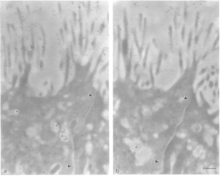
On ultrathin sections from the large intestine (proximal colon), immunoreactivity for /31,4-GT was detectable in the Golgi apparatus of both goblet and absorptive cells (Figure la and b). Within the Golgi apparatus cistemal stack, the labelling was restricted to several cisternae on the trans side of the stack.
Cl S
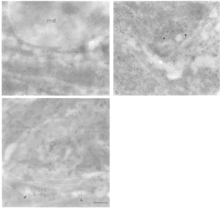
Fig. 1. Localization of |31,4-GT and /3-galactose residues on sections from bovine proximal colon. In both goblet cells (a) and absorptive cells (b), 01,4-GT
immunoreactivity detected with mab H5 is restricted to trans regions of the Golgi apparatus. Note the absence of staining over goblet cell mucus droplets (md) and unstained regions of the absorptive cell lrans-Go\g\ network (arrowheads). The distribution of /3-galactose residues, as detected by RCL I followed by asialofetuin—gold, mirrors that of /31,4-GT in the rram-Golgi apparatus, as shown in an absorptive cell (c) Note the staining of vesicles at the as side of the Golgi apparatus with RCL I (arrowheads). The cis side of the Golgi apparatus is labelled 'cis' in this and all subsequent figures. Bars 0 22 /im (a—c).
Localization of galactosyltransferase with monoclonal antibodies
However, elements of the fram-tubular network (trans-Go\g\ network) did not display immunoreactive sites for /31,4-GT (Figure lb). All other cellular organelles, apical and basolateral plasma membranes, and goblet cell mucus droplets were also not stained by the antibody. /3-Galactose residues, visualized with Ricinus communis lectin I (RCL I) followed by asialofetuin-gold, were similarly restricted to the trans region of the Golgi apparatus (Figure lc), indicating that this is the site where galactosylation occurs.
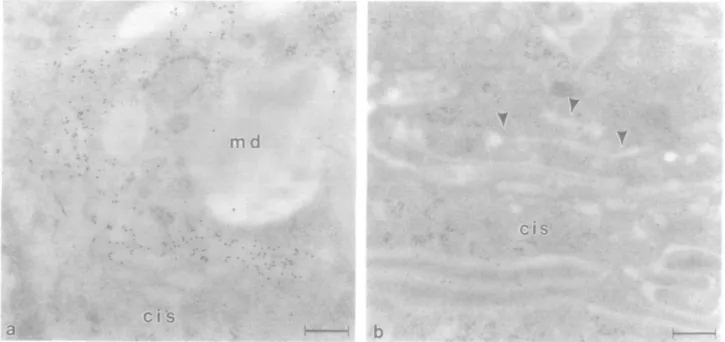
On ultrathin sections from the small intestine (duodenum) of the same animals, trans-Go\g\ apparatus cisternae of goblet cells were intensely stained by mab H5 while, surprisingly, adjacent absorptive cell Golgi apparatus was not stained (Figure 2a and b). As in the large intestine, no immunoreactivity was detectable in other organelles, plasma membranes or goblet cell
mucus. However, trans regions of the absorptive cell Golgi apparatus were intensely stained with RCL I followed by asialofetuin—gold (not shown), indicating the presence of abundant /3-galactose residues. RCL I also labelled apical and basolateral plasma membranes, lysosomes and apical cyto-plasmic vesicles of the small intestinal absorptive cells. Thymus
On sections from the thymus, the only cells displaying immuno-reactivity for mab H5 were the epithelial-reticular cells; all other cells, including the abundant lymphocytes, were not stained. Within the epithelial-reticular cells, the staining was restricted to trans cisternae of the Golgi apparatus (Figure 3a). As observed for the intestinal epithelial cells, portions of the
md
C I S
ci s
Fig. 2. Immunocytochemical detection of /31,4-GT with mab H5 on sections from bovine duodenum. Gold particle label is restricted to trans cisternae of the goblet cell Golgi apparatus (a). Note the absence of staining in the goblet cell mucus droplets (md). /31,4-GT was not detectable in absorptive cell Golgi apparatus from the same animals (b). Arrowheads indicate portions of the trans-Go\g\ network (GERL elements). Bars 0.21 /un (a) and 0.3 /im (b).
C I S
CI S
-I
Fig. 3. Detection of 01,4-GT and 0-galactose residues on sections from bovine thymus. Immunoreactivity for 01,4-GT using mab H5 is restricted to irons regions of the Golgi apparatus in epithelial-reticular cells (a). Note immunostaining present in typical GERL element (arrowheads), while other portions of the /ranj-Golgi network are unstained (arrows). /3-Galactose residues, as detected with RCL I followed by asialofetuin—gold, were observed over trans cisternae of the Golgi apparatus (b). Bars 0.21 j»m (a, b).
D J.Taatjes et at.
franj-Golgi network were not stained by the antibody (Figure 3a); however, in contrast, typical GERL rigid elements were usually stained (Figure 3a). Incubation of sections with RCL I followed by asialofetuin—gold resulted in staining of trans-Golgi apparatus cisternae (Figure 3b). Although thymocytes did not display immunoreactive sites for /31,4-GT, they did display RCL I binding sites in the trans region of the Golgi apparatus and along the plasma membrane (not shown).
Trachea
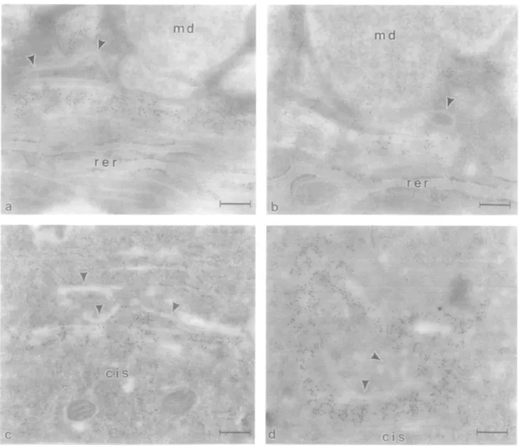
Several trachea! cell types displayed intense immunoreactivity in the Golgi apparatus for anti-/31,4-GT monoclonal antibodies. Owing to the presence of a variety of cell types, as well as good ultrastructural preservation, we decided to focus our attention on this ciliated pseudostratified columnar epithelium. In goblet cells, several cisternae at the trans aspect of the Golgi apparatus were intensely stained with mab H5 or HI62 (Figure 4a and b).
Portions of the trans-Go\g\ network, as well as mucus droplets, were not stained (Figure 4a and b). Similarly, in ciliated cells and brush cells, portions of the trans region of the Golgi apparatus displayed intense immunoreactivity for /31,4-GT (Figure 4c and d), although the intensity of immunostaining was usually greater in brush cells. Classical GERL elements were usually not stained with the monoclonal anti-/31,4-GT antibodies (Figures 4a—d). Typically, 2—4 cisternae at the cis side of the Golgi apparatus were not stained, followed by 3—4 stained cisternae at the middle to trans region of the stack, and finally unstained cisternae or tubules of the trans-Go\g\ network. This pattern of staining was evident in all three of the epithelial cell types examined in the trachea (goblet, ciliated and brush cells). However, owing to variability in the orientation of the Golgi apparatus in different cells in different sections, exact delineation of the subcompartmentation of /31,4-GT immuno-reactivity in all cells, as well as comparisons amongst cells, are difficult. Nevertheless, the overall pattern of immunostaining amongst the different cells was similar.
4
* * •
m d
md
re r
r e r
c i s
ci s
Fig. 4. Immunolocalization of /31,4-GT in bovine tracheal epithelium. In goblet cell Golgi apparatus (a, b), gold particle label is distributed in several cisternae in the middle/mmi regions of the stack. Note the absence of staining in typical GERL elements of the franj-Golgi network (arrowheads) and in mucus droplets (md). Similar results were obtained with mab H5 (a) or mab H162 (b). In ciliated cells (c) and brush cells (d), immunostaining is observed in several (rara-Golgi apparatus cisternae. However, note that typical GERL ngid elements and portions of the /ranj-Golgi network (arrowheads) are not stained, rer. rough endoplasmic reticulum. Bars 0.23 nm (a) and 0.22 /im (b—d).
Localization of galactosjltransfera.se with monoclonal antibodies /3-Galactose residues, as visualized with Datura stramonium
lectin (DSL) followed by asialofetuin—gold, were restricted to regions of the trans-Golgi network, lysosomes and plasma membrane. Within the Golgi apparatus, DSL binding sites were generally observed more towards the trans side (including
GERL elements) than immunoreactivity for /31,4-GT (Figure 5a and b).
Some sections were processed in a double staining procedure to localize /31,4-GT immunoreactivity and /3-galactose residues on the same section. As shown in Figure 6, immunoreactivity
Cl S
.. T
C I S
Fig. 5. Serial sections (not consecutive) incubated with mab H5 followed by protein A-gold (a) or DSL followed by asialofetuin-gold (b). Immunoreactivity
is restricted to several rra/u-Golgi apparatus cistemae from a trachea] ciliated cell (a). Note that typical GERL elements (arrowheads) are not stained for |S1,4-GT. Binding sites for DSL (b) are observed in the distal trans region of the Golgi apparatus, including GERL elements (arrowheads). Additionally, lysosomes (asterisk) are also stained with DSL/asialofetuin-gold, but not with mab H5. Vesicles and tubules at the trans aspect of the Golgi apparatus are not stained by mab H5 (arrows in 'a'), but do display DSL binding sites (arrows in 'b'). m, mitochondrion. Bar 0.25 /un (a, b).
D.J.Taatjes et al.
for /31,4-GT (small gold particles) and Gal/3 l,4GlcN Ac residues [detected with DSL followed by asialofetuin-gold (large gold particles)] overlap within the Golgi apparatus. However, the stainings for enzyme and product were not entirely superimposable; immunoreactivity for /31,4-GT is more widely distributed into middle cisternae of the Golgi apparatus stack. Furthermore, DSL binding sites persist in the trans-Golgi network (especially in the GERL element) which are not immunoreactive with the anti-/Sl,4-GT antibodies (Figure 6). DSL binding sites were also observed in the apical and basolateral plasma membranes, lysosomes and cytoplasmic vesicles.
Of all the numerous cell types examined, immunoreactivity with the three monoclonal antibodies was observed at the plasma membrane in only a subpopulation of tracheal epithelial cells. Based on morphological characteristics, most prominent-ly a tuft of apical microvilli, we believe that these represent trachea] brush cells. Figure 7 shows three serial (not con-secutive) sections of the apical plasma membrane of such a cell stained with mab H5. These serial sections demonstrate that the staining at the apical plasma membrane is not the result of a processing artifact, as the same cell is stained from sections processed separately on different grids. Moreover, labelling at the apical plasma membrane of these cells was also observed with mab H162 and mab H12. Indeed, in serial sections the same cell was found to be stained at the apical plasma membrane by both mab H5 and mab H12 (Figure 8). Interestingly, the label was preferentially associated with the microvillar projections of the apical plasma membrane, with little label observed over the smooth membrane regions between individual microvilli (Figures 7 and 8). These cells also contained apical cytoplasmic vesicles that were also stained by the monoclonal antibodies.
The immunocytochemicaJ results from all of the tissues examined are summarized in Table I.
Discussion
The use of immunocytochemical studies to localize a specific antigen rely for their interpretation on the specificity of the antibodies. In the present investigation we have used a panel of well characterized monoclonal antibodies previously demon-strated to recognize by Western blot analysis three different polypeptide epitopes within the non-glycosylated recombinant bovine /31,4-GT polypeptide expressed in E.coli. When /31,4-GT was present in cells in a quantity sufficient for detection by post-embedding immunoelectron microscopy, these monoclonal antibodies were found to be reliable and specific reagents for the localization of both Golgi apparatus and cell surface /31,4-GT.
Subcompartmentation of (31,4-GT in epithelial cell Golgi apparatus
One of the primary aims of this study was to compare the distribution of /31,4-GT in the Golgi apparatus of various epithelial cell types. To date, the Golgi apparatus distribution of/31,4-GT has been documented for only a limited number of cell types: HeLa cells (Roth and Berger, 1982; Strous etal, 1991), HepG2 cells (Geuze etal., 1985), myotubes (Tassin
etal., 1985), rat gastric mucous neck cells and rat testis interstitial cells (Suganuma etal., 1991). In the latter three studies, the low resolution of the immunoperoxidase technique 584
C I S
C I S
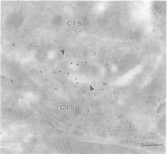
Fig. 6. Double staining for (31,4-GT and /3-galactose residues on the same
thin section from bovine trachea Immunoreactivity for mab H5 (small gold particles) and DSL binding sites (large gold particles) are found over the trans region of a ciliated cell Golgi apparatus. This Golgi apparatus is horseshoe shaped with cis regions found at both the bottom and top of the micrograph, with trans region in between. Note that the staining with DSL ensues in more distal rra/is-Golgi apparatus cisternae as compared to /31,4-GT immunoreactivity and that GERL elements (arrowheads) are stained with DSL, but not with mab H5 Bar 0 21 /im
makes it difficult to interpret the precise localization of the enzyme as it relates to the structural organization of the Golgi apparatus.
In the present study, we found that the distribution of /31,4-GT was virtually the same in the Golgi apparatus from all of the different epithelial cells examined. Several unlabelled cisternae at the cis side were followed by several labelled cisternae in the middle/fra/u region, followed by mostly unlabelled structures as part of the trans-Golgi network. Typical rigid elements at the trans aspect of the cisternal stack indicative of GERL elements (Novikoff and Novikoff, 1977) were generally unlabelled, as previously observed in HeLa cells by Roth and Berger (1982). However, the rigid GERL elements were labelled by the monoclonal anti-/31,4-GT antibodies in thymic epithelial-reticular cells. Such localizations probably re-flect the enormous diversity in Golgi apparatus structure noted in various cell types and under different experimental condi-tions. Without the aid of extensive serial sectioning analysis and exact control of experimental procedures, it is impossible to precisely compare the distribution of antibody binding sites in different Golgi apparatus. Moreover, the number and extent of Golgi apparatus subcompartments is highly subjective and often arbitrarily defined. Indeed, a still unresolved question concerns the extent or lack of overlap of sequentially acting glycosyltransferases within the trans-Golgi apparatus. Double labelling procedures for the localization of galactosyl- and sialyltransferase at the light microscopic level have provided conflicting results (Berger and Hesford, 1985; Berger etal., 1987b; Taatjes etal., 1987). Clearly, double labelling at the electron microscopic level of two sequentially acting glyco-syltransferases will be required to settle this conflict and may aid in establishing more precise definitions of Golgi apparatus subcompartments.
Localization of galactosyltransferase with monoclonal antibodies
md
md
m
'md
(Tl
Fig. 7. Three serial sections (not consecutive) showing the apical cytoplasm and plasma membrane of the same tracheal brush cell. Binding sites for mab H5
are observed in the apical plasma membrane in all three sections. Note the absence of staining in the neighbouring goblet cell mucus droplets (md) and along the lateral plasma membrane (arrowheads) separating the brush and goblet cell, m, mitochondrion. Bars 0.22 ftm (a-c)
D J.TaatJes et aL
•
4
I #
«^^^g,. ^. ' ^ • ^ ^ ^
b
Fig. 8. Two serial sections (not consecutive) showing the apical cytoplasm and plasma membrane of a tracheal brush cell. Binding sites for both mab H5 (a) and mab H12 (b) are observed in the apical plasma membrane of the same brush cell. Apical cytoplasmic vesicles (asterisks) are also stained. Note the absence of staining along the lateral plasma membrane (arrowheads) separating the brush cell from its neighbour. Bar 0.29 /im (a, b).
Table I. Subcellular distribution of j31,4-GT in bovine epithelial cells Tissue Proximal colon Duodenum Thymus Trachea Cell type Absorptive Goblet Absorptive Goblet Epithelial-reticular Ciliated Goblet Brush" cw-Golgi middle Golgi -•H -H -H trans- TGN APM Golgi + + + + ± + -+ - -+"
This table is based on observed immunostaining obtained with the
monoclonal anti-bovine /31,4-GT antibodies. Division of the Golgi apparatus into cis, middle, trans and rrans-Golgi network subcompartments is highly subjective; see the text for details.
TGN, rra/u-Golgi network; APM, apical plasma membrane.
( - ) , no detectable immunolabel; ( + ) , immunolabel present; ( ± ) , label present in portions of this Golgi apparatus subcompartment.
Tentatively identified as 'brush' cell based on morphological characteristics
bLabel present at APM on only a portion of cells. 586
Nevertheless, our present results indicate that the subcom-partmentaJ distribution of /31,4-GT in the Golgi apparatus is restricted to the trans region across many different cell types, and within similar cells in different organs. This contrasts with the differential distribution of a2,6-sialyltransferase observed in the Golgi apparatus of rat intestinal absorptive and goblet cells (Roth et al., 1986). In goblet cells, a2,6-sialyltransferase was found to be compartmentalized to trans cisternae of the Golgi apparatus, while in adjacent absorptive cells this immuno-reactivity for a2,6-siaJyltransferase was observed throughout the Golgi apparatus cisternal stack (with the exception of the fenestrated first cis cisterna). However, in the present study we did find a regional distribution within the intestinal tract of binding sites for the monoclonal anti-/31,4-GT antibodies. In the large intestine, the Golgi apparatus was stained in both absorptive and goblet cells. In contrast, in the small intestine (duodenum) from the same animals absorptive cell Golgi apparatus was not stained. The Golgi apparatus and apical plasma membrane of absorptive cells were stained with RCL I, indicating the presence of /3-galactose residues (Goldstein and Poretz, 1986). However, since RCL I is not a specific probe for /31,4-linked galactose residues (Goldstein and Poretz, 1986), this lectin binding could represent activity from a host of other
Localization of galactosyltransferase with monoclonal antibodies galactosyltransferases. /31,4-GT is constitutively expressed in
most if not all somatic cells. Why then were we unable to im-munocytochemically detect /31,4-GT in duodenal absorptive cells? Although not clear at this point, the answer may lie in the limitations of immunocytochemical techniques. Perhaps this enzyme is maintained in much lower quantities within the Golgi apparatus of duodenal absorptive cells as compared to absorptive cells from the large intestine, or the turnover time of this enzyme is much faster in these cells.
&1,4-GT does not appear to be expressed at the cell surface of most bovine epithelial cells
In addition to its primary location in the Golgi apparatus, /31,4-GT immunoreactivity has also been reported to be present at the cell surface of many tissues and cell types [Berger et al., 1981; Pestalozzi etal, 1982; Davis etai, 1984; Roth etal., 1985; Shaper et al., 1985; see also the recent review by Shur (1991)]. Most epithelial cells displayed cell surface /31,4-GT immunoreactivity when assessed at either the light or electron microscopic levels (Pestalozzi et al., 1982; Davis et al., 1984; Roth etal., 1985; Shaper etal., 1985), while human fibro-blasts and HeLa cells in culture showed only Golgi apparatus-associated immunoreactivity (Berger etal., 1981; Roth and Berger, 1982). Indeed, the immunohistochemical localization of /31,4-GT at the cell surface was taken as corroborative evidence for earlier suggestions of ecto-galactosyltransferase based on enzyme activity measurements and autoradiographic incorporation of radioactive substrates (Porter and Bernacki, 1975; Weiser etal., 1978; Cummings etal., 1979). Interest-ingly, the immunohistochemical studies are linked by the use of affinity-purified polyclonal antibodies raised against a purified soluble form of the enzyme derived from human milk (Berger etal., 1981) or bovine colostrum (Shaper etal., 1985). Although the polyclonal anti-/31,4-GT antibodies raised against the human milk enzyme were affinity purified (Berger et al., 1981), they were subsequently found, via sensitive radiobinding assays, to contain antibody species recognizing blood group-related carbohydrate structures present on the human /31,4-GT polypeptide (Childs et al., 1986; Feizi et al., 1987). Thus, pre-absorption of the affinity-purified anti-/31,4-GT antibody with blood group substances resulted in the abolition of immuno-fluorescence on sections from human intestine and stomach (Childs et al., 1986; Feizi et al., 1987). We have found similar results with a monospecific affinity-purified polyclonal anti-body raised against affinity-purified bovine milk /31,4-GT. Although the antibody decorated the plasma membrane and mucus droplets on identical sections from bovine intestine as used in this study with the monoclonal antibodies, pre-absorption of the antibody with asialomucin virtually abolished all cell surface and mucus droplet staining (D.J.Taatjes, unpublished). These findings might not be totally unexpected since human /31,4-GT is a glycoprotein containing highly im-munogenic carbohydrate structures (Childs et al., 1986; Amano et al., 1991). From these studies, it would appear that the majority of the staining observed at the cell surface was due to recognition of carbohydrate epitopes and not /31,4-GT polypeptide.
There are several ways available to potentially avoid antibodies raised against /31,4-GT which may in actuality recognize carbohydrate epitopes present on the polypeptide. The availability of cDNA clones for /31,4-GT (Shaper et al., 1986) would allow the production of non-glycosylated
recombinant proteins in bacteria, which could be then used as affinity matrices for the purification of polyclonal antisera. By definition, the resulting affinity-purified antibodies would recognize only protein epitopes. We have used this strategy in a previous study for the selection of polypeptide-specific antibodies against rat a2,6-sialyltransferase (Taatjes etal., 1988). Similarly, the recombinant /31,4-GT could be used as an immunogen for eliciting an antibody response in injected animals. Indeed, Berger and colleagues (Watzele et al., 1991) recently used such techniques for the production of a polyclonal rabbit antibody directed against a non-glycosylated fusion protein of human /31,4-GT produced in E.coli. The polyclonal antiserum decorated a peri-nuclear pattern, indicative of the Golgi apparatus, upon immunofluorescence labelling of HeLa and CaCo-2 cells in culture. Alternatively, one could chemically deglycosylate purified /31,4-GT and use this form of the enzyme as an immunogen for injection into rabbits. This method was applied by Berger and colleagues (1987a); however, they still could not demonstrate exclusive protein specificity with the resulting antisera. Moreover, the achieve-ment of total chemical deglycosylation is difficult to establish unequivocally. Finally, monoclonal antibodies could be raised against affinity-purified /31,4-GT and selected for their ability to recognize non-glycosylated recombinant protein produced in bacteria. This is the approach we have used in the present paper. Thus, we have visualized immunocytochemically three different epitopes located within the /31,4-GT polypeptide (Ulrich etal., 1986; Russo, 1990). Berger and colleagues (1986) have also produced a series of monoclonal antibodies against human milk /31,4-GT. While some were shown to recognize a Golgi apparatus-like pattern in HeLa cells by im-munofluorescence staining, others were found to stain cytoskeletal elements, nuclei or contact sites of the cell surface (Berger et al., 1986).
Recently, Suganuma and co-workers (1991) reported the immunoelectron microscopic localization of /31,4-GT in a spectrum of murine tissues. They used a rat IgM monoclonal antibody produced against the enzyme purified from mouse F9 cells and observed staining using immunoperoxidase method-ology in trans-Golgi apparatus cisternae, surprisingly in the nuclear envelope, and in the plasma membrane of F9 EC cells, PYS-2 parietal endoderm cells, STO fibroblasts, gastric mucosal epithelial cells, intestinal goblet cells, spermatocytes, Sertoli cells, spermatids and epididymal epithelial cells (Suganuma et al., 1991). In addition, a general plasma membrane staining was observed on most epithelial cells. In contrast, of all the epithelial cells we examined in the present study, staining was observed at the plasma membrane from only a minority of bovine tracheal epithelial cells. The staining tended to be re-stricted to a subregion of the plasma membrane comprising the microvillar projections. Importantly, this immunoreactivity was observed with all three of the /31,4-GT monoclonal anti-bodies, arguing strongly against a carbohydrate determinant being responsible for this plasma membrane staining. Although we cannot rule out the formal possibility that the three different monoclonal antibodies recognize an unrelated cell surface protein, the most direct explanation for these observations is that in these cells the immunoreactivity at the plasma mem-brane may represent an ecto-galactosyltransferase present in quantities sufficient for detection with our methods. Indeed, im-munostaining of serial sections with two of the monoclonal antibodies demonstrated binding to the apical plasma membrane of the same epithelial cell, arguing in favour of the recognition
D.J.Taatjes et at.
of (31,4-GT. These results, together with those described above, do not conclusively prove the existence of an ecto-galactosyltransferase; rather a combination of approaches, including molecular biology, biochemistry and immuno-cytochemistry, may probably be required to solve this con-troversial issue (Berger, 1991; Suganuma, 1991).
Materials and methods Reagents
Slaphylococcal protein A was obtained from Pharmacia Fine Chemicals (Piscataway, NJ), RCL I was from Vector Laboratories (Burlingame, CA), and affinity-punfied rabbit anti-mouse IgG was from Organon Teknika-Cappel (West Chester, PA). TetrachJorauric acid, tnsodium citrate and paraformalde-hyde were from Merck (Darmstadt, FRG), tannic acid (Aleppo tannin, no 1740) from Mallinckrodt (St Louis, MO) and glutaraldehyde (vacuum distilled) came from Fluka (Buchs, Switzerland). Ovalbumin, bovine serum albumin (RIA grade, fraction V), Tnton X-100, Tween 20, fetuin, asialofeluin, asialomucin, peroxidase, ovomucoid (trypsin inhibitor) and DSL were purchased from Sigma (St Louis, MO). All other reagents were of the highest available purity.
Antibodies
Three mouse monoclonal antibodies (designated H5, H12 and HI62) were developed against a soluble form of bovine UDP-galactose:A'-acetylglucosamine galactosyltransferase purified to apparent homogeneity by a combination of affinity and immunoadsorption chromatography, as previously described (Ulnch et al , 1986). By a combination of competitive binding assays and Western blot analysis with |31,4-GT, these monoclonal antibodies were found to recognize distinct epitopes of the polypeptide (Ulrich et al., 1986). Moreover, recent studies have demonstrated that each of these antibodies can recognize, by Western blot analysis, three different peptide epitopes on the recombinant bovine /31,4-GT polypeptide expressed in E.coli, indicating that the three different epitopes are contained within the primary structure (Russo, 1990; Russo et al., 1992).
For comparison, a monospecific affinity-purified rabbit polyclonal antibody raised against affinity-purified bovine milk /91,4-GT (Shaper el al , 1985) was also used for immunocytochemical localization experiments.
Tissue processing
Bovine tissues were freshly obtained from a local slaughterhouse in Basel, Switzerland. Small pieces from the trachea, thymus, small and large intestine were excised and quickly immersed in 3% formaldehyde (freshly prepared from paraformaldehyde), 0.1% glutaraldehyde in 0.01 M phosphate buffer, 0.15 M NaCl, pH 7.4 [phosphate-buffered saline (PBS)] for 2 h at room temperature. During the fixation period, the tissue pieces were minced into smaller pieces of - 1 mm3 After two quick rinses with PBS, free aldehyde groups were
quenched by immersing the tissue pieces in 50 mM ammonium chloride in PBS for 1 h. Afterwards, the pieces were rinsed with PBS and stored in PBS overnight at 4°C. The next day, the tissue pieces were dehydrated in a series of graded ethanols at progressively lower temperature and embedded in Lowicryl K4M (Carlemalm et al., 1982) at -35°C as previously described (Roth et al , 1981).
Preparation of colloidal gold-labelled reagents
Colloidal gold particles were prepared with an average diameter of 10 nm according to the tannic acid/citrate reduction method (Slot and Geuze, 1985) and with an average diameter of 15 nm according to the citrate reduction method (Frens, 1973). Protein A was complexed with 10 nm colloidal gold particles according to standard protocols (Roth et al., 1978). Asialofetuin was complexed with 10 or 15 nm colloidal gold particles as previously described (Taatjes et al., 1990).
Immunolocalization of 01,4-galactosyllransferase
Immunoreactive sites for 01,4-GT were detected with the protein A—gold technique (Roth et al , 1978). Ultrathin sections were cut with glass knives and collected on 150 mesh nickel grids coated with parlodion and carbon. Grids were floated section side down on drops of 0.5% ovalbumin in PBS for 20 min, followed by transfer to droplets of one of the ajiti-galactosyltransferase
antibodies and incubated overnight (14-21 h) at 4°C. The antibodies were used at the following concentrations [diluted with PBS containing 0.1 % bovine serum albumin (BSA)]: mab H5 (5 Mg/ml), mab H12 (0.3 /ig/ml) and mab H162 (50 /xg/ml). Since mab H12 tended to produce somewhat high background, we also used this antibody diluted with PBS containing 1% BSA, 0.075% Triton X-100 and 0.075% Tween 20 (PBS+ + +) to a concentration of 12.5 /ig/ml. This resulted in a virtual absence of background gold particles. Following two nnses with PBS (5 min each), the grids were either placed directly onto droplets of protein A-gold or incubated for 1 h at room temperature on droplets of affinity-purified rabbit anti-mouse IgG (10 /ig/ml). Incubation of grids on the secondary antibody rabbit anti-mouse proved to not be obligatory, since its omittance nevertheless resulted in specific immunostaining with the monoclonal antibodies The grids were incubated for 1 h on droplets of protein A—gold (10 nm colloidal gold particles), diluted with PBS+ + + to yield an optical density (OD) of 0.06 at 525 nm. Following rinses with PBS and distilled water, the grids were air dried. Finally, the sections were contrasted with 3% aqueous uranyl acetate for 5 min and lead acetate for 45 s.
The monospecific affinity-purified rabbit polyclonal antibody against /31,4-GT was used at a concentration of 50 jig/ml, followed by protein A—gold as described above for the monoclonal antibodies.
Localization of fi-galactose residues
0-Galactose residues were detected on ultrathin sections using lectins in a two-step cytochemical affinity technique as previously described (Taatjes et al., 1990). Sections were floated on drops of PBS for 5 min, followed by transfer to droplets of RCL I [nominal specificity for (3-galactose (Goldstein and Poretz, 1986)] or DSL [nominal specificity for Gal|31,4GlcNAc (Goldstein and Poretz, 1986)] for 45 min at room temperature. RCL I was used at a concentration of 75 /ig/ml and DSL at a concentration of 25 ^g/ml. Following two rinses with PBS (5 min each), the grids were incubated for 30 min on droplets of asialofetuin—gold complex. The asialofetuin-gold complexes (10 or 15 nm) were diluted with PBS+ + + to yield an optical density of 0.3 at 525 nm. Finally, the grids were nnsed with PBS and distilled water, and contrasted as described above.
Double labelling procedure
Immunoreactivity for galactosyltransferase and lectin recognition of /3-galactose residues were performed on the same sections in a double staining procedure Grids were floated on drops of PBS for 5 min, followed by incubation on droplets of DSL (25 /ig/ml) for 30 min at room temperature. After two rinses with PBS, the grids were transferred to droplets of asialofetuin—gold complex (15 nm particles; diluted with PBS+ + + to yield an OD525 m of 0.3) for
30 min at room temperature. This was followed by rinses with PBS and distilled water, and air drying. The dried grids were then floated for 20 min on drops of PBS containing 0.5% ovalbumin, followed by incubation overnight at 4°C on droplets of mab H5 (5 /ig/ml). After two rinses with PBS, the grids were placed on droplets of protein A-gold complex (10 nm particles; diluted with PBS+ + + to yield an OD323 m of 0.06) for 1 h at room temperature. Finally,
the grids were rinsed, air dried and contrasted as described above.
Specificity control incubations
For antibody incubations: (l) grids were incubated on PBS containing 0.1 % BSA overnight at 4°C, followed by incubation on droplets of protein A-gold complex for 1 h; (ii) grids were incubated on PBS containing 0.1% BSA overnight at 4°C, followed by sequential incubation on droplets of affinity-purified rabbit anti-mouse IgG for 1 h and protein A—gold complex for 1 h; (lii) antibodies were preabsorbed with various glycoproteins prior to use. Briefly, asialomucin, asialofetuin, fetuin, ovomucoid or peroxidase were reconstituted in PBS containing 0.1 % BSA to a concentration of 200 jig/ml. Equal volumes of each of these glycoprotein solutions were mixed with double-strength antibody concentrations 30 min prior to use for incubations
For lectin incubations: (i) grids were floated on drops of PBS for 30 min, followed by incubation on droplets of asialofetuin—gold complex for 30 min.
Acknowledgements
We would like to thank Michele von Turkovich and Audrey Avens for excellent technical assistance. D.J.T is a Parker B.Francis Fellow in Pulmonary Research. This work was supported in part by National Institutes of Health Grants GM46488 (to D.J.T.), CA45799 and GM3831O (to J.H.S ), and by Grant no. 31-26273.89 from the Swiss National Science Foundation (to J.R ). 588
Localization of galactosyltransferase with monoclonal antibodies Abbreviations
BSA, bovine serum alburrun; DSL, Datura stramonium lectin: (31.4-GT. 01.4-galactosyltransferase; mab, monoclonal antibody; PBS, phosphate-buffered saline; RCL I, Ricinus communis lectin I
References
Amano.J., Straehl.P., Berger.E.G., Kochibe.N. and Kobata.A. (1991) Structures of mucin-type sugar chains of the galactosyltransferase purified from human milk. Occurrence of the ABO and Lewis blood group determinants. J. Biol. Chem., 266, 11461-11477.
Berger.E.G. (1991) Letter to the editor. J. Histochem. Cytochem,, 39, 1439. Berger.E.G. and Hesford,F. (1985) Localization of galactosyl- and sialyltransferase by immunofluorescence: evidence for different sites. Proc. Natl. Acad. Sd. USA, 82, 4736-^1739.
Berger.E.G., Mandel.T. and Schilt,U. (1981) Immunohistochemical localization of galactosyltransferase in human fibroblasts and HeLa cells. J. Histochem. Cytochem., 29, 364-370.
Berger.E.G., Aegerter,E., Mandel,T. and Haun,H.-P. (1986) Monoclonal antibodies to soluble, human milk galactosyltransferase (lactose-synthase A protein). Carbohydr. Res., 149, 23-33.
Berger.EG., Muller.U., Aegerter.E. and Strous.G.J. (1987a) Biology of galactosyltransferase: recent developments. Biochem. Soc. Trans., 15, 610-613.
Berger.E.G., Thurnher.M and Muller.U. (1987b) Galactosyltransferase and sialyltransferase are located in different subcellular compartments in HeLa cells. Exp Cell Res., 173, 267-273.
Carlemalm.E., Garavito,R.M. and Villiger.W. (1982) Resin development for electron microscopy and an analysis of embedding at low temperature. J. Microsc. (Oxford), 126, 123-143.
Childs,RA., Berger.E.G., Thorpe.S.J., Aegerter.E. and Feizi.T. (1986) Blood-group-related carbohydrate antigens are expressed on human milk galactosyltransferase and are immunogeruc in rabbits. Biochem. J., 238, 605-611.
Cummings.R.D., Cebula.T.A. and Roth.S. (1979) Characterization of a galactosyltransferase in plasma membrane-enriched fractions from BALB/c 3T12 cells. J. Biol. Chem., 254, 1233-1240.
Davis.B.W., Berger.E.G., Locher,G.W., Zeller.M and Goldhirsch,A. (1984) Immunohistochemical localization of galactosyltransferase in the normal ovary and fallopian tube. J. Histochem, Cytochem., 32, 92—96.
Dunphy.W.G. and Rothman.J.E. (1985) Compartmental organization of the Golgi stack. Cell, 42, 13-21.
Dunphy.W.G., Brands.R. and Rothman.J.E. (1985) Attachment of terminal /V-acerylglucosamine to asparagine-linked oligosaccharides occurs in central cisternae of the Golgi stack. Cell, 40, 463^172.
Feizi.T., Thorpe.S.J. and Childs.R.A. (1987) Blood group genetic markers on human milk galactosyltransferase: relevance to the immunohistochemical approach to enzyme localization. Biochem. Soc. Trans., 15, 614-617. Frens.G. (1973) Controlled nucleation for the regulation of the particle size in
monodisperse gold suspensions. Nature Phys. Sd., 241, 20—22.
Geuze.H.J., Slot.J W., Strous.G.J A.M., Hasilik.A. and von Figura.K. (1985) Possible pathways for lysosomal enzyme delivery. J. Cell Biol., 101, 2253-2262.
Goldstein,I.J. and Poretz.R.D. (1986) Isolation, physicochemical character-ization, and carbohydrate-binding specificity of lectins. In: Liener.I.E., Sharon,N. and Goldstein,I.J. (eds), The Lectins. Properties, Functions, and Applications in Biology and Medicine. Academic Press, Orlando, pp. 33-248.
Novikoff.A.B. and Novikoff,P.M. (1977) Cytochemical contributions to differentiating GERL from the Golgi apparatus. Histochem. J., 9, 525-551. Pestalozzi.D.M., Hess.M. and Berger.E.G. (1982) Immunohistochemical evidence for cell surface and Golgi localization of galactosyltransferase in human stomach, jejunum, liver and pancreas. J. Histochem. Cytochem., 30,
1146-1152.
Porter.C.W. and Bemacki.R.J. (1975) Ultrastructural evidence for ectoglyco-syltransferases. Nature, 256, 648-650.
RothJ. (1987) Subcellular organization of glycosylation in mammalian cells. Biochim. Biophys. Ada, 906, 405-436.
Roth,J. and Berger.E.G. (1982) Immunocytochemical localization of galacto-syltransferase in HeLa cells; co-distribution with thiamine pyrophosphatase in trans Golgi cistemae. J. Cell Biol., 93, 223-229.
RothJ., Bendayan.M. and Orci.L. (1978) Ultrastructural localization of intra-cellular antigens by the use of protein A-gold complex. J. Histochem. Cytochem., 26, 1074-1081.
Roth.J., Bendayan.M., Carlemalm.E., Villiger.W. and Garavito.M. (1981) Enhancement of structural preservation and immunocytochemical staining in low temperature embedded pancreatic tissue. J. Histochem. Cvtochem.. 29. 663-671.
Roth.J., Lentze,M.J. and Berger.E.G (1985a) Immunocytochemical demon-stration of ectogalactosyltransferase in absorptive intestinal cells. J. Cell Biol., 100, 118-125
Roth,J , Taatjes.D.J , Lucocq.J M., Weinstein.J. and Paulson.J.C. (1985b) Demonstration of an extensive (ram-tubular network continuous with the Golgi apparatus stack that may function in glycosylation. Cell, 43, 287-295. Roth,J., Taatjes.D.J., WeinsteinJ., Paulson.J.C., Greenwell.P and Watkins.W M. (1986) Differential subcompartmentation of terminal glyco-sylation in the Golgi apparatus of intestinal absorptive and goblet cells. J. Biol. Chem., 261, 14307-14312.
Russo.R. (1990) PhD Thesis, Johns Hopkins University.
Russo.R.N , Shaper, N.L., Taatjes.D.J. and ShaperJ.H (1992) |31,4-galacto-syltransferase. A short NH2-terminal fragment that includes the cytoplasmic
and transmembrane domain is sufficient for Golgi retention. J. Biol. Chem.,
267, 9241-9247.
Shaper.N.L., Mann.P.L and ShaperJ.H. (1985) Cell surface galactosyltrans-ferase: Immunochemical localization. J. Cell. Biochem., 28, 229-239. Shaper.N.L., Shaper.J.H., Meuth.J.L., Fox.J.L., Chang.H., Kirsch.I.R. and
Hollis,G F. (1986) Bovine galactosyltransferase: Identification of a clone by direct immunological screening of a cDNA expression library Proc. Natl Acad. Set USA, 83, 1573-1577
Shur,B.D. (1991) Cell surface /31,4 galactosyltransferase: twenty years later Glycobiology, 1, 563-575
Slot.J.W. and Geuze.H.J. (1985) A new method of preparing gold probes for multiple-labelling cytochemistry. Eur. J. Cell Biol., 38, 87-93.
Strous.G.J , Berger.E.G., van Kerkhof.P., Bosshart,H., Berger.B. and Geuze.H.J. (1991) Brefeldin A induces a microtubule-dependent fusion of galactosyltransferase-containing vesicles with the rough endoplasmic reuculum. Biol. Cell., 71, 25-31.
Suganuma.T. (1991) Letter to the editor. J. Histochem. Cytochem., 39, 1440 Suganuma,T., Muramatsu.H., Muramatsu.T., Ihida.K., Kawano,J.-I. and
Murata,F. (1991) Subcellular localization of jV-acetylglucosaminide /SI,4 galactosyltransferase revealed by immunoelectron microscopy. J. Histochem. Cytochem., 39, 299-309.
Taatjes.D.J , Roth.J., WeinsteinJ., Paulson.J.C., Shaper.N.L. and ShaperJ.H. (1987) Codistribution of galactosyl- and sialyltransferase: reorganization of trans Golgi apparatus elements in hepatocytes in intact liver and cell culture. Eur. J. Cell Biol., 44, 187-194.
Taatjes.D.J., Roth.J., WeinsteinJ. and Paulson.J.C. (1988) Post-Golgi apparatus localization and regional expression of rat intestinal sialyl-transferase detected by immunoelectron microscopy with polypeptide epitope-purified antibody. J. Biol. Chem., 263, 6302-6309.
Taatjes.D.J., Barcomb.L., Leslie.K.O. and Low.R.B. (1990) Lectin binding patterns to terminal sugars of rat lung alveolar epithelial cells. J. Histochem Cytochem., 38, 233-244.
Tassin.A.M., Paintrand.M., Berger.E.G. and Bomens.M. (1985) The Golgi apparatus remains associated with microtubule organizing centers during myogenesis. J. Cell Biol., 101, 630-638.
UlrichJ.T., Schenck.J.R., Rittenhouse.H.G., Shaper.N.L. and ShaperJ.H. (1986) Monoclonal antibodies to bovine UDP-galactosyltransferase. Characterization, cross-reactivity, and utilization as structural probes. J. Biol. Chem., 261, 7975-7981.
Watzele.G., Bachofner.R. and Berger.E.G (1991) Immunocytochemical localization of the Golgi apparatus using protein-specific antibodies to galactosyltransferase. Eur. J. Cell Biol., 56, 451-458.
Weiser.M.M., Neumeier.M M., Quaroni.A. and Kirsch,K. (1978) Synthesis of plasmalemmal glycoproteins in intestinal epithelial cells. Separation from villus and crypt cell surface membranes; Glycosyltransferase activity of surface membrane. J. Cell Biol., Tl, 122-134.